Comprehensive molecular characterization of surgical vs. dietary weight loss: impact on
mammary tumor burden
by
Steven S. Doerstling
Honors Thesis
Department of Nutrition
The University of North Carolina at Chapel Hill
Gillings School of Global Public Health
2017
Approved:
_____________________________
Advisor: Dr. Stephen D. Hursting
_____________________________
ACKNOWLEDGEMENTS
I would like to convey my sincerest gratitude to Dr. Stephen Hursting and Emily Rossi
for their unwavering support over the past two years. It would be remiss not to express
special thanks to Emily, my award-winning graduate student mentor, whose unmatched
guidance and dedication to my academic and personal development has left an
indelible impression on my career and feelings toward turquoise. I am of course
indebted to my wonderful parents, family, and friends. I could not have asked for a
TABLE OF CONTENTS
Chapters
I. ABSTRACT……… 4-5 II. INTRODUCTION……… 6-10
III. HYPOTHESIS, SPECIFC AIMS………..……….……… 11
IV. METHODS………...……….… 12-16 V. RESULTS..………... 17-27 VI. DISCUSSION………... 28-33 VII. REFERENCES………... 34-35 Tables 1. Serum Hormones and Cytokines………... 19
2. Pathway Analysis of Differentially Methylated Genes………. 24
3. DNA Methylation Concordance between Mouse and Human Studies……… 29-30 Figures 1. Animal Study Design, Body Weight, Body Fat Percentage………..… 18
2. Mammary Fat Pad Adipocyte Size and CLS Density……….…… 21
3. Ex vivo Tumor Volume………... 22
I. Abstract
Background: Obesity is a widespread health concern and established risk factor for basal-like breast cancer. However, studies are conflicted on the benefits of weight loss
relevant to breast cancer prevention. Recent studies suggest that certain methods of
robust weight loss with long-term maintenance, such as bariatric surgery, might in fact
be able to reverse obesity-associated breast cancer risk.
Hypothesis: We hypothesize that weight loss by sleeve gastrectomy will generate sufficient metabolic normalization to reverse obesity-driven mammary tumor burden
more effectively than weight loss by diet alone.
Methods: Mice were fed a low-fat control (CON) or high-fat diet-induced obesity (DIO) regimen for 15 weeks to model chronic obesity. DIO mice were randomized to continue
on a high-fat diet (Obese) or undergo weight loss by either sleeve gastrectomy (~70%
excision of the stomach) in combination with switching to a low-fat diet or by switching
to a low-fat diet alone, resulting in formerly obese (FOB)-Surg and FOB-Diet mice,
respectively. Additionally, a subset of normal weight (NW) Control mice (Con, n=25)
was maintained on a low-fat diet throughout the study. NW Control, FOB-Diet, and
Obese mice underwent a sham procedure to control for the insult of surgery. FOB-Surg
and FOB-Diet mice did not lose a significantly different amount of weight and body fat;
both groups had significantly lower weight and percent body fat than Obese mice. Eight
weeks after surgical procedures and diet switches, all mice on study were orthotopically
Results: At the end of study, ex vivo tumor volume in FOB-Surg mice was not
significantly different from NW Control mice and significantly different from Obese mice.
However, tumor volume in FOB-Diet mice was significantly different from NW Control
mice and not significantly different from Obese mice. In addition, FOB-Surg mice had
levels of serum tumor necrosis factor-alpha (TNF-a), insulin, and mammary adipocyte
size that were not significantly different from NW Control mice and significantly different
from FOB-Diet and Obese mice. Lastly, pathway analysis of mammary tissue gene
expression revealed redundant upregulation of genes in extracellular matrix remodeling
and growth factor signaling in Obese vs. FOB-Surg and FOB-Diet vs. FOB-Surg mice.
Conclusions: Our results demonstrate that surgical weight loss imparted a plurality of metabolic advantages, functional genomic changes, and successful reversal of
obesity-associated mammary tumor burden that were not similarly achieved by dietary weight
II. Introduction
The predominance of obesity persists in the United States, with recent
epidemiologic studies reporting a prevalence of 37.7% in the adult population (1). A
robust body of evidence demonstrates that obesity significantly increases the risk of
developing breast cancer in postmenopausal women (2). Furthermore, obese women
experience poorer breast cancer outcomes by virtue of increased tumor grade at clinical
presentation, disease recurrence, and substandard response to certain therapies (3).
Accordingly, rigorous scientific investigation has been dedicated to interrogating the
mechanisms complicit in this obesity-cancer link.
Obesity is attendant to a spectrum of physiological aberrations resulting from
chronic positive energy intake (4). In obesity, excess energy is converted to
triacylglycerol and stored in various adipose tissue depots throughout the body. The
obese state is thus associated with profound expansion of the adipose tissue, which
occurs by adipocyte hyperplasia (increase in adipocyte number) or hypertrophy
(increase in adipocyte size). In obese adults, hypertrophy predominates as the
mechanism to support the growth in adipose tissue (5). Adipocyte hypertrophy, and the
associated fatty acid spill over, results in insulin resistance, elevated serum
triglycerides, local tissue hypoxia, inflammation, and altered sex hormone levels (6). In
consequence, obesity is a strong risk factor for a host of diseases including
cardiovascular disease, type II diabetes, and a variety of cancers including those of the
breast, pancreas, colorectum, endometrium, esophagus, and kidney (7).
Several key obesity-driven physiological disruptions have been identified as
include growth factor signaling– particularly insulin-like growth factor 1 (IGF-1)– and
inflammatory cytokines and adipokines secreted from immune cells and adipocytes (8).
Obesity is known to induce insulin resistance, resulting in compensatory
hyperinsulinemia, which causes enhanced synthesis of IGF-1. IGF-1 is a potent mitogen
that activates a receptor-tyrosine kinase signaling cascade resulting in cell growth,
survival, proliferation, and evasion of apoptosis; thus, IGF-1 and intracellular growth
factor signaling are heavily implicated in breast cancer risk and progression (9). Obesity
confers its pro-inflammatory effects in part as a result of adipose tissue remodeling.
Positive energy balance and triglyceride storage causes adipocyte hypertrophy. In
consequence, adipocytes can outgrow supporting vasculature, resulting in hypoxia, and
expand the margins permitted by the outer cellular membrane, resulting in apoptosis
(7). Moreover, it is well-documented that adipose tissue in obese subjects exhibits
increased secretions of several pro-inflammatory cytokines, including interleukin-6
(IL-6), tumor necrosis factor-a (TNF-a), and monocyte chemoattractant protein-1 (MCP-1)
(7). These cellular phenomena contribute to substantial recruitment of immune cells to
the adipose tissue as well as proliferation of macrophage populations native to adipose
depots (6, 7). In this inflammatory milieu, activated macrophages supply significant
secretions of cytokines and engage in phagocytosis of necrotic engorged adipocytes,
forming visible histopathological signatures known as crown-like structures (CLS) (10).
This cycle can self-perpetuate by way of cytokine-induced lipolysis, which results in
release of fatty acids that activate toll-like receptors on immune cells to sustain elevated
Tissues proximal to adipose tissue, such as the mammary epithelium, are
vulnerable to this obesity-associated inflammatory cascade, which promotes
carcinogenic events and cancer progression through several molecular levers. For
instance, increased IL-6 secretions promote cell proliferation through the Janus Kinase
(JAK)/ Signal transducer and activator of transcription 3 (STAT3) pathway (12), and
TNF-a secretions promote cell proliferation, angiogenesis, and evasion of apoptosis via
nuclear factor-kappa B (NF-kB) signaling (13). In addition, reactive oxygen and nitrogen
species originating from adipose tissue macrophages jeopardize genomic integrity by
inducing structural DNA alterations (7). Therefore, it is critical to target the progression
of obesity-associated adipose dysregulation in order to reduce the barrage of survival
signals from adipose tissue coterminous with potential sites of malignant disease.
Given the indisputable positive associations between obesity and breast cancer,
it would follow that obesity reversal can achieve corresponding reversal of
obesity-associated cancer risk. However, the scientific community has failed to reach a
consensus on the efficacy of weight loss on cancer risk and outcomes. Despite a
handful of studies demonstrating intermediate reductions in biomarkers of cancer risk
with intermittently promising outcomes, results are often inconsistent (14), thereby
precluding an evidence-based recommendation of certain weight loss strategies for
targeted breast cancer risk reduction. This is largely due to the low success rate of
achieving significant weight loss and an even lower rate of long-term weight loss
maintenance. Indeed, we and others have explored obesity-reversal in preclinical
mouse models, and formerly-obese mice often exhibit persistent dysregulation in
Although weight loss, in particular weight loss exclusively by low-fat diet, has not
demonstrated success as a comprehensive risk reduction method, weight loss by
bariatric surgery has shown auspicious results for reversal of obesity-associated tumor
burden. Christou et al. showed that in comparison to morbidly obese control subjects,
individuals who underwent bariatric surgery had a remarkable five-fold reduction in
5-year incidence of all cancers and breast cancer (17). Bariatric surgery describes a
variety of surgical procedures that modify the anatomy of the digestive tract in an effort
to impart weight loss and resolve insulin resistance and hyperglycemia, for which it is
often exceptionally effective. The two most common bariatric surgery procedures are
Roux-en Y gastric bypass (RYGB) and vertical sleeve gastrectomy (VSG) (18). RYGB is
regarded as the “gold standard” bariatric surgery procedure due to its extensive
documented success in generating long-term weight loss and mitigation of
obesity-associated comorbidities. Until recently, RYGB was the most common bariatric
operation, but due to the inherent complexity of constructing multiple anastomoses for
each procedure and the associated surgical complications, VSG surpassed RYGB as
the most common procedure in 2013, boasting a straightforward surgical protocol, low
complication rate, and impressive clinical outcomes (19, 20). Weight loss achieved by
VSG is comparable to RYGB, and both procedures give rise to greater weight loss and
better hemoglobin A1c (HbA1c) outcomes, a diagnostic tool for diabetes, than standard
medical therapy (21, 22). Furthermore, VSG does not pose a risk of vitamin and mineral
absorption posed by RYGB, an important consideration for obese women of
A variety of mechanisms are under investigation to explain the efficacy of
bariatric surgery with respect to endocrine health and cancer outcomes; top candidates
include caloric restriction due to sensitive food reward signals, caloric malabsorption in
the small intestine, gut hormone alterations, and bile acid and gut microbiota changes
(18, 23). Furthermore, bariatric surgery has been shown to drastically modulate the
epigenome, in particular DNA methylation. Studies have demonstrated profound
remodeling of methylation profiles in gene clusters relevant to obesity caused by
bariatric surgery. In one study comparing DNA methylation profiles in subcutaneous
adipose tissue of women before and after undergoing bariatric surgery, researchers
reported differential methylation in genes with established involvement in
obesity-associated adipose tissue dysfunction (24). Additional research has revealed that
bariatric surgery can reverse obesity-associated DNA methylation signatures to levels
comparable to never-obese subjects in skeletal muscle (25), and of particular
importance to obesity-breast cancer link, adipose tissue (26). Given the potency of
weight loss, metabolic improvements, and epigenetic modulation achieved exclusively
by bariatric surgery, we sought to compare VSG and dietary weight loss as
obesity-reversal interventions and characterize their respective cancer-protective effects in a
III. Hypothesis, Specific Aims
Hypothesis: Considering the robust degree of longitudinal weight loss, endocrine health resolution, and epigenetic remodeling achieved by bariatric surgery, we
hypothesize that weight loss by sleeve gastrectomy will generate sufficient metabolic
normalization to reverse obesity-driven mammary tumor burden more effectively than
weight loss by diet alone.
Aim 1: Compare the effectiveness of bariatric surgery and weight loss by diet alone to reduce obesity-associated metabolic perturbations, adipose tissue dysfunction, inflammation, and tumor burden.
Aim 2: Characterize the DNA methylation profiles in the mammary fat pad to determine exclusive regulation by weight loss intervention and test concordance with obesity-associated methylation in the Normal Breast Study.
IV. Methods
Animal Study Design: Husbandry and Diet
All animal study protocols were approved and coordinated in compliance with guidelines
issued by the University of North Carolina at Chapel Hill Institutional Care and Use
Committee (IACUC). Ninety four female 6-8 week old C57BL/6 mice (a well
characterized energy balance-responsive mouse model) were purchased from Charles
River Laboratories International, Inc. (Wilmington, MA). Upon arrival, mice were housed
two per cage on a 12-hour light/dark cycle and offered food and water ad libitum. A diet
of standard chow was administered to all mice for a one week acclimation period. Next,
mice were randomized to two groups, receiving either a control (CON; 10% kcal from
fat) diet (n=22; Product # D12450J; Research Diets, Inc.) or a diet-induced obesity
(DIO; 60% kcal from fat) regimen (n=72; Product # D12492; Research Diets, Inc.) to
generate a normal-weight (NW) control or obese phenotype, respectively. Body weight
and food intake were measured weekly. After 15 weeks on diet, with the weights of NW
CON and DIO mice significantly different from each other (p<0.001), obese mice were
then randomized to continue the DIO diet (Obese) or receive a surgical or diet weight
loss intervention, resulting in formerly obese (FOB)-Surg and FOB-Diet groups,
respectively. FOB-Surg mice were subject to sleeve gastrectomy (~80% excision of the
stomach), and NW Control, Obese, and FOB-Diet mice received a sham procedure.
Three days post-operation, both FOB-Surg and FOB-Diet began the same low-fat
control diet as the NW Control group. Eight weeks after surgery when body weights of
mice were relatively stabile, all mice were orthotopically injected with 3.5 x 10⁴ E0771
breast cancer originally isolated from a spontaneous medullary breast adenocarcinoma
in a C57BL/6 mouse (27). In vivo tumor growth was measured two times per week with
skinfold calipers and in vivo tumor area was determined using the formula πr2. Four
weeks following orthotopic injection, tumors in 50% of Obese mice (the fastest growing
group) reached the requisite size defined by the IACUC protocol; therefore, all mice on
study were sacrificed. Mammary tumors, tumor-adjacent and tumor-distal mammary fat
pad were excised and sectioned to either be formalin fixed or flash frozen in liquid
nitrogen and stored at -80°C until further analysis. Ex vivo tumor volume was calculated
using the formula 1/6π x D1 x D2 x D3 (where D is equal to ex vivo diameter of the
tumor). End of study blood was collected by cardiac puncture, allowed to clot at room
temperature for 30 minutes and centrifuged for 10 minutes at 1000 x g to isolate serum,
and stored at -80°C.
Sleeve Gastrectomy and Sham Procedures
Sleeve gastrectomy and sham procedures were performed by trained animal surgeons
according to a validated protocol (28). Briefly, vertical sleeve gastrectomy (VSG)
involved excision of ~80% of the lateral stomach. The sham procedure, performed on
NW Control, Obese, and FOB-Diet mice to control for the physiological insult of surgery,
was executed by first isolating the stomach and then applying manual pressure with
forceps for five seconds. The excision and pressure were applied along a line
continuous with the esophagus and pylorus. All surgeries occurred within a four day
window, and mice within all study groups were randomized to the day of operation.
consistent across all groups. Additionally, all mice received a three-day liquid diet
(Osmolite OneCal) before being reintroduced to solid food. Antibiotics were given to all
mice three days post-op. All mice were weighed daily and food intake was quantified for
one week post-op.
Quantitative Magnetic Resonance Imaging Analysis
Quantitative magnetic resonance imaging (qMRI) (Echo Medical Systems, Houston, TX)
was used to measure the body composition for all groups (n=6-9 mice/group) at the end
of study. Lean body mass, fat body mass, and free water were quantified. Body fat
percentage was calculated by dividing the fat body mass by the body weight measured
with a digital scale.
Metabolic Analysis of Serum Hormones, Cytokines, and Adiponectin
One week prior to tumor injection, serum was collected from mice fasted 4-6 hours by
submandibular bleed. Serum hormones, cytokines, and adipokines including insulin,
leptin, adiponectin, IL-6, TNFa, and resistin, were measured using Milliplex Mouse
Metabolic Hormone Magnetic bead Panel (MMHMAG-44K), Bio-Plex ProTM Mouse
Adiponectin Assay, and Mouse Cytokine Panel A 6- Plex, respectively (Bio-Rad
Laboratories; Hercules, California). Insulin-like growth factor 1 (IGF- 1) concentrations
were measured using R&D Systems IGF-1 Bead-Based Single-plex Luminex assay
Mammary Fat Pad Adipocyte Size and Crown Like Structure Analysis
Hematoxylin and eosin (H&E) staining on 4-micron thick sections from formalin-fixed,
paraffin embedded distal mammary fat pad tissue was processed, scanned and imaged
using Aperio CS2 Digital Pathology Scanner (Leica Biosystems, Wetzlar, Germany) at
40X magnification. Representative snapshots (n=9-11 mice/group; 3 snapshots were
sample) were randomly selected from whole tissue images zoomed in at 8.8X (300 μM)
utilizing ImageScope Viewing Software Version 12.0 (Leica Biosystems). Mammary fat
pad average adipocytes size and number of adipocytes were quantified using ImageJ
Version 1.51e (National Institute of Health, Bethesda, Maryland). An adipocyte tool
macro (MRI Adipocyte Tools.txt) was downloaded from
(http://dev.mri.cnrs.fr/projects/imagej-macros/wiki/Adipocytes_Tool) and imported into
ImageJ. Furthermore, the number of crown-like structures (CLS) (10) was quantified
from whole tissue H&E stained distal mammary fat pad sections (n=10-15 mice/group).
Briefly, the number of CLS were counted in a blinded fashion, and CLS density
measures were achieved by dividing the number of CLS by the total slide area eligible
for analysis using ImageScope Viewing Software Version 12.0 (Leica Biosystems).
DNA methylation analysis
Genome-wide methylation profiles for the distal mammary fat pad were determined by
reduced representation bisulfite sequencing (RBBS). DNA was extracted from a random
sample (n=4 mice/group) of distal mammary tissues using TRI Reagent (Sigma-Aldrich)
according to the manufacturer's instructions. Library preparation and sequencing were
Sequencing Facility. Alignment and differential methylation analysis were conducted as
previously described (15).
RNA-Seq
Total RNA was extracted from the flash-frozen tumor-adjacent and tumor-distal
mammary fat pad samples collected at the end of the study using TRI Reagent
(Sigma-Aldrich) according to the manufacturer's instructions. RNA libraries were prepared using
the Illumina TruSeq Stranded Total RNA Sample Preparation kit according to
manufactures instructions. The libraries were sequenced using a 2x76 bases paired end
protocol on the Illumina HiSeq 2000 instrument. The reads were mapped to mouse
genome (mm10) by TopHat (version 2.0.7). The number of fragments in each known
gene from RefSeq database (UCSC Genome Browser 2013) was enumerated using
HTSeq-count from HTSeq package (version 0.5.3p9). Differential expression was
performed using DESeq2.
Pathway Analysis
WebGestalt (www.webgestalt.org) (29) over representation enrichment analysis (ORA)
of KEGG pathways and gene ontology biological process curated gene sets was
performed for selected pairwise comparisons of RNA-Seq and RRBS data.
Statistical Analysis
All values are represented as mean ± standard deviation (STDEV). One-way analysis of
assess the effects of diet and weight loss on body weight and fat percentage, tumor
volume, serum hormone and cytokine concentrations, and mammary fat pad adipocyte
size and CLS density. Results were analyzed using GraphPad Prism software
(Graphpad Software Inc., La Jolla, CA) and p ≤ 0.05 was considered statistically
significant.
V. Results
Weight Loss Interventions by Surgery (FOB-Surg) or Diet Alone (FOB-Diet) are Equally Effective at Reducing Body Weight and Fat Mass
Mice were fed a low fat control or a diet-induced obesity regimen for 15 weeks in order
to establish a normal weight control or obese phenotype, respectively. Obese mice had
significantly higher body weight at the time of sleeve gastrectomy or sham procedure
relative to control mice (p < 0.0001). Eight weeks following the operations and diet
switch, body weight between FOB-Surg and FOB-Diet mice was not significantly
different (Figure 1A), and neither group was significantly different from NW Control
mice, however all were significantly different from Obese mice prior to tumor cell
injection (p < 0.0001 for all comparisons, NW Control vs. Obese, FOB-Surg vs. Obese,
FOB-Diet vs. Obese). At end of study, body fat percentage (Figure 1B) was not
statistically different among NW Control, FOB-Surg and FOB-Diet mice, and all groups
were significantly lower than Obese mice (p < 0.0001 for all comparisons, NW Control
vs. Obese, FOB-Surg vs. Obese, FOB-Diet vs. Obese), indicating successful and
Figure 1. Surgical and dietary weight loss generate comparable reductions in body weight and body fat percentage. (A) Body weight of mice throughout the course of study. (B) Combined violin and box plots of body fat percentage at end of study. Differences in significance denoted by different letters (a,b) p-value <0.05.
Surgical Weight Loss in Mice More Effectively Reduces Circulating Growth Factors and Pro-Inflammatory Mediators in Serum
Seven weeks after surgical procedures and diet switch when weights were stabilized,
serum was collected from mice (n=10-12 mice/group) by submandibular bleed for
multiplex metabolite analyses. For all metabolites measured except adiponectin, levels
in NW Control mice were significantly lower than Obese mice.Insulin levels in
FOB-Surg mice were significantly different from both FOB-Diet and Obese mice and not
significantly different from NW Control mice; insulin levels in FOB-Diet exhibited
intermediate reductions from Obese levels but were not significantly different. For
Insulin-like growth factor-1 (IGF-1) and resistin, FOB-Surg mice displayed significantly
non-ratio, interleukin-6 (IL-6), and monocyte chemoattractant protein-1 (MCP-1), both
FOB-Surg and FOB-Diet mice displayed significantly lower levels relative to Obese mice.
Lastly, FOB-Surg mice exhibited significantly lower levels of tumor necrosis factor alpha
(TNFa) than both FOB-Diet and Obese mice; levels of TNFa in FOB-Diet mice were not
statistically different from only Obese mice (Table 1).
NW Control FOB-Surg FOB-Diet Obese
Insulin (ng/mL) 0.74 ± 0.25a,b 0.56 ± 0.22a 1.04 ± 0.35b,c 1.35 ± 0.49c
IGF-1 (ng/mL) 42.2 ± 13.8a 39.0 ± 15.9a 51.4 ± 18.6a,b 76.7 ± 29.6b
Leptin (ng/mL) 2.41 ± 0.89a 2.16 ± 1.20a 4.77 ± 4.21a 11.5 ± 0.05b
Adiponectin (µg/mL) 12.0 ± 2.99 11.1 ± 5.01 12.6 ± 2.91 9.68 ± 5.13
Leptin:Adiponectin 2.22E-06 ± 1.34E-06a 2.34E-06 ± 1.29E-06a 4.95E-06 ± 5.29E-06a 3.50E-05 ± 1.30E-05b
Resistin (ng/mL) 11840 ± 4143a 6732 ± 2237a 12528 ± 4717a,b 20214 ± 10323b
TNF! (pg/mL) 11.5 ± 7.31a 18.8 ± 14.1a 41.2 ± 14.1b 56.9 ± 18.2b
IL-6 (pg/mL) 30.6 ± 8.21a 39.5 ± 17.5a 50.8 ± 24.0a 89.4 ± 20.6b
MCP-1 (pg/mL) 99.8 ± 5.34a 102.2 ± 22.4a 136.8 ± 50.4a 192.3 ± 51.9b Cytokines
Hormones
Table 1. Serum hormones and cytokines
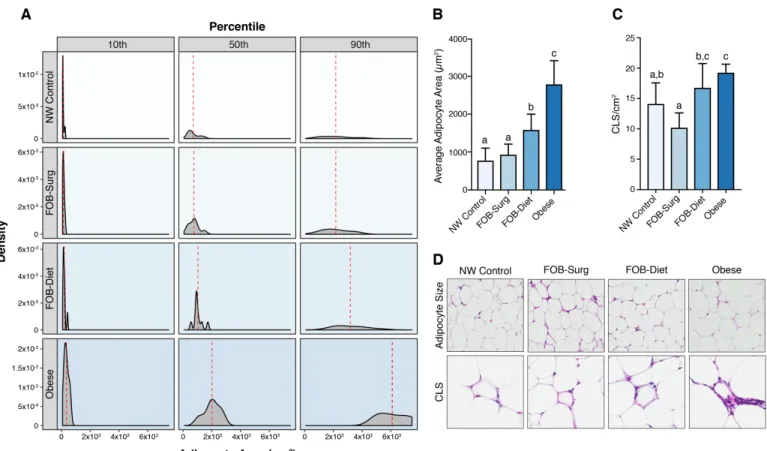
Surgical Weight Loss in Mice Reverses Adipocyte Hypertrophy and Crown-Like Structure Density in the Mammary Tissue
Adipocyte area in the mammary fat pad displayed significant fluctuation by phenotype.
Both surgical and dietary weight loss resulted in reduced adipocyte area, with the most
profound differences relative to Obese mice occurring at the 50th and 90th percentile of
adipocyte area (Figure 2A). More precisely, FOB-Surg mice exhibited an average
adipocyte area not significantly different from NW Control mice and significantly different
from both Diet and Obese Mice (p<0.0001), while average adipocyte area of
FOB-Diet mice displayed significant reduction relative to Obese mice (p<0.0001) but
remained significantly different from both NW Control (p<0.0001) and FOB-Surg
(p<0.05) (Figure 2B). Furthermore, density of crown-like structures in the mammary fat
pad was lowest in FOB-Surg mice and significantly lower than that of FOB-Diet and
Obese mice. Of note, crown-like structure density in FOB-Diet mice was not statistically
different from Obese mice (Figure 2C). Representative images of H&E stained
mammary fat pad depicting adipocyte size and CLS are displayed (Figure 2D).
Surgical Weight Loss, but not Weight Loss by Diet Alone, Reverses the Pro-Tumorigenic Effects of Obesity
Intriguingly, ex vivo tumor volume of FOB-Surg mice was not statistically different
fromNW Control mice and significantly different than Obese mice. However, FOB-Diet
mice had tumors that were not significantly different from Obese mice and significantly
different than NW Control mice (Figure 3). Therefore, FOB-Surg mice, but not FOB-Diet
Figure 2. Surgical weight loss results in more robust reductions in adipocyte hypertrophy and crown-like structure density in the mammary fat pad. (A) Panel
displaying density functions for 10th, 50th, and 90th percentiles of adipocyte area across
Figure 3. Ex vivo tumor volume reveals unique cancer-protective effects of surgical versus dietary weight loss. Differences in significance denoted by different letters (a,b,c) p-value <0.05.
The Methylation of Genes in the Mammary Tissue of Mice that Lost Weight via Surgery Displays a Pattern that is Distinct from FOB-Diet and Obese mice
The complete RRBS dataset was filtered by pairwise comparisons between all study
groups with p<0.0001 and false discovery rate (FDR)<0.0001 as filtering criteria for
differential methylation. Genes with differential methylation according to these criteria at
any gene feature (e.g. promoter, intron) were included. Gene lists were entered into
WebGestalt for overrepresentation enrichment analysis (ORA) in curated KEGG
pathways (Table 2). The genes harboring differential methylation between Obese and
FOB-Surg mice were highly enriched for a variety of pathways implicated in mammary
carcinogenesis and a tumorigenic microenvironment. Interestingly, there were markedly
fewer genes displaying differential methylation according to the criteria above in Obese
vs. FOB-Diet mice relative to Obese vs. FOB-Surg mice (2011 and 4258 genes,
redundancy in pathways represented by differentially methylated genes between
FOB-Diet vs FOB-Surg mice and Obese vs FOB-Surg mice, suggesting that Obese and
FOB-Diet mice share a significant number of similar DNA methylation features that
explain overlapping pathway enrichment relative to FOB-Surg mice.
The Expression of Genes in the Mammary Tissue of Mice that Lost Weight via Surgery Displays a Pattern that is Distinct from FOB-Diet and Obese mice
RNA Sequencing data was filtered to create gene lists for all pairwise comparisons
between groups; differential expression with group-specific directionality was achieved
by selecting genes with log2(fold change) > 0.58 (which is equivalent to fold change >
1.5) for each inter-group comparison. Gene lists were entered into WebGestalt for
overrepresentation enrichment analysis (ORA) in curated KEGG pathways. There was
significant overlap between pathways upregulated in Obese vs. FOB-Surg mice and
activation by obesity and persist despite weight loss by diet alone (Figure 4A,B). These
pathways are well-characterized in the context of obesity and breast cancer risk and
suggest broad activation of canonical cell signaling cascades involved in cell
proliferation, growth, and extracellular matrix function. Next, comparisons of the two
weight loss groups to Obese mice reveals significant overlap in pathway
characterization reflecting upregulated genes in FOB-Surg (Figure 4C) and FOB-Diet
(Figure 4E) mice vs. Obese mice. There were remarkably few upregulated genes in
Obese vs. FOB-Diet mice relative to other comparisons. The resulting ORA revealed a
Observed Genes in Pathway
Percent of All
Genes in Pathway P-value FDR
Signaling pathways regulating pluripotency of stem cells* 47 33.6 1.34E-07 2.00E-05
Breast cancer* 47 32.4 4.36E-07 3.75E-05
Pathways in cancer* 99 25.3 5.02E-07 3.75E-05
Basal cell carcinoma* 22 1.36E-05 8.13E-04
Rap1 signaling pathway 56 26.5 3.76E-05 1.87E-03
Insulin secretion* 27 33.3 7.21E-05 2.57E-03
Ras signaling pathway 58 25.7 7.74E-05 2.57E-03
Wnt signaling pathway 41 28.1 1.07E-04 2.92E-03
Hippo signaling pathway* 42 27.3 1.82E-04 4.31E-03
Focal adhesion* 51 25.1 3.61E-04 6.75E-03
Differentially methylated Obese vs FOB-Diet (n = 2011)
Insulin secretion* 18 22.2 5.39E-05 6.38E-03
Breast cancer* 26 17.9 7.09E-05 6.38E-03
Pathways in cancer* 53 13.5 8.53E-05 6.38E-03
Signaling pathways regulating pluripotency of stem cells* 20 14.3 6.26E-06 1.87E-03
Focal adhesion* 23 11.3 6.12E-05 5.15E-03
Hippo signaling pathway* 19 12.3 8.62E-05 5.15E-03
Breast cancer* 18 12.4 1.22E-04 6.08E-03
Basal cell carcinoma* 10 18.2 1.86E-04 7.94E-03
Respective gene sets were subjected to over representation enrichment analysis (ORA) for specific KEGG pathways as compared to the mouse genome by applying a hypergeometric test and threshold minimum of five genes represented in the pathway. Asterisk indicates redundant pathway among the comparisons. FDR: false discovery rate.
Table 2. KEGG pathways enriched for differentially methylated genes
Differentially methylated Obese vs FOB-Surg (n = 4258) Pathway
correspondingly small number of enriched pathways (Figure 4D), perhaps telling of
latent obesity-associated gene expression patters in FOB-Diet but not FOB-Surg mice.
Lastly, the genes that were upregulated in FOB-Surg vs. FOB-Diet mice were enriched
for pathways implicated in a spectrum of inflammatory processes (Figure 4F).
Considering the potential inconsistencies between this pathway analysis and the serum
metabolite data in Table 1 showing FOB-Surg mice having lower levels of inflammatory
markers relative to FOB-Diet mice, gene-level analyses were pursued.
Interestingly, explorations of redundant pathways upregulated in both FOB-Diet and
FOB-Surg mice revealed group-exclusive expression profiles of distinct molecular
functions. For example, the “Cytokine-cytokine receptor interaction” KEGG pathway was
shown to be enriched for genes upregulated in FOB-Diet vs. FOB-Surg mice and vice
versa. In this pathway, Lep, the gene encoding the adipokine leptin, was upregulated in
FOB-Diet vs.FOB-Surg mice, whereas Lepr, the gene encoding leptin receptor, was
upregulated in FOB-Surg vs. FOB-Diet mice, perhaps suggesting resolution of leptin
resistance. The proinflammatory chemokine Ccl7 and chemokine receptor Ccr1 (30)
were both upregulated in FOB-Diet vs. FOB-Surg, and the anti-inflammatory cytokine
interleukin-10 receptor Il10ra (31) and anti-obesigenic chemokine Cxcr4 (32) were both
Figure 4. Pathway analysis of differentially expressed genes in the mammary fat pad. Selected KEGG pathways showing enrichment for differentially expressed (fold change > 1.5) genes with corresponding p-values from hypergeometric test for
overrepresentation enrichment analysis (ORA). Pathways displayed for genes (A) upregulated in Obese vs FOB-Surg mice, (B) upregulated in FOB-Surg vs Obese mice, (C) upregulated in FOB-Diet vs FOB-Surg mice, (D) upregulated in Obese vs FOB-Diet mice, (E) upregulated in FOB-Surg vs FOB-Diet mice, and (F) upregulated in FOB-Diet vs Obese mice.
DNA Methylation and Gene Expression Profiles of Genes Altered After Surgical Weight Loss Displays Partial Concordance with Human Data Sets
Genes exhibiting significant hyper- and hypomethylation in Obese vs NW Control,
FOB-Surg, or FOB-Diet mice were selected by calculating the difference in average percent
methylation of the groups and attributing significance according to false discovery rate <
0.05 and p-value < 0.05. These differentially methylated features were compared to
DNA methylation data obtained from mammary tissue cataloged in the Normal Breast
Study showing concordant hyper- or hypomethylation in obese subjects. Displayed are
results of concordance analysis (Table 3) showing robust alignment of genes implicated
in a variety of pathways relevant to carcinogenesis and regulation of cell proliferation.
Additionally, gene ontology (GO) and KEGG pathways regulated by differential gene
expression were compared to a microarray gene expression study of white adipose
tissue (WAT) of obese vs. lean subjects and subjects after vs. before undergoing
bariatric surgery performed by Henegar et al. (33). For concordance analysis, Obese vs.
NW Control mice were used in comparison with obese vs. lean subjects. Our data
exhibited overlap in up- and down-regulated pathways, with six out of eleven GO and
five out of ten KEGG pathways showing identical regulation for comparisons between
FOB-Surg mice were used in comparison with analysis of samples after vs. before bariatric
surgery. Again, our data displayed unique similarity with directional regulation of GO
and KEGG pathways, with eight out of fourteen GO and five out of 12 KEGG pathways
showing identical regulation.
VI. Discussion
The results of our preclinical study using a mouse model of premenopausal
basal-like breast cancer demonstrate that weight loss by bariatric surgery, but not
weight loss by diet alone, is able to reverse obesity-driven transformations in
metabolism, inflammation, DNA methylation, gene expression, and mammary tumor
burden. Consistent with established trends in the literature, Obese mice, relative to NW
Control mice, exhibited significantly higher levels of the following obesity-associated
cancer promoting perturbations: serum hormones (insulin, IGF-1, leptin:adiponectin,
resistin), circulating inflammatory markers (TNF-a, IL-6, MCP-1), body weight, body fat
percentage, adipocyte size, and CLS density in the mammary fat pad. FOB-Surg mice,
which achieved an obese phenotype by consuming a high fat diet for 15 weeks and
underwent a vertical sleeve gastrectomy procedure followed by a switch to a low fat
diet, displayed significant reductions in the obesity-associated cancer promoting
perturbations from Obese mice and not significantly different from NW Control mice in
all of these measures. FOB-Diet mice, which likewise achieved an obese phenotype
and received a sham procedure followed by a switch to a low-fat diet to lose weight,
percentage, leptin:adiponectin, IL-6, and MCP-1, although mammary adipocyte size
remained significantly higher than NW Control mice. FOB-Surg mice, but not FOB-Diet
mice, exhibited average adipocyte size, CLS density, circulating TNF-a, and ex vivo
mammary tumor volume was not significantly different from NW Control mice, while the
same measures in FOB-Diet mice, (with the exception of adipocyte size, which was
intermediate), were not significantly different from Obese mice. Therefore, we conclude
that surgical weight loss imparted a plurality of metabolic advantages and successful
reversal of obesity-associated mammary tumor burden that were not similarly achieved
by dietary weight loss.
In order to provide a comprehensive molecular comparison of surgical and
dietary weight loss relevant to breast cancer risk and progression, we performed parallel
mRNA sequencing and reduced representation bisulfite sequencing of the mammary fat
pad to delineate functional changes in the transcriptome and epigenome. Intriguingly,
there were many more genes with differentially methylated features in Obese vs.
FOB-Surg mice (n=4258 genes) than Obese vs. FOB-Diet mice (n=2011 genes), suggesting
that surgical weight loss was more effective at generating a DNA methylation profile
distinct from Obese mice. Differentially methylated genes in Obese vs. FOB-Surg mice
and FOB-Diet vs. FOB-Surg mice were enriched for similar canonical signaling
pathways, pointing to DNA methylation as a potential mechanism by which
obesity-driven cellular changes remain active in FOB-Diet mice but not FOB-Surg mice.
Next, differentially expressed genes were subjected to unbiased pathway
analysis to highlight coherent gene clusters in the context of curated signaling
genes, pathways upregulated in Obese vs. FOB-Surg mice and FOB-Diet vs. FOB-Surg
mice exhibited substantial redundancy, suggesting that obesity-associated gene
expression profiles in the mammary fat pad are responsible for characterizing
differential gene expression between mice that underwent dietary vs. surgical weight
loss. Genes and pathways that were upregulated by the weight loss interventions (that
is, upregulated genes and pathways in FOB-Diet vs. Obese mice and FOB-Surg vs.
Obese mice) showed considerable similarity in their biological function. Namely, the
majority of the pathways involved macronutrient metabolism and metabolic machinery
of core anabolic and catabolic pathways, such as fatty acid metabolism, tricarboxylic
acid (TCA) cycle, insulin signaling, pyruvate metabolism, and peroxisome
proliferator-activated receptor (PPAR) signaling. These results provide robust evidence for the
centrality of metabolic control in obesity-reversal interventions. Interestingly, genes
upregulated in FOB-Surg vs. FOB-Diet mice are heavily enriched for pathways
implicated in a variety of inflammatory processes, despite powerful anti-inflammatory
effects of surgical weight loss substantiated by circulating cytokine levels and CLS
density in the mammary fat pad. However, curated signaling pathways often assemble
genes that both positively and negatively regulate a given process under a common
term. Therefore, further gene-level analysis is warranted to understand the distinct
molecular mechanisms of inflammation that are regulated by the two weight loss
interventions respectively.
Lastly, the sequencing results of our animal study demonstrate considerable
similarity with similar molecular investigations in humans. The results of our RRBS
Breast Study. This study generated correlation coefficients between methylation levels
and obesity, and accordingly each methylation probe can be observed for up- or
downregulation in obesity. Concordance analysis between these two datasets revealed
a number of gene features that exhibit concordant reduction or enrichment of CpG
methylation in obese vs. nonobese women in comparions to Obese vs. NW Control
mice, Obese vs. FOB-Surg mice, and failure to reverse these trends in FOB-Diet mice.
The resulting genes are therefore critical targets under epigenetic control that in part
mediate the differential outcomes between the two weight loss groups.
This is the first study to compare the effects of surgical vs. dietary weight loss on
mammary tumor burden in a preclinical mouse model of basal-like breast cancer. In
light of a recent study by our group that discovered persistent obesity-associated
inflammation, DNA methylation, and tumor outcomes in formerly-obese mice that lost
weight by diet (15), we report that similar obesity-associated measures are successfully
reversed via weight loss by sleeve gastrectomy alone. Leveraging the power of
next-generation sequencing platforms, RNA-Seq revealed upregulation of macronutrient
metabolic machinery as a consistent transcriptomic phenomenon between both weight
loss groups. However, we observed an upregulation of certain molecular processes
implicated in the innate immune and inflammatory response in FOB-Surg mice
compared to FOB-Diet, which are perhaps at play in the exclusive cancer-protective
effects observed in FOB-Surg mice. Lastly, genome-wide DNA methylation profiles are
distinct between the two weight loss groups, highlighting the importance of epigenetic
reprogramming in response to ambient nutrient load and whole-organism physiology.
with persistent obesity-associated expression are needed to obviate the burden of
undergoing bariatric surgery while mimicking the procedure’s unique cancer-protective
effects.
VII. References
1. K. M. Flegal, D. Kruszon-Moran, M. D. Carroll, C. D. Fryar, C. L. Ogden, Trends in obesity among adults in the united states, 2005 to 2014. JAMA 315, 2284-2291 (2016).
2. S. B. Matthews, H. J. Thompson, The Obesity-Breast Cancer Conundrum: An Analysis of the Issues. Int J Mol Sci 17, (2016).
3. S. Jiralerspong, P. J. Goodwin, Obesity and Breast Cancer Prognosis: Evidence, Challenges, and Opportunities. J Clin Oncol 34, 4203-4216 (2016).
4. A. G. Renehan, M. Zwahlen, M. Egger, Adiposity and cancer risk: new mechanistic insights from epidemiology. Nat Rev Cancer 15, 484-498 (2015).
5. R. Drolet et al., Hypertrophy and hyperplasia of abdominal adipose tissues in women. Int
J Obes (Lond) 32, 283-291 (2008).
6. M. J. Khandekar, P. Cohen, B. M. Spiegelman, Molecular mechanisms of cancer development in obesity. Nat Rev Cancer 11, 886-895 (2011).
7. M. Wagner, E. S. Samdal Steinskog, H. Wiig, Adipose tissue macrophages: the
inflammatory link between obesity and cancer? Expert Opin Ther Targets 19, 527-538 (2015).
8. A. M. Lorincz, S. Sukumar, Molecular links between obesity and breast cancer. Endocr
Relat Cancer 13, 279-292 (2006).
9. P. F. Christopoulos, P. Msaouel, M. Koutsilieris, The role of the insulin-like growth factor-1 system in breast cancer. Mol Cancer 14, 43 (2015).
10. S. Cinti et al., Adipocyte death defines macrophage localization and function in adipose tissue of obese mice and humans. J Lipid Res 46, 2347-2355 (2005).
11. D. A. Gutierrez, M. J. Puglisi, A. H. Hasty, Impact of increased adipose tissue mass on inflammation, insulin resistance, and dyslipidemia. Curr Diab Rep 9, 26-32 (2009). 12. P. C. Heinrich et al., Principles of interleukin (IL)-6-type cytokine signalling and its
regulation. Biochem J 374, 1-20 (2003).
13. B. Hoesel, J. A. Schmid, The complexity of NF-kappaB signaling in inflammation and cancer. Mol Cancer 12, 86 (2013).
14. T. Byers, R. L. Sedjo, Does intentional weight loss reduce cancer risk? Diabetes Obes
Metab 13, 1063-1072 (2011).
16. B. F. Zamarron et al., Macrophage Proliferation Sustains Adipose Tissue Inflammation in Formerly Obese Mice. Diabetes 66, 392-406 (2017).
17. N. V. Christou, M. Lieberman, F. Sampalis, J. S. Sampalis, Bariatric surgery reduces cancer risk in morbidly obese patients. Surg Obes Relat Dis 4, 691-695 (2008).
18. A. D. Miras, C. W. le Roux, Mechanisms underlying weight loss after bariatric surgery.
Nat Rev Gastroenterol Hepatol 10, 575-584 (2013).
19. A. M. Carlin et al., The comparative effectiveness of sleeve gastrectomy, gastric bypass, and adjustable gastric banding procedures for the treatment of morbid obesity. Ann
Surg 257, 791-797 (2013).
20. N. T. Nguyen, J. E. Varela, Bariatric surgery for obesity and metabolic disorders: state of the art. Nat Rev Gastroenterol Hepatol 14, 160-169 (2017).
21. A. P. Courcoulas et al., Three-Year Outcomes of Bariatric Surgery vs Lifestyle
Intervention for Type 2 Diabetes Mellitus Treatment: A Randomized Clinical Trial. JAMA
Surg 150, 931-940 (2015).
22. P. R. Schauer et al., Bariatric surgery versus intensive medical therapy for diabetes--3-year outcomes. N Engl J Med 370, 2002-2013 (2014).
23. K. K. Ryan et al., FXR is a molecular target for the effects of vertical sleeve gastrectomy.
Nature 509, 183-188 (2014).
24. M. C. Benton et al., An analysis of DNA methylation in human adipose tissue reveals differential modification of obesity genes before and after gastric bypass and weight loss. Genome Biol 16, 8 (2015).
25. R. Barres et al., Weight loss after gastric bypass surgery in human obesity remodels promoter methylation. Cell Rep 3, 1020-1027 (2013).
26. I. Dahlman et al., The fat cell epigenetic signature in post-obese women is characterized by global hypomethylation and differential DNA methylation of adipogenesis genes. Int J
Obes (Lond) 39, 910-919 (2015).
27. K. Sugiura, C. C. Stock, Studies in a tumor spectrum. II. The effect of
2,4,6-triethylenimino-s-triazine on the growth of a variety of mouse and rat tumors. Cancer 5, 979-991 (1952).
28. H. E. Wilson-Perez et al., Vertical sleeve gastrectomy is effective in two genetic mouse models of glucagon-like Peptide 1 receptor deficiency. Diabetes 62, 2380-2385 (2013). 29. J. Wang, D. Duncan, Z. Shi, B. Zhang, WEB-based GEne SeT AnaLysis Toolkit
(WebGestalt): update 2013. Nucleic Acids Res 41, W77-83 (2013).
30. N. Mukaida, S. Sasaki, T. Baba, Chemokines in cancer development and progression and their potential as targeting molecules for cancer treatment. Mediators Inflamm 2014, 170381 (2014).
31. A. P. Hutchins, D. Diez, D. Miranda-Saavedra, The IL-10/STAT3-mediated anti-inflammatory response: recent developments and future challenges. Brief Funct
Genomics 12, 489-498 (2013).
32. L. Yao et al., Deficiency in adipocyte chemokine receptor CXCR4 exacerbates obesity and compromises thermoregulatory responses of brown adipose tissue in a mouse model of diet-induced obesity. FASEB J 28, 4534-4550 (2014).