Age Estimation from Pulp/Tooth Area Ratio Using Digital
Panoramic Radiography
Shirin Sakhdari1 , Sandra Mehralizadeh1, Maryam Zolfaghari2, Majid Madadi3.
1Assistant Professor, Department of Oral and Maxillofacial Radiology, School of Dentistry, Islamic Azad University, Tehran, Iran
2Dentist, Private Office, Semnan, Iran 3Dentist,Private Office, Urmia, Iran
Corresponding author: Sh. Sakhdari, Assistant Professor, Department of Oral and Maxillofacial Radiology, School of Dentistry, Islamic Azad University, Tehran, Iran
sh_sakhdari@dentaliau.ac.ir
Received: 19 Mar 2014 Accepted: 9 Oct 2014
Abstract
Background and Aim: To estimate the age in forensic identification, when none of the other identification methods are feasible, the teeth are used. The aim of the present study was to estimate the age from pulp/tooth area ratio (AR) by digital panoramic radiography in patients referred to a radiology clinic.
Materials and Methods: In this diagnostic study, digital panoramic radiographs of 120 cases were assessed. The chronological age was calculated by subtracting the date of birth from the date of radiographs and the AR was calculated with "AutoCAD" software. Using the regression model, the age was estimated. In this study, the role of sex was also assessed.
Results: The mean difference between the chronological and the estimated age was 0.11 years in male group. The correlation coefficient was -0.180 and the correlation between age and AR was not statistically significant (p= 0.169). The mean difference between the chronological and estimated age was 0.36 years in female group. Correlation coefficient was -0.336 and the correlation between age AR was significant (p= 0.004). Negative correlation indicates that AR decreases by aging.
Conclusion: According to the results, AR cannot be used for age estimation alone; but it can be used in combination with other indices for this purpose.
Key Words: Digital panoramic radiography, Age estimation, AutoCAD
Journal of Islamic Dental Association of IRAN (JIDAI) Winter 2015 ;27, (1)
Introduction
Age estimation is very important in forensic medicine, not only for identification of victims, but also in crimes and accidents. It is also used for purposes other than forensic uses, such as signing up for school, recruitment, marriage and some social activities [1]. Forensic dentistry is defined as the use of dental science for legal purposes. In other words, dental records are used for the benefit of administration of justice [2]. As present, in some cases, the judge delivers the verdict based on dental evidence and records. In cases where none
of the identification methods are feasible, teeth come to the rescue [3]. In 1925, Bodeckar stated that aging causes significant changes in tooth structure; these changes can be used for age estimation. The first efficient method for the identification of the age of unknown corps was introduced by Gustafson in 1950 in Scandinavia [4].
In 1995, Kvaal introduced a method by which, the tooth age was estimated based on the amount of secondary dentin and the degree of pulp lucency on radiographs [5]. Since then, numerous studies have
investigated age estimation of living individuals using radiography [6]. Different methods have been presented for age estimation such as the use of tooth structure (wear, cementum deposition, etc.), observation of dental collagen, using Gustafson parameters (the degree and translucency of secondary dentin) and Lamendin technique; each having limitations [6, 7]. Some of these methods can only be used for dental remnants or in the elderly. Some others are complicated or invasive and sometimes require tooth extraction [8]. In some other cases, microscopical slices need to be prepared from the tooth structure [9, 10]. Use of radiography and methods that enable analysis of radiographic images, measurement of tooth structures and assessment of details are easy and practical in living individuals. However, these methods have limitations as well [11, 12].
Cameriere in 2004 measured the AR in a canine tooth on digital panoramic and periapical radiographs to assess the status of secondary dentin [13]. In previous studies, the AR was reported as the only parameter that had a significant correlation with age. Thus, the correlation of AR with age has been the subject of many investigations and the efficacy of this ratio has been evaluated in some ethnic groups [6].
Cameriere et al, in their recent report assessed age estimation using AR of mandibular premolars on panoramic radiographs [14]. Some limitations have been described for this method in the literature such as the nature of panoramic radiography and its innate distortion [15]. Panoramic radiography has unique characteristics making it suitable for many investigations [16]. At present, digital panoramic radiography is considered a suitable alternative to conventional methods.
Considering the controversies in the results of previous studies on age estimation with this technique and lack of a similar study in Iran, this preliminary study aimed to assess the efficacy of the method presented by Cameriere for age estimation from the AR in patients requiring a panoramic radiograph presenting to a maxillofacial radiology clinic.
Materials and Methods
In this diagnostic study, 120 digital panoramic
radiographs of patients over 12 years of age were selected considering the error rate of 10% of the actual value in similar studies, 5% confidence interval and 80% accuracy of results. Taking into account the regression model statistical method and considering the number of independent variables, 60 male and 60 female patients were studied. Patient’s age at the time of radiography was calculated by subtracting the date of radiography from the patient’s birth date (year and month). Panoramic radiographs were obtained, recorded and evaluated. The images were transferred to AutoCAD software (2011) and the area of the maxillary right canine tooth was cropped. In this study, only the maxillary right canine teeth that were fully erupted and sound were evaluated and teeth with root fillings or coronal restorations or crowns, broken teeth, carious teeth and rotated teeth were excluded. Next, for each tooth, a minimum of 20 points were marked on the tooth periphery and 10 points were marked on the pulp periphery on the image. Using the "Area" option, based on the measured values, the AR was estimated by the software (Figure 1).
Figure 1. Steps of measurement of AR by the AutoCAD software
After data collection, patient’s age was estimated based on this ratio using logistic regression analysis. A comparison was made between the estimated age and the chronological (actual) age and the role of related factors (sex) in this ratio was investigated.
All measurements were made by an oral and maxillofacial radiologist. Two weeks after the termination of assessments, 30 radiographs were randomly selected and evaluated again by the same observer to ensure the accuracy of measurements.
In this study, patients were evaluated in two groups of males and females (n=60).
Results
The minimum and maximum difference between the actual age and the estimated age in males was 0.07 and 33.28 years, respectively. The mean AR was calculated to be 0.144±0.044. In 60 male subjects, the regression formula for age estimation in each individual was calculated as follows:
Age=24×AR+37.7
The correlation coefficient was found to be -0.180; statistically, the correlation between AR and age was not significant (p=0.169) (Diagram 1).
Diagram 1. Distribution of actual age in comparison with AR in males
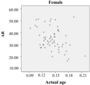
The minimum and maximum difference between the actual age and the estimated age in females was 0.36 and 17.15 years, respectively. The mean AR was calculated to be 0.143±0.023. In 60 female subjects, the regression formula for age estimation in each individual was calculated as follows: Age=-144× AR+55
The correlation coefficient was found to be -0.336 and this correlation was statistically significant (p=0.004) (Diagram 2). The negative correlation coefficient showed that AR decreased by aging. Two weeks after the termination of assessments, 30 radiographs were randomly selected and evaluated again by the same observer. The ICC was calculated to be 0.912, which is close to one and indicates high reproducibility of results.
Discussion
Study of the morphological parameters of teeth on radiographs for age estimation has higher reliability than many other methods [13]. Dimensions of teeth do not change significantly during growth and development and thus, they can
Diagram 2. Distribution of actual age in comparison with AR in females
be easily used for age estimation. The
advantages of radiography further add to the value of this method [17]. Soomer believes that methods in which, the teeth are sectioned or measurements are made on sound teeth are more accurate than other methods [7]. However, the simplicity of radiography and its practicality make this method unique. Several methods have been introduced for age estimation using dental radiographs. Most of these techniques focus on tooth development such as apex closure or tooth formation [17, 18]. A different technique is to estimate AR; this ratio indirectly determines the deposition of secondary dentin. Secondary dentin is not influenced by the environmental factors and therefore, this method is very accurate. Preliminary studies have demonstrated that the amount of secondary dentin has a close association with chronological age and can be indirectly measured using radiography [6]. Brkic et al, in their study concluded that teeth of both jaws are reliable for age estimation; however, the correlation between the maxillary teeth and age is more significant [19]. Moreover, the growth layers in the maxillary teeth are more regular and
0.00 0.10 0.20 0.30 0.40 20.00
40.00 60.00 80.00
male
Actual age
A
R
0.09
Female
A
R
0.12 0.15 0.18 0.21 10.00
20.00 30.00 40.00 50.00 60.00
Actual age
more distinct [20]. Although according to the results of Paewinsky, the mesiodistal width of the maxillary lateral incisor showed a significant association with patient’s age [11], canine tooth is the most suitable tooth for this purpose due to having a large pulp chamber, less wear, and chance of staying longer in the dental arch. Thus, in the current study, maxillary canine teeth were used [15]. Bosman et al. adopted the Kavval’s method for age estimation using panoramic radiographs and concluded that this method was accurate enough for age estimation [12]. Cameriere et al, also used Kavval’s method for age estimation using panoramic radiographs of maxillary canines in their preliminary study and offered a regression equation for age estimation. They reported that the actual age had a significant correlation with AR and another variable (ratio of pulp width to root width) and these two variables were capable of accurately estimating the age in 85% of cases [13]. In our study, the regression equation underesti-mated or overestiunderesti-mated the age in males. In fe-males, this equation was capable of estimating the actual age in 16% of cases. In our study, no varia-ble other than AR was evaluated and this may ex-plain the difference in results.
In the aforementioned study, the effect of some confounders on calculations, such as the size of tooth, the angle between the film and the X ray beam and the innate magnification of radiography, was discussed [13]. These factors were also taken into account in the current study. Moreover, in contrast to previous studies [9-13, 15, 18], digital panoramic radiographs were used to eliminate the processing steps, increase accuracy and save time. A few studies believe that the Kavval method is not suitable [21, 22]; however, studies are ongoing to confirm its accuracy and efficacy.
Cameriere in his study in 2012 on mandibular premolars suggested a regression equation for age estimation from the AR. The results revealed that when higher numbers of teeth were entered into the equation, the difference between the actual and estimated age decreased. He believed that digital periapical radiographs could be more efficient for age estimation due to having higher accuracy and resolution. One major reason for measurement errors on panoramic radiographs is difficulty detecting the reference points on radiographs and subsequently difficult drawing of reference lines for measurements [14]. In the current study,
radiographs had adequate resolution and
calculation of AR was done with high accuracy. In the aforementioned study, the maximum difference between the actual and estimated age was 6.02 years. In our study, this value was 0.36 and 0.11 years for females and males, respectively.
In a similar study by Saxena in 2011, the mean difference between the actual and estimated age in males and females and within the age groups was not significant. In our study, the difference between the actual and estimated age was not significant either. The regression equation formulated in this study estimated age with favorable accuracy; this result is in contrast to the findings of previous studies. In the study by Saxena, more favorable results were obtained by classification of age groups and their separate assessment [15]. In the current study, age classification was not performed and this may explain the difference in results of the two studies. In a study by Singaraju in 2009 a significant difference was not found in the mean age calculated based on AR and the actual age of patients between different age groups [9]. This result is in accord with our findings. . Presence of a good correlation between the actual and estimated age revealed that AR was a suitable index for age estimation in different groups; whereas, in the current study, the correlation between AR and the actual age was not good. In a study by Zaher in 2011, the AR in the maxillary lateral incisors was introduced to be a suitable index for age estimation. In their study, periapical radiographs were used and the maximum difference between the actual and estimated age was found to be 5.08 years [6]. These results are in line with our findings.
In all similar studies, the actual mean age was compared with the estimated mean age and no significant difference was noted. In fact, classification of age groups is responsible for such insignificant differences. Even in our study that age of patients had a wide range in male and female groups, this difference was small.
However, age estimation based on AR had 33.8 years difference with the actual age in some cases; which indicates the inefficacy of this index for age estimation in particular cases. In the studies conducted with the use of logistic regression model, the numerical mean values were compared and matched. This method does not seem to be accurate for age estimation. In our study, the AR showed a weak correlation with actual age of
patients; this indicates that AR cannot be a suitable criterion for age estimation. Therefore, although regression equations are helpful for assessment of the correlation between the mean age and the mean AR, they may be inaccurate for age estimation in particular cases. Factors related to the skeletal morphology highly depend on the ethnicity, race and environment [14] and studies with similar methodology in different populations have yielded different results. Using a higher number of teeth simultaneously for age estimation may yield different results. Moreover, studies on larger sample sizes can better elucidate the value of AR.
Conclusion
In males, the regression equation underestimated or overestimated the actual age. In females, this equation accurately estimated the age in 16% of cases. AR as a single index cannot be used for age estimation of an individual; however it can be used in combination with other indexes for this purpose.
References
1. Willems G. A review of the most commonly used dental age estimation techniques. J Forensic Odontostomatol. 2001 June;19(1):9-17.
2. Shahidi P. Dentist And Law. 1st ed. Tehran: Moalef Co; 1993 [Persian].
3. Kashyap VK, Koteswara Rao NR. A modified Gustafson method of age estimation from teeth. Forensic Sci Int. 1990 Oct;47(3):237-47.
4. Xiaohu X, Philipsen HP, Jablonski NG, Pang KM, Zhu J. Age estimation from the structure of adult human teeth: Review of the literature. Forensic Sci Int. 1992 Apr; 54(1):23-8.
5. Kvaal SI, Kolltveit KM, Thomsen IO, Solheim T. Age estimation of adults from dental radiographs. Forensic Sci Int. 1995 Jul;74(3):175-85.
6. Zaher JF, Fawzy IA, Habib SR, Ali MM. Age estimation from pulp/tooth area ratio in maxillary
incisors among Egyptians using dental
radiographic images. J Forensic Leg Med. 2011 Feb; 18(2):62-5.
7. Soomer H, Ranta H, Lincoln MJ, Penttila A, Leibur E. Reliability and validity of eight dental age estimation methods for adults. J Forensic Sci. 2003 Jan;48(1):149-52.
8. Kvaal S, Solheim T. A non-destructive dental-method for age estimation. J ForensicOdontosto-matol. 1994 June; 12(1):6-11.
9. Singaraju S, Sharada P. Age estimation using pulp/tooth area ratio: A digital image analysis. J
Forensic Dent Sci. 2009May;1(1):37-41.
10. Lamendin H, Baccino E, Humbert JF, Tavernier JC, Nossintchouk RM, Zerilli A. A simple technique for age estimation in adult corpses: the two criteria dental method. J Forensic Sci. 1992 Sept; 37(5):1373-9.
11. Paewinsky E, Pfeiffer H, Brinkmann B. Quantification of secondary dentine formation
from orthopantomograms-a contribution to
forensic age estimation methods in adults. Int J legal Med. 2005 Jan; 119(1):27-30.
12. Bosmans N, Ann P, Aly M, Willems G. The application of Kvaal's dental age calculation technique on panoramic dental radiographs. Forensic Sci Int. 2005 Oct; 153(2-3):208-12. 13. Cameriere R, Ferrante L, Cingolani M. Variations in pulp/tooth area ratio as an indicator of age: A preliminary study. J Forensic Sci. 2004 Mar;49(2):317-9.
14. Cameriere R, De Luca S, Alemán I, Ferrante L, Cingolani M. Age estimation by pulp/tooth ratio in lower premolars by orthopantomography. Forensic Sci Int. 2012 Jan;214(1-3):105-12.
15. Saxena S. Age estimation of indian adults from orthopantomographs. Braz Oral Res. 2011 May-Jun; 25(3):225-9.
16. White SC, pharaoh MJ editors. Oral Radiology: Principles and interpretation. 6th ed. St. Louis: Mosby; 2009.
17. Mirhashemi SAH, Jabbarian R. The use of dental radiography in age determination: Concepts and methods. J Dent Med. 2014 Sum;27 (2):137-43.
18. Cameriere R, Ferrante L, Liversidge HM, Prieto JL, Brkic H. Accuracy of age estimation in children using radiograph of developing teeth. Forensic Sci Int. 2008 Apr;176(2-3):173-7.
19. Brkic H, Milicevic M, Petrovecki M. Age
estimation methods using anthropological
parameters on human teeth–(A0736). Forensic Sci Int. 2006 Oct16;162(1-3):13-6.
20. Fancy S. Preparation of mammalian teeth for age determination by cementum layers: A Review. Wildlife Soc Bull. 1980 Autumn;8(3):242-8. 21. Landa MI, Garamendi PM, Botella MC, Alemán I. Application of the method of Kvaal et al. to digital orthopantomograms. Int J legal Med. 2009 Mar; 123(2):123-8.
22. Meinl A, Tangl S, Pernicka E, Fenes C, Watzek G. On the applicability of secondary dentin formation to radiological age estimation in young adults. J Forensic Sci. 2007 Mar;52(2):438-41.