NEUROIMMUNE FUNCTION IN DIFFERENT MODELS OF STRESS AND WITHDRAWAL
Andrew Parker Kendle
A thesis submitted to the faculty at the University of North Carolina at Chapel Hill in partial fulfillment of the requirements for graduating with Honors in the Department of Biology in the
College of Arts and Sciences
Chapel Hill 2014
Approved by:
ABSTRACT
The neuroimmune system has become a major area of research into the pathology of alcoholism and stress. However, variation in neuro-cytokine responses to stress and withdrawal has impeded progress in defining the precise role the neuroimmune system plays. By measuring cytokine mRNA and protein levels along with obtaining behavioral data, the presented research further examines the function of the neuroimmune system with respect to ethanol exposure. Results broaden the body of literature in which the neuroimmune system has been shown to be altered by stress and withdrawal, importantly in adolescent animals. The increasing variety of methods and rat strains shown to have neuroimmune activation elicited by ethanol may point to a phenomenon that is more translationally applicable and clinically relevant.
INTRODUCTION
The health effects of alcohol abuse and alcoholism have been well documented. Alcohol abuse disorder as defined by the DSM-IV has an estimated 5% prevalence and causes 79,000 deaths annually in the United States (Grant et al. 2004, Bouchery et al. 2011). Additionally, 75% of chronic alcoholics show significant brain damage at autopsy (Vetreno et al. 2011). Disorders related to excessive alcohol use impose staggering socio-economic costs on the United States. The estimated national cost of alcohol abuse combining mortality, disease and injury, property damage, crime and lost productivity was $223.5 billion in 2006 (Bouchery et al. 2011). These statistics place alcohol abuse disorder as one of the most harmful and expensive preventable health disorders in the country.
A major reason for the deficit in clinical treatment options is the incomplete understanding of the mechanisms through which ethanol exerts its pathologic effects on the brain. A mounting body of research implicates the neuroimmune system as an important player in all behavioral stages of addiction (intoxication, withdrawal, craving) as well as the neurotoxic effects of ethanol (Coller et al. 2012, Crews et al. 2011, He et al. 2008, Knapp et al. 2011). More specifically, this response is at least in part the pro-inflammatory action of glial cells in the CNS which may be activated by ethanol via the gut-brain axis or in local brain tissue (Mayfield et al. 2013). Activation of
microglial toll-like receptor (TLR) by lipopolysaccharide or endogenous signals such as high-mobility group box 1 (HMGB1), have been shown to be necessary for this glial
pro-inflammatory response (Fernandez-Lizarbe et al. 2009, Crews et al. 2012, Breese et al. 2013). This TLR signaling cascade culminates in the production of the transcription factor nuclear factor kappa-light-chain-enhancer of activated B cells (NF-κB), which induces production of pro-inflammatory cytokines such as interleukin-1 beta (IL-1β), tumor necrosis factor alpha (TNFα), monocyte chemotactic protein-1 (MCP-1), etc. (Zou et al. 2010). These cytokines have been shown to contribute to neuroinflammatory and behavioral effects in models of alcohol use disorder (Knapp et al. 2011). Some data have shown that cycled ethanol exposure may engender a more robust withdrawal than constant ethanol (Breese et al. 2005, 2011, Overstreet et al. 2002). Also, stress is known to be an important factor in alcoholic relapse by inducing anxiety-like behavior. Stress influences some of the same pro-inflammatory CNS effects as withdrawal, possibly via a corticotrophin releasing factor-dependent pathway (Breese et al. 2005, 2011, Sinha et al. 2007, Knapp et al. 2011).
This paper addresses experiments in which the hypothesis tested was that the
cycled ethanol use and withdrawal combined with stress. Fischer rats were chosen because they exhibit a pronounced cytokine response to stress (Porterfield et al. 2011). Different brain regions were examined for cytokines in regard to their varying levels of involvement in stress and withdrawal (Porterfield et al. 2011, Johnson et al. 2005, O’Connor et al. 2003). Adolescent mice were examined due to the importance of this stage in the development of alcoholism and data reporting differences between adolescent and adult neuroimmune response (Kane et al. 2013).
METHODS
Animals
Young adult male Fischer 344 rats obtained from Harlan Sprague Dawley (Dublin, VA) were 42 days of age and weighed 90-100g upon arrival. Adolescent male Fischer 344 rats
obtained from Harlan Sprague Dawley (Dublin, VA) were 23 days of age and weighed 30g upon arrival. All rats were then group housed and fed RMH3000 rat chow (TestDiets, Richmond, IN) for 2 to 3 days to acclimate them to the new environment (temperatures 70 to 72°F; humidity 40 to 60%; and a 12-h light/12-h dark cycle). Animals were then singly housed for the 1-2 days immediately prior to study initiation.
Diets
Ethanol withdrawal was initiated by switching rats from ED to CD on the morning of the behavioral test.
Stress Protocol
Restraint stress was applied by confining rats for 60 minutes in conical decapicones (Braintree Scientific, Braintree, MA) with rubber bands around the base of the rat’s tail to secure the animal in place. This stress was chosen because it has previously been shown to alter
ethanol- and withdrawal- induced behaviors (Breese et al. 2005, Knapp et al. 2007). After stress, rats were sacrificed by decapitation and tissues collected either immediately or two hours
following the end of stress, as noted for each group.
Behavioral Tests
A social interaction test was utilized to quantify anxiety-like behavior during withdrawal. Rats were placed in an open field (60x60 cm, with 16 10x10 cm squares marked on the floor) 5-6 hours after removal of ethanol. Rat pairs matched by weight and treatment when possible were placed in the arena, and time spent in social interaction was recorded during a 5-minute session, with lower scores taken as anxiety-like behavior (File et al. 1978, Breese et al. 2005). Locomotor activity (measured as count of line crosses) was recorded during the same sessions as a control for sedative or stimulant effects.
Tissue Collection
PCR
Total RNA was extracted from homogenized microdissected brain regions using Trizol. RNA was converted to cDNA by RT-PCR as described elsewhere. SYBR green PCR master mix (Applied Biosystems, Foster City, CA) was used for real time analysis. The cycle time values were normalized with β-actin to ascertain relative differences in mRNA expression between groups. Calculated values were expressed as relative change from controls, which were set as 100%. Relevant primer sequences are listed in Table A1 of the appendix.
ELISA
Brain region samples for ELISA were homogenized in Iscove’s Modified Dulbecco’s Medium (Invitrogen) with a complete protease inhibitor cocktail (Roche Diagnostics,
Indianapolis, IN). Homogenized samples were centrifuged 10 minutes at 12,000g and 4°C. Supernatants were collected and stored at -80°C. ELISA kits for IL-1β and TNFα were
purchased from R&D Systems (Minneapolis, MN) with standard curves diluted down three times further in 1:1 dilutions; for HMGB1 from IBL-International (Hamburg, Germany); and for MCP-1 from BD Biosciences (Franklin Lakes, NJ). Other ELISA procedures were performed
according to manufacturers’ directions. Cytokine levels were corrected by protein concentration using Pierce BCA Protein assay (Thermo Scientific, Rockford, IL).
Statistics
RESULTS
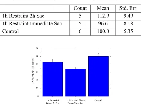
Fischer Young Adults: Cytokines Immediately and 2h Following Stress
The relationship between stress and cytokines was investigated. Cortical mRNA levels of
various cytokines in young adult Fischer rats were determined after 1 hour restraint stress with
immediate and 2 hour sac as described. MCP-1 showed a moderate but not significant decrease
immediately with a return to control after 2 hours, TNFα showed a large decrease immediately
and partially rebounded levels after 2 hours, and IL-1β showed no significant changes at either
time (Tables 1 and 2, Fig. 1). While the MCP-1 levels showed no significant change, the pattern
exhibited was similar to the significant change seen for TNFα.
Table 1. Cortical MCP-1 mRNA following Restraint Stress (% Control)
Count Mean Std. Err.
1h Restraint 2h Sac 5 103.7 13.0
1h Restraint Immediate Sac 5 78.3 5.32
Control 6 100.0 6.70
Table 2. Cortical IL-1β mRNA following Restraint Stress (% Control)
Count Mean Std. Err.
1h Restraint 2h Sac 5 112.9 9.49
1h Restraint Immediate Sac 5 96.6 8.18
Control 6 100.0 5.35
Fig. 1. Cortical TNFα mRNA expression following restraint stress. A significant decrease immediately was followed
by a partial return to control after 2h (ANOVA test p<0.05). Error bars ± 1 standard error.
Expression of IL-1β and TNFα protein were also investigated in the plasma, cortex, and hypothalamus of the same animals. In the plasma, IL-1β exhibited a significant increase at both sac times following restraint stress (Fig. 2a). In the cortex, IL-1β was significantly lowered only at 2 hours following restraint stress (Fig. 2b). In the hypothalamus, IL-1β was significantly higher at both sac times following restraint stress (Fig. 3c). TNFα showed no significant changes (Tables 3, 4 and 5).
Table 3. Plasma TNFα Protein following Restraint Stress (pg/mL)
Count Mean Std. Err.
1h Restraint 2h Sac 6 2.99 0.49
1h Restraint Immediate Sac 7 2.77 0.98
Control 6 1.24 0.70
Table 4. Cortical TNFα Protein following Restraint Stress (pg/mg)
Count Mean Std. Err.
1h Restraint 2h Sac 6 0.39 0.026
1h Restraint Immediate Sac 7 0.39 0.029
Control 6 0.37 0.039
*
* *
*
Table 5. Hypothalamic TNFα Protein following Restraint Stress (pg/mg)
Count Mean Std. Err.
1h Restraint 2h Sac 5 0.019 0.049
1h Restraint Immediate Sac 5 0.101 0.042
Control 6 0.076 0.026
Fischer Adolescents: Cycled Ethanol, Withdrawal and Stress
Further data relating to the combination of withdrawal and stress was collected. Cortical mRNA levels of various cytokines in adolescent Fischer rats were determined after the 4.5%
cycled ethanol treatment followed by 24 hr. withdrawal (WD), 1 hour restraint stress and 2 hour
delayed sac as described. TNFα was elevated with cycled ethanol and lowered with ethanol and
stress, IL-1β was elevated only with cycled ethanol alone, and MCP-1 and HMGB1 showed no
change (Figure 3, Tables 6 and 7). No rats were stressed without being given cycled ethanol.
Figure 3:Cortical Cytokine mRNA with cycled EtOH, WD and restraint stress. a) TNFα was significantly raised
with EtOH alone and significantly lowered with EtOH + stress. b) IL-1β was significantly raised with EtOH alone,
and returned to control with EtOH + stress. Error bars ± 1 standard error.
Table 6. Cortical MCP-1 mRNA following Cycled Ethanol, WD and Restraint Stress (% control)
Count Mean Std. Err. 4.5% Cycled EtOH, No Stress 5 118.3 2.65 4.5% Cycled EtOH, Stress 6 101.1 9.42
Control 5 100.0 8.24
*
Table 7. Cortical HMBG1 mRNA following Cycled Ethanol, WD and Restraint Stress (% control)
Count Mean Std. Err. 4.5% Cycled EtOH, No Stress 5 118.3 3.00 4.5% Cycled EtOH, Stress 6 104.7 6.82
Control 5 100.0 8.51
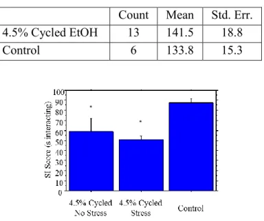
Locomotor and social interaction test scores were collected 5-6 hours into withdrawal (1
day prior to the stress manipulation). No significant difference was observed in locomotor scores
between the cycled ethanol group and the control group (Table 8). Social interaction showed a
significant reduction in interaction time in both the ethanol group and the ethanol with stress
group (Figure 4).
Table 8. Locomotor scores taken before stress protocol.
Count Mean Std. Err. 4.5% Cycled EtOH 13 141.5 18.8
Control 6 133.8 15.3
Figure 4: Social interaction scores. Time spent in social interaction was significantly lowered for both cycled EtOH
and cycled EtOH + restraint stress groups. Error bars ± 1 standard error.
*
DISCUSSION
The results of these experiments add to the growing body of literature pertaining to the interface between stress and withdrawal in chronic ethanol use. The cytokine data elucidates certain nuances in neuroimmune function, such as the stress/withdrawal challenge used in an adolescent rat. These results clearly demonstrate significant anxiety-like behavior paired with neuroimmune change with withdrawal in adolescents. This combination of age and short duration of drinking may be of great interest in the prevention of alcoholism. In addition, the cytokine data for young adult rats provide additional knowledge about the role of stress in Fischer rats over time, though more studies must be done for a clearer picture.
anxiety-like behavior), and plasma. IL-1β was raised at both sac times in plasma, lowered at the 2h sac in cortex, and importantly raised at both sac times in hypothalamus; TNFα showed no significant change. IL-1β has been shown repeatedly to be raised in plasma following stressors (Johnson et al. 2005). The reason for the lack of change in TNFα may be regulation by other cytokines, as it has previously been reported to not be raised unless IL-6 is blocked (Nukin et al. 1998). Data on IL-6 levels in a similar stress and animal model, or perhaps the same procedure done with IL-6 inhibited, would be needed to ascertain the validity of this action in this model. IL-1β protein has been shown to be lowered in cortex and raised in hypothalamus, though only with acute stressors in Sprague-Dawley rats (O’Connor et al. 2003, Johnson et al. 2005,
Porterfield et al. 2011). The use of different rat strains and different stressors in these studies is important as it indicates a response that is relevant to a broad variety of stimuli.
protein activity causes changes that eventually lead to a downregulation of TNFα mRNA by the time data was obtained. Further studies of TNFα protein or mRNA turnover rate must be
performed in order to better understand this result. For the behavioral test, locomotor scores showed no significant difference, meaning that differential locomotor scores did not confound anxiety-like behavior data. Social interaction scores showed significant anxiety-like behavior in the ethanol groups experiencing withdrawal, as seen in literature (Knapp et al. 2011, Breese et al. 2011, Overstreet et al. 2002). Because this study into adolescent Fischer rats was preliminary, there was no control group for stress without ethanol, thus further interpretations of the stress-alone variable are not presented here.
REFERENCES
Bouchery EE, Harwood HJ, Sacks JJ, Simon CJ, Brewer RD, 2011: Economic costs of excessive alcohol consumption in the U.S., 2006. Am J Prev Med 41(5):516-524.
Breese GR, Knapp DJ, Werner DF, Crews FT, Whitman BA, 2013: The cytokine mRNA increase induced by withdrawal from chronic ethanol in the sterile environment of brain is mediated by CRF and HMGB1.Alcohol Clin Exp Res 1-12.
Breese GR, Overstreet DH, Knapp DJ, 2005: Conceptual framework for the etiology of alcoholism: a ‘‘kindling’’/stress hypothesis. Psychopharmacology (Berl) 178 (4), 367–380.
Breese GR, Sinha R, Heilig M, 2011:Chronic alcohol neuroadaptation and stress contribute to susceptibility for alcohol craving and relapse. Pharmacol Ther 129 (2), 149–171.
Coller JK, Hutchinson MR, 2012: Implications of central immune signaling caused by drugs of abuse: mechanisms, mediators and new therapeutic approaches for prediction and treatment of drug
dependence. Pharmacol & Therapeu 134:219-245
Crews FT, Qin L, Sheedy D, Vetreno RP, Zou J, 2012: High mobility group box 1/toll-like receptor danger signaling increases brain neuroimmune activation in alcohol dependence. Biol Psychiatry.
Crews FT, Zou J, Qin L, 2011: Induction of innate immune genes in brain create the neurobiology of addiction. Brain Behav Immun 25:S4–S12.
Dawson DA, Goldstein RB, Grant BF, 2007: Rates and correlates of relapse among individuals in remission from DSM-IV alcohol dependence: a 3-year follow-up. Alcohol Clin Exp Res 31(12)2036-2045.
Fernandez-Lizarbe S, Pascual M, Guerri C, 2009: Critical role of TLR4 response in the activation of microglia induced by ethanol. J Immunol 183(7):4733-44.
File SE, Hyde JR, 1978: Can social interaction be used to measure anxiety? Br J Pharmacol. 62 (1), 19–24.
Grant BF, Dawson DA, Stinson FS, Chou P, Dufour MC, Pickering RP, 2004: The 12-month prevalence and trends in DSM-IV alcohol abuse and dependence: United States, 1991-1992 and 2001-2002. Drug Alcohol Depend 74 (3):223-234.
He J, Crews FT, 2008: IncreasedMCP-1 and microglia in various regions of the human alcoholic brain. Exp Neurol 210:349–358.
Johnson JD, Campisi J, Sharkey CM, Kennedy SL, Nickerson M, Greenwood BN, Fleshner M, 2005: Catecholamines mediate stress-induced increases in peripheral and central inflammatory cytokines.
Neuroscience 135:1295-1307.
Kane CJM, Phelan KD, Douglas JC, Wagoner G, Johnson JW, Xu J, Phelan PS, Drew PD, 2013: Effects of ethanol on immune response in the brain: region-specific changes in adolescent versus adult mice. Alcohol Clin Exp Res. 1-8.
Knapp DJ, Overstreet DH, Angel RA, Navarro M, Breese GR, 2007: The amygdala regulates the antianxiety sensitization effect of flumazenil during repeated chronic ethanol or repeated stress.
Knapp DJ, Whitman BA, Wills TA, Angel RA, Overstreet DH, Criswell HE, Ming Z, Breese GR, 2011: Cytokine involvement in stress may depend on corticotrophin releasing factor to sensitize ethanol withdrawal anxiety. Brain Behav Immun 25:S146–S154.
Mayfield J, Ferguson L, Harris RA, 2013: Neuroimmune signaling: a key component of alcohol abuse. Curr Op Neurobio 23:513-5520.
Nukina H, Sudo N, Komaki G, Yu X, Mine K, Kubo C, 1998: The restraint stress-induced elevation in plasma interleukin-6 negatively regulates the plasma TNF-alpha level. Neuroimmunomodulation
5:323-327.
O’Connor KA, Johnson JD, Hansen MK, Wieseler Frank JL, Marksimova E, Watkins LR, Maier SF, 2003: Peripheral and central proinflammatory cytokine response to a severe acute stressor. Brain Res
991:123-132.
Overstreet DH, Knapp DJ, Breese GR, 2002: Accentuated decrease in social interaction in rats subjected to repeated ethanol withdrawals. Alcohol Clin Exp Res. 26:1259-1268.
Porterfield VM, Zimomra ZR, Caldwell EA, Camp RM, Gabella KM, Johnson JD, 2011: Rat strain differences in restraint stress induced brain cytokines. Neuroscience 188:48-54.
Sinha R, Li CS, 2007: Imaging stress- and cue-induced drug and alcohol craving: association with relapse and clinical implications. Drug Alcohol Rev. 26 (1) 25–31.
Vetreno RP, Hall JM, Savage LM, 2011: Alcohol-related amnesia and dementia: animal models have revealed the contributions of different etiological factors on neuropathology, neurochemical
dysfunction and cognitive impairment. Neurobiol Learn Mem 96(4):596-608.
APPENDICES