A Comparison of Impulse Oscillometry and Spirometry Values
in Patients with Gastroesophageal Reflux Disease
ABSTRACT BACKGROUND
The relationship between gastroesophageal reflux (GERD) and airway dis-eases is still a matter of debate. Oscillometry is an objective, independent tool for the evaluation of airway resistance. The main purpose of this study is to compare spirometry and oscillometry results before and after treatment by a proton pump inhibitor (PPI) in a group of GERD patients who have no respira-tory symptoms.
METHODS
This study was performed on patients with endoscopically diagnosed reflux esophagitis who had no pulmonary symptoms. Patients received omeprazole 40 mg, twice a day for 12 weeks. Spirometry and oscillometry were performed before and after treatment. Impulse oscillometry (IOS) was performed by a force oscillation instrument. We recorded respiratory resistance at 5 Hz (R5) and 20 Hz (R20), resonant frequency (Fres), and distal capacitive reactance (X5) for each patient.
RESULTS
Included were 30 patients (17 males; 13 females) whose mean age was 32 years. According to the Los Angeles Classification, 16 patients had grades B or C esophagitis and 14 had grade A. Although all patients had normal spirometry results, 50% had increased airway resistance according to oscil-lometric findings. After treatment with omeprazole, only 16.3% had abnormal oscillometry results (p=0.004). Spirometry results [forced expiratory volume at the first second (FEV1); forced vital capacity (FVC); FEV1/FVC; and mean forced expiratory flow 25%-75% (FEF 25%-75%)] showed significant further improvement compared to pretreatment normal values (p<0.001 for all). CONCLUSION
Abnormal airway resistance may be present in GERD patients even when there is no obvious respiratory symptom. Oscillometry seems to be more sensitive than spirometry in reporting abnormal pulmonary function in patients with GERD.
KEYWORDS
Oscilometry; Spirometry; Gastroesophageal reflux 1. Department of Pulmonology, School
of Medicine, Ahvaz Jundishapur University of Medical Sciences, Ahvaz, Iran
2. Department of Gastroenterology,
Research Institute for Infectious
Diseases of the Digestive System,
Ahvaz Jundishapur University of Medical Sciences, Ahvaz, Iran
Esmaeil Eidani1, Seyed Jalal Hashemi2*, Hanieh Raji1, Masoumeh Hosaini Askarabadi2
* Corresponding Author: Seyed Jalal Hashemi, MD
Department of Gastroenterology and Hep -atology, Ahvaz Jundishapur University of
Medical Sciences, P.O. Box89, Ahvaz, Iran Telefax: +98 611 2216504
Email: [email protected] Received: 24 Aug. 2012
Accepted: 10 Dec. 2012
Please cite this paper as:
Eidani E, Hashemi SJ, Raji H, Hosaini Askarabadi M. Comparison of Impulse Oscillometry and Spirometry Values in Patients with Gastroesophageal Reflux Disease. Middle East J Dig Dis 2013;5:22-8.
INTRODUCTION
Gastroesophageal reflux disease (GERD) is a common gastrointestinal (GI) disease that affects
about one third of the Western world.1,2 The reported
prevalence of GERD in Iran ranges from 15.4%
to 39.7%.3-6 The most most common endoscopic
diagnosis in a referral center of Iran is endoscopic esophagitis.7 GERD can lead to several increas
-ingly recognized ‘atypical’ or ‘extra-esophageal’
manifestations such as laryngitis, dental ero-sions, hoarseness, non-cardiac chest pain, chronic cough, asthma, recurrent pneumonia, subglottic stenosis, and laryngeal cancer.8,9 The prevalence
of GERD in asthmatic patients is reported to be
as high as 70.6% to 80% in the Western popula
-tion.9-11 However, it is difficult to establish a causal relationship.12,13 Both asthma and GERD are com
-mon worldwide diseases. In Iran, the prevalence of wheezing varies throughout different parts
of the country and ranges from 7.6% to 40.2%,
whereas the prevalence of asthma is estimated to be 3.2%.14,15
The efficacy of proton pump inhibitors (PPIs) on asthma outcomes in patients with GERD is
controversial.16-19 Some investigators have
pro-posed that anti-reflux surgery may be more effec
-tive than medical therapy.20 Microaspiration of
gastric acid and acid induced vagal nerve stimu-lation are considered to be important underlying mechanisms.21 Most patients with GERD may
have some degree of respiratory abnormality. In order to evaluate these abnormalities more sensi-tive tools that are not dependent on patient effort are needed. Recently, the impulse oscillation
sys-tem (IOS) has been introduced as a new syssys-tem for
the forced oscillation test. It is a noninvasive test, not dependent on the patient’s effort and measures airway resistance and compliance using electrical impulses.22 It has been shown that the accuracy
of IOS is comparable to plethysmography.23
Re-sults may be abnormal in patients with increased airway resistance despite normal spirometry test results.24 The present study aims to assess airway
abnormalities in GERD patients before and after
anti-reflux therapy by using both the oscillometry
and spirometry tests.
MATERIALS AND METHODS
Between November 2010 and February 2011
we selected cases from among patients who at-tended the Gastrointestinal Clinic at Ahwaz Imam
Khomeini Hospital and had complaints of reflux
symptoms. The diagnosis of GERD was estab
-lished by endoscopy. Patients who smoked in ad-dition to those with known respiratory diseases, ischemic heart disease, heart failure, liver
dis-orders, or malignancies were excluded from the study. Also excluded were pregnant or lactating
females and those who refused to have an upper GI endoscopy performed.
All enrolled patients underwent an upper GI
en-doscopy conducted by an expert gastroenterologist using Olympus 160 equipment. Reflux esophagitis was defined as the presence of a mucosal break at the distal part of the esophagus and classified
according to the Los Angeles Classification Sys
-tem.25 Excluded were patients with normal endos
-copy results or those with any other endoscopic
diagnosis except for reflux esophagitis.
All patients with reflux esophagitis underwent
IOS testing by a force oscillation instrument (En
-rich Jaeger, Hoechberg, Germany). For the IOS
test, patients were instructed to remain in an up-right position, with their heads in neutral
align-ment or mildly extended. Tight clothing or belts
were removed prior to the IOS. Patients were asked to hold the oral plastic piece of the oscillom-eter tightly between their teeth, above the tongue, with their lips and nose completely closed. Short-time electrical impulses within a power range of
0-100 Hz at a duration of 45 ms and intervals of
0.2 s generated by a loudspeaker in the patients’ mouths were used.
Total impedance of the respiratory system
de-termines the magnitude and phases of flow oscilla
-tion that result from pressure fluctua-tion. Accord
-ing to the relationship between flow and pressure,
“respiratory impedance” can be subdivided into “resistance and reactance” where “resistance”
rep-resents the total effect of the lung and chest wall and “reactance’ shows the net effect of two
op-posite forces (elastic and inertial). We measured
respiratory impedance for 30 s during tidal breath-ing. Respiratory resistance was measured at a low
frequency (5Hz=R5) for total respiratory resis
-tance and at a high frequency (20Hz or R20) for
proximal respiratory resistance. Respiratory reac
-tance was measured at the 5 Hz frequency (Xr5). Resonant frequency (Fres) was determined as the
oscillatory frequency at which the respiratory re
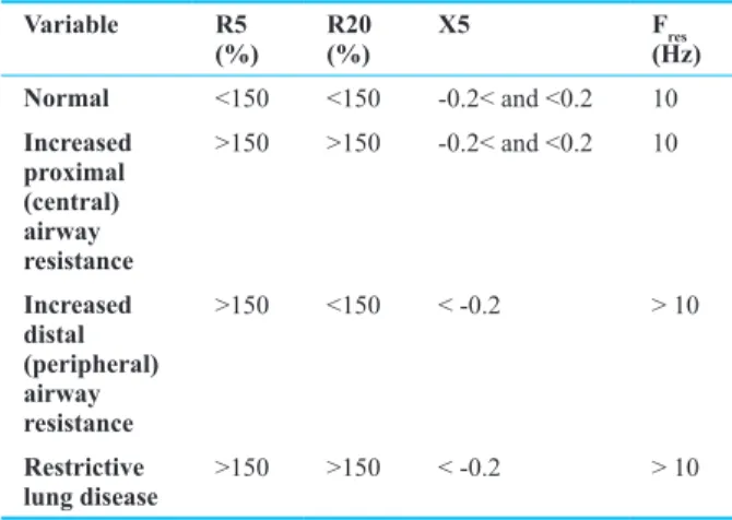
-actance was zero. The system’s software calcu-lated all values. After comparing R5 and R20 to normal values, they were reported as the percent of predicted value. Oscillometry parameters are interpreted according to Table 1.
Spirometry was performed at the beginning of the study for each patient in order to measure
forced vital capacity (FVC), forced expiratory vol
-ume at the first second (FEV1), forced expiratory
flow 25%-75% (FEF 25%-75%), and peak expira
-tory flow rate (PEFR). FEV1 >80% of predicted value, FVC >80% of predicted value, FEV1/FVC >70%, an FEF 25%-75% value that was >80%, and a PEFR >80% were all considered normal.
After baseline IOS and spirometry, patients were
given omeprazole (40 mg, Abidi Co., Ltd., Tehran,
Iran) twice a day for 12 weeks. Patients were fol
-lowed at the clinic every four weeks. Patient com-pliance to therapeutic treatment was monitored by telephone contact. Spirometry and oscillometry were performed again at the end of treatment.
The Research Ethics Committee of Ah
-waz Jundishapour University of Medical
Sci-ences approved this study on 09/23/2010 with
the registration number 3135 and IRCT code of
201102035749.
Statistical Analysis
Data are presented as mean±standard deviation (SD) or percentage as appropriate. Chi-square and Fisher’s ex -act tests were used for comparison of dichotomous data. Assessment of mean between both groups was performed using the paired sample independent t-test. A p-value less
than 0.05 was considered significant.
RESULTS
There were 13 females (43%) and 17 males (57%) in this study. Patients’ mean age was 32±2 years (range: 18-47). There were 14 (46.6%) patients with grade A esopha
-gitis, 15 (50%) had grade B esophagitis and one (3.3%)
had grade C esophagitis.
Prior to treatment, patients’ mean percent
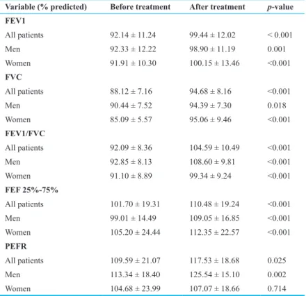
pre-dicted spirometry measurements were as follows: FEV1 (92.14±11.24); FVC (88.12±7.16); FEV1/FVC (92.09±8.36); FEF 25%-75% (92.09±8.36); and PEFR (109.59±21.07), all of which were within normal limits (Table 1).Oscillometry results at baseline indicated that 50% of patients had R5 levels that were >150% of pre
-dicted, of whom 65% of these were male. Additionally, there were 54% of patients who had R20 levels >150% of predicted, of whom 71% were male. All X5 values ranged from -0.2 and 0.0 KPa/L/s (Table 2). Based on these findings, approximately 50% of patients demon
-strated increased proximal (central) airway resistance.
There was no restrictive pattern observed.
All patients completed the treatment period with no
significant complications reported. Spirometry mea -surements improved following 12 weeks of treatment
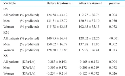
by omeprazole (Table 2). The oscillometry test results showed the following: R5 >150% in 17% of patients and normal R20 and X5 levels in all patients. Furthermore, a significant improvement in mean R5, R20, and X5 values were observed (Table 3). Treatment by omeprazole did not significantly affect PEFR and R5 values in females, nor did it affect X5 values in males. The level of improve
-Table 1: Interpretation of oscillometry test results. Variable R5
(%) R20 (%) X5 F(Hz)res Normal <150 <150 -0.2< and <0.2 10
Increased proximal (central) airway resistance
>150 >150 -0.2< and <0.2 10
Increased distal (peripheral) airway resistance
>150 <150 < -0.2 > 10
Restrictive
ment in spirometric indices and oscillometric indices did
not significantly differ in patients with GERD grade A compared to those with GERD grades B and C as seen in Figure 1: FEV1 (p=0.9); FVC (p=0.46); FEV1/FVC (p=0.57); FEEF 25%-75% (p=0.15); FEER (p=0.10); R5 (p=0.89); R20 (p=0.16); and R5 (p=0.79).
DISCUSSION
Because the oscillometry test does not take long to perform and is not dependent on patient effort, it may be
of more benefit than spirometry, particularly for children
and the elderly. In the present study we have used both
oscillometry and spirometry to evaluate GERD patients
who had no respiratory symptoms. We observed that all
patients had normal respiratory values. However the os
-cillometry findings revealed that airway resistance was high at 5 Hz and 20 Hz frequencies (R5 and R20) in at
least 50% of patients. According to our results patients
with GERD, even in the absence of respiratory symp -toms, might have increased airway resistance. Several studies reported the superiority of IOS for detecting
ab-normal airway resistance. Evans compared spirometry
and IOS parameters in normal volunteers at rest and after
hyperventilation with cold air and an exercise challenge. He observed that oscillometry test results were more sen -sitive than spirometry in the detection of post-challenge increased airway resistance.24,26,27 In an interesting study by Bidad et al., IOS was more sensitive than spirometry in diagnosing asthma among pregnant women.28 A study by Kanda compared IOS and spirometry in a group of
pa-tients with COPD or asthma and normal individuals who
were non-smokers. In that study IOS was more sensitive than spirometry for detecting abnormal airway resis-tance.24 It has been reported that IOS has increased
sen-sitivity in detecting certain occupational induced airway hypersensitivities.29 Thus, it is reasonable to presume that
patients with GERD have subtle airway hyper-respon -siveness, of which a minority of them progress to asthma.
We evaluated the effect of treatment with high dose omeprazole on the objective parameters. After treatment,
16.3% of patients still had abnormal oscillometry results. However at the end of treatment all patients had normal
Table 2: Comparison of spirometry test results before and after treatment. Variable (% predicted) Before treatment After treatment p-value FEV1
All patients 92.14 ± 11.24 99.44 ± 12.02 < 0.001
Men 92.33 ± 12.22 98.90 ± 11.19 0.001
Women 91.91 ± 10.30 100.15 ± 13.46 <0.001
FVC
All patients 88.12 ± 7.16 94.68 ± 8.16 <0.001
Men 90.44 ± 7.52 94.39 ± 7.30 0.018
Women 85.09 ± 5.57 95.06 ± 9.46 <0.001
FEV1/FVC
All patients 92.09 ± 8.36 104.59 ± 10.49 <0.001
Men 92.85 ± 8.13 108.60 ± 9.81 <0.001
Women 91.10 ± 8.89 99.34 ± 9.24 <0.001
FEF 25%-75%
All patients 101.70 ± 19.31 110.48 ± 19.24 <0.001 Men 99.01 ± 14.49 109.05 ± 16.85 <0.001 Women 105.20 ± 24.44 112.35 ± 22.57 <0.001
PEFR
All patients 109.59 ± 21.07 117.53 ± 18.68 0.025
Men 113.34 ± 18.40 125.54 ± 15.10 0.002
Women 104.68 ± 23.99 107.07 ± 18.66 0.714
spirometry results. Although oscillometry was more sen-sitive in detecting increased airway resistance, spiromet-ric indices that were already within normal limits at the beginning of the study improved by the end of treatment.
Oscillometry measurements showed a significant im -provement in airway resistance as well.
Numerous clinical trials have studied the correlation
between GERD and asthma. Although they have evaluat
-ed the role that GERD treatment plays in r-educing asthma
symptoms, this treatment remains debatable.30 Numerous
reviewed articles have used spirometry to assess pulmo-nary function.
There are several theories regarding the mechanism
of respiratory hypersensitivity in the setting of GERD,
which include microaspiration of gastric secretions,31 a
vagally mediated neurologic reflex-induced broncho -spasm,32 a sensitized vagal neural system-induced airway
hyper-responsiveness.12,33 The last issue has been
sup-ported by observations of the overlap with GERD and irritable bowel syndrome (IBS), which may show a com -mon underlying mechanism.34
The first limitation of the current study is that it is an
open-label trial. The lack of a control group may lead to
some difficulties in interpreting the results. Additionally,
Table 3: Comparison of oscillometry test results before and after treatment. Variable Before treatment After treatment p-value R5
All patients (% predicted) 124.58 ± 43.12 112.77 ± 36.76 0.004
Men (% predicted) 131.31 ± 42.79 120.51 ± 37.10 0.030
Women (% predicted) 115.78 ± 43.65 102.65 ± 35.15 0.072
R20
All patients (% predicted) 140.95 ± 26.47 128.02 ± 22.26 <0.001
Men (% predicted) 150.62 ± 16.77 137.78 ± 11.86 0.002
Women (% predicted) 128.30 ± 31.83 115.25 ± 26.41 0.013
X5
All patients (KPa/L/s) -0.283 ± 0.193 -0.168 ± 0.173 0.004
Men (KPa/L/s) -0.305 ± 0.172 -0.201 ± 0.219 0.072 Women (KPa/L/s) -0.254 ± 0.214 -0.125 ± 0.072 0.026
Fig. 1: Comparisons of differences in spiromeric and oscilometric values before and after treatment in patients with GERD A and GERD B.
the final sample size was small.
In conclusion, there appears to be mild increased
air-way resistance in some patients with GERD even in the
absence of respiratory symptoms. Oscillometry may be
more sensitive than spirometry in the identification of these subtle abnormalities. However to establish the ef
-ficacy of GERD treatment on pulmonary functions, it is
necessary to conduct additional randomized, controlled studies with larger populations.
ACKNOWLEDGMENT
We would like to express gratitude to Dr. Masoumeh Hossaini, resident of internal medicine, who made this
study possible by mean of usage of data and results of
her final thesis with the registration number of 261/d in
Ahwaz Joundishapour University of Medical Sciences.
CONFLICT OF INTEREST
The authors declare no conflict of interest related to
this work.
REFERENCES
1. Locke GR TN, Fett SL, Zinsmeister AR, Melton LJ 3rd. Prevalence and clinical spectrum of gastroesophageal re-flux: a population-based study in Olmsted County, Minne-sota. Gastroenterology 1997;112:1448 -56.
2. Howard PJ, Heading RC. Epidemiology of gastroesopha-geal reflux disease. World J Surg 1992;16:288 -93. 3. Ehsani MJ, Maleki I, Mohammadzadeh F, Mashayekh A.
Epidemiology of gastroesophageal reflux disease in Teh-ran, Iran. J Gastroenterol Hepatol 2007;22:1419-22. 4. Saberi-Firoozi M, Khademolhosseini F, Yousefi M,
Mehra-bani D, Zare N, Heydari ST. Risk factors of gastroesopha-geal reflux disease in Shiraz, southern Iran. World J Gas-troenterol 2007;13:5486-91.
5. Mostaghni A, Mehrabani D, Khademolhosseini F, Masoumi SJ, Moradi F, Zare N, et al. Prevalence and risk factors of gastroesophageal reflux disease in Qashqai migrating no-mads, southern Iran. World J Gastroenterol 2009;15:961-5. 6. Delavari A, Birjandi F, Elahi E, Saberifiroozi M. The Prev-alence of Gastroesophageal Reflux Disease (GERD) in the Islamic Republic of Iran: A Systematic Review. Middle East J Dig Dis 2012;4:5-15.
7. Ghajarie Sepanlou S, Abdollahzadeh N, Noori F, Malekza-dehF, Malekzadeh R. Time Trends of Gastro-esophageal Reflux Disease (GERD) and Peptic Ulcer Disease (PUD) in Iran. Middle East J Dig Dis 2010;2:78-83.
8. Shaker R, Castell DO, Schoenfeld PS, Spechler SJ. Night-time heartburn is an under-appreciated clinical problem that
impacts sleep and daytime function: the results of a Gallup survey conducted on behalf of the American Gastroentero-logical Association. Am J Gastroenterol 2003;98:1487-93. 9. Bisaccioni C, Aun MV, Cajuela E, Kalil J, Agondi RC,
Giavina-Bianchi P. Comorbidities in severe asthma: fre-quency of rhinitis, nasal polyposis, gastroesophageal reflux disease, vocal cord dysfunction and bronchiectasis. Clinics (Sao Paulo) 2009;64:769-73.
10. Sontag SJ, O’Connell S, Khandelwal S, Miller T, Nem-chausky B, Schnell TG, et al. Most asthmatics have gastro-esophageal reflux with or without bronchodilator therapy. Gastroenterology 1990;99:613-20.
11. Goldman J, Bennett JR. Gastro-oesophageal reflux and re-spiratory disorders in adults. Lancet 1988;2:493-5. 12. Astarita C, Gargano D, Cutajar M, Napolitano A, Manguso
F, Abbate GF. Gastroesophageal reflux disease and asthma: an intriguing dilemma. Allergy 2000;55 Suppl 61:52-5. 13. Boskabady MH, Kolahdoz GH. Prevalence of asthma
symptoms among the adult population in the city of Mash-had (north-east of Iran). Respirology 2002;7:267-72. 14. Zobeiri M. Prevalence, risk factors and severity of
asth-ma symptoms in children of Kerasth-manshah, IRAN: ISAAC phase I, II. Acta Med Iran 2011;49:184-8.
15. Shakurnia AH, Assar S, Afra M, Latifi M. Prevalence of asthma among schoolchildren in Ahvaz, Islamic Republic of Iran. East Mediterr Health J 2010;16:651-6.
16. Harding SM, Richter JE, Guzzo MR, Schan CA, Alexan-der RW, Bradley LA. Asthma and gastroesophageal reflux: acid suppressive therapy improves asthma outcome. Am J Med 1996;100:395-405.
17. Katz PO CD, Chen Y, Andersson T, Sostek MB. Intragas-tric. acid suppression and pharmacokinetics of twice-daily esomeprazolee:a randomized, three-way crossover study. Aliment Pharmacol Ther 2004;20:399-406.
18. Richter JE. Gastroesophageal reflux disease and asthma: the two are directly related. Am J Med 2000;108 Suppl 4a:153S-8S.
19. Matyasova Z, Novotna B, Matulova M, Dolina J, Kroupa R, Lanikova Z, et al. The relation of GERD, bronchial asthma and the upper respiratory tract. Vnitr Lek 2005;51:1341-50. 20. Bowrey DJ, Peters JH, DeMeester TR. Gastroesophageal
re-flux disease in asthma: effects of medical and surgical antire-flux therapy on asthma control. Ann Surg 2000;231:161-72. 21. Canning BJ, Mazzone SB. Reflex mechanisms in gastro-esophageal reflux disease and asthma. Am J Med 2003;115 Suppl 3A:45S-8S.
22. Frei J, Jutla J, Kramer G, Hatzakis GE, Ducharme FM, Da-vis GM. Impulse oscillometry: reference values in children 100 to 150 cm in height and 3 to 10 years of age. Chest 2005;128:1266-73.
23. Hellinckx J, Cauberghs M, De Boeck K, Demedts M. Evaluation of impulse oscillation system: comparison with forced oscillation technique and body plethysmography. Eur Respir J 2001;18:564-70.
24. Kanda S, Fujimoto K, Komatsu Y, Yasuo M, Hanaoka M, Kubo K. Evaluation of respiratory impedance in asthma and COPD by an impulse oscillation system. Intern Med
49:23-30.
25. Lundell LR, Dent J, Bennett JR, Blum AL, Armstrong D, Galmiche JP, et al. Endoscopic assessment of oesophagitis: clinical and functional correlates and further validation of the Los Angeles classification. Gut 1999;45:172-80. 26. Evans TM, Rundell KW, Beck KC, Levine AM, Baumann
JM. Impulse oscillometry is sensitive to bronchoconstric-tion after eucapnic voluntary hyperventilabronchoconstric-tion or exercise. J Asthma 2006;43:49-55.
27. Evans TM, Rundell KW, Beck KC, Levine AM, Baumann JM. Airway narrowing measured by spirometry and im-pulse oscillometry following room temperature and cold temperature exercise. Chest 2005;128:2412-9.
28. Bidad K, Heidarnazhad H, Kazemnejad A, Pourpak Z. Im-pulse oscillometry in comparison to spirometry in pregnant asthmatic females. Eur Respir J 2008;32:1673-5.
29. Gube M, Brand P, Conventz A, Ebel J, Goeen T, Holzinger K, et al. Spirometry, impulse oscillometry and capnovolu-metry in welders and healthy male subjects. Respir Med 2009;103:1350-7.
30. Kiljander TO, Junghard O, Beckman O, Lind T. Effect of esomeprazolee 40 mg once or twice daily on asthma: a ran-domized, placebo-controlled study. Am J Respir Crit Care Med 2010;181:1042-8.
31. Boyle JT, Tuchman DN, Altschuler SM, Nixon TE, Pack AI, Cohen S. Mechanisms for the association of gastro-oesophageal relux and bronchospasm. Am Rev Respir Dis 1985;131:S16-20.
32. Mansfield LE, Stein MR. Gastroesophageal relux and asth-ma: a possible relex mechanism. Ann Allergy 1978;41:224-6. 33. Harding SM, Schan CA, Guzzo MR, Alexander RW,
Bradley LA, Richter JE. Gastroesophageal relux-induced bronchoconstriction. Is microaspiration a factor? Chest 1995;108:1220-7.
34. Yarandi SS, Nasseri-Moghaddam S, Mostajabi P, Malekza-deh R. Overlapping gastroesophageal reflux disease and irritable bowel syndrome: increased dysfunctional symp-toms. World J Gastroenterol 2010;16:1232-8.