552
|
wileyonlinelibrary.com/journal/cam4 Cancer Medicine. 2020;9:552–561.O R I G I N A L R E S E A R C H
High pretreatment plasma Epstein‐Barr virus (EBV) DNA level
is a poor prognostic marker in HIV‐associated, EBV‐negative
diffuse large B‐cell lymphoma in Malawi
Nathan D. Montgomery
1,2,3|
Cara Randall
1,3|
Matthew Painschab
2,3,4|
Ryan Seguin
3|
Bongani Kaimila
3|
Edwards Kasonkanji
3|
Takondwa Zuze
3|
Robert Krysiak
3|
Marcia K. Sanders
2|
Avian Elliott
6|
Melissa B. Miller
1|
Coxcilly Kampani
3|
Fred Chimzimu
3|
Maurice Mulenga
3|
Blossom Damania
2,5|
Tamiwe Tomoka
3|
Yuri Fedoriw
1,2,3|
Dirk P. Dittmer
2,5|
Satish Gopal
2,3,4This is an open access article under the terms of the Creative Commons Attribution License, which permits use, distribution and reproduction in any medium, provided the original work is properly cited.
© 2019 The Authors. Cancer Medicine published by John Wiley & Sons Ltd.
1Department of Pathology & Laboratory Medicine, University of North Carolina, Chapel Hill, NC, USA
2Lineberger Comprehensive Cancer Center, University of North Carolina, Chapel Hill, NC, USA
3UNC Project‐Malawi, Lilongwe, Malawi
4Department of Medicine, Division of Hematology & Oncology, University of North Carolina, Chapel Hill, NC, USA 5Department of Microbiology &
Immunology, University of North Carolina, Chapel Hill, NC, USA
6UNC Healthcare, Chapel Hill, NC, USA
Correspondence
Nathan D. Montgomery, Department of Pathology & Laboratory Medicine, The University of North Carolina at Chapel Hill, CB#7525, Chapel Hill, NC 27599-7525, USA. Email: [email protected]
Funding information
AIDS Malignancy Consortium; Center for AIDS Research, University of North Carolina at Chapel Hill; National Institutes of Health, Grant/Award Number: CA019014, CA190152 and CA210285
Abstract
Plasma Epstein‐Barr virus (EBV) DNA measurement has established prognostic util-ity in EBV‐driven lymphomas, where it serves as a circulating tumor DNA marker. The value of plasma EBV measurement may be amplified in sub‐Saharan Africa (SSA), where advanced imaging and molecular technologies for risk stratification are not typically available. However, its utility in diffuse large B‐cell lymphoma (DLBCL) is less certain, given that only a subset of DLBCLs are EBV‐positive. To explore this possibility, we measured plasma EBV DNA at diagnosis in a cohort of patients with DLBCL in Malawi. High plasma EBV DNA at diagnosis (≥3.0 log10 copies/mL) was
associated with decreased overall survival (OS) (P = .048). When stratified by HIV status, the prognostic utility of baseline plasma EBV DNA level was restricted to HIV‐ positive patients. Unexpectedly, most HIV‐positive patients with high plasma EBV DNA at diagnosis had EBV‐negative lymphomas, as confirmed by multiple methods. Even in these HIV‐positive patients with EBV‐negative DLBCL, high plasma EBV DNA remained associated with shorter OS (P = .014). These results suggest that EBV reactivation in nontumor cells is a poor prognostic finding even in HIV‐positive pa-tients with convincingly EBV‐negative DLBCL, extending the potential utility of EBV measurement as a valuable and implementable prognostic marker in SSA.
K E Y W O R D S
diffuse large B‐cell lymphoma, Epstein‐Barr virus, HIV, prognosis, sub‐Saharan Africa
1
|
INTRODUCTION
Epstein‐Barr virus (EBV) is an oncogenic herpesvirus implicated in many lymphomas, as well as some solid
tumors.1 For a subset of lymphomas, such as endemic
forms of Burkitt lymphoma, EBV is present in the tumor cells of nearly all cases.2 However, for other lymphomas,
EBV‐negative and EBV‐positive forms of disease are rec-ognized.3,4 The likelihood that a patient's DLBCL will be
EBV‐associated is primarily related to underlying immune status, with human immunodeficiency virus (HIV) infec-tion, iatrogenic immunosuppression, and advanced age all increasing the likelihood of EBV positivity.5
DLBCL is the most common subtype of lymphoma worldwide, and in sub‐Saharan Africa (SSA), the incidence of this and other aggressive B‐cell lymphomas is increas-ing, primarily due to HIV and population aging.6-9 Prior
studies have suggested EBV‐positivity in approximately 80%‐90% of cases of HIV‐associated DLBCL with immu-noblastic cytology and approximately 30% of cases with centroblastic cytology.5,10 By comparison, less than 10% of
DLBCL arising in HIV‐negative individuals is associated with EBV.5
Accurate risk stratification of DLBCL remains chal-lenging in low‐income countries, as there is generally no access to positron emission tomography (PET) scans, cell‐ of‐origin subtyping assays, or prognostically informative cytogenetic or immunophenotypic studies. While often under‐appreciated, risk stratification is critically important in these settings. Intensive chemotherapy is often associ-ated with treatment‐relassoci-ated morbidity and mortality due to poor supportive care, conferring substantial risks to pa-tients that may be more pronounced than in high‐income countries.
Plasma EBV measurement is a clinically valuable and highly implementable biomarker for many EBV‐associ-ated malignancies.11-13 We and others have suggested that
plasma EBV measurement may be particularly informative in SSA and other low‐income settings, where EBV‐associ-ated lymphomas are common.14-16 This hypothesis has
gen-erally been confirmed for lymphomas that are invariably EBV‐positive, like endemic Burkitt lymphoma and HIV‐as-sociated classic Hodgkin lymphoma.14,15,17,18 In such cases,
plasma EBV functions as a sensitive circulating tumor DNA (ctDNA) marker, with both prognostic and monitoring util-ity. Conversely, the value of plasma EBV measurement for DLBCL, which is EBV‐associated in a minority of cases, is less clear. We therefore sought to correlate pretreatment plasma EBV level with survival in a cohort of patients with DLBCL in Malawi.
2
|
METHODS
2.1
|
Study participants
All patients included in this study were enrolled in the Kamuzu Central Hospital (KCH) Lymphoma Study. Details of this study have been described previously19,20; but briefly,
the KCH Lymphoma Study is an ongoing prospective ob-servational cohort based at a national teaching hospital in
Lilongwe, Malawi. KCH is one of two referral centers for cancer care in Malawi, with a catchment area of approxi-mately 9 million people.
For this analysis, all patients were adults (≥18 years) diagnosed with DLBCL between June 1, 2013 and May 31, 2016. All DLBCL diagnoses were confirmed by tissue bi-opsy, supported by manual immunohistochemistry (IHC) and a weekly clinicopathologic teleconference attended by pathologists and oncologists in the United States and Malawi.20 Subsequently, tissue blocks were sent to the
University of North Carolina (UNC) at Chapel Hill for diagnostic confirmation and additional ancillary studies, as described previously and below.20 In addition to a
con-firmed diagnosis of DLBCL, the only other inclusion crite-rion for this study was availability of a pretreatment plasma EBV level (see below).
First‐line chemotherapy for patients with DLBCL in this cohort was CHOP (cyclophosphamide, doxorubicin, vincris-tine, prednisone), along with concurrent antiretroviral ther-apy in HIV‐positive patients. All participants were followed until death, or administrative censoring on November 1, 2018, with none lost to follow‐up.
2.2
|
Plasma EBV measurement
Plasma EBV measurements were performed in the UNC Vironomics Core Facility, as previously described.14 Briefly,
anti‐coagulated plasma was collected prior to initiation of cytotoxic chemotherapy and then stored at −80°C, prior to shipment to UNC. Quantitative plasma EBV measurement was performed using a quantitative real‐time polymerase chain reaction (qPCR) assay with primer pairs designed to amplify a conserved region of the EBV EBNA3C gene, cor-responding to positions 88933‐89033 of the EBVI reference genome and positions 89374‐89836 of the EBVII reference genome. The linear detection range for this assay is 2.0‐8.0 log10 copies/mL, with values <2.0 log10 copies/mL reported
as “not detected”.
2.3
|
Tumor EBV staining
50% positive cells (patient 25 in Table 3). In all cases, results were compared to appropriate controls, including confirmation of adequate RNA preservation for interpreta-tion of negative EBER ISH (Epstein‐Barr encoded RNA in situ hybridization) stains.
2.4
|
Tumor EBV polymerase chain reaction
DNA was extracted on a Maxwell® 16 MDx instrument (Promega) using 5 × 10 μm scrolls prepared from the diag-nostic formalin‐fixed, paraffin embedded tissue block. Then, EBV was detected by a qPCR assay targeting the BamH1W segment of EBV, as previously described.21 Cases were
con-sidered positive if the fluorescence signal crossed the critical threshold in fewer than 40 cycles.
2.5
|
Statistical analysis
Patient clinical characteristics were compared by Mann‐ Whitney U‐Test (for continuous data) or by Fisher Exact Test (categorical data). Survival curves were compared and hazard ratios were calculated by log rank test. All statistical analyses were performed using GraphPad Prism 8.
2.6
|
Informed consent and IRB approval
Informed consent was obtained in English or Chichewa, the two national languages in Malawi. This study was approved by the UNC Institutional Review Board and the Malawi National Health Sciences Research Committee and is regis-tered with clinicaltrials.gov under NCT02835911.
3
|
RESULTS
3.1
|
Patients
A pretreatment plasma EBV measurement was available in 44 patients diagnosed with DLBCL during the study period,
including 25 HIV‐positive and 19 HIV‐negative individu-als. Clinical characteristics of these patients, stratified by HIV status, are shown in Table 1. As in previous descrip-tions of this cohort,19,22 HIV‐positive patients were younger
than their HIV‐negative counterparts (45.4 vs 56.2 years,
P = .03), with similar male predominance in both groups
(19:6 vs 12:7, P = .51).
3.2
|
Pretreatment plasma EBV
measurements and survival
Pretreatment plasma EBV DNA level was also similar be-tween HIV‐positive and HIV‐negative patients (median 2.81 log10 copies/mL vs < 2.0, P = .22, Table 1). In addition, a
similar proportion of HIV‐positive and HIV‐negative pa-tients had negative pretreatment plasma EBV DNA studies, falling below the assay's validated limit of detection of 2.0 log10 copies/mL (10/25 vs 12/19, P = .22, Table 1). Amongst
those patients with a positive plasma EBV result, the median pretreatment plasma EBV DNA level was similar in HIV‐ positive and HIV‐negative patients (3.91 vs 3.94, P = .70, data not shown). Finally, there was also no difference in the fraction of HIV‐positive vs HIV‐negative patients with a “high” pretreatment plasma EBV DNA level, defined here as ≥3.0 log10 copies/mL (10/25 vs 6/19, P = .75). This “high”
threshold was chosen to define a cutpoint robustly above the limit of detection and above the level typically seen in cancer patients with non‐EBV‐associated tumors.23
Next, we sought to determine whether pretreatment EBV DNA level was correlated with outcomes in Malawian pa-tients with DLBCL. First, we evaluated all study participants, irrespective of HIV status. In the full cohort, high pretreat-ment plasma EBV DNA was correlated with a significant decrease in overall survival (OS) (P = .048 and hazard ratio = 2.1, 95% CI = 0.9‐4.8) (Figure 1A).
In order to determine whether the survival disadvantage of a high pretreatment plasma EBV level might be related to HIV status, HIV‐negative and HIV‐positive patients were
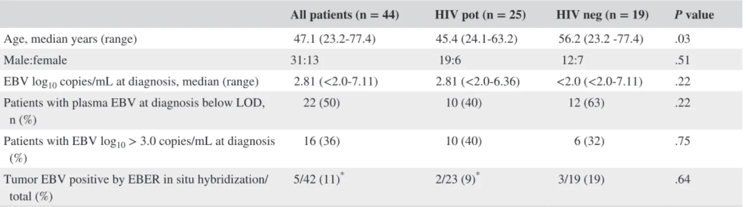
TABLE 1 Clinical characteristics and pretreatment plasma EBV DNA level of all DLBCL patients
All patients (n = 44) HIV pot (n = 25) HIV neg (n = 19) P value
Age, median years (range) 47.1 (23.2‐77.4) 45.4 (24.1‐63.2) 56.2 (23.2 ‐77.4) .03
Male:female 31:13 19:6 12:7 .51
EBV log10 copies/mL at diagnosis, median (range) 2.81 (<2.0‐7.11) 2.81 (<2.0‐6.36) <2.0 (<2.0‐7.11) .22 Patients with plasma EBV at diagnosis below LOD,
n (%) 22 (50) 10 (40) 12 (63) .22
Patients with EBV log10 > 3.0 copies/mL at diagnosis
(%) 16 (36) 10 (40) 6 (32) .75
Tumor EBV positive by EBER in situ hybridization/
total (%) 5/42 (11)
* 2/23 (9)* 3/19 (19) .64
Abbreviations: EBER, EBV encoded RNA; EBV, Epstein‐Barr virus; LOD, limit of detection.
analyzed separately. Whereas there was no correlation be-tween pretreatment plasma EBV DNA level and survival in HIV‐negative patients (Figure 1B, P = .67), high pretreat-ment plasma EBV DNA was associated with markedly shorter OS in HIV‐positive patients (Figure 1C, P = .005, hazard ratio = 3.8, 95% CI = 1.1‐12.5). Emphasizing the relation-ship between pretreatment plasma EBV level and survival in HIV‐positive patients, the median OS for patients with high pretreatment plasma EBV DNA was just 16 days, compared to 1534 days for patients with low pretreatment plasma EBV DNA (Table 2, P = .005). In addition, all HIV‐positive pa-tients who died in the first 100 days after diagnosis had a pretreatment plasma EBV level ≥3.0 log10 copies/mL (Table
2, P = .0002), despite having similar CD4 counts at DLBCL
diagnosis (198 ± 178 × 109/L in patients dying in <100 days
vs 173 ± 154 × 109/L in patients living > 100 days, P = .83).
HIV‐positive patients with high pretreatment plasma EBV were also more likely to have Eastern Cooperative Oncology Group (ECOG) performance status ≥2 and Ann Arbor Stage IV disease (Table 2, P = .002 and P = .049 respectively). In addition, the ratio of patient lactate dehydrogenase (LDH) to the laboratory's upper limit of normal was higher in HIV‐ positive patients with a high pretreatment EBV level, though there was no difference in the fraction of patients with an el-evated LDH between HIV‐positive and HIV‐negative groups (Table 2, P = .02 and P = 1.0, respectively). Both interna-tional prognostic index score and age‐adjusted internainterna-tional prognostic index score tended to be higher in HIV‐positive
patients with a high pretreatment EBV level, though these differences were not statistically significant (P = .09 and 0.12, respectively). Finally, there were no differences in mean age, gender, time since HIV diagnosis, CD4 count, HIV viral load, peripheral white blood cell count, antiretroviral therapy (ART) status if HIV‐positive, or treatment‐related mortality between patients with a high or low pretreatment plasma EBV level (Table 2).
3.3
|
Correlation between tumor EBER
ISH and plasma EBV detection
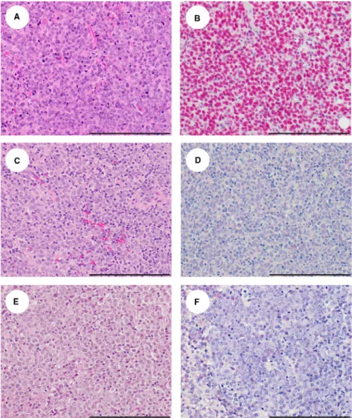
To determine whether the high plasma EBV detected in some patients correlated with EBV status in the tumor, we evalu-ated tumor EBV status by EBER ISH. Interpretable EBER ISH results were available on 42/44 patients. Images of rep-resentative lymphomas from patients in this cohort are shown in Figure 2. A similar proportion of DLBCLs were EBER
ISH positive in HIV‐positive (2/23, 9%) and HIV‐negative (3/19, 16%) patients, with 4/5 positive cases occurring in in-dividuals 54 years of age or older, including all 3 HIV‐nega-tive cases (Table 1, data not shown). Amongst HIV posiHIV‐nega-tive cases, there were too few EBER‐positive DLBCLs to make meaningful clinical comparisons between EBER‐positive (n = 2) and EBER‐negative cases (n = 21). However, there was no obvious difference in mean CD4 count between these groups (105 ± 78 × 109/L in EBER‐positive DLBCL vs
179 ± 154 × 109/L in EBER‐negative DLBCL).
Notably, tumor EBER‐positive cases in the cohort (n = 5) were substantially less numerous than cases with a high pre-treatment plasma EBV DNA level (n = 16). While 3 of 5 EBER‐positive cases had a pretreatment plasma EBV DNA level ≥ 3.0 log10 copies/mL, high pretreatment plasma EBV
DNA was also detected in 12/37 tumor EBER‐negative DLBCLs, raising the possibility that plasma EBV DNA may not be tumor‐derived in many DLBCL cases.
TABLE 2 Clinical characteristics of HIV‐positive patients with DLBCL stratified by pretreatment plasma EBV DNA level
All HIV‐positive DLBCL HIV‐positive patients with tumor EBV quadruple negative DLBCL
n
EBV log10 < 3.0 copies/mL (n = 15)
EBV log10 > 3.0 copies/mL
(n = 10) Pvalue n
EBV log10 < 3.0 copies/mL (n = 8)
EBV
log10 > 3.0 cop-ies/mL (n = 7) Pvalue
Median survival (days) 25 1534 16 .005 15 1391 15 .01
Death within 100 d, n (%) 25 0 (0) 7 (70) .0002 15 0 (0) 5 (33) .007
Age median years, (range) 25 47.2 (30.3‐63.2) 44.5 (24.1‐53.6) .16 15 49.3 (30.3‐60.1) 44.6 (32.8‐49.9) .09
M:F 25 11:4 8:2 1.0 15 6:2 5:2 1
ECOG PS > 2, n (%) 25 1 (7) 7 (70) .002 15 1 (13) 5 (71) .04
Ann Arbor stage IV, n (%) 25 4 (27) 7 (70) .049 15 2 (25) 4 (57) .31
IPI > 3, n (%) 24 2 (14) 5 (50) .09 15 1 (13) 2 (29) .57
Age adjusted IPI > 3, n (%) 24 1 (7) 4 (40) .12 15 1 (13) 2 (29) .57 Lactate dehydrogenase
ratio (Patient:ULN), mean ± SD
24 2.0 ± 1.5 4.8 ± 4.4 .02 15 2.5 ± 2.1 5.1 ± 5.2 .18
Lactate dehydrogenase level greater than ULN, n (%)
24 12 (86) 9 (90) 1 15 7 (88) 6 (86) 1
Time since HIV diagnosis,
years, mean ± SD 23 3.0 ± 4.6 2.8 ± 3.4 .79 14 2.5 ± 3.4 1.3 ± 2.4 .82 CD4 count, cells × 109/L,
mean ± SD 25 197 ± 158 155 ± 161 .20 15 177 ± 110 189 ± 184 .54
HIV viral load at diag-nosis, log10 copies/µL, mean ± SD
25 2.4 ± 2.4 2.5 ± 2.4 .90 15 1.8 ± 1.7 2.1 ± 2.1 .89
ART naive (<3 mo) at
diag-nosis, n (%) 25 7 (47) 5 (50) 1.0 15 3 (38) 5 (71) .31
White blood cell count,
109/L, mean ± SD 25 5.5 ± 15 6.5 ± 2.6 .29 15 6 0 ± 1.2 7.5 ± 2.5 .14
To assess other potential sources of plasma EBV DNA in patients with tumor EBER‐negative DLBCL, we also eval-uated EBER staining in background, non‐neoplastic lym-phocytes. Rare non‐neoplastic EBER‐positive lymphocytes were identified in 6 tumor EBV‐negative DLBCLs, in each case representing <1% of all lymph node cells. Cases with non‐neoplastic EBER‐positive lymphocytes were no more likely than cases without non‐neoplastic EBER‐positive lym-phocytes to have a plasma EBV DNA level above the assay's validated limit of detection or ≥3.0 log10 copies/mL (P = .64
and .66, respectively).
3.4
|
Evaluation of tumor EBV status
by orthogonal methods
The existence of rigorous pathology protocols in the KCH Pathology Laboratory, along with confirmation of RNA preservation in 42/44 cases, decreases the likelihood of false negative EBER ISH staining in this cohort. To independently confirm EBV status in the DLBCL, we used two IHC stains, LMP1 (latent membrane protein 1) and EBNA1 (Epstein‐Barr
nuclear antigen 1). LMP1 and EBNA1 are co‐expressed with EBER in EBV's type II and III latency programs, which are typically observed in DLBCL.24,25 As such, these stains
pro-vided an orthogonal method of EBV detection. Insufficient tissue remained for testing in 5 cases, leaving 20 cases of HIV‐positive DLBCL for evaluation, 18 of which had an in-terpretable EBER stain for comparison. LMP1/EBNA1 re-sults in tumor cells were concordant with EBER ISH in 17/18 cases (95%). The lone discordant case (patient 9) exhibited positive EBER staining but was negative for LMP1/EBNA1.
To further exclude falsely negative EBV detection in HIV‐ associated DLBCL cases in our cohort, we next attempted to evaluate EBV status by real‐time qPCR performed on FFPE tissue scrolls (Table 3). Tissue EBV qPCR was concordant with EBER ISH results in 18/18 cases and with LMP1/ EBNA1 IHC in 18/20 cases. Both cases with discordant qPCR and LMP1/EBNA1 staining results were qPCR posi-tive and stain negaposi-tive. One of these cases ( patient 9) was also EBER ISH positive, as mentioned above. In the other, EBER ISH results were uninterpretable due to failed RNA preservation (patient 5).
3.5
|
Pretreatment plasma EBV and survival
in HIV‐positive, tumor EBV negative cases
15 of 25 (60%) cases of HIV‐associated DLBCL in this co-hort were negative by all four tissue‐based methods used to detect EBV in this study (EBER ISH, LMP1 IHC, EBNA1 IHC, Tissue PCR). We refer to these cases as “tumor EBV‐ quadruple‐negative” in order to highlight that there is no evidence to indicate that EBV is present in the neoplas-tic cells in these lymphomas (Table 3). Nevertheless, 7 of 15 (47%) tumor EBV‐quadruple‐negative lymphomas were from patients with a high pretreatment plasma EBV DNA level, favoring the conclusion that plasma EBV is not tumor‐derived in these individuals. Notably, even in
these tumor EBV quadruple negative cases, a high pretreat-ment plasma EBV DNA level remained associated with shorter OS (Figure 1D) (P = .014, hazard ratio = 4.1 with 95%CI 1.0‐16.4). In general, clinical features were similar in tumor EBV‐quadruple‐negative patients compared to the full HIV‐positive cohort (Table 2).
4
|
DISCUSSION
Plasma EBV has been studied intensely for its clinical util-ity as a predictive or prognostic biomarker in EBV‐driven lymphoma.26,27 In SSA, this biomarker has been most
exten-sively evaluated in endemic Burkitt lymphoma and classic
TABLE 3 Tumor EBV status evaluated by multiple methods
Patient number
Pretreatment plasma EBV
≥3.0 EBER ISH LMP1 IHC EBNA1 IHC
Tissue EBV PCR
Tumor EBV quadruple
negative LYM ISH/IHC concordant
EBER/ PCR con-cordant
1 NO NEG NEG NEG NEG YES 5 YES YES 1
2 NO NEG NEG NEG NEG YES 9 YES YES 2
3 NO NEG QNS QNS QNS N/A 24 QNS QNS
4 NO NEG NEG NEG NEG YES 34 YES YES 3
5 NO Uninterpretable NEG NEG POS NO 44 Unint.
6 NO NEG NEG NEG NEG YES 86 YES Equivocal
7 NO NEG QNS QNS QNS N/A 116 QNS QNS
8 NO NEG QNS QNS QNS N/A 127 QNS QNS
9 NO POS NEG NEG POS NO 243 NO
10 NO NEG NEG NEG NEG YES 302 YES YES 4
11 NO NEG QNS QNS QNS N/A 15 QNS QNS
12 NO NEG NEG NEG NEG YES 145 YES YES 5
13 NO NEG NEG NEG NEG YES 159 YES YES 6
14 NO NEG NEG NEG NEG YES 256 YES YES 7
15 NO NEG QNS QNS QNS N/A 81 QNS QNS
16 YES NEG NEG NEG NEG YES 1 YES YES 8
17 YES NEG NEG NEG NEG YES 192 YES Equivocal
18 YES NEG NEG NEG NEG YES 204 YES YES 9
19 YES NEG NEG NEG NEG YES 318 YES YES 10
20 YES NEG NEG NEG NEG YES 153 YES YES 11
21 YES NEG NEG NEG NEG YES 202 YES YES 12
22 YES NEG NEG NEG NEG YES 154 YES YES 13
23 YES Uninterpretable POS POS POS NO 121 Unint.
24 YES POS POS POS POS NO 313 YES
25 YES NEG (<10%) NEG
(<10%) NEG (< 10%) NEG NO
a 282 YES YES 14
Note: “Uninterpretable” indicates that RNA was inadequately preserved for interpretation of EBER stain.
Abbreviations: EBER, Epstein‐Barr encoded RNA; EBNA1, Epstein‐Barr nuclear antigen 1; EBV, Epstein‐Barr virus; IHC, immunohistochemistry; ISH, in situ hybridization; LMP1, latent membrane protein 1; NEG, negative; PCR, polymerase chain reaction; POS, positive; QNS, quantity not sufficient for testing.
aDenotes that the case was classified as not “tumor EBV quadruple negative” due to the small percentage of tumor cells (<10%) positive by EBER, LMP1, and
Hodgkin lymphoma, both of which are EBV positive in most cases.14,15,17,18 Plasma EBV is also a promising lymphoma
marker in HIV‐positive patients,28 though its utility has been
primarily explored in patients with EBV‐positive tumors. We sought to explore the prognostic utility of plasma EBV in DLBCL in Malawi. This question is of particular rele-vance for low‐income countries, which have a high incidence of DLBCL, but do not have advanced capabilities like PET scans, that are routinely used for DLBCL risk stratification in high‐income countries. Measuring plasma viral loads is readily implementable in low‐resource settings, especially given that existing laboratory capacity has been developed to support HIV RNA monitoring in public sector ART pro-grams. Although high pretreatment plasma EBV DNA has been reported in patients with EBER‐negative DLBCL,29,30
these earlier studies excluded HIV‐positive patients. Here, we extend those observations to HIV‐infected individuals in Malawi and further show that a high pretreatment plasma EBV DNA level is associated with poor overall survival in HIV‐positive, tumor EBV‐negative patients.
More than 90% of the world's population is estimated to be infected with EBV,31-34 and once infected, EBV persists as a
latent infection for the remainder of an individual's lifetime.35
Latently infected cells are rare in normal individuals, represent-ing fewer than 1 in 10,000 peripheral blood leukocytes.36 As a
result, EBV DNA is typically undetectable or present only at a very low level in the plasma of normal adults.27,37,38 In patients
with EBV‐driven malignancies, particularly in the post‐trans-plant setting, plasma EBV may rise as a ctDNA marker.39-42 In
contrast, detection of high plasma EBV among patients in our cohort with EBV tumor‐negative DLBCL strongly suggests that the virus is derived from non‐tumor cells.
However, from this small study, it is not possible to deter-mine underlying mechanisms for poor outcomes in patients with a high pretreatment plasma EBV DNA level. Our find-ings are reminiscent of cytomegalovirus (CMV) reactivation being associated with worse outcomes in critically ill pa-tients.43,44 Analogously, EBV reactivation in nontumor cells
of HIV‐positive patients with DLBCL could reflect underly-ing illness or immune dysfunction not reflected in standard CD4 count assessment. This interpretation is supported by the observation that HIV‐positive patients with high pretreat-ment plasma EBV DNA levels were more likely to have an ECOG performance status ≥2, with a similar trend persisting in tumor EBV‐quadruple‐negative DLBCL. It is also possible that EBV reactivation in nontumor cells directly contributes to worse outcomes for these patients, perhaps by redirecting an already impaired immune response in HIV‐infected pa-tients. Finally, although we have evaluated tumor EBV status by multiple, independent methods, it is possible that some quadruple‐negative cases are EBV‐driven, despite negative ISH, IHC, and PCR studies.
To our knowledge, there are no prior studies explor-ing the prognostic significance of plasma EBV levels in HIV‐positive patients with tumor EBV‐negative DLBCL. However, there have been two prior publications showing that high plasma EBV DNA is associated with inferior out-comes in tumor EBV‐negative DLBCL in HIV‐negative patients.29,30 In our cohort, we did not observe differences
in OS related to plasma EBV measurement in HIV‐neg-ative patients, although interpretation may be limited by the relatively small number of HIV‐negative individuals in our cohort. Interestingly, a recent study demonstrated that the presence of EBV‐positive, non‐neoplastic bystander cells are associated with increased IPI score and decreased OS in patients with tumor EBV‐negative DLBCL, though HIV status was not reported.45 Additionally, studies from
The Cancer Genome Atlas found the tumor immune mi-croenvironment was altered significantly by EBV‐positive bystander cell infiltration in HIV‐negative solid tumors,46
suggesting that EBV reactivation in nontumor cells may in-fluence tumor biology.
The primary limitation of the current study is the rela-tively small sample size, particularly for tumor EBV‐quadru-ple‐negative cases. Given these small numbers, multivariate analyses were not possible. Of note, we recently validated a real‐time plasma EBV DNA assay in Malawi and hope to fur-ther replicate these initial observations in a larger cohort of patients.
Accurate risk stratification of DLBCL is challenging in SSA and other low‐income countries, where there is often no capacity for cytogenetic or immunophenotypic characteriza-tion, sequencing studies, or cell‐of‐origin classification. The development of blood‐ and plasma‐based molecular diagnos-tic tests hold promise in this setting, pardiagnos-ticularly as up‐front capital equipment costs decline. Many centers in SSA have already developed infrastructure for PCR‐based viral load as-says, demonstrating that these technologies are highly imple-mentable in low‐resource settings. Our experience with EBV measurement in Malawi suggests that this virus may also be an attractive target for blood‐based molecular assay development in SSA to improve outcomes across diverse lymphoma sub-types. Intriguingly, this single highly implementable biomarker might help identify patients with EBV‐positive lymphomas at highest risk for lymphoma relapse or progression as suggested by our previous work,10,11 as well as patients with
EBV‐neg-ative DLBCL at highest risk for early death, as suggested by these new analyses.
ACKNOWLEDGMENTS
UNC Center for AIDS Research. BD is a Lymphoma and Leukemia Society Fellow.
ORCID
Nathan D. Montgomery https://orcid. org/0000-0003-1765-6623
DATA AVAILABILITY STATEMENT
The data that support the findings of this study are available from the corresponding author upon reasonable request.
REFERENCES
1. Shannon‐Lowe C, Rickinson AB, Bell AI. Epstein‐Barr virus‐associated lymphomas. Philos Trans R Soc B: Biol Sci. 2017;372(1732):20160271.
2. Shiramizu B, Barriga F, Neequaye J, et al. Patterns of chromosomal breakpoint locations in Burkitt's lymphoma: relevance to geography and Epstein‐Barr virus association. Blood. 1991;77(7):1516‐1526. 3. Swerdlow SHCE, Harris NL, Jaffe ES, et al. eds. WHO
Classification of Tumours of Haematopoietic and Lymphoid Tissues. Revised, 4th ed. Lyon: IARC; 2017; No. 2.
4. Menon MP, Pittaluga S, Jaffe ES. The histological and biological spectrum of diffuse large B‐cell lymphoma in the World Health Organization classification. Cancer J. 2012;18(5):411‐420. 5. Linke‐Serinsoz E, Fend F, Quintanilla‐Martinez L. Human
im-munodeficiency virus (HIV) and Epstein‐Barr virus (EBV) re-lated lymphomas, pathology view point. Semin Diagn Pathol. 2017;34(4):352‐363.
6. A clinical evaluation of the International Lymphoma Study Group classification of non‐Hodgkin's lymphoma. The Non‐Hodgkin's Lymphoma Classification Project. Blood. 1997;89(11):3909‐3918. 7. Naresh KN, Raphael M, Ayers L, et al. Lymphomas in sub‐Saharan
Africa–what can we learn and how can we help in improving diag-nosis, managing patients and fostering translational research? Br J
Haematol. 2011;154(6):696‐703.
8. Parkin DM, Nambooze S, Wabwire‐Mangen F, Wabinga HR. Changing cancer incidence in Kampala, Uganda, 1991–2006. Int
J Cancer. 2010;126(5):1187‐1195.
9. Chokunonga E, Borok MZ, Chirenje ZM, Nyakabau AM, Parkin DM. Trends in the incidence of cancer in the black population of Harare, Zimbabwe 1991–2010. Int J Cancer. 2013;133(3):721‐729. 10. Cesarman E. Pathology of lymphoma in HIV. Curr Opin Oncol.
2013;25(5):487‐494.
11. Kanakry JA, Li H, Gellert LL, et al. Plasma Epstein‐Barr virus DNA predicts outcome in advanced Hodgkin lymphoma: correla-tive analysis from a large North American cooperacorrela-tive group trial.
Blood. 2013;121(18):3547‐3553.
12. Welch JJG, Schwartz CL, Higman M, et al. Epstein‐Barr virus DNA in serum as an early prognostic marker in children and adolescents with Hodgkin lymphoma. Blood Adv. 2017;1(11):681‐684. 13. Leung S‐F, Zee B, Ma BB, et al. Plasma Epstein‐Barr viral
de-oxyribonucleic acid quantitation complements tumor‐node‐metas-tasis staging prognostication in nasopharyngeal carcinoma. J Clin
Oncol. 2006;24(34):5414‐5418.
14. Westmoreland KD, Montgomery ND, Stanley CC, et al. Plasma Epstein‐Barr virus DNA for pediatric Burkitt lymphoma diagno-sis, prognosis and response assessment in Malawi. Int J Cancer. 2017;140(11):2509‐2516.
15. Westmoreland KD, Stanley CC, Montgomery ND, et al. Hodgkin lymphoma, HIV, and Epstein‐Barr virus in Malawi: longitudi-nal results from the Kamuzu Central Hospital Lymphoma study.
Pediatr Blood Cancer. 2017;64(5):e26302.
16. Gulley ML, Morgan DR. Molecular oncology testing in resource‐ limited settings. J Mol Diagn. 2014;16(6):601‐611.
17. Kabyemera R, Masalu N, Rambau P, et al. Relationship between non‐Hodgkin's lymphoma and blood levels of Epstein‐Barr virus in children in north‐western Tanzania: a case control study. BMC
Pediatr. 2013;13:4.
18. Orem J, Sandin S, Mbidde E, Mangen FW, Middeldorp J, Weiderpass E. Epstein‐Barr virus viral load and serology in child-hood non‐Hodgkin's lymphoma and chronic inflammatory condi-tions in Uganda: implicacondi-tions for disease risk and characteristics. J
Med Virol. 2014;86(10):1796‐1803.
19. Gopal S, Fedoriw Y, Kaimila B, et al. CHOP chemotherapy for ag-gressive non‐hodgkin lymphoma with and without HIV in the an-tiretroviral therapy era in Malawi. PLoS ONE. 2016;11(3):e0150445. 20. Montgomery ND, Liomba NG, Kampani C, et al. Accurate
real‐time diagnosis of lymphoproliferative disorders in Malawi through clinicopathologic teleconferences: a model for pa-thology services in sub‐Saharan Africa. Am J Clin Pathol. 2016;146(4):423‐430.
21. Ryan JL, Fan H, Glaser SL, Schichman SA, Raab‐Traub N, Gulley ML. Epstein‐Barr virus quantitation by real‐time PCR targeting multiple gene segments: a novel approach to screen for the virus in paraffin‐embedded tissue and plasma. J Mol Diag. 2004;6(4):378‐385.
22. Painschab MS, Kasonkanji E, Zuze T, et al. Mature outcomes and prognostic indices in diffuse large B‐cell lymphoma in Malawi: a prospective cohort. Br J Haematol. 2018;184(3):364‐372. 23. Kanakry JA, Hegde AM, Durand CM, et al. The clinical
signif-icance of EBV DNA in the plasma and peripheral blood mono-nuclear cells of patients with or without EBV diseases. Blood. 2016;127(16):2007‐2017.
24. Nguyen‐Van D, Keane C, Han E, et al. Epstein‐Barr virus‐pos-itive diffuse large B‐cell lymphoma of the elderly expresses EBNA3A with conserved CD8 T‐cell epitopes. Am J Blood Res. 2011;1(2):146‐159.
25. Liu F, Asano N, Tatematsu A, et al. Plasmablastic lymphoma of the elderly: a clinicopathological comparison with age‐related Epstein‐Barr virus‐associated B cell lymphoproliferative disorder.
Histopathology. 2012;61(6):1183‐1197.
26. Kimura H, Kwong YL. EBV viral loads in diagnosis, monitoring, and response assessment. Front Oncol. 2019;9:62.
27. Kanakry J, Ambinder R. The biology and clinical utility of EBV monitoring in blood. Curr Top Microbiol Immunol. 2015;391:475‐499.
28. Muncunill J, Baptista M‐J, Hernandez‐Rodríguez Á, et al. Plasma Epstein‐Barr virus load as an early biomarker and prognostic factor of human immunodeficiency virus‐related lymphomas. Clin Infect
Dis. 2019;68(5):834‐843.
30. Okamoto A, Yanada M, Inaguma Y, et al. The prognostic signifi-cance of EBV DNA load and EBER status in diagnostic specimens from diffuse large B‐cell lymphoma patients. Hematol Oncol. 2017;35(1):87‐93.
31. Cohen JI. Epstein‐Barr virus infection. New Engl J Med. 2000;343(7):481‐492.
32. Hesse J, Ibsen KK, Krabbe S, Uldall P. Prevalence of antibod-ies to Epstein‐Barr virus (EBV) in childhood and adolescence in Denmark. Scand J Infect Dis. 1983;15(4):335‐338.
33. Lai PK, Mackay‐Scollay EM, Alpers MP. Epidemiological stud-ies of Epstein‐Barr herpesvirus infection in Western Australia. J
Hygiene. 1975;74(3):329‐337.
34. Sumaya CV, Henle W, Henle G, Smith MH, LeBlanc D. Seroepidemiologic study of Epstein‐Barr virus infections in a rural community. J Infect Dis. 1975;131(4):403‐408.
35. Young LS, Rickinson AB. Epstein‐Barr virus: 40 years on. Nat Rev
Cancer. 2004;4(10):757‐768.
36. Miyashita EM, Yang B, Lam KM, Crawford DH, Thorley‐Lawson DA. A novel form of Epstein‐Barr virus latency in normal B cells in vivo. Cell. 1995;80(4):593‐601.
37. Pajand O, Pourakbari B, Mahjob F, Aghamohammadi A, Mamishi N, Mamishi S. Detection of Epstein‐Barr virus DNA in plasma and lymph node biopsy samples of pediatric and adult patients with Hodgkin lymphoma. Pediatr Hematol Oncol. 2011;28(1):10‐15. 38. Wagner HJ, Fischer L, Jabs WJ, Holbe M, Pethig K, Bucsky P.
Longitudinal analysis of Epstein‐Barr viral load in plasma and pe-ripheral blood mononuclear cells of transplanted patients by real‐time polymerase chain reaction. Transplantation. 2002;74(5):656‐664. 39. Au WY, Pang A, Choy C, Chim CS, Kwong YL. Quantification
of circulating Epstein‐Barr virus (EBV) DNA in the diagnosis and monitoring of natural killer cell and EBV‐positive lymphomas in immunocompetent patients. Blood. 2004;104(1):243‐249. 40. Meerbach A, Wutzler P, Hafer R, Zintl F, Gruhn B. Monitoring
of Epstein‐Barr virus load after hematopoietic stem cell
transplantation for early intervention in post‐transplant lymphop-roliferative disease. J Med Virol. 2008;80(3):441‐454.
41. Lin J‐C, Wang W‐Y, Chen KY, et al. Quantification of plasma Epstein‐Barr virus DNA in patients with advanced nasopharyngeal carcinoma. New Engl J Med. 2004;350(24):2461‐2470.
42. Rooney CM, Loftin SK, Holladay MS, Brenner MK, Krance RA, Heslop HE. Early identification of Epstein‐Barr virus‐associated post‐transplantation lymphoproliferative disease. Br J Haematol. 1995;89(1):98‐103.
43. Domart Y, Trouillet JL, Fagon JY, Chastre J, Brun‐Vezinet F, Gibert C. Incidence and morbidity of cytomegaloviral infection in patients with mediastinitis following cardiac surgery. Chest. 1990;97(1):18‐22.
44. Limaye AP, Kirby KA, Rubenfeld GD, et al. Cytomegalovirus reactivation in critically ill immunocompetent patients. JAMA. 2008;300(4):413‐422.
45. Ohashi A, Kato S, Okamoto A, et al. Reappraisal of Epstein‐ Barr virus (EBV) in diffuse large B‐cell lymphoma (DLBCL): comparative analysis between EBV‐positive and EBV‐negative DLBCL with EBV‐positive bystander cells. Histopathology. 2017;71(1):89‐97.
46. Selitsky SR, Marron D, Mose LE, Parker JS, Dittmer DP. Epstein‐ Barr virus‐positive cancers show altered B‐cell clonality.
mSys-tems. 2018;3(5):e00081-18.