European Respiratory Society guidelines
for the diagnosis of primary ciliary
dyskinesia
Jane S. Lucas
1,2, Angelo Barbato
3, Samuel A. Collins
1,2, Myrofora Goutaki
4,5,
Laura Behan
1,2, Daan Caudri
6,7, Sharon Dell
8,9, Ernst Eber
10,
Estelle Escudier
11,12, Robert A. Hirst
13, Claire Hogg
14, Mark Jorissen
15,
Philipp Latzin
5, Marie Legendre
11,12, Margaret W. Leigh
16, Fabio Midulla
17,
Kim G. Nielsen
18, Heymut Omran
19, Jean-Francois Papon
20,21, Petr Pohunek
22,
Beatrice Redfern
23, David Rigau
24, Bernhard Rindlisbacher
25,
Francesca Santamaria
26, Amelia Shoemark
14, Deborah Snijders
3,
Thomy Tonia
4, Andrea Titieni
19, Woolf T. Walker
1,2, Claudius Werner
19,
Andrew Bush
14and Claudia E. Kuehni
4@ERSpublications
International ERS guidelines recommend a combination of tests to diagnose primary ciliary dyskinesiahttp://ow.ly/sJhH304InBN
Cite this article as:Lucas JS, Barbato A, Collins SA,et al. European Respiratory Society guidelines for the diagnosis of primary ciliary dyskinesia. Eur Respir J 2017; 49: 1601090 [https://doi.org/10.1183/ 13993003.01090-2016].
ABSTRACT The diagnosis of primary ciliary dyskinesia is often confirmed with standard, albeit complex and expensive, tests. In many cases, however, the diagnosis remains difficult despite the array of sophisticated diagnostic tests. There is no“gold standard”reference test. Hence, a Task Force supported by the European Respiratory Society has developed this guideline to provide evidence-based recommendations on diagnostic testing, especially in light of new developments in such tests, and the need for robust diagnoses of patients who might enter randomised controlled trials of treatments. The guideline is based on pre-defined questions relevant for clinical care, a systematic review of the literature, and assessment of the evidence using the GRADE (Grading of Recommendations, Assessment, Development and Evaluation) approach. It focuses on clinical presentation, nasal nitric oxide, analysis of ciliary beat frequency and pattern by high-speed video-microscopy analysis, transmission electron microscopy, genotyping and immunofluorescence. It then used a modified Delphi survey to develop an algorithm for the use of diagnostic tests to definitively confirm and exclude the diagnosis of primary ciliary dyskinesia; and to provide advice when the diagnosis was not conclusive. Finally, this guideline proposes a set of quality criteria for future research on the validity of diagnostic methods for primary ciliary dyskinesia.
This article has supplementary material available from erj.ersjournals.com
Received: May 31 2016 | Accepted after revision: Aug 25 2016
These guidelines were endorsed by the ERS Science Council and Executive Committee in September 2016.
The guidelines published by the European Respiratory Society (ERS) incorporate data obtained from a comprehensive and systematic literature review of the most recent studies available at the time. Health professionals are encouraged to take the guidelines into account in their clinical practice. However, the recommendations issued by this guideline may not be appropriate for use in all situations. It is the individual responsibility of health professionals to consult other sources of relevant information, to make appropriate and accurate decisions in consideration of each patient’s health condition and in consultation with that patient and the patient’s caregiver where appropriate and/or necessary, and to verify rules and regulations applicable to drugs and devices at the time of prescription.
Affiliations: 1Primary Ciliary Dyskinesia Centre, University Hospital Southampton NHS Foundation Trust, Southampton, UK. 2University of Southampton Faculty of Medicine, Academic Unit of Clinical and Experimental Medicine, Southampton, UK. 3Primary Ciliary Dyskinesia Centre, Dept of Woman and Child Health (SDB), University of Padova, Padova, Italy. 4Institute of Social and Preventive Medicine, University of Bern, Bern, Switzerland. 5Dept of Paediatrics, Inselspital, University Hospital of Bern, University of Bern, Bern, Switzerland.6Telethon Kids Institute, The University of Western Australia, Subiaco, Australia.7Dept of Pediatrics/Respiratory Medicine, Erasmus University, Rotterdam, The Netherlands. 8Division of Respiratory Medicine, The Hospital for Sick Children, Toronto, ON, Canada. 9Dept of Pediatrics and Institute of Health Policy Management and Evaluation, University of Toronto, Toronto, ON, Canada. 10Division of Paediatric Pulmonology and Allergology, Dept of Paediatrics and Adolescent Medicine, Medical University of Graz, Graz, Austria.11Service de Génétique et Embryologie Médicales, Centre de Référence des Maladies Respiratoires Rares, Hôpital Armand Trousseau, Assistance Publique - Hôpitaux de Paris (AP-HP), Paris, France.12Inserm UMR_S933, Sorbonne Universités (UPMC Univ Paris 06), Paris, France. 13Centre for PCD Diagnosis and Research, Dept of Infection, Immunity and Inflammation, University of Leicester, Leicester Royal Infirmary, Leicester, UK. 14Depts of Paediatrics and Paediatric Respiratory Medicine, Imperial College and Royal Brompton Hospital, London, UK.15ENT Dept, University Hospitals Leuven, Leuven, Belgium.16University of North Carolina at Chapel Hill, Chapel Hill, NC, USA.17Paediatric Dept, Sapienza University of Rome, Rome, Italy. 18Danish PCD & chILD Centre, CF Centre Copenhagen, Paediatric Pulmonary Service, Dept of Paediatrics and Adolescent Medicine, Copenhagen University Hospital, Rigshospitalet, Copenhagen, Denmark. 19Dept of Pediatrics, University Hospital Muenster, Münster Germany. 20AP-HP, Hôpital Kremlin-Bicetre,
service d’ORL et de chirurgie cervico-faciale, Le Kremlin-Bicetre, France.21Faculté de Médecine, Université Paris-Sud, Le Kremlin-Bicêtre, France.22Paediatric Dept, Second Faculty of Medicine, Charles University and Motol University Hospital, Prague, Czech Republic. 23PCD Family Support Group, Milton Keynes, UK. 24Iberoamerican Cochrane Center, Barcelona, Spain. 25Kartagener Syndrom und Primäre Ciliäre Dyskinesie
e. V., Steffisburg, Switzerland.26Pediatric Pulmonology, Dept of Translational Medical Sciences, Federico II University, Azienda Ospedaliera Universitaria Federico II, Naples, Italy.
Correspondence: Jane S Lucas, Primary Ciliary Dyskinesia Centre, University Hospital Southampton NHS Foundation Trust, Southampton, SO16 6YD, UK. E-mail: [email protected]
Introduction
Primary ciliary dyskinesia (PCD) represents a clinical and genetic heterogeneous group of respiratory ciliopathies, with reduced mucociliary clearance of the airways. The various mutations result in different clinical and pathological patterns, contributing to the challenges of diagnosis. There is no single gold standard diagnostic test for PCD [1]. Current diagnosis requires a combination of technically demanding investigations, including nasal nitric oxide (nNO), high-speed video microscopy analysis (HSVA) and transmission electron microscopy (TEM). Historically, clinicians used the saccharine test to screen for PCD, but this is no longer advocated [2]. Furthermore, more sophisticated diagnostic tests that might improve diagnostic accuracy (genotyping, immunofluorescence of ciliary proteins and electron microscopy tomography) are becoming increasingly available.
The availability of tests varies across Europe [3]. This has partially improved recently in response to collaborations including a former Task Force (2006–2009) of the European Respiratory Society (ERS) [2–4] and Framework Programme (FP)-7 funded BESTCILIA. The ERS Task Force published a consensus statement in 2009 [2] to guide diagnostic testing and BESTCILIA has recently introduced diagnostic testing into three countries where services did not previously exist. Since the 2009 statement, a number of groups and consortia have investigated the accuracy of various diagnostic tests for PCD, providing the opportunity to advance the state of diagnostics by developing evidence-based guidelines. Therefore, in 2014 a new PCD ERS Task Force consisting of adult and paediatric physicians from pulmonology and ear, nose and throat disciplines along with diagnostic scientists was established. It aimed to develop evidence-based guidelines for the diagnosis of PCD. This is important for the appropriate clinical management and prognosis of individual patients with suspected or eventually confirmed PCD to ensure patients with PCD are correctly
Support statement: J.S. Lucas, K.G. Nielsen, C.E Kuehni, C. Hogg, M.W Leigh and H. Omran received funding from the European Union Seventh Framework Programme (FP7/2007–2013) under grant agreement n8305404 (BESTCILIA). J.S. Lucas, L. Behan, W.T. Walker and S.A. Collins were supported by the NIHR Respiratory Biomedical Research Unit at the University Hospital Southampton NHS Foundation Trust (Southampton, UK) and AAIR Charity. A. Bush is an NIHR Senior Investigator and was supported by the NIHR Respiratory Disease Biomedical Research Unit at the Royal Brompton and Harefield NHS Foundation Trust and Imperial College London (both London, UK). M. Goutaki was supported by the following national grants: Bernese Lung League, Milena- Pro Kartagener foundation and Swiss National Foundation 32003B_162820/1. S. Dell and M.W. Leigh received funding from the NIH (U54HL096458) through the Genetic Disorders of Mucociliary Clearance Consortium, an initiative of the NIH Office of Rare Diseases Research at the National Center for Advancing Translational Science, and the National Heart, Lung and Blood Institute.
diagnosed, whilst avoiding the problems of false-positive diagnoses. It should also ensure a definitive diagnosis before PCD patients are enrolled in randomised controlled clinical trials of treatment.
Methods
The methods are described in detail in the supplementary material.
Task Force composition
In brief, the panel consisted of a multidisciplinary group of clinicians and scientists with recognised expertise in the diagnosis of PCD; junior members/trainees affiliated to European PCD centres were active members of the committee (table S1). Methodologists from the ERS provided expertise in guideline development following the GRADE (Grading of Recommendations, Assessment, Development and Evaluation) approach for diagnostic tests [5]. Panel members disclosed potential conflicts of interest according to ERS policies at the start of the Task Force and prior to publication of this manuscript.
Patient-important outcomes
The GRADE approach emphasises the importance of recommendations based on the impact on patient-important outcomes [6]. The patient representatives to the Task Force fully endorsed that an accurate diagnosis was an important outcome, because it leads to a better recognition of their problems by physicians and more effective treatment, and thus improves their health and quality of life. This was confirmed by our questionnaire survey of 352 PCD patients from 25 countries and 20 in-depth interviews [7]. However, diagnostic accuracy studies do not provide direct evidence for the improvement of patient-important outcomes; consequently, the confidence in results of test accuracy studies can be judged, at best, as moderate.
Formulation of the topics and questions
The Task Force members agreed that six facets of PCD diagnostics should be evaluated: clinical symptoms, nNO, HSVA, TEM, genotype and immunofluorescence labelling of ciliary proteins. We evaluated each test to see whether it should be included in the diagnostic pathway for PCD, using a“PICO”structured question: “Patients suspected of having PCD,Investigated by [nNO, TEM,etc.], whenComparing patients with a final positive or negative diagnostic outcome, what was the diagnostic accuracy (Outcome) of the test?”The PICO questions for each test were finalised during several rounds of teleconferences and email discussions (table S2).
The essential inclusion criterion for studies was that they must have included consecutive patients referred for PCD testing in whom the PCD diagnosis was then either confirmed or excluded. We excluded studies if patients had already had previous diagnostic testing. In the absence of such studies, in the narrative review we discussed case–control studies that compared PCD patients with healthy controls or patients suffering from other respiratory diseases (e.g.cystic fibrosis). Results from such studies cannot be generalised to the clinical situation, where patients with PCD must be distinguished from patients referred for similar complaints, but without PCD. Thus, the results from case–control studies are far less relevant for clinical care. The main limitation for this project was the lack of a gold standard diagnostic test for PCD. In the absence of this, we compared the diagnostic performance indicators (e.g.sensitivity and specificity) to the authors’final decision regarding positive/negative PCD diagnosis based on all available tests.
The Task Force also agreed on a list of less structured questions relevant to PCD diagnostics for the narrative discussion. As the evidence for these questions were not formally graded they were not used for recommendations.
Literature search methods
We searched Medline and Embase databases (accessed through Ovid) from January 1, 1996 to March 14, 2016. Full details are provided in the supplementary material. In brief, titles and abstracts were screened; the full text was then reviewed for papers that potentially fulfilled criteria for inclusion. These manuscripts were checked for completeness by the Task Force panel to ensure all data fulfilling thea prioriinclusion criteria were present. PRISMA flow diagrams show the search process for each Working Group (figure S1).
Quality of evidence and strength of recommendations
The four tests, where evidence-based recommendations were made, were all acceptable to the patient, feasible and acceptable.
Consensus statement for confirming or excluding PCD
We used a modified Delphi survey to reach a consensus regarding the use of diagnostic tests to definitively confirm and exclude the diagnosis of PCD, and to provide advice regarding patients who do not have a definitive diagnosis but diagnostic tests suggest that the diagnosis is highly likely or inconclusive. The methods are detailed in the supplementary material.
Results
The results of the evidence assessment gave rise to the recommendations listed in table 1.
TABLE 1Evidence-based recommendations for the use of each of the six tests considered for primary ciliary dyskinesia (PCD) diagnosis
Which patients should be referred for diagnostic testing? Based on moderate confidence in the evidence:
1. We recommend that patients are tested for PCD if they have several of the following features: persistent wet cough; situs anomalies; congenital cardiac defects; persistent rhinitis; chronic middle ear disease with or without hearing loss; a history in term infants of neonatal upper and lower respiratory symptoms or neonatal intensive care admittance (strong recommendation)
2. Patients with normal situs presenting with other symptoms suggestive of PCD (as listed in recommendation 1) should be referred for diagnostic testing (strong recommendation)
3. Siblings of patients should be tested for PCD, particularly if they have symptoms suggestive of PCD (as listed in recommendation 1) (strong recommendation)
4. We recommend the use of combinations of distinct PCD symptoms and predictive tools (e.g.PICADAR) to identify patients for diagnostic testing (weak recommendation)
In patients suspected of having PCD should nasal nitric oxide be used as a diagnostic tool? Based on moderate confidence in the evidence, we recommend that:
1. Nasal nitric oxide measurement should be used as part of the diagnostic work-up of school children aged >6 years and adults suspected of having PCD, preferably using a chemiluminescence analyser with a velum closure technique (strong recommendation)
2. In children aged <6 years suspected of having PCD, we suggest nasal nitric oxide measurement using tidal breathing as part of the diagnostic work-up (weak recommendation)
Remark: we suggest that patients presenting with a strong clinical history should undergo further testing, even if nasal nitric oxide is normal (weak recommendation)
In patients suspected of having PCD should HSVA be used as a diagnostic tool? Based on low confidence in the evidence, we recommend:
1. HSVA, including ciliary beat frequency and beat pattern analysis, should be used as part of the diagnostic work-up of patients suspected of having PCD (weak recommendation)
2. Ciliary beat frequency should not be used without assessment of ciliary beat pattern in diagnosing PCD (strong recommendation) 3. To improve diagnostic accuracy of HSVA, CBF/P assessment should be repeated after ALI culture (strong recommendation) In patients suspected of having PCD should TEM be used as a diagnostic tool?
Based on low confidence in the evidence, we recommend:
1. Ciliary ultrastructure analysis by TEM should be used as part of the diagnostic work-up of patients suspected of having PCD (strong recommendation)
2. Further diagnostic investigations should be performed in patients with normal ultrastructure if the clinical history is strong (strong recommendation)#
3. In patients with hallmark ciliary ultrastructure defects for PCD further confirmatory diagnostic investigations are not required (strong recommendation)
In patients suspected of having PCD, should genotyping be used as a diagnostic tool?
There were no studies that fulfilled inclusion criteria to answer this question. Statements to assist the clinician are made in the genetics sections but these are not evidence based. Therefore, we could not make formal recommendations as for other diagnostic procedures. However, we have provided a list of Task Force statements on genetics, which is based upon agreement between experts rather than published evidence.
In patients suspected of having PCD, should immunofluorescence be used as a diagnostic tool?
There were no studies that fulfilled inclusion criteria to answer this question. Statements to assist the clinician are made in the immunofluorescence sections but these are not evidence based. Therefore, we could not make formal recommendations as for other diagnostic procedures. However, we have provided a list of Task Force statements on immunofluorescence, based upon agreement between experts rather than published evidence.
Clinical features
Summary of recommendations
Which patients should be referred for diagnostic testing?
Based onmoderateconfidence in the evidence:
1) We recommend that patients are tested for PCD if they have several of the following features: persistent wet cough; situs anomalies; congenital cardiac defects; persistent rhinitis; chronic middle ear disease with or without hearing loss; a history in term infants of neonatal upper and lower respiratory symptoms or neonatal intensive care admittance (strong recommendation).
2) Patients with normal situs presenting with other symptoms suggestive of PCD should be referred for diagnostic testing (strong recommendation).
3) Siblings of patients should be tested for PCD, particularly if they have symptoms suggestive of PCD (strong recommendation).
4) We recommend the use of combinations of distinct PCD symptoms and predictive tools (e.g.PICADAR) to identify patients for diagnostic testing (weak recommendation).
Review of evidence directly addressing the question“in patients suspected of having PCD, which clinical features predict a positive diagnosis”?
Our search identified 1269 studies of which two directly answered the question and were included in the quantitative synthesis (figure S1) and an additional six contributed to the narrative review. We excluded 1217 publications based on titles and abstracts. After full text review we excluded 44 of the remaining 52 studies because they did not fulfil the inclusion criteria (table S3).
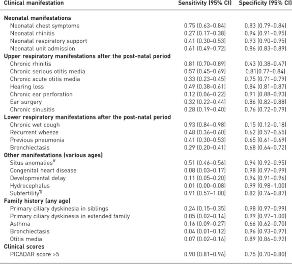
Two studies, BEHAN et al. [10] and SHAPIRO et al. [11], were suitable to provide evidence for our recommendations. They included 1408 patients (table 2).
BEHANet al.[10] analysed data from 868 consecutive paediatric and adult patients. 227 with inconclusive or incomplete results were excluded, leaving 641 for analysis. All patient data were collected through a proforma completed by a clinician prior to the diagnostic testing. They reported sensitivity and specificity of a large range of clinical features (table 2 and table S3). Wet cough did not discriminate well between PCD positive and negative patients (sensitivity 0.93, specificity 0.15). Because it was the main reason for referral, it was present in virtually all patients. Neonatal chest symptoms and neonatal rhinitis had a high specificity (0.83 and 0.94), but a lower sensitivity (0.27 and 0.75). The sensitivity and specificity for 25 clinical features are summarised in table 2 and described in detail in the supplementary material.
In addition to reporting on single symptoms, BEHANet al.[10] developed a seven-point questionnaire-based prediction tool (PICADAR), to help predict the likelihood that a patient referred for evaluation of persistent wet cough has PCD. PICADAR was internally and externally (in a second cohort) validated and is the first clinical prediction tool developed for PCD. The score ranged from 0 to 14. Sensitivity and specificity of a score of >5 were 0.90 and 0.75, respectively, clearly better than single symptoms.
SHAPIROet al.[11] analysed data from 767 consecutive paediatric and adult patients. Information on situs was determined by physicians at local consortium sites through review of radiology, surgery and cardiology reports and radiology images from participant medical records. Patients were divided into three situs categories: situs solitus, situs inversus and situs ambiguous (including heterotaxy).
Situs abnormalities were reported by BEHANet al.[10] and SHAPIROet al.[11] for a total of 1048 patients. The pooled sensitivity and specificity for the two papers (for any situs abnormality) was 0.508 and 0.939, respectively (table S3).
Narrative review of additional evidence
BEUCHERet al.[15] compared patients with abnormal and normal electron microscopy in a retrospective cohort of 89 children suspected of PCD, excluding 21 uncertain cases and found that only situs inversus differed significantly between the groups. PIFFERI et al. [16] compared clinical symptoms in 98 patients with primary PCDversus secondary ciliary dyskinesia and statistically significant differences were found for situs inversus and severity of bronchiectasis. In the only publication that focused on neonates, MULLOWNEYet al. [17] compared neonatal symptoms between 46 PCD patients and 46 controls with a history of neonatal respiratory distress syndrome, and found that lobar collapse, situs inversus and prolonged oxygen need were more common in infants with PCD. The combination of situs inversus, lobar collapse or oxygen need for >2 days had 87% (95% CI 74–94) sensitivity and 96% (85–99) specificity for PCD. A systematic review by GOUTAKI et al. [18] describes other case–control or case series studies on prevalence of clinical symptoms in PCD, which do not fulfil the inclusion criteria for this study. All studies are from developed countries, and it is probable that the predictive value of some symptoms would be different depending on geographical region; for example, sensitivity and specificity of bronchiectasis will be different in sub-Saharan Africa where bronchiectasis due to tuberculosis is common.
Key unanswered questions and research needs
Further research is needed using prospective cohort studies of patients referred with suspicion of PCD in whom clinical features are assessed in a standardised way before they are diagnosed. Analyses must be stratified by age. In particular, there is a need for prospective studies of neonates with neonatal respiratory distress syndrome. In addition, it might be helpful to combine information into clinical prediction scores,
TABLE 2Summary of reported clinical manifestations in studies included in the quantitative analysis
Clinical manifestation Sensitivity (95% CI) Specificity (95% CI)
Neonatal manifestations
Neonatal chest symptoms 0.75 (0.63–0.84) 0.83 (0.79–0.84)
Neonatal rhinitis 0.27 (0.17–0.38) 0.94 (0.91–0.95)
Neonatal respiratory support 0.41 (0.30–0.53) 0.93 (0.90–0.95) Neonatal unit admission 0.61 (0.49–0.72) 0.86 (0.83–0.89) Upper respiratory manifestations after the post-natal period
Chronic rhinitis 0.81 (0.70–0.89) 0.43 (0.38–0.47)
Chronic serious otitis media 0.57 (0.45–0.69) 0.81(0.77–0.84) Chronic acute otitis media 0.33 (0.23–0.45) 0.75 (0.71–0.79)
Hearing loss 0.49 (0.38–0.61) 0.84 (0.81–0.87)
Chronic ear perforation 0.12 (0.06–0.22) 0.91 (0.88–0.93)
Ear surgery 0.32 (0.22–0.44) 0.86 (0.82–0.88)
Chronic sinusitis 0.28 (0.19–0.40) 0.76 (0.72–0.79)
Lower respiratory manifestations after the post-natal period
Chronic wet cough 0.93 (0.84–0.98) 0.15 (0.12–0.18)
Recurrent wheeze 0.48 (0.36–0.60) 0.62 (0.57–0.65)
Previous pneumonia 0.41 (0.30–0.53) 0.65 (0.61–0.69)
Bronchiectasis 0.29 (0.20–0.41) 0.68 (0.64–0.72)
Other manifestations (various ages)
Situs anomalies# 0.51 (0.46–0.56) 0.94 (0.92–0.95)
Congenital heart disease 0.08 (0.03–0.17) 0.98 (0.97–0.99)
Developmental delay 0.11 (0.05–0.20) 0.94 (0.91–0.96)
Hydrocephalus 0.01 (0.00–0.08) 0.99 (0.98–1.00)
Subfertility¶ 0.91 (0.57–1.00) 0.82 (0.74–0.87)
Family history (any age)
Primary ciliary dyskinesia in siblings 0.24 (0.15–0.35) 0.98 (0.97–0.99) Primary ciliary dyskinesia in extended family 0.05 (0.02–0.14) 0.99 (0.97–1.00)
Asthma 0.16 (0.09–0.27) 0.66 (0.62–0.70)
Bronchiectasis 0.04 (0.01–0.12) 0.96 (0.93–0.97)
Otitis media 0.07 (0.02–0.16) 0.89 (0.86–0.92)
Clinical scores
PICADAR score >5 0.90 (0.81–0.96) 0.75 (0.70–0.80)
using state of the art approaches [17]. Validity of the different clinical features is also likely to vary depending on the population under evaluation. For instance, positive and negative predictive values of the symptoms strongly depend on the prevalence of the disease in the referral population and will be poorer in populations with a lower prevalence (i.e. in primary or secondary care compared to PCD referral centres). Sensitivity and specificity (shown in the table 2) do not depend on prevalence. Nevertheless, the mix in PCD phenotypes, and therefore the usefulness of different symptoms for prediction of PCD, is likely to differ between patients attending specialised clinics (e.g.ear nose and throat, pulmonology, and cardiology). For instance, while chronic cough will not distinguish between patients with and without PCD in a pulmonology clinic, chronic ear, nose and throat symptoms will not be distinctive in an ear, nose and throat clinic where (nearly) every patient has these complaints, and cardiac defects will not distinguish in a cardiology setting. Another factor to consider is that clinical features might differ between genetic variants (e.g.patients with CCNO variants show no situs anomalies but increased female infertility), so results vary with differences in prevalence of specific mutations in the evaluated population. Therefore, studies must be performed in specific healthcare settings and study populations, and consider age, sex and genetic abnormalities.
Summary
Relevant literature answering our question was extremely scarce (only two papers, of which one reported only on situs inversus), and results did not take severity of symptoms into account or stratify by age. Overall confidence in their results is moderate mainly because diagnostic performance does not inform downstream consequences of further clinical management based on the assessment of these symptoms.
Results suggest that clinical symptoms may help to distinguish between patients with and without PCD, but the positive predictive value (how many patients with a specific symptom have the disease) of single symptoms is low. Instead, the combination of suggestive symptoms might discriminate better, but this needs further study in different populations.
Wet cough starting in early childhood has been used as the initial selection criterion in most PCD studies. Therefore, it has a low discriminative value due to its high prevalence in both PCD positive and negative individuals recruited to these studies.
Nasal nitric oxide
Summary of recommendations
In patients suspected of having PCD, should nNO be used as a diagnostic tool?
Based onmoderateconfidence in the evidence, we recommend that:
1) nNO measurement should be used as part of the diagnostic work-up of schoolchildren aged >6 years and adults suspected of having PCD, preferably using a chemiluminescence analyser with a velum closure technique (strong recommendation).
2) In children aged <6 years suspected of having PCD, we suggest nNO measurement using tidal breathing as part of the diagnostic work-up (weak recommendation).
We suggest that patients presenting with a strong clinical history should undergo further testing, even if nNO is normal (weak recommendation).
Explanation of the diagnostic test
nNO is extremely low in PCD when compared to healthy and disease controls, for unknown reasons [19]. The accuracy of nNO as a diagnostic test in PCD varies by type of analyser, sampling method and age of patient [20].
Current guidelines recommend aspiration of gas from one nostril with gas entrainedviathe other naris to measure nNO using a stationary chemiluminescence analyser during a velum closure, such as breath hold or oral exhalation against resistance. The reading should be obtained from a technically acceptable plateau reading [21]. Whilst measurement during velum closure by chemiluminescence analyser is considered the “gold standard”, this manoeuvre is not possible in all situations. In young children measurements during tidal breathing have been reported [22, 23]. Electromechanical portable analysers [23] are used where stationary chemiluminescence analysers are not available. There is currently no consensus over what threshold constitutes a positive or negative cut-off.
Review of evidence directly addressing the question“in patients suspected of having PCD, should nNO be used as a diagnostic tool?”
Our search identified 98 studies, of which 23 met inclusion criteria for qualitative assessment. Of these, four papers (n=588 patients) assessed nNO in a cohort of patients suspected of PCD who eventually received either a positive or negative diagnosis, directly addressing the question (table 3 and table S4). The other 19 papers were excluded from informing the recommendations, but contributed to the narrative review (table S5).
MARTHIN et al. [24] measured nNO during breath hold, exhalation against resistance and tidal breathing. Sensitivity ranged from 0.92 (for breath hold) to 1.0 (oral exhalation against resistance) and specificity ranged from 0.80 (tidal breathing) to 0.96 (breath hold) [24]. LEIGHet al.[25] developed a threshold of 77 nL·min−1 using data from a PCD specialist centre and then trialled this cut-off in 155 consecutive patients at other sites. Comparing PCD-positive to indeterminate patients provided a sensitivity of 0.99 and specificity of 0.75 [25]. Lower specificity was because the diagnostic protocol only included electron microscopy and genetics, thus missing a number of true PCD cases (“indeterminate”rather than PCD negative). BEYDONet al.[22] reported a sensitivity of 0.91 and specificity of 0.86 for velum closure (cut-off 82 nL·min−1) and 0.90 and 0.97 for tidal breathing (40 nL·min−1; mean of five peaks). JACKSONet al.[26] used a cut-off of 30 nL·min−1and reported sensitivity and specificity of 0.91 and 0.96, respectively, to distinguish 34 PCD-positive from 267 PCD-negative patients. Further methodological details of these four studies are included in table S4.
Overall confidence in this evidence is moderate mainly because diagnostic performance is not informative of downstream consequences of further clinical management.
Narrative review of additional evidence
A number of studies that did not meet the inclusion criteria for making recommendations addressed important issues. Several studies used alternative methods for the measurement of nNO that do not use the American Thoracic Society/ERS guideline “gold standard” (velum closure, stationary analyser) [21]. Tidal breathing manoeuvres are useful, especially in those unable to perform a velum closure, but may be less discriminative. The study by MARTHINet al.[23] of consecutive referrals found breath hold sensitivity of 0.92 and specificity of 0.96 compared to tidal breathing values of 0.93 and 0.80 (thresholds were breath hold– 52.5 nL·min−1and tidal breathing–47.4 nL·min−1). However, BEYDONet al.[22] found increased accuracy of tidal breathing (37.9 nL·min−1 threshold; sensitivity 0.94, specificity 0.92) versus velum closure
(82.2 nL·min−1, sensitivity 0.91, specificity 0.86). Measurement using portable analysers was assessed in two case–control studies. Using a portable analyser, MARTHINet al. [23] compared PCD patients to those with cystic fibrosis and healthy controls. They found a sensitivity and specificity of 1.0 and 0.95, respectively, for breath hold (64 nL·min−1threshold) and 1.0 and 1.0, respectively, for tidal breathing (43 nL·min−1) [23].
HARRISet al.[27] studied 13 PCD and 37 disease control/healthy patients using tidal breathing and a portable analyser with a cut-off of 30 nL·min−1. Sensitivity and specificity were 1.0 and 0.95, respectively [27].
Measurement of nNO in young children is possible; however, discrimination between PCD patients and controls is reduced as nNO is inversely proportional to age in healthy patients <12 years of age [28, 29]. One study showed that velum closure was possible in children as young as 3.9 years [20]. However, the majority of very young children are unable to co-operate with velum closure and so tidal breathing
TABLE 3Summary of diagnostic accuracy of nasal nitric oxide from measurements in consecutive patients suspected of primary ciliary dyskinesia (PCD)
First author [ref.] Study population Sampling method Threshold nL·min−1
Sensitivity (95% CI)
Specificity (95% CI)
MARTHIN[24] 117 referrals (PCD: n=14) Breath hold: n=58 52.5 0.92 (0.62–0.998) 0.96 (0.85–0.995) Oral exhalation against
resistance: n=37
72.6 1.0 (0.54–1.0) 0.94 (0.79–0.99)
Tidal breathing: n=97 47.4 0.93 (0.66–0.998) 0.80 (0.69–0.88) LEIGH[25] 155 referrals (PCD: n=71;
indeterminate: n=84)
Oral exhalation, velum closure: n=155
77 0.99 (0.92–0.9996) 0.75 (0.64–0.84)
BEYDON[22] 86 referrals: (PCD: n=49; non-PCD: n=37)
Velum closure: n=74 82.2 0.91 (0.79–0.98) 0.90 (0.78–0.97)
0.86 (0.68–0.96) 0.97 (0.86–0.999)
Tidal breathing (five peaks): n=86
40
JACKSON[26] 301 referrals: (PCD: n=34; non-PCD: n=267)
Velum closure (breath hold or oral exhalation): n=301
measurements have to be used. The studies by MARTHINet al.[23] and BEYDONet al.[22] suggest that tidal breathing may produce similar accuracy to velum closure in adults; however, this has not been shown in children. The study of 117 consecutive referrals of all ages by MARTHIN et al. [23] found that the false-positive rate for children <6 years of age using tidal breathing was 39%.
There is increasing evidence that some genetic mutations causing PCD may be associated with subtle beating abnormalities and nNO levels within the normal range. This includes two studies of PCD individuals with mutations encoding radial spoke head proteins, and one study in PCD individuals with abnormal nexin link composition due to GAS8 mutations associated with an nNO higher than usually seen in PCD [30–32]. This may partially explain the variability of the results and low diagnostic performance of nNO in some studies.
Key unanswered questions and research needs
Current evidence has shown that nNO is a useful test as part of the diagnostic process; however, there is no consensus on appropriate thresholds. Likewise, standardised protocols and thresholds need to be developed for tidal breathing, portable analysers and measurements, and normative data in younger children, particularly those <6 years of age. Further work on genotype–phenotype correlation can help in the interpretation of nNO levels in cases of diagnostic uncertainty in order to reduce the number of false-negative test results. There is no evidence that can lead us to recommend which patients with a normal nNO should be referred for further testing and this requires evaluation.
Summary
nNO is a highly accurate test for PCD when measuredviaa stationary chemiluminescence analyser using velum closure techniques with a sensitivity and specificity of 0.90–1.0 and 0.75–0.97, respectively. Tidal breathing technique or use of portable analysers are less sensitive and specific but may contribute to the diagnostic decision. Different studies have used different methods and cut-off values making it difficult to provide definite thresholds.
nNO is not sufficiently accurate to rule PCD in or out in isolation but considering that it is relatively easy to perform, is noninvasive and is affordable, the panel considered that it should be used as part of the diagnostic work-up of patients suspected of having PCD.
High-speed video analysis
Summary of recommendations
In patients suspected of having PCD, should HSVA be used as a diagnostic tool?
Based onlowconfidence in the evidence, we recommend:
1) HSVA, including ciliary beat frequency and beat pattern analysis, should be used as part of the diagnostic work-up of patients suspected of having PCD (weak recommendation).
2) Ciliary beat frequency should not be used without assessment of ciliary beat pattern in diagnosing PCD (strong recommendation).
To improve diagnostic accuracy of HSVA, CBF/p assessment should be repeated after air–liquid interface culture (strong recommendation).
Explanation of the test
PCD is related to abnormal ciliary function [33], which can be analysedex vivoby assessment of ciliary activity in respiratory epithelium from the nose or bronchus. Ciliated cells can be observed immediately after sampling [34–36] and again after a period of culturing to differentiate PCD from secondary dyskinesia [37–40]. A video attached to a microscope records at high speeds (120–500 fps), and is replayed slower (30–60 fps) to review ciliary beat pattern and measure ciliary beat frequency [41]. Most studies have used analysis by expert microscopists, whilst several studies used computer analysis in an attempt to reduce subjectivity/observer bias. HSVA provides a permanent record that can be used for audit, expert advice or research.
Review of evidence directly addressing the question“in patients suspected of having PCD, should HSVA be used as a diagnostic tool?”
PAPONet al. [42] measured 12 parameters of ciliary beat pattern, including ciliary beat frequency. The distance travelled by the cilia tip weighted by the percentage of beating edges had the best sensitivity (0.96) and specificity (0.95) to distinguish 10 PCD positive from 15 PCD negative patients [42]. Using this parameter in nine patients with previously inconclusive diagnoses, it was possible to support the diagnosis of PCD in four cases and exclude it in two. JACKSONet al. [26] found a sensitivity and specificity of 1.00 and 0.93, respectively, for the combination of ciliary beat frequency measurement and beat pattern evaluation in a cohort of 625 referrals, including 60 PCD positive patients.
Overall confidence in this evidence is low, mainly because diagnostic performance is not informative of downstream consequences of further clinical management based on the assessment of HSVA and because of study limitations (HSVA was widely used as part of the reference standard and lack of blinding).
Narrative review of additional evidence
To date, there is no standardised method for cell processing and analysis. Respiratory epithelium can be collected using brush, curette or forceps, usually from the nose [43]. Ciliary function varies under differing conditions; for example, temperature and pH, with some centres measuring at 37oC [26, 40, 42, 44] and others at lower temperatures [35, 37, 45]. This will affect ciliary function and all centres need to define their own normative data until a consensus is reached to allow standardisation of methods and reporting between centres.
Equivocal results or abnormal results require repeat sampling or re-analysis following cell culture [38, 40, 46, 47] since secondary defects are common. In a series of 712 patients, JORISSENet al.[47] assessed ciliary beat frequency and ciliary coordination in suspension culture (i.e.spheroids). 642 (20%) non-PCD patients demonstrated abnormal ciliary activity before culture, but after culture 100% had normal ciliary activity. Conversely, in biopsies of 70 PCD patients (evaluable in 56), 20% had a normal ciliary beat frequency and 10% had coordinated ciliary activity before culture. After culture, normal ciliary beat frequency was found in 7% of PCD patients but ciliary function was never normally coordinated, making this parameter more sensitive and specific than ciliary beat frequency measurement [47]. HIRSTet al.[40] reported ciliary beat frequency and pattern before and after air–liquid interface cultures in 158 patients. Before culture, most PCD and non-PCD patients exhibited a degree of functional ciliary abnormalities. After air–liquid interface culture, normal ciliary beat pattern was observed in all non-PCD patients, whilst in PCD patients ciliary beat pattern was uniformly abnormal. PIFFERIet al. [46] assessed the results of ciliary beat pattern and frequency analysis after suspension culture (i.e. spheroids) in nine subjects with inconclusive results on nasal brushings. After culture, four patients had abnormal ciliary beat pattern suggesting PCD diagnosis, two had secondary dyskinesia ciliary beat pattern and three remained inconclusive. Culture techniques are limited by success rates ranging from 54% to 83% [26, 37, 40].
Ciliary beat frequency measurement does not adequately differentiate PCD from non-PCD unless combined with ciliary beat pattern assessment. STANNARDet al. [48] found a sensitivity and specificity of 0.97 and 0.95, respectively, for the percentage of dyskinetic epithelial edges whilst ciliary beat frequency alone only yielded a sensitivity and specificity of 0.87 and 0.77, respectively. Moreover, ciliary beat frequency may be slow, normal or increased in PCD depending on the genotype [45].
Some ultrastructural defects and genetic mutations causing PCD may be associated with specific patterns of ciliary beating. In a cohort of 56 children, CHILVERS et al. [44] reported virtually immotile cilia in patients with either a: combined inner and outer dynein arm (IDA and ODA) defect or an isolated ODA defect; stiff beat pattern in patients with either an isolated IDA or radial spoke with an IDA defect; and circular beating cilia in patients with a ciliary transposition defect. RAIDT et al.[45] studied ciliary beat pattern according to the genetic variants of PCD. Although numbers associated with some genes were extremely small, the data supports the linking of a PCD causing gene with particular ciliary beat patterns: cilia from patients with mutations in ODA causing genes (DNAH5, DNAI1, DNAI2, ARMC4) showed minimal residual movements. In patients with DNAH11 mutations (normal ultrastructure), cilia exhibited
TABLE 4Summary of diagnostic accuracy of high-speed video analysis (HSVA) from evaluation in consecutive patients suspected of primary ciliary dyskinesia (PCD)
First author [ref.] Study population
Cilia assessment method Sensitivity (95% CI) Specificity (95% CI)
PAPON[42] 25 referrals (10 PCD positive)
HSVA beat frequency and quantitative measurements of beat pattern
0.96 (0.89–0.98) 0.95 (0.91–0.98)
JACKSON[26] 625 referrals (60 PCD positive)
a hyperkinetic ciliary beat pattern with reduced proximal axonemal bending. Some genetic defects, such as GAS8 mutations, can result in subtle defects that are hardly detectable by HSVA [30].
Key unanswered questions and research needs
Current evidence suggests that HSVA is a useful test as part of the diagnostic process, but there is no consensus on appropriate cell processing and method of ciliary assessment. Likewise, standardised protocols and thresholds need to be developed forex vivoanalysis of ciliary beat pattern. Further work on genotype/ultrastructural phenotype correlation can help in interpretation of HSVA parameters in cases of diagnostic uncertainty.
Summary
HSVA is an accurate test for PCD when performed by experienced observers combining ciliary beat frequency measurement and pattern analysis (sensitivity and specificity of 0.95–1.00 and 0.93–0.95, respectively). Culturing the respiratory cells may contribute to improve the accuracy of HSVA, in particular to rule out false positives.
HSVA is not sufficiently standardised to rule PCD in isolation in or out. Considering that optimal conditions in functional evaluation of cilia remains to be defined, the panel considered that HSVA should be performed by experienced staff as part of the diagnostic work-up of patients suspected of having PCD. This might impair the availability of the test in many centres.
Transmission electron microscopy
Summary of recommendations
In patients suspected of having PCD, should TEM be used as a diagnostic tool? Based onlowconfidence in the evidence, we recommend:
1) Ciliary ultrastructure analysis by transmission electron microscopy should be used as part of the diagnostic work-up of patients suspected of having PCD (strong recommendation).
2) Further diagnostic investigations should be performed in patients with normal ultrastructure if the clinical history is strong (strong recommendation).
3) In patients with hallmark ciliary ultrastructure defects for PCD further confirmatory diagnostic investigations are not required (strong recommendation).
Explanation of the diagnostic test
In 1976, AFZELIUS [33] demonstrated that TEM could be used to detect ultrastructural defects of cilia in patients with primary ciliary dyskinesia. Subsequently, for many years TEM was considered the “gold standard”diagnostic test for PCD. However, several genetic studies have demonstrated that an increasing number of distinct genetic PCD sub-types (e.g.due to DNAH11 mutations) cannot be diagnosed by TEM [49]. Thus, TEM cannot rule out PCD diagnosis.
Respiratory epithelium is usually sampled from the inferior turbinate of the nose by brush or curette biopsy or from the lower respiratory tract during bronchoscopy. The epithelium is chemically fixed with glutaraldehyde, processed and embedded into blocks, which are sectioned with an ultramicrotome. Staining with heavy metals (lead and uranyl) provides contrast. Assessment of cilia from healthy cells in transverse sections is made using TEM [50, 51]. The number of cilia and cells analysed varies between centres; unless sufficient numbers are assessed, defects caused by mutations in genes that cause intermittent defects are likely to be missed. Considerable expertise is required to perform TEM and interpret results; expenditure for equipment and running costs are high.
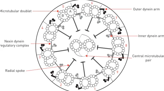
The normal ultrastructural ciliary arrangement in a transverse section is a circle of nine microtubule doublets, each with a pair of dynein arms, plus a central pair of microtubules (figure 1). There are a number of ultrastructural phenotypes associated with a diagnosis of PCD (figure 2). The majority of cases are due to a lack of dynein arms; other defects include disorganisation of the microtubular doublets or loss of the central microtubular pair (table 5). Some patients with PCD have apparently normal ciliary ultrastructure, as seen by TEM. Secondary ciliary dyskinesia can be associated with transient ultrastructural abnormalities, such as compound cilia, axonemal blebs or additional tubules, which must not be confused with PCD.
Review of evidence directly addressing the question“in patients suspected of having PCD, should TEM be used as a diagnostic tool?”
a cohort of patients suspected of PCD who eventually received either a positive or negative diagnosis, contributing to the evidence for recommendations (figure S1).
Tables 5 and 6 summarise the 11 studies addressing the question. The sensitivity calculated from each study ranged between 0.71 and 1.00 and the specificity between 0.92 and 1.00. There were five false-positive patients in two studies. PAPONet al. [42] identified two false-positive results; one was a child with severe asthma without recurrent upper airway infection who had short or absent ODA concerning 44% of nasal cilia and 90% of bronchial cilia, the other an adult with situs inversus and nasal polyposis without lower airway symptoms who had absent IDA without microtubular disorganisation ( personal communication). MUNKHOLM et al. [57] identified three false positives in individuals each eventually thought to have secondary ciliary dyskinesia, two of whose clinical phenotype improved becoming asymptomatic and a third who had severe asthma. Total specificity, when combining all 11 studies, was >0.99.
The quality of evidence was rated low because diagnostic performance is not informative of downstream consequences of further clinical management and because of study limitations (frequent use of TEM in the reference standard and lack of blinding). However, the very low rate of false positives and the very high specificity of TEM led to strong recommendations.
Narrative review of additional evidence
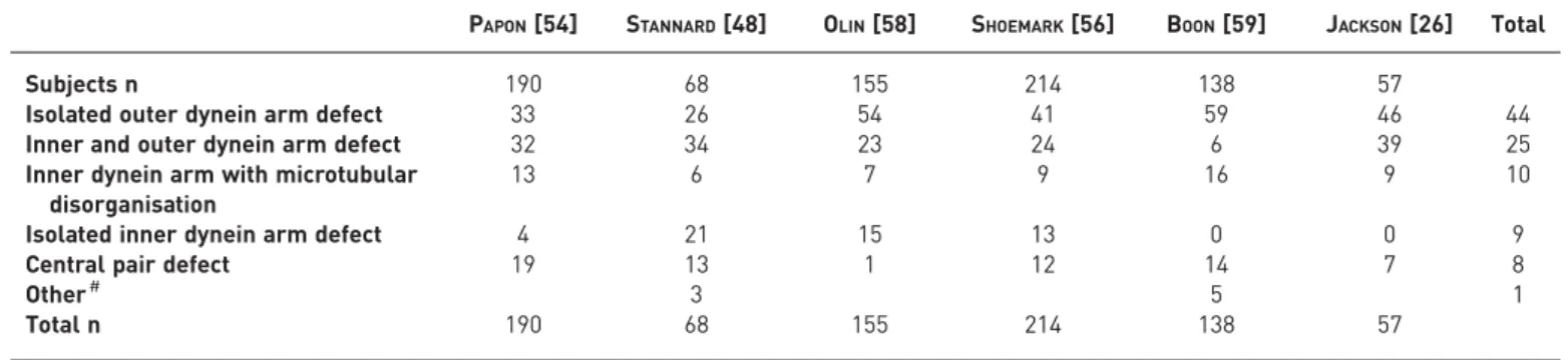
Assessment of the proportion of TEM defects in patients with PCD was made following a review of all manuscripts ( post 1996) describing a cohort of more than 50 individuals [26, 42, 48, 56, 58, 59] (table 4). ODA defects (26–59%) and combined ODA and IDA defects (6–39%) were the most commonly observed. The recommendations below refer to common hallmark defects (absence of ODA, combined absence of IDA and ODA, IDA absence combined with microtubular disarrangement). Isolated IDA defects by TEM are controversial. Several studies acknowledge that IDA are difficult to visualise by TEM [42, 60, 61] and repeat analysis has been recommended before confirming a diagnosis [62]. For central pair defects the ciliary defect is usually present in a minority of cilia making the diagnosis difficult especially since patients do not have situs inversus.
Evidence for add on techniques to improve electron microscopy in the diagnosis of PCD was reviewed. Computer-assisted analysis has been reported to enhance the visualisation of dynein arms and consequently improve the sensitivity of electron microscopy [60, 61]. Electron tomography is an advanced TEM technique allowing visualisation of structures in three-dimension. A series of TEM images are acquired by tilting the specimen stage at regular increments around two perpendicular axes. Images from both tilt series are then aligned into a single three-dimensional high-resolution projection. If a structural feature is repeated within a tomogram, it can be enhanced through sub-tomographic averaging; a technique in which software extracts the chosen common features and makes comparison by cross correlation. Electron tomography has been shown to improve three-dimensional visualisation and resolution of cilia allowing identification of patients withHYDINandDNAH11gene defects in a research setting [63]. The use of tomography for diagnosis has not been evaluated.
B
3 4
5
6
7
8
9
1 2
A Microtubular doublet
Nexin dynein regulatory complex
Central microtubular pair Inner dynein arm Outer dynein arm
Radial spoke
Our evidence review considered only conclusive results. Reported rates of inconclusive results ranged from 1.7% to 28.6% [26, 37, 39, 58]. This was attributed to poor sampling technique or the presence of secondary changes to the cilia. Seven of the 11 studies reported measures to avoid sampling during or immediately after an upper respiratory tract infection to improve adequacy and minimise secondary ciliary ultrastructural change.
Cell culture techniques that induce ciliogenesis from human biopsies are used in a number of PCD diagnostic centres. Two techniques to induce basal cell proliferation and ciliated cell differentiation have been described for PCD diagnosis [37, 40]. JORISSENet al.[37] established a submerged culture technique and the air–liquid interface technique was first described for PCD diagnosis by HIRSTet al. [40]. Both methods have shown that the TEM axoneme structure is conserved after cell culture in normal and PCD subjects, and they have been shown to reduce secondary damage [37, 40]. TEM following culture has the potential to aid diagnosis of reduced generation of multiple motile cilia [64].
Key unanswered questions and research needs
Basic scientific research must improve the technique of TEM and identify PCD in those with “normal ultrastructure”. The diagnostic community requires standardised protocols and consensus on terminology,
a) c)
b) d)
especially regarding the number and proportion of cilia required to make a diagnosis. True relevance and prevalence of IDA and other rare defects needs to be confirmed.
Summary
TEM is a highly specific test to confirm a diagnosis of PCD and is a key part of the diagnostic work. However, some patients with PCD have apparently normal ultrastructure and therefore TEM should not be used in isolation to exclude a diagnosis.
All 11 studies were retrospective analyses of cohorts of patients with clinical suspicion of PCD, the largest of which spanned time-periods of 20 years [54, 56]. Further downgrading was due to the use of TEM as the reference standard and lack of blinding, resulting in grading of evidence as low.
Genetics
In patients suspected of having PCD, should genotyping be used as a diagnostic tool? There were no studies that fulfilled the inclusion criteria to answer this question.
Explanation of the diagnostic test
PCD is a genetically heterogeneous disorder. As with autosomal recessive disorders in general, disease is more likely in offspring from consanguineous relationships, and has a 1:4 probability from any conception where both parents are healthy carriers. To date, mutations in more than 30 genes have been reported to cause PCD (table 7). A more detailed explanation of the PCD-associated genes is presented in the supplementary material.
To establish the genetic diagnosis, non-ambiguous bi-allelic mutations in autosomal recessive PCD and hemizygous mutations in X-linked PCD should be identified. The majority of reported mutations are nonsense, frameshift or splice mutations while missense mutations are identified in a minority of cases. Most of the mutations are private, but founder mutations (e.g. in DNAI1 [105] andDNAH5 [95]) and
TABLE 5Characteristics of the ultrastructural defects described in nine studies directly addressing the PICO question using transmission electron microscopy to diagnose primary ciliary dyskinesia
PAPON[54] STANNARD[48] OLIN[58] SHOEMARK[56] BOON[59] JACKSON[26] Total
Subjects n 190 68 155 214 138 57
Isolated outer dynein arm defect 33 26 54 41 59 46 44
Inner and outer dynein arm defect 32 34 23 24 6 39 25
Inner dynein arm with microtubular disorganisation
13 6 7 9 16 9 10
Isolated inner dynein arm defect 4 21 15 13 0 0 9
Central pair defect 19 13 1 12 14 7 8
Other# 3 5 1
Total n 190 68 155 214 138 57
Data are presented as percentage, unless otherwise stated.#: include ciliary aplasia, disorientation and extra microtubules.
TABLE 6Sensitivity and specificity of the 11 studies directly addressing the PICO question using transmission electron microscopy to diagnose primary ciliary dyskinesia
First author [ref.] Study population n Conclusive diagnostic result reached n Sensitivity (95% CI) Specificity (95% CI)
JORISSEN[37] 812 468 0.71 (0.61–0.81) 1.0 (0.99–1.0)
PIFFERI[52] 64 62 0.75 (0.48–0.93) 1.0 (0.93–1.0)
PIFFERI[39] 59 56 0.77 (0.50–0.93) 1.0 (0.91–1.0)
HIRST[53] 231 187 1.0 (0.88–1.0) 1.0 (0.98–1.0)
PAPON[54] 1149 793 0.82 (0.77–0.86) 1.0 (0.99–1.0)
OLM[55] 24 24 0.92 (0.62–1.0) 1.0 (0.74–1.0)
PAPON[42] 34 28 0.83 (0.52–0.98) 1.0 (0.79–1.0)
SHOEMARK[56] 1182 1031 0.88 (0.83–0.91) 1.0 (1.0–1.0)
HIRST[40] 165 122 0.96 (0.87–1.0) 1.0 (0.95–1.0)
MUNKHOLM[57] 239 61 0.83 (0.61–0.95) 0.92 (0.79–0.98)
TABLE 7Overview of primary ciliary dyskinesia causing genes and their associated findings by transmission electron microscopy (TEM) and immunofluorescence analyses
Gene [Ref.] Locus TEM Immunofluorescence
DNAH5 [65] 5p15 ODA Absent DNAH5 and DNAH9 [67, 94, 95]
DNAH11 [49] 7p15–21 Normal DNAH11 is absent in patients with DNAH11 loss-of
function mutations, DNAH5 and DNALI1 present [96, 97]
DNAI1 [66] 9p21-p13 ODA DNAH5 staining may be present proximally but
absent distally,
DNAH9 absent within the ciliary axonemes [67, 94]
DNAI2 [67] 17q25.1 ODA DNAH5, DNAI2 and DNAH9 absent or aberrant [67]
NME8 (TXNDC3) [68] 7p14.1 ODA Not reported
DNAL1 [69] 14q24.3 ODA Not reported
CCDC151 [70] 19p13.2 ODA DNAH5, CDC151, CCDC114 and ARMC4 absent,
DNALI1 present [70]
CCDC114 [71] 19q13.33 ODA CCDC114 severely reduced, DNAH5 absent, DNALI1
undisturbed [71]
ARMC4 [72] 10p21 ODA Reduced ARMC4 staining along cilia, complete distal loss
of DNAH5, DNAH5 only on proximal ciliary end, DNALI1 present [72, 98]
CCDC103 [73] 17q12 ODA+IDA DNAH5, DNAH9 and DNALI1 are missing or
reduced in a small number of patients [73]
DYX1C1 (DNAAF4) [74] 15q21 ODA+IDA DNAH5, DNAH9 and DNAI2 absent [74]
SPAG1 [75] 8q22 ODA+IDA Absent DNAH5 and DNALI1 [75]
LRRC6 [76] 8q24 ODA+IDA LRRC6, DNALI1 and DNAI2 absent or very
reduced [76, 82, 99]
DNAAF2 (KTU) [77] 14q21.3 ODA+IDA DNAH5 and DNAI2 absent distally with some
residual staining, DNAH9 and DNALI1 absent [77]
DNAAF1 (LRRC50) [78, 79] 16q24 ODA+IDA DNAH5, DNAH9 and DNALI1 absent [79]
C21orf59 [80] 21q22.1 ODA+IDA DNAH5 and DNALI1 absent [80]
DNAAF3 [81] 19q13 ODA+IDA DNAH5, DNAH9 and DNALI1 absent [81]
ZMYND10 [82] 3p21.3 ODA+IDA DNAH5, DNAI2 and DNALI1 absent [82, 100]
DNAAF5 (HEATR2) [83] 7p22.3 ODA+IDA DNAI1, DNAH5 and DNALI1 absent,
HEATR2 reduced [83, 101]
HYDIN [84] 16q22 Normal/subtle: increased
frequency of transposition defects
Normal IDA (DNALI1) and ODA (DNAH5) [84]
RSPH1 [32] 21q22.3 Intermittent central pair/transposition defects
RSPH1 and RSPH9 absent, RSPH4A present [32, 86, 102]
RSPH3 [85] 6q25.3 Intermittent central pair/near
absence of radial spokes
RSPH3 and RSPH11 absent, RSPH1, RSPH4A and RSPH23 present (RSPH9 not reported),
DNALI1 present [85]
RSPH9 [86] 6p21 Intermittent central pair defect/
transposition
Absent RSPH9, RSPH1 and RSPH4A present [86]
RSPH4A [86] 6q22 Intermittent central pair defect/
transposition
RSPH4A, RSPH9 and RSPH1 absent [86]
DRC1 (CCDC164) [87] 2p23 Normal/subtle: N-DRC links missing with occasional MT disorganisation
GAS8 and LRRC48 absent from ciliary axonemes [87]
GAS8 (DRC4) [30] 16q24.3 Normal/subtly abnormal: increased frequency of MT misalignment
DNALI1 and DNAH5 present, GAS8 absent [30]
CCDC65 (DRC2) [88] 12q13.12 Normal/N-DRC links missing with occasional MT disorganisation
CCDC65 and GAS8 reduced [88]
CCDC39 [89] 3q26 MT disorganisation+IDA Absent CCDC39 protein, ODA normal distribution (DNAH5,
DNAI2, DNAH9), DNALI1 (IDA) absent, GAS8 in cytoplasm but absent from axoneme [89, 103]
CCDC40 [90] 17q25 MT disorganisation+IDA Absent CCDC39 protein, RSPH4A and ROPN1 L/RSP11
present in axonemes [90, 103]
RPGR# [91] Xp21.1 Variable Normal, DNAH5 and DNALI1 present [104]
OFD1¶ [92] Xp22 Unknown Not reported
CCNO [64] 5q11.2 Reduction of cilia number DNAH5 present, rootletin mislocated in deeper regions of cytoplasm, CCNO not detectable [64]
MCIDAS [93] 5q11.2 Reduction of cilia number MCIDAS, CCNO, DNAH5, CCDC39 and CCDC78 absent [93]
mutational hot spots (e.g. CCNO [106]) have been reported. The ranking of the effect of the mutations should follow international recommendations [107]: benign (class 1), likely benign (class 2), unknown significance (class 3), likely pathogenic (class 4), pathogenic (class 5).
The associations between genotype and structural defects documented by TEM and/or immunofluorescence are well established, but much less is known about gene HSVA associations. With studies based on small numbers of patients and often a limited number of videos per patient [45], there is insufficient data to correlate mutations within a gene with dyskinesia phenotype. To date, our knowledge suggests that disease-causing mutations in DNAH5 are always associated with predominantly static cilia whilst mutations in different regions of DNAH11 can lead to either static cilia or hyper frequent, stiff cilia [45, 108]. Therefore, to confirm the genetic cause of PCD, the ultrastructural defect and gene should correlate; in the future, we may be able to use gene mutation ciliary pattern correlations as further support.
In principle, all DNA sequencing technologies can be applied for genetic testing in patients with a confirmed PCD or a high suspicion of PCD (see supplementary material). However, due to the high number and the huge size of PCD genes high-throughput techniques are now widely used. The yield of allele-specific approaches is low in PCD given the high genetic and allelic heterogeneity. Detected mutations should be confirmed by Sanger sequencing and checked for segregation in the parents. Genetic laboratories have to be aware that large heterozygous genomic deletions have been reported in PCD individuals that might be missed by DNA sequencing technologies. Homozygous and heterozygous intragenic large duplications and deep intronic mutations are also missed by sequencing techniques. The detection of intragenic deletions and duplications will benefit from the fine set-up of targeted next generation sequencing panels; however, this approach requires specific development and sensitivity assessment. All techniques, especially the second line approaches, benefit from the knowledge of the ultrastructural defect of the patient in order to assess the relevance of the molecular findings. The≈30 PCD genes implicated to date encompass >700 exons and thus it is not unusual to identify a heterozygous variant in a gene that is obviously not responsible for the disease of the patient based on ultrastructural data. Cell and whole organism models can be used to confirm that a gene is disease causing.
Review of evidence directly addressing the question“in patients suspected of having PCD, should genotyping be used as a diagnostic tool?”
Searches identified 462 studies, of which 95 met inclusion criteria for qualitative assessment (table S8). Most studies included patients with confirmed PCD with the aim of identifying novel genes rather than diagnostic cohorts. There were no studies that fulfilled the inclusion criteria for quantitative assessment.
Narrative review of additional evidence
In populations with confirmed or highly suspected PCD diagnosis, it is possible to identify genetic causation in 50–75% of cases [109, 110]. The sensitivity of genetic testing as a first-line diagnostic test for PCD is currently unknown but is likely to be low. With the identification of further PCD genes and high-throughput sequencing technologies, PCD genetic testing as “stand alone” testing might be considered in the future. Genotyping is useful in instances where confirmation of the diagnosis is difficult by other approaches (e.g. DNAH11, CCNO, MCIDAS and RSPH genes mutations). The detection of bi-allelic disease-causing mutations in autosomal recessive PCD or hemizygous mutations in X-linked PCD is highly specific.
Most studies to identify novel PCD gene defects used ultrastructural defects detected by routine TEM as the starting point for the genetics search; therefore, the likelihood to identify mutations in PCD with ultrastructural defects is higher than in PCD devoid of ultrastructural defects. This underscores the need not to rely on TEM as the sole diagnostic test for PCD.
Reports of mutations in specific genes typically relate to small numbers of patients and are not necessarily ethnically representative; therefore, the contribution of each gene should be interpreted with caution. Studies testing forDNAH5 and DNAI1 mutation suggest that these mutations account for ∼50–70% of cases of ODA defects [94, 95, 111, 112]. Mutations inCCDC39orCCDC40[89, 90, 103, 113] account for almost all PCD individuals with microtubular disorganisation and absence of IDA. Of 58 unrelated PCD patients with normal ultrastructure, 22% had bi-allelic mutations inDNAH11[96]. Mutations in the genes encoding radial spoke head and stalk proteins (RSPH1, RSPH3, RSPH4A, RSPH9), HYDIN and nDRC proteins (DRC1, CCDC65, GAS8) can cause PCD with normal or subtly abnormal ultrastructure (table 7); to date, the contribution of these genes to the prevalence of PCD has not been determined.
A systematic population-based genetic Israeli study revealed that RGMC may be more frequent (6%) in their particular PCD populations than previously estimated [106].
HYDIN, do not result in laterality defects. In addition, to date all PCD individuals carrying bi-allelic mutations in genes encoding nDRC proteins such as CCDC164, CCDC65 and GAS8 did not exhibit any situs abnormalities.
Given the large size of the regions sequenced in PCD patients, it is not unusual to identify one or several rare missense variants that are not linked to the disease. Great care should be taken to interpret those variants. It is important to perform segregation analysis for those variants and their interpretation can rely on expert labs. In some cases immunofluorescence microscopy has been proven to be a useful tool to determine pathogenicity of missense mutations in PCD individuals with mutations in RSPH4A and RSPH9 that encode radial spoke head proteins [86]. However, immunofluorescence analysis can be normal if the mutated protein is still expressed and correctly assembled within the axonemes, such as reported for DNAH11missense mutations [97].
We did not find evidence to either confirm or refute genotyping as a diagnostic test for PCD. Whilst there is a need for evidence of the utility of genotyping in a diagnostic setting, table 8 summarises the Task Force assessment of currently published evidence in genetic testing in PCD.
Key unanswered questions and research needs
The role of genetic testing is not well defined in the PCD diagnostic pathway. We need studies to investigate the accuracy and limitations of genetics as a diagnostic tool for PCD. The standards for diagnostic testing for PCD need defining.
Summary
We were unable to determine the accuracy of genetic testing due to lack of suitable studies. Several studies have identified the genes responsible in patients with confirmed PCD, suggesting that genetic testing identifies the gene in∼65% of cases; this is likely to increase as more genes are identified. The question of diagnostic accuracy should be revisited as new data become available.
Immunofluorescence
In patients suspected of having PCD, should immunofluorescence be used as a diagnostic tool? There were no studies that fulfilled inclusion criteria to answer this question.
Explanation of the diagnostic test
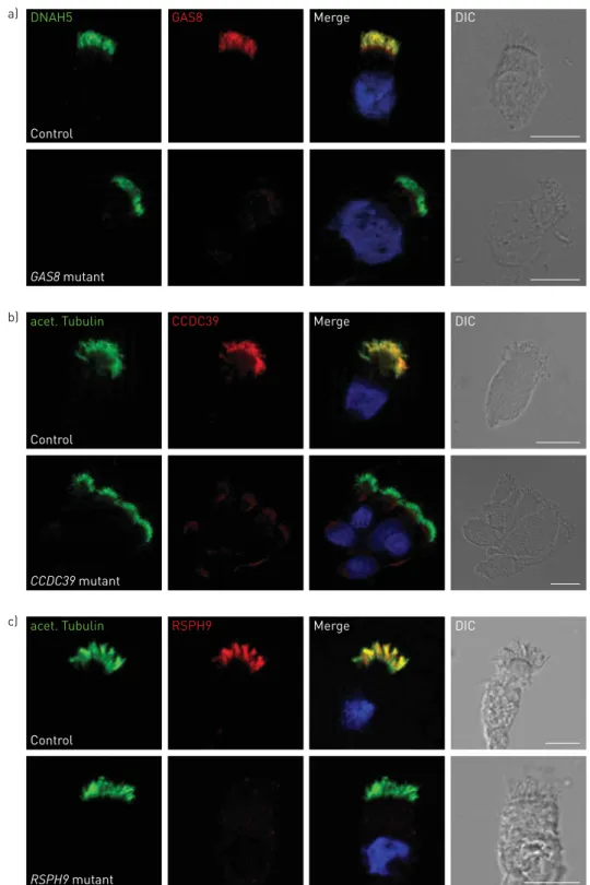
Labelling of ciliary proteins was developed to improve understanding of the impact of disease-causing genes on ciliary proteins [116]. Specific antibodies with secondary fluorescent tags localise to proteins in human respiratory epithelial cells and are visualised by fluorescent or confocal microscopy. A number of antibodies against ciliary proteins are available, including antibodies targeting the ODA, IDA, radial spoke head and dynein regulatory complex proteins. An example of this technique is shown in figure 3.
Respiratory epithelial cells in suspension are placed onto glass slides, air dried and fixed. The cells are incubated with antibodies to ciliary protein not implicated in PCD (e.g. acetylated tubulin) to label the axoneme and combined with antibodies of interest produced in a different species (e.g. anti-DNAH5 to identify ODA structures).
TABLE 8Summary of the Task Force consensus on the published evidence on genetic testing in primary ciliary dyskinesia (PCD) diagnostics
Whilst further evidence in a diagnostic setting is required, experts of the Task Force agreed:
1. Genetic testing to confirm diagnosis can be performed in PCD individuals diagnosed by other means
(e.g.HSVA, TEM, IF) or in individuals with high clinical suspicion for PCD (typical clinical findings, low nNO) and no availability of other investigations, such as HSVA, TEM or IF. A negative genetic test does not exclude PCD.
2. Genetic testing can also be performed to establish diagnosis in patients highly suspected of PCD and in whom HSVA, TEM or IF failed to confirm the diagnosis, as can be the case for patients withDNAH11,CCNO,MCIDASorRSPHgene mutations.
3. Genetic testing and interpretation of results should follow national and international best practice guidelines [114, 115]. 4. Genetic diagnosis has to be consistent with the clinical and TEM/IF/HSVA phenotype, or diagnosis reconsidered if the picture is
inconsistent.
5. Allelic segregation analysis within the family (especially in both parents) is important to confirm the genotype in the probands (to differentiate between homozygosity and hemizygosity, and between compound heterozygosity and a complex allele).
6. Genetic testing in probands and in their relatives is helpful for genetic counselling to inform reproductive choices. 7. In the future genetic testing might be important for genotype specific therapy.
a)
b)
c)
DNAH5
acet. Tubulin
GAS8
CCDC39
Merge
Merge
DIC
DIC
acet. Tubulin RSPH9 Merge DIC
Control
GAS8 mutant
Control
Control CCDC39 mutant
RSPH9 mutant
Review of evidence directly addressing the question“in patients suspected of having PCD, should immunofluorescence be used as a diagnostic tool?”
Our search identified 276 studies (figure S1). No studies reported use of immunofluorescence antibodies in a diagnostic setting and we were, therefore, unable to establish the accuracy of IF as a diagnostic test. 40 studies contributed to our understanding of the potential to use immunofluorescence diagnostically, as summarised below (table S9).
Narrative review of additional evidence
Although the literature focuses on research to understand the downstream effects of mutations in PCD-related genes (table 7), several centres now use immunofluorescence to aid diagnosis [117]. This is likely to increase as more antibodies become available, and once data for the accuracy of the tests becomes available. In the two largest patient-based cohort studies, DNAH5 immunofluorescence was tested in 16 PCD patients and a further 17 families who had ODA defects observed by TEM; mislocalisation of the protein was reported in all cases. Furthermore, DNAH5 protein was present in patients with cystic fibrosis and in healthy controls [94, 95]. A number of studies have used immunofluorescence to examine protein mislocalisation related to genetic mutations (table 7), providing indicators to the antibodies that might be used and findings expected when immunofluorescence is used as a diagnostic test. Immunofluorescence can identify mislocalisation of proteins in PCD patients with a range of mutations, providing information on the pathogenicity of a mutation [86]. However, most manuscripts report immunofluorescence findings from small numbers of patients for each gene and mutation specific findings are not yet known.
With immunofluorescence it is possible to identify almost all ultrastructural abnormalities detectable by TEM and also some cases where the TEM is apparently normal or subtly abnormal [30, 86, 97]. The sensitivity and specificity of immunofluorescence is unknown but will reflect the combination and quality of antibodies. In the authors’ experience, a number of antibodies do not work and validation including appropriate disease and healthy controls is required before they are used diagnostically. Immunofluorescence analysis can be normal if the mutated protein is still expressed within the axoneme [97].
We did not find evidence to either confirm or refute immunofluorescence as a diagnostic test for PCD. Whilst further evidence in a diagnostic setting is required, a summary of the Task Force findings from published evidence are shown in table 9.
Key unanswered questions and research needs
We need validation studies to investigate the accuracy and limitations of immunofluorescence as a diagnostic tool for PCD in diagnostic cohort studies. Each applied antibody needs validation in studies including appropriate PCD disease and healthy controls.
Summary
We were unable to determine the accuracy of immunofluorescence testing due to lack of suitable studies. Task force experts agree that immunofluorescence can be useful in clinical settings. Immunofluorescence is cheaper and easier than other diagnostic tests, providing a potential test for resource-limited settings.
Confirming or excluding a diagnosis of PCD
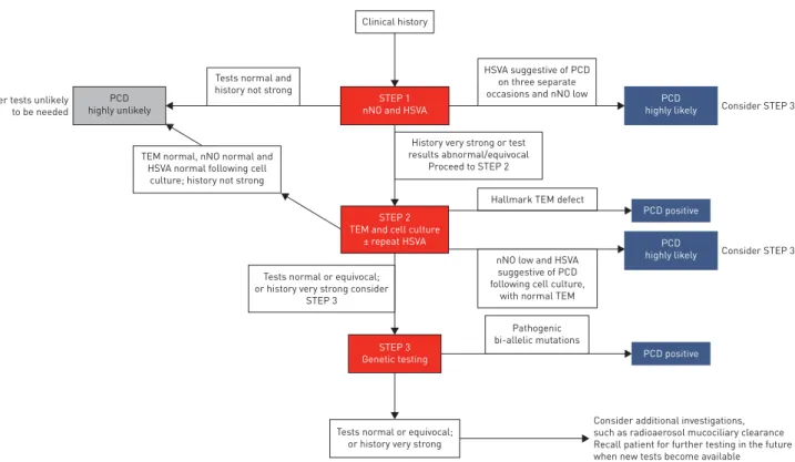
The Delphi Consensus Survey comprised four consecutive online surveys, each building on former rounds. The outcomes of each round are summarised in table S11. Experts from the ERS Task Force agreed (⩾80% of respondents) on the following, which enabled us to propose a diagnostic algorithm (figure 4).
Positive diagnosis
For patients with a supportive history of PCD, the following results are confirmatory of a positive diagnosis of PCD.
1) Hallmark ciliary ultrastructure defects for PCD (absence of ODA, combined absence of IDA and ODA, IDA absence combined with microtubular disarrangement) assessed by TEM.
2) Non-ambiguous bi-allelic mutations in PCD causing genes.
The Task Force did not reach consensus (80%) that any other test in isolation or in combination could provide a conclusive positive diagnosis.
Highly likely diagnosis