ROLES OF THE NF-κB PATHWAY IN GLIOBLASTOMA STEM CELLS AND CHORDOMA
Amanda L. Rinkenbaugh
A dissertation submitted to the faculty of the University of North Carolina at Chapel Hill in partial fulfillment of the requirements for the degree of Doctor of Philosophy in the
Department of Pathology and Laboratory Medicine in the School of Medicine.
Chapel Hill 2016
Approved by:
Albert S. Baldwin, Jr.
© 2016
ABSTRACT
Amanda L. Rinkenbaugh: Roles of the NF-κB Pathway in Glioblastoma Stem Cells and Chordoma
(Under the direction of Albert S. Baldwin, Jr.)
The NF-κB pathway consists of a family of five transcription factors: RelA/p65, RelB, c-Rel, p100/p52, and p105/p50. Originally discovered for its involvement in inflammation and immune signaling, aberrant constitutive NF-κB activation is seen in many tumor types. NF-κB-dependent target gene regulation mediates several hallmarks of cancer, including survival, suppression of apoptosis, and invasion. This work examines NF-κB signaling in both glioblastoma and chordoma samples. In the first project, NF-κB is found to mediate cancer stem cell maintenance in glioblastoma explants. Both genetic and pharmacological NF-κB inhibition impair neurosphere formation at limiting dilutions. Use of
an ex vivo brain slice co-culture model confirmed the in vitro findings, providing a novel
ACKNOWLEDGEMENTS
TABLE OF CONTENTS
LIST OF FIGURES ... iii
LIST OF ABBREVIATIONS ... iv
CHAPTER I: INTRODUCTION ... 1
1.1 Summary ... 1
1.2 NF-κB Signaling ... 2
1.3 NF-κB in Cancer ... 3
1.3.1 NF-κB activation in cancer ... 3
1.3.2 Chronic inflammation as a precursor to cancer ... 5
1.4 Glioblastoma ... 6
1.5 Chordoma ... 7
1.6 Cancer Stem Cells ... 9
1.6.1 NF-κB activation in CSCs ... 11
1.6.2 Connections between NF-κB signaling, cytokines, and CSCs ... 12
1.6.3 Interactions between NF-κB and the tumor microenvironment ... 14
1.6.4 Contributions by the NF-κB pathway to invasion and metastasis ... 15
1.7 NF-κB as a Therapeutic Target ... 17
CHAPTER II: IKK/NF-κB SIGNALING CONTRIBUTES TO
GLIOBLASTOMA STEM CELL MAINTENANCE ... 42
2.1 Summary ... 42
2.2 Introduction ... 44
2.3 Materials and Methods ... 47
2.4 Results ... 51
2.5 Discussion ... 55
REFERENCES ... 66
CHAPTER III: NF-κB SIGNALING CONTRIBUTES TO PROLIFERATION AND INVASION IN CHORDOMA ... 73
3.1 Summary ... 73
3.2 Introduction ... 74
3.3 Materials and Methods ... 76
3.4 Results ... 78
3.5 Discussion ... 80
REFERENCES ... 88
CHAPTER IV: CONCLUSIONS AND FUTURE DIRECTIONS ... 90
4.1 Conclusions and Future Directions ... 90
LIST OF FIGURES
Figure 1.1 Domain organization of NF-κB transcription factor family members ... 21
Figure 1.2 Domain organization of IκB family members ... 22
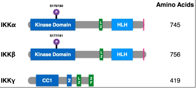
Figure 1.3 Domain organization of IKK family members ... 23
Figure 1.4 Canonical and non-canonical NF-κB signaling pathways ... 24
Figure 1.5 Overview of NF-κB pathway involvement in cancer stem cell biology ... 25
Figure 2.1 NF-κB is preferentially activated in CD133+ glioblastoma stem cells ... 58
Figure 2.2 Pharmacological inhibition of the IKK/NF-κB pathway decreases neurosphere formation ... 59
Figure 2.3 Genetic inhibition of the IKK/NF-κB pathway decreases neurosphere formation ... 61
Figure 2.4 Multiple NF-κB subunits contribute to neurosphere formation ... 62
Figure 2.5 TAK1 activates the NF-κB pathway to promote glioblastoma stem cell function ... 63
Figure 2.6 TGF-β is one source of NF-κB activation in GBM ... 64
Figure 2.7 Inhibition of the IKK/NF-κB pathway decreases glioblastoma growth/ survival ex vivo ... 65
Figure 3.1 NF-κB activation in human chordoma samples ... 82
Figure 3.2 Increased nuclear localization of p65 in chordoma samples ... 83
Figure 3.3 Heterogeneous p65 staining across chordoma cases. ... 84
Figure 3.4 Inhibition of NF-κB impairs proliferation of chordoma cell lines. ... 85
Figure 3.5 IKK inhibition impairs chordoma cell invasion ... 86
LIST OF ABBREVIATIONS
AML: acute myeloid leukemia APC: adenomatous polyposis coli cIAP: cellular inhibitor of apoptosis CML: chronic myeloid leukemia CSC: cancer stem cells
DEN: diethylnitrosamine
DLBCL: diffuse large B cell lymphoma ECM: extracellular matrix
EGF: epidermal growth factor
EGFR: epidermal growth factor receptor EMT: epithelial-mesenchymal transition FGF: fibroblast growth factor
GBM: glioblastoma multiforme GSC: glioblastoma stem cell
IκB: nuclear factor of kappa light polypeptide gene enhancer in B-Cells inhibitor IKK: inhibitor of kappaB kinase
IL-6: interleukin-6 LPS: lipopolysaccharide LZ: leucine zipper
NF-κB: nuclear factor of kappa light polypeptide gene enhancer in B-Cells NIK: NF-κB-inducing kinase
PGE2: prostaglandin E2
PTEN: phosphatase and tensin homolog POSTN: periostin
RANK: receptor activator of NF-κB RHD: Rel homology domain
ROS: reactive oxygen species
STAT: signal transducer and activator of transcription TAK1: TGF-β activated kinase 1
TAD: transcription activation domain TLR: toll-like receptor
CHAPTER I
INTRODUCTION
1.1 Summary
The NF-κB transcription factor pathway is a crucial regulator of inflammation and immune response. Additionally, aberrant NF-κB signaling has been identified in many types of cancer. Downstream of key oncogenic pathways, such as RAS, BCR-ABL, and Her2, NF-κB regulates transcription of target genes that promote cell survival and proliferation, inhibit apoptosis, and mediate invasion and metastasis. The cancer stem cell model posits that a subset of tumor cells (cancer stem cells) drive tumor initiation, exhibit resistance to treatment, and promote recurrence and metastasis. This chapter examines the evidence for a role for NF-κB signaling in glioblastoma and chordoma, with a particular emphasis on cancer stem cell biology.
________________________
1.2 NF-κB Signaling
leads to its cleavage into p52, producing an active RelB-p52 dimer that moves to the nucleus and regulates transcription (Figure 1.4) (Ghosh and Hayden, 2012).
1.3 NF-κB in Cancer
1.3.1 NF-κB activation in cancer
et al., 2009). Expression of the superrepressor form of IκBα (serines 32/36 mutated to alanines, preventing phosphorylation and degradation and leading to decreased NF-κB activity; IκBα-SR) and genetic deletion of IKKβ or RelA in RAS-driven lung tumor and melanoma models strongly suppressed tumor growth (Bassères et al., 2010; Meylan et al., 2009; Yang et al., 2010).
Once activated, NF-κB regulates a wide variety of target genes that overlap heavily with the hallmarks of cancer (Hanahan and Weinberg, 2011). Proliferation is one of the most basic characteristics of a cancer cell and NF-κB is involved through regulation of CyclinD1, Cyclin E, and c-Myc. NF-κB promotes survival and inhibits apoptosis through several mechanisms (Baldwin, 2012). These include transcriptional regulation of the cellular inhibitor of apoptosis (cIAPs) 1, 2, and XIAP, as well as Bcl-2 and Bcl-xL (Chu et al., 1997; Ramakrishnan et al., 2010; Wang et al., 1998). Perhaps as expected, NF-κB regulates a number of cytokines that contribute to tumor-promoting inflammation such as: TNFα, IL-1, IL6, MCP1, COX2, and iNOS. Other NF-κB targets contribute to epithelial-mesenchymal transition (vimentin, Twist), remodeling the extracellular matrix through induction of angiogenesis (IL8, VEGF), and promotion of invasion and metastasis (MMP2, MMP9, uPA) (Bassères and Baldwin, 2006).
2010; Boehm et al., 2007; Orlowski and Baldwin, 2002). Rearrangements of the NFKB2 locus (gene name for the p100 subunit) that lead to loss of the inhibitory IκB-like domain and increased p52 production are found in some B cell lymphomas (Neri et al., 1991). C11orf95-RELA fusions have been described as driver events in ependymomas (Parker et al., 2014), while monoallelic deletions of IκBα were identified in a subset of glioblastoma (Bredel et al., 2011). Mutations in upstream proteins that lead to aberrant, constitutive NF-κB activation have been identified. For example, in certain subtypes of lymphoma, translocations can affect MALT1 and BCL10, while CARD11 features a variety of point mutations. All three of these proteins interact to form a complex that drives NF-κB activation (Lim et al., 2012). Growth factor receptors, including EGFR and Her2, are frequently overexpressed in cancer and activate similar pathways, including NF-κB (Merkhofer et al., 2010; Tanaka et al., 2011).
IKK exhibits NF-κB-independent functions that promote growth and survival functions important to a variety of cancer cells. For example, IKKα and IKKβ promote mTOR activation, via kinase activity (Dan et al., 2007; 2008; 2014; Xu et al., 2013). Another example is that IKKα was found to phosphorylate the CDK inhibitor p27 downstream of Her2 to promote cancer stem cell self-renewal (Zhang et al., 2013). IKKβ was reported to phosphorylate the tumor suppressor p53 to promote its instability (Xia et al., 2009).
1.3.2 Chronic inflammation as a precursor to cancer
cancer with targeted IKKβ deletion in either the epithelial or myeloid compartments, NF-κB mediated survival of intestinal epithelial cells, while NF-κB activation in myeloid cells drove production of growth factors that promoted tumor proliferation (Greten et al., 2004). In a model of hepatocellular carcinoma driven by treatment with the carcinogen diethylnitrosamine (DEN), NF-κB is again activated in the myeloid compartment, this time to drive IL6 production and subsequent STAT3 activation in the hepatocytes (Maeda et al., 2005). Interestingly, this model shows that deletion of IKKβ or IKKγ actually leads to enhanced tumor development. The liver initially shows more cell death following DEN treatment; however because hepatocytes are highly regenerative, the cell death triggers proliferation of the remaining cells (Sakurai et al., 2006). It is thought that a cycle of injury, cell death, and proliferation drives tumor formation in this model (DiDonato et al., 2012). NF-κB was shown to be activated in cancer-associated fibroblasts promoting the expression of inflammatory cytokines, although the role of this response in promoting tumorigenesis is controversial (Erez et al., 2010; Koliaraki et al., 2015; Pallangyo et al., 2015). Work examining tumor-associated macrophages has shown that NF-κB signaling maintains a tumor-promoting, immunosuppressive (or M2) phenotype and inhibits a tumor-suppressing (or M1) phenotype (Hagemann et al., 2008; Saccani et al., 2006). Taken together, these studies start to describe a complex microenvironment with multiple cell types interacting to drive tumorigenesis and place NF-κB as a central mediator between these various components.
1.4 Glioblastoma
care treatment currently involves a combination of surgery, radiation, and chemotherapy, however median survival for these patients is only 14.6 months. Improvements in GBM treatment have not been frequent or substantial. In addition to the difficulties that accompany developing any new cancer therapy, GBM provides the extra hurdle of blood-brain barrier penetrance. While some drugs may show anti-tumorigenic activity in vitro, many are unable to cross the blood-brain barrier, and thus never reach the cancer cells they are supposed to target. The approval of temozolomide as a new chemotherapeutic treatment option in 2005 was one of the biggest additions in several decades of GBM research, yet the added survival benefit was a mere 2.5 months (Stupp et al., 2005).
Histopathologically, GBM is described as a heterogeneous tumor type with high levels of angiogenesis and invasion. The Cancer Genome Atlas (TCGA) analyzed around 200 GBMs and based on gene expression patterns clustered them into four subtypes: neural, proneural, classical, and mesenchymal (Verhaak et al., 2010). The most common genetic alterations associated with these tumors include EGFR amplification and mutation, loss of PTEN, and loss of p53, all of which have been associated with increased NF-κB activity (Brennan et al., 2013; Cooks et al., 2013; Gustin et al., 2001; Kaus et al., 2010; Tanaka et al., 2011; Verhaak et al., 2010). In GBM, NF-κB has been reported to regulate survival, invasion, and resistance to both radiation and chemotherapy (Atkinson et al., 2009; Bredel et al., 2006; Fukushima et al., 2012; Holmes et al., 2012; Li et al., 2010; Raychaudhuri et al., 2007; Tanaka et al., 2011; Xi et al., 2015).
1.5 Chordoma
been well-studied and the molecular underpinnings of these tumors remain fairly uncharacterized when compared to other tumor types, such as glioblastoma. Though the tumors are relatively slow-growing, they present other clinical challenges as the anatomical location in the central nervous system makes surgery more difficult. Additionally, they are resistant to chemotherapy and radiation (Chugh et al., 2007; Forsyth et al., 1993). Furthermore, they tend to recur and are both locally invasive and capable of metastasis, particularly to the lungs, bone, and liver. Current treatment generally includes radical surgery and high-dose radiation; however treatment is not standardized due to low patient volume and lack of molecular characterization.
One of the major molecular determinants that has been associated with chordoma is brachyury expression. Brachyury (gene name T) is a T-box transcription factor expressed in the notochord during development, as well as specifically expressed in chordoma (Vujovic et al., 2006). Its expression is actually a requirement for the validation of pathology specimens or cell lines as bona fide chordomas. In familial chordoma, germline gene duplication of the T locus was identified in several families (Yang et al., 2009). Subsequently, a SNP within T
was found to be present in 86% of chordoma patients versus 56% in controls (Pillay et al., 2012). Interestingly, this allele appears to affect both expression levels and DNA binding of the transcription factor, consistent with a functional role in chordoma development (Papapetrou et al., 1997).
allow the tumor cells to disseminate from the primary tumor. Many factors have been associated with this process including the matrix metalloproteinases (MMPs). MMPs are able to break down a wide variety of substrates, including collagen, gelatin, fibronectin, and laminin. Epidermal overexpression of type I collagenase (MMP1) in a carcinogenesis model led to a significant increase in tumor incidence (D'Armiento et al., 1995). Conversely, deletion of MMP7 in the Min/+ model of colon cancer led to a marked decrease in tumor formation (Wilson et al., 1997). Both of these results demonstrate involvement of the MMP family in promotion of cancer progression, but do not specifically involve invasive phenotypes. More recent studies have found that MMP9 expression is upregulated in invasive skin cancer lesions and MMP7 progressively accumulates as pancreatic tumors become metaplastic (Crawford et al., 2002; Kupferman et al., 2000). In chordoma, both MMP1 and MMP2 expression has been correlated with increased infiltration of bone as well as poor prognosis in patient specimens (Naka et al., 2004; 2008).
1.6 Cancer Stem Cells
many solid tumors including those of the brain, prostate, breast, colon, and pancreas (Al-Hajj et al., 2003; Collins et al., 2005; Li et al., 2007; Ricci-Vitiani et al., 2006; Singh et al., 2003; 2004). In addition to being responsible for primary tumor formation, CSCs are also generally thought to drive metastasis and exhibit increased resistance to radiation and chemotherapy. Due to their stem-like characteristics, these cells are also capable of differentiation into multiple lineages, which accounts for some of the heterogeneity seen in tumors (Reya et al., 2001). While CSCs are frequently depicted at the top of a hierarchically arranged tumor, there is evidence that plasticity allows for the conversion of bulk tumor cells into CSCs (Chen et al., 2010).
Several assays allow for the study of CSCs. In vitro experiments focus on sphere formation under stem cell permissive conditions, such as serum-free media supplemented with essential growth factors and low-adherence plates. Ideally, these experiments are performed at limiting dilutions to best assess self-renewal from single cells. Additionally, in vivo assessments of tumor formation remain the gold standard for true CSC function, again
to dissect the differences between the cell types. Proliferation, survival, and gene expression analyses are commonly measured.
1.6.1 NF-κB activation in CSCs
One of the earliest examples of NF-κB involvement in CSCs came from primary AML samples, where the CD34+ cells showed enhanced NF-κB DNA binding that was not seen in regular hematopoietic stem cells (Guzman et al., 2001). Since that initial report, elevated or constitutive NF-κB activity has been seen in many tumor types. Prostate CSCs were found to express higher levels of acetylated and total p65, as well as a decrease in IκBα expression when compared to parental tumors (Rajasekhar et al., 2011). In glioblastoma, CSCs exhibited increased nuclear localization of p65 as compared with cells cultured under monolayer conditions (Garner et al., 2013). Tumorsphere-forming cells showed increased phosphorylation of p65, again consistent with elevated NF-κB signaling in this population of cells. In that study, inhibition of NF-κB reduced self-renewal and blocked xenograft tumor growth using a limiting dilution approach (Song et al., 2012). In addition to direct evidence of preferential NF-κB activation in CSC subsets of tumors, several groups have taken an unbiased approach of profiling gene expression and defining CSC-associated signatures. This has revealed an inflammatory signature, which can frequently be tightly associated with NF-κB regulation, in a variety of tumors such as glioblastoma, breast, prostate, and ovarian cancers (Birnie et al., 2008; Korkaya et al., 2011; Leizer et al., 2010; Liu et al., 2007; Murohashi et al., 2010; Tafani et al., 2011).
Her2-driven breast cancer, both canonical and non-canonical NF-κB pathways contribute to stemness and tumor formation. Expression of IκBα-SR impaired the formation of luminal epithelial tumors. Use of an NF-κB-GFP reporter allele localized activation to the luminal progenitors (Pratt et al., 2009). Another analysis of IκBα-SR in a Her2 mouse model found changes in a gene signature associated with stem cells, then specifically showed NF-κ B-dependent changes in the specific stem cell factors Nanog and Sox2 (Figure 1.5) (Liu et al., 2010). Knock-in of a kinase dead IKKα led to decreased self-renewal and senescence under mammary stem cell culture conditions (Cao et al., 2007). In the Her2 breast cancer model, IKKα was found to phosphorylate p27 leading to its nuclear export and promoting CSC proliferation and expansion (Zhang et al., 2013). One of the canonical alterations that occurs during colorectal tumorigenesis is loss of APC. Myant and colleagues found that APC loss drives RAC1 activity to mediate ROS production and NF-κB activation, ultimately leading to an expansion of Lgr5+ CSCs (Myant et al., 2013).
1.6.2 Connections between NF-κB signaling, cytokines, and CSCs
levels of TNFα than normal hematopoietic stem cells. Canonical NF-κB activation positively regulates expression of IL3 and granulocyte/macrophage colony-stimulating factor common β-chain receptor (CSF2RB) to promote proliferation and survival of CML stem cells (Gallipoli et al., 2013). Similar findings in a mouse model of acute myeloid leukemia (AML) described a feedback loop between TNFα and NF-κB, confirmed by correlations in patient samples (Kagoya et al., 2014). TNFα treatment of MCF7 breast cancer cells increased their mammosphere-forming capacity through upregulation of NF-κB and subsequently Slug (Figure 1.5) (Storci et al., 2010). In colorectal cancer, levels of prostaglandin E2 (PGE2) correlated with CSC markers in human tumor samples. Treatment of either a genetic or xenograft mouse model with PGE2 led to CSC expansion through upregulation of several signaling pathways including NF-κB (Wang et al., 2015). In glioblastoma, IL-17 receptor was found to be co-expressed with multiple CSC markers, including CD133, Nestin, and Sox2, as well as a source of NF-κB activation (Parajuli et al., 2016).
mammosphere formation, but addition of exogenous IL6 or IL-1β rescues the defect
(Kendellen et al., 2013). In CML, increased levels of IL6 drive CML progenitors into the
myeloid lineage, sustaining CML development (Reynaud et al., 2011). IL6, IL8, and MCP1 similarly contribute to the survival and self-renewal of glioblastoma CSCs (Figure 1.5) (Parajuli et al., 2016; Wang et al., 2009).
1.6.3 Interactions between NF-κB and the tumor microenvironment
component of the extracellular matrix that has been identified in the niche of both normal and cancer stem cells. Generally thought to be produced by stromal fibroblasts, POSTN promotes metastasis to the lung in a breast cancer model by supporting the growth and expansion of CSCs (Malanchi et al., 2012). Another group found that breast CSCs express higher levels of POSTN in vitro than their non-CSC counterparts. POSTN drives an ERK-NF-κB signaling axis, driving production of IL6 and IL8, which in turn contribute to CSC maintenance through STAT3 activation (Lambert et al., 2016). Breast cancer also exhibits a circuit of progestin-driven RANKL (receptor activator of NF-κB ligand) expression, leading to NF-κB activation. Deletion of the RANKL receptor RANK decreases the CD49fhi-CSC population and tumor incidence (Figure 1.5) (Schramek et al., 2010).
1.6.4 Contributions by the NF-κB pathway to invasion and metastasis
NF-κB frequently cooperates with additional signaling pathways to mediate these oncogenic effects. Coordinated activity between NF-κB and STAT3 has been previously mentioned in this review. Concurrent constitutive signaling from NF-κB and STAT3 in glioblastoma CSCs regulates expression of a set of genes (NOTCH1, HES5, JAG1, NUMBL, DTX3, DVL3, and RBPJ) that drive activation of Notch signaling, a third CSC-associated pathway (Figure 2) (Garner et al., 2013). Another experiment, suggesting an important interaction between the CSCs and the bulk tumor cells, found that NF-κB activity in the non-CSCs upregulates JAG1 to stimulate Notch signaling in proximal breast non-CSCs (Yamamoto et al., 2013).
The majority of the findings discussed here have focused on the canonical NF-κB pathway, particularly the p65 subunit. However, there is also evidence that the non-canonical pathway contributes to CSC phenotypes. In breast cancer, knockdown of IKKα, p100/p52, or RelB all produced a decrease in mammosphere formation (Kendellen et al., 2013). Eva1, found to be overexpressed in glioblastoma CSCs, drives NIK stabilization and p100 processing, potentially by promoting ubiquitination and degradation of TRAF2 and cIAP (Ohtsu et al., 2016). RelB has been described as an oncogenic driver in mesenchymal glioma, regulating Olig2 expression and promoting tumor growth and invasion (Lee et al., 2013).
1.7 NF-κB as a Therapeutic Target
and/or metastasis in vivo. A combination of idarubicin and MG132, a proteasome inhibitor, induced cell death in AML stem cells, partially through NF-κB inhibition (Guzman et al., 2002). While proteasome inhibition will impact several pathways in a cell, NF-κB inhibition is a well-established effect of MG132 treatment as it blocks IκBα degradation and effectively sequesters NF-κB subunits in the cytoplasm. The same group went on to identify the compound parthenolide as selectively inducing apoptosis in leukemia stem cells as opposed to normal hematopoietic stem cells, through a mechanism of increased reactive oxygen species, p53 activation, and NF-κB inhibition (Guzman et al., 2005). A subsequent in silico screen for additional drugs with specificity towards AML stem cells identified two other compounds, celastrol and 4-hydroxy-2-nonenal, and once again they found NF-κB inhibition to be part of the mechanism of action (Hassane et al., 2008). Parthenolide has also shown preferential activity in breast CSCs compared to the bulk tumor cells (Zhou et al., 2007). Use of SN50, a peptide inhibitor that blocks nuclear import of NF-κB and other transcription factors, decreases the sphere formation ability and tumorigenic capacity of glioma CSCs (Zhang et al., 2014). Others have found that inhibition of NF-κB promotes more rapid differentiation and progression to senescence in glioblastoma CSCs (Nogueira et al., 2011).
studies have found an impact of IKK/NF-κB inhibition on tumor growth. While this work didn’t specifically analyze CSC effects, if CSCs are primarily driving tumor initiation, we could interpret these results as having some effect on the CSC population. Direct IKKβ inhibitors showed efficacy in a mutant KRas, p53-null model of lung cancer (Bassères et al., 2014; Xue et al., 2011). In addition to inhibitors targeting the kinase activity, the NF-κB pathway can be inhibited by peptides encompassing the NEMO-binding domain (NBD) that block association of the IKK catalytic subunits with NEMO/IKKγ. Recently, use of an NBD peptide slowed tumor growth in both a human glioma xenograft and a genetic mouse model of glioma (Friedmann-Morvinski et al., 2016). The NBD peptide has also shown efficacy in a canine model of DLBCL (Gaurnier-Hausser et al., 2011; Habineza Ndikuyeze et al., 2014).
effective treatment against CSCs, but it could also restore sensitivity to other therapeutic options.
1.8 Conclusions
Figure 1.1 Domain organization of NF-κB transcription factor family members
Figure 1.2 Domain organization of IκB family members
Figure 1.3 Domain organization of IKK family members
Figure 1.4 Canonical and non-canonical NF-κB signaling pathways
REFERENCES
Akinleye, A., Chen, Y., Mukhi, N., Song, Y., and Liu, D. (2013). Ibrutinib and novel BTK inhibitors in clinical development. J. Hematol. Oncol. 6, 59.
Al-Hajj, M., Wicha, M.S., Benito-Hernandez, A., Morrison, S.J., and Clarke, M.F. (2003). Prospective identification of tumorigenic breast cancer cells. Proc. Natl. Acad. Sci. U.S.A. 100, 3983–3988.
Alvero, A.B., Fu, H.H., Holmberg, J., Visintin, I., Mor, L., Marquina, C.C., Oidtman, J., Silasi, D.-A., and Mor, G. (2009). Stem-like ovarian cancer cells can serve as tumor vascular progenitors. Stem Cells 27, 2405–2413.
An, J., and Rettig, M.B. (2005). Mechanism of von Hippel-Lindau protein-mediated suppression of nuclear factor-kappaB activity. Mol. Cell. Biol. 25, 7546–7556.
An, J., Fisher, M., and Rettig, M.B. (2004). VHL expression in renal cell carcinoma sensitizes to bortezomib (PS-341) through an NF-κB-dependent mechanism. Oncogene 24, 1563–1570.
Asano, T., Yao, Y., Zhu, J., Li, D., Abbruzzese, J.L., and Reddy, S.A.G. (2004). The PI3-kinase/Akt signaling pathway is activated due to aberrant Pten expression and targets transcription factors NF-κB and c-Myc in pancreatic cancer cells. Oncogene 23, 8571–8580. Asiedu, M.K., Beauchamp-Perez, F.D., Ingle, J.N., Behrens, M.D., Radisky, D.C., and Knutson, K.L. (2013). AXL induces epithelial-to-mesenchymal transition and regulates the function of breast cancer stem cells. Oncogene 33, 1316–1324.
Atkinson, G.P., Nozell, S.E., Harrison, D.K., Stonecypher, M.S., Chen, D., and Benveniste, E.N. (2009). The prolyl isomerase Pin1 regulates the NF-kappaB signaling pathway and interleukin-8 expression in glioblastoma. Oncogene 28, 3735-3745.
Baldwin, A.S. (2012). Regulation of cell death and autophagy by IKK and NF-κB: critical mechanisms in immune function and cancer. Immunol. Rev. 246, 327–345.
Barberà, M.J., Puig, I., Domínguez, D., Julien-Grille, S., Guaita-Esteruelas, S., Peiró, S., Baulida, J., Francí, C., Dedhar, S., Larue, L., et al. (2004). Regulation of Snail transcription during epithelial to mesenchymal transition of tumor cells. Oncogene 23, 7345–7354.
Bassères, D.S., and Baldwin, A.S. (2006). Nuclear factor-κB and inhibitor of κB kinase pathways in oncogenic initiation and progression. Oncogene 25, 6817–6830.
3546.
Belguise, K., Guo, S., Yang, S., Rogers, A.E., Seldin, D.C., Sherr, D.H., and Sonenshein, G.E. (2007). Green tea polyphenols reverse cooperation between c-Rel and CK2 that induces the aryl hydrocarbon receptor, Slug, and an invasive phenotype. Cancer Res. 67, 11742– 11750.
Beroukhim, R., Mermel, C.H., Porter, D., Wei, G., Raychaudhuri, S., Donovan, J., Barretina, J., Boehm, J.S., Dobson, J., Urashima, M., et al. (2010). The landscape of somatic copy-number alteration across human cancers. Nature 463, 899–905.
Birnie, R., Bryce, S.D., Roome, C., Dussupt, V., Droop, A., Lang, S.H., Berry, P.A., Hyde, C.F., Lewis, J.L., Stower, M.J., et al. (2008). Gene expression profiling of human prostate cancer stem cells reveals a pro-inflammatory phenotype and the importance of extracellular matrix interactions. Genome Biol. 9, R83.
Boehm, J.S., Zhao, J.J., Yao, J., Kim, S.Y., Firestein, R., Dunn, I.F., Sjostrom, S.K., Garraway, L.A., Weremowicz, S., Richardson, A.L., et al. (2007). Integrative genomic approaches identify IKBKE as a breast cancer oncogene. Cell 129, 1065–1079.
Bonnet, D., and Dick, J.E. (1997). Human acute myeloid leukemia is organized as a hierarchy that originates from a primitive hematopoietic cell. Nature Med. 3, 730–737. Bradford, J.W., and Baldwin, A.S. (2014). IKK/nuclear factor-kappaB and oncogenesis: roles in tumor-initiating cells and in the tumor microenvironment. Adv. Cancer Res. 121, 125–145. Brassesco, M.S., Roberto, G.M., Morales, A.G., Oliveira, J.C., Delsin, L.E.A., Pezuk, J.A., Valera, E.T., Carlotti, C.G., Rego, E.M., de Oliveira, H.F., et al. (2013). Inhibition of NF-κB by dehydroxymethylepoxyquinomicin suppresses invasion and synergistically potentiates temozolomide and γ-radiation cytotoxicity in glioblastoma cells. Chemother. Res. Pract. 2013, 1–16.
Bredel, M., Bredel, C., Juric, D., Duran, G.E., Yu, R.X., Harsh, G.R., Vogel, H., Recht, L.D., Scheck, A.C., and Sikic, B.I. (2006). Tumor necrosis factor-alpha-induced protein 3 as a putative regulator of nuclear factor-kappaB-mediated resistance to O6-alkylating agents in human glioblastomas. J. Clin. Oncol. 24, 274–287.
Bredel, M., Scholtens, D.M., Yadav, A.K., Alvarez, A.A., Renfrow, J.J., Chandler, J.P., Yu, I.L.Y., Carro, M.S., Dai, F., Tagge, M.J., et al. (2011). NFKBIA deletion in glioblastomas. N. Engl. J. Med. 364, 627–637.
Brennan, C.W., Verhaak, R.G.W., McKenna, A., Campos, B., Noushmehr, H., Salama, S.R., Zheng, S., Chakravarty, D., Sanborn, J.Z., Berman, S.H., et al. (2013). The somatic genomic landscape of glioblastoma. Cell 155, 462–477.
Calabrese, C., Poppleton, H., Kocak, M., Hogg, T.L., Fuller, C., Hamner, B., Oh, E.Y., Gaber, M.W., Finklestein, D., Allen, M., et al. (2007). A perivascular niche for brain tumor stem cells. Cancer Cell 11, 69–82.
Cao, L., Fan, X., Jing, W., Liang, Y., Chen, R., Liu, Y., Zhu, M., Jia, R., Wang, H., Zhang, X., et al. (2015). Osteopontin promotes a cancer stem cell-like phenotype in hepatocellular carcinoma cells via an integrin-NF-κB-HIF-1α pathway. Oncotarget 6, 6627–6640.
Cao, Y., Luo, J.-L., and Karin, M. (2007). IkappaB kinase alpha kinase activity is required for self-renewal of ErbB2/Her2-transformed mammary tumor-initiating cells. Proc. Natl. Acad. Sci. U.S.A. 104, 15852–15857.
Charles, N., Ozawa, T., Squatrito, M., Bleau, A.-M., Brennan, C.W., Hambardzumyan, D., and Holland, E.C. (2010). Perivascular nitric oxide activates notch signaling and promotes stem-like character in PDGF-induced glioma cells. Cell Stem Cell 6, 141–152.
Chefetz, I., Alvero, A., Holmberg, J., Lebowitz, N., Craveiro, V., Yang-Hartwich, Y., Yin, G., Squillace, L., Gurrea Soteras, M., Aldo, P., et al. (2014). TLR2 enhances ovarian cancer stem cell self-renewal and promotes tumor repair and recurrence. Cell Cycle 12, 511–521. Chen, R., Nishimura, M.C., Bumbaca, S.M., Kharbanda, S., Forrest, W.F., Kasman, I.M., Greve, J.M., Soriano, R.H., Gilmour, L.L., Rivers, C.S., et al. (2010). A hierarchy of self-renewing tumor-initiating cell types in glioblastoma. Cancer Cell 17, 362–375.
Chu, Z.L., McKinsey, T.A., Liu, L., Gentry, J.J., Malim, M.H., and Ballard, D.W. (1997). Suppression of tumor necrosis factor-induced cell death by inhibitor of apoptosis c-IAP2 is under NF-kappaB control. Proc. Natl. Acad. Sci. U.S.A. 94, 10057–10062.
Chua, H.L., Bhat-Nakshatri, P., Clare, S.E., Morimiya, A., Badve, S., and Nakshatri, H. (2006). NF-κB represses E-cadherin expression and enhances epithelial to mesenchymal transition of mammary epithelial cells: potential involvement of ZEB-1 and ZEB-2. Oncogene 26, 711–724.
Chugh, R., Tawbi, H., Lucas, D.R., Biermann, J.S., Schuetze, S.M., and Baker, L.H. (2007). Chordoma: the nonsarcoma primary bone tumor. The Oncologist 12, 1344–1350.
Collins, A.T., Berry, P.A., Hyde, C., Stower, M.J., and Maitland, N.J. (2005). Prospective identification of tumorigenic prostate cancer stem cells. Cancer Res. 65, 10946–10951. Connelly, L., Robinson-Benion, C., Chont, M., Saint-Jean, L., Li, H., Polosukhin, V.V., Blackwell, T.S., and Yull, F.E. (2007). A transgenic model reveals important roles for the NF-kappa B alternative pathway (p100/p52) in mammary development and links to tumorigenesis. J. Biol. Chem. 282, 10028–10035.
Crawford, H.C., Scoggins, C.R., Washington, M.K., Matrisian, L.M., and Leach, S.D. (2002). Matrix metalloproteinase-7 is expressed by pancreatic cancer precursors and regulates acinar-to-ductal metaplasia in exocrine pancreas. J. Clin. Invest. 109, 1437–1444. Cusack, J.C., Liu, R., Houston, M., Abendroth, K., Elliott, P.J., Adams, J., and Baldwin, A.S. (2001). Enhanced chemosensitivity to CPT-11 with proteasome inhibitor PS-341: implications for systemic nuclear factor-kappaB inhibition. Cancer Res. 61, 3535–3540. D'Armiento, J., DiColandrea, T., Dalal, S.S., Okada, Y., Huang, M.T., Conney, A.H., and Chada, K. (1995). Collagenase expression in transgenic mouse skin causes hyperkeratosis and acanthosis and increases susceptibility to tumorigenesis. Mol. Cell. Biol. 15, 5732–5739. Dalerba, P., Dylla, S.J., Park, I.-K., Liu, R., Wang, X., Cho, R.W., Hoey, T., Gurney, A., Huang, E.H., Simeone, D.M., et al. (2007). Phenotypic characterization of human colorectal cancer stem cells. Proc. Natl. Acad. Sci. U.S.A. 104, 10158–10163.
Dan, H.C., Adli, M., and Baldwin, A.S. (2007). Regulation of mammalian target of rapamycin activity in PTEN-inactive prostate cancer cells by IkappaB kinase alpha. Cancer Res. 67, 6263–6269.
Dan, H.C., Cooper, M.J., Cogswell, P.C., Duncan, J.A., Ting, J.P.Y., and Baldwin, A.S. (2008). Akt-dependent regulation of NF-kappaB is controlled by mTOR and Raptor in association with IKK. Genes Dev. 22, 1490–1500.
Dan, H.C., Ebbs, A., Pasparakis, M., Van Dyke, T., Bassères, D.S., and Baldwin, A.S. (2014). Akt-dependent activation of mTORC1 complex involves phosphorylation of mTOR (mammalian target of rapamycin) by IkappaB kinase alpha (IKKalpha). J. Biol. Chem. 289, 25227–25240.
Di Minin, G., Bellazzo, A., Dal Ferro, M., Chiaruttini, G., Nuzzo, S., Bicciato, S., Piazza, S., Rami, D., Bulla, R., Sommaggio, R., et al. (2014). Mutant p53 reprograms TNF signaling in cancer cells through interaction with the tumor suppressor DAB2IP. Mol. Cell 56, 617–629. DiDonato, J.A., Mercurio, F., and Karin, M. (2012). NF-κB and the link between inflammation and cancer. Immunol. Rev. 246, 379–400.
Erez, N., Truitt, M., Olson, P., and Hanahan, D. (2010). Cancer-associated fibroblasts are activated in incipient neoplasia to orchestrate tumor-promoting inflammation in an NF-κ B-dependent manner. Cancer Cell 17, 135–147.
Es-haghi, M., Soltanian, S., and Dehghani, H. (2015). Cooperation of Nanog, NF-κΒ, and CXCR4 in a regulatory network for directed migration of cancer stem cells. Tumor Biol. 37, 1559-1965.
Differ. 10, 353–367.
Finco, T.S., Westwick, J.K., Norris, J.L., Beg, A.A., Der, C.J., and Baldwin, A.S. (1997). Oncogenic Ha-Ras-induced signaling activates NF-kappaB transcriptional activity, which is required for cellular transformation. J. Biol. Chem. 272, 24113–24116.
Forsyth, P.A., Cascino, T.L., Shaw, E.G., Scheithauer, B.W., O'Fallon, J.R., Dozier, J.C., and Piepgras, D.G. (1993). Intracranial chordomas: a clinicopathological and prognostic study of 51 cases. J. Neurosurg. 78, 741–747.
Friedmann-Morvinski, D., Narasimamurthy, R., Xia, Y., Myskiw, C., Soda, Y., and Verma, I.M. (2016). Targeting NF-kappaB in glioblastoma: A therapeutic approach. Science Advances 2, e1501292–e1501292.
Fukushima, T., Kawaguchi, M., Yorita, K., Tanaka, H., Takeshima, H., Umezawa, K., and Kataoka, H. (2012). Antitumor effect of dehydroxymethylepoxyquinomicin, a small molecule inhibitor of nuclear factor-κB, on glioblastoma. Neuro-Oncology 14, 19–28.
Gallipoli, P., Pellicano, F., Morrison, H., Laidlaw, K., Allan, E.K., Bhatia, R., Copland, M., Jørgensen, H.G., and Holyoake, T.L. (2013). Autocrine TNF-α production supports CML stem and progenitor cell survival and enhances their proliferation. Blood 122, 3335–3339. Garner, J.M., Fan, M., Yang, C.H., Du, Z., Sims, M., Davidoff, A.M., and Pfeffer, L.M. (2013). Constitutive activation of signal transducer and activator of transcription 3 (STAT3) and nuclear factor-kappaB signaling in glioblastoma cancer stem cells regulates the Notch pathway. J. Biol. Chem. 288, 26167–26176.
Gaurnier-Hausser, A., Patel, R., Baldwin, A.S., May, M.J., and Mason, N.J. (2011). NEMO-binding domain peptide inhibits constitutive NF-kappaB activity and reduces tumor burden in a canine model of relapsed, refractory Diffuse Large B-Cell Lymphoma. Clin. Can. Res. 17, 4661–4671.
Ghosh, S., and Hayden, M.S. (2012). Celebrating 25 years of NF-κB research. Immunol. Rev. 246, 5–13.
Greten, F.R., Eckmann, L., Greten, T.F., Park, J.M., Li, Z.-W., Egan, L.J., Kagnoff, M.F., and Karin, M. (2004). IKKβ links inflammation and tumorigenesis in a mouse model of colitis-associated cancer. Cell 118, 285–296.
Gustin, J.A., Maehama, T., Dixon, J.E., and Donner, D.B. (2001). The PTEN tumor suppressor protein inhibits tumor necrosis factor-induced nuclear factor-kappaB activity. J. Biol. Chem. 276, 27740–27744.
Guzman, M.L., Neering, S.J., Upchurch, D., Grimes, B., Howard, D.S., Rizzieri, D.A., Luger, S.M., and Jordan, C.T. (2001). Nuclear factor-kappaB is constitutively activated in primitive human acute myelogenous leukemia cells. Blood 98, 2301–2307.
Jordan, C.T. (2005). The sesquiterpene lactone parthenolide induces apoptosis of human acute myelogenous leukemia stem and progenitor cells. Blood 105, 4163–4169.
Guzman, M.L., Swiderski, C.F., Howard, D.S., Grimes, B.A., Rossi, R.M., Szilvassy, S.J., and Jordan, C.T. (2002). Preferential induction of apoptosis for primary human leukemic stem cells. Proc. Natl. Acad. Sci. U.S.A. 99, 16220–16225.
Habineza Ndikuyeze, G., Gaurnier-Hausser, A., Patel, R., Baldwin, A.S., May, M.J., Flood, P., Krick, E., Propert, K.J., and Mason, N.J. (2014). A Phase I clinical trial of systemically delivered NEMO binding domain peptide in dogs with spontaneous Activated B-Cell like Diffuse Large B-Cell Lymphoma. PLoS ONE 9, e95404.
Hagemann, T., Lawrence, T., McNeish, I., Charles, K.A., Kulbe, H., Thompson, R.G., Robinson, S.C., and Balkwill, F.R. (2008). “Re-educating” tumor-associated macrophages by targeting NF-kappaB. J. Exp. Med. 205, 1261–1268.
Han, Y.P., Tuan, T.L., Wu, H., Hughes, M., and Garner, W.L. (2001). TNF-alpha stimulates activation of pro-MMP2 in human skin through NF-(kappa)B mediated induction of MT1-MMP. J. Cell. Sci. 114, 131–139.
Hanahan, D., and Weinberg, R.A. (2011). Hallmarks of cancer: the next generation. Cell 144, 646–674.
Hassane, D.C., Guzman, M.L., Corbett, C., Li, X., Abboud, R., Young, F., Liesveld, J.L., Carroll, M., and Jordan, C.T. (2008). Discovery of agents that eradicate leukemia stem cells using an in silico screen of public gene expression data. Blood 111, 5654–5662.
Hayden, M.S., and Ghosh, S. (2008). Shared principles in NF-κB signaling. Cell 132, 344– 362.
Helbig, G., Christopherson, K.W., Bhat-Nakshatri, P., Kumar, S., Kishimoto, H., Miller, K.D., Broxmeyer, H.E., and Nakshatri, H. (2003). NF-kappaB promotes breast cancer cell migration and metastasis by inducing the expression of the chemokine receptor CXCR4. J. Biol. Chem. 278, 21631–21638.
Hemmati, H.D., Nakano, I., Lazareff, J.A., Masterman-Smith, M., Geschwind, D.H., Bronner-Fraser, M., and Kornblum, H.I. (2003). Cancerous stem cells can arise from pediatric brain tumors. Proc. Natl. Acad. Sci. U.S.A. 100, 15178–15183.
Himelstein, B.P., Lee, E.J., Sato, H., Seiki, M., and Muschel, R.J. (1997). Transcriptional activation of the matrix metalloproteinase-9 gene in an H-ras and v-myc transformed rat embryo cell line. Oncogene 14, 1995–1998.
Huang, S., Robinson, J.B., Deguzman, A., Bucana, C.D., and Fidler, I.J. (2000). Blockade of nuclear factor-kappaB signaling inhibits angiogenesis and tumorigenicity of human ovarian cancer cells by suppressing expression of vascular endothelial growth factor and interleukin 8. Cancer Res. 60, 5334–5339.
Huber, M.A., Azoitei, N., Baumann, B., Grünert, S., Sommer, A., Pehamberger, H., Kraut, N., Beug, H., and Wirth, T. (2004). NF-κB is essential for epithelial-mesenchymal transition and metastasis in a model of breast cancer progression. J. Clin. Invest. 114, 569–581.
Iliopoulos, D., Hirsch, H.A., and Struhl, K. (2009). An epigenetic switch involving NF-kappaB, Lin28, Let-7 microRNA, and IL6 links inflammation to cell transformation. Cell 139, 693–706.
Iliopoulos, D., Hirsch, H.A., Wang, G., and Struhl, K. (2011). Inducible formation of breast cancer stem cells and their dynamic equilibrium with non-stem cancer cells via IL6 secretion. Proc. Natl. Acad. Sci. U.S.A. 108, 1397–1402.
Kagoya, Y., Yoshimi, A., Kataoka, K., Nakagawa, M., Kumano, K., Arai, S., Kobayashi, H., Saito, T., Iwakura, Y., and Kurokawa, M. (2014). Positive feedback between NF-κB and TNF-α promotes leukemia-initiating cell capacity. J. Clin. Invest. 124, 528–542.
Kanegae, Y., Tavares, A.T., Izpisúa Belmonte, J.C., and Verma, I.M. (1998). Role of Rel/NF-kappaB transcription factors during the outgrowth of the vertebrate limb. Nature 392, 611–614.
Kaus, A., Widera, D., Kassmer, S., Peter, J., Zaenker, K., Kaltschmidt, C., and Kaltschmidt, B. (2010). Neural stem cells adopt tumorigenic properties by constitutively activated NF-kappaB and subsequent VEGF up-regulation. Stem Cells Dev. 19, 999–1015.
Kendellen, M.F., Bradford, J.W., Lawrence, C.L., Clark, K.S., and Baldwin, A.S. (2013). Canonical and non-canonical NF-κB signaling promotes breast cancer tumor-initiating cells. Oncogene 33, 1297–1305.
Kieran, M., Blank, V., Logeat, F., Vandekerckhove, J., Lottspeich, F., Le Bail, O., Urban, M.B., Kourilsky, P., Baeuerle, P.A., and Israel, A. (1990). The DNA binding subunit of NF-kappaB is identical to factor KBF1 and homologous to the rel oncogene product. Cell 62, 1007–1018.
Kim, H.J., Litzenburger, B.C., Cui, X., Delgado, D.A., Grabiner, B.C., Lin, X., Lewis, M.T., Gottardis, M.M., Wong, T.W., Attar, R.M., et al. (2007). Constitutively active type I insulin-like growth factor receptor causes transformation and xenograft growth of immortalized mammary epithelial cells and is accompanied by an epithelial-to-mesenchymal transition mediated by NF-kappaB and Snail. Mol. Cell. Biol. 27, 3165–3175.
networks: attacking cancer's inflammatory roots. Clin. Can. Res. 17, 6125–6129.
Kumar, M., Allison, D.F., Baranova, N.N., Wamsley, J.J., Katz, A.J., Bekiranov, S., Jones, D.R., and Mayo, M.W. (2013). NF-κB regulates mesenchymal transition for the induction of non-small cell lung cancer initiating cells. PLoS ONE 8, e68597.
Kupferman, M.E., Fini, M.E., Muller, W.J., Weber, R., Cheng, Y., and Muschel, R.J. (2000). Matrix metalloproteinase 9 promoter activity is induced coincident with invasion during tumor progression. Am. J. Pathol. 157, 1777–1783.
Lambert, A.W., Wong, C.K., Ozturk, S., Papageorgis, P., Raghunathan, R., Alekseyev, Y., Gower, A.C., Reinhard, B.M., Abdolmaleky, H.M., and Thiagalingam, S. (2016). Tumor cell-derived periostin regulates cytokines that maintain breast cancer stem cells. Mol. Cancer Res. 14, 103–113.
Lee, D.W., Ramakrishnan, D., Valenta, J., Parney, I.F., Bayless, K.J., and Sitcheran, R. (2013). The NF-κB RelB protein is an oncogenic driver of mesenchymal glioma. PLoS ONE 8, e57489.
Leizer, A.L., Alvero, A.B., Fu, H.H., Holmberg, J.C., Cheng, Y.-C., Silasi, D.-A., Rutherford, T., and Mor, G. (2010). Regulation of inflammation by the NF-κB pathway in ovarian cancer stem cells. Am. J. Reprod. Immunol. 65, 438–447.
Li, C.W., Xia, W., Huo, L., Lim, S.O., Wu, Y., Hsu, J.L., Chao, C.H., Yamaguchi, H., Yang, N.K., Ding, Q., et al. (2012). Epithelial-mesenchymal transition induced by TNF-alpha requires NF-kappaB-mediated transcriptional upregulation of Twist1. Cancer Res. 72, 1290–1300.
Li, C., Heidt, D.G., Dalerba, P., Burant, C.F., Zhang, L., Adsay, V., Wicha, M., Clarke, M.F., and Simeone, D.M. (2007). Identification of pancreatic cancer stem cells. Cancer Res. 67, 1030–1037.
Li, J., Gong, L.-Y., Song, L.-B., Jiang, L.-L., Liu, L.-P., Wu, J., Yuan, J., Cai, J.-C., He, M., Wang, L., et al. (2010). Oncoprotein Bmi-1 renders apoptotic resistance to glioma cells through activation of the IKK-Nuclear Factor-κB Pathway. Am. J. Pathol. 176, 699–709. Li, Y., Zhou, Q.-L., Sun, W., Chandrasekharan, P., Cheng, H.S., Ying, Z., Lakshmanan, M., Raju, A., Tenen, D.G., Cheng, S.-Y., et al. (2015). Non-canonical NF-κB signalling and ETS1/2 cooperatively drive C250T mutant TERT promoter activation. Nat. Cell Biol. 17, 1327–1338.
Lim, K.-H., Yang, Y., and Staudt, L.M. (2012). Pathogenetic importance and therapeutic implications of NF-κB in lymphoid malignancies. Immunol. Rev. 246, 359–378.
Liu, R., Wang, X., Chen, G.Y., Dalerba, P., Gurney, A., Hoey, T., Sherlock, G., Lewicki, J., Shedden, K., and Clarke, M.F. (2007). The prognostic role of a gene signature from tumorigenic breast-cancer cells. N. Engl. J. Med. 356, 217–226.
Long, H., Xie, R., Xiang, T., Zhao, Z., Lin, S., Liang, Z., Chen, Z., and Zhu, B. (2012). Autocrine CCL5 signaling promotes invasion and migration of CD133+ ovarian cancer stem-like cells via NF-κB-mediated MMP-9 upregulation. Stem Cells 30, 2309–2319.
Maeda, S., Kamata, H., Luo, J.-L., Leffert, H., and Karin, M. (2005). IKKβ couples hepatocyte death to cytokine-driven compensatory proliferation that promotes chemical hepatocarcinogenesis. Cell 121, 977–990.
Malanchi, I., Santamaria-Martínez, A., Susanto, E., Peng, H., Lehr, H.-A., Delaloye, J.-F., and Huelsken, J. (2012). Interactions between cancer stem cells and their niche govern metastatic colonization. Nature 481, 85–89.
Mani, S.A., Guo, W., Liao, M.-J., Eaton, E.N., Ayyanan, A., Zhou, A.Y., Brooks, M., Reinhard, F., Zhang, C.C., Shipitsin, M., et al. (2008). The epithelial-mesenchymal transition generates cells with properties of stem cells. Cell 133, 704–715.
Mayo, M.W., Wang, C.Y., Cogswell, P.C., Rogers-Graham, K.S., Lowe, S.W., Der, C.J., and Baldwin, A.S. (1997). Requirement of NF-kappaB activation to suppress p53-independent apoptosis induced by oncogenic Ras. Science 278, 1812–1815.
Merkhofer, E.C., Cogswell, P., and Baldwin, A.S. (2010). Her2 activates NF-kappaB and induces invasion through the canonical pathway involving IKKalpha. Oncogene 29, 1238– 1248.
Meylan, E., Dooley, A.L., Feldser, D.M., Shen, L., Turk, E., Ouyang, C., and Jacks, T. (2009). Requirement for NF-kappaB signalling in a mouse model of lung adenocarcinoma. Nature 462, 104–107.
Min, C., Eddy, S.F., Sherr, D.H., and Sonenshein, G.E. (2008). NF-kappaB and epithelial to mesenchymal transition of cancer. J. Cell. Biochem. 104, 733–744.
Moreira, D., Zhang, Q., Hossain, D.M.S., Nechaev, S., Li, H., Kowolik, C.M., D'Apuzzo, M., Forman, S., Jones, J., Pal, S.K., et al. (2015). TLR9 signaling through NF-κB/RELA and STAT3 promotes tumor-propagating potential of prostate cancer cells. Oncotarget 6, 17302– 17313.
Murohashi, M., Hinohara, K., Kuroda, M., Isagawa, T., Tsuji, S., Kobayashi, S., Umezawa, K., Tojo, A., Aburatani, H., and Gotoh, N. (2010). Gene set enrichment analysis provides insight into novel signalling pathways in breast cancer stem cells. Br. J. Cancer 102, 206– 212.
colorectal cancer initiation. Cell Stem Cell 12, 761–773.
Naka, T., Boltze, C., Kuester, D., Schulz, T.-O., Samii, A., Herold, C., Ostertag, H., and Roessner, A. (2004). Expression of matrix metalloproteinase (MMP)-1, MMP-2, MMP-9, cathepsin B, and urokinase plasminogen activator in non–skull base chordoma. Am. J. Clin. Pathol. 122, 926–930.
Naka, T., Kuester, D., Boltze, C., Schulz, T.-O., Samii, A., Herold, C., Ostertag, H., and Roessner, A. (2008). Expression of matrix metalloproteinases-1, -2, and -9; tissue inhibitors of matrix metalloproteinases-1 and -2; cathepsin B; urokinase plasminogen activator; and plasminogen activator inhibitor, type I in skull base chordoma. Hum. Pathol. 39, 217–223. Neri, A., Chang, C.C., Lombardi, L., Salina, M., Corradini, P., Maiolo, A.T., Chaganti, R.S., and Dalla-Favera, R. (1991). B cell lymphoma-associated chromosomal translocation involves candidate oncogene lyt-10, homologous to NF-kappaB p50. Cell 67, 1075–1087. Nguyen, L.V., Vanner, R., Dirks, P., and Eaves, C.J. (2012). Cancer stem cells: an evolving concept. Nat. Rev. Cancer 12, 133–143.
Nogueira, L., Ruiz-Ontañon, P., Vazquez-Barquero, A., Lafarga, M., Berciano, M.T., Aldaz, B., Grande, L., Casafont, I., Segura, V., Robles, E.F., et al. (2011). Blockade of the NF-κB pathway drives differentiating glioblastoma-initiating cells into senescence both in vitro and in vivo. Oncogene 30, 3537–3548.
Ohtsu, N., Nakatani, Y., Yamashita, D., Ohue, S., Ohnishi, T., and Kondo, T. (2016). Eva1 maintains the stem-like character of glioblastoma-initiating cells by activating the noncanonical NF-κB signaling pathway. Cancer Res. 76, 171–181.
Orlowski, R.Z., and Baldwin, A.S. (2002). NF-kappaB as a therapeutic target in cancer. Trends Mol. Med. 8, 385–389.
Palafox, M., Ferrer, I., Pellegrini, P., Vila, S., Hernandez-Ortega, S., Urruticoechea, A., Climent, F., Soler, M.T., Muñoz, P., Viñals, F., et al. (2012). RANK induces epithelial-mesenchymal transition and stemness in human mammary epithelial cells and promotes tumorigenesis and metastasis. Cancer Res. 72, 2879–2888.
Pallangyo, C.K., Ziegler, P.K., and Greten, F.R. (2015). IKKβ acts as a tumor suppressor in cancer-associated fibroblasts during intestinal tumorigenesis. J. Exp. Med. 212, 2253–2266. Papapetrou, C., Edwards, Y.H., and Sowden, J.C. (1997). The T transcription factor functions as a dimer and exhibits a common human polymorphism Gly-177-Asp in the conserved DNA-binding domain. FEBS Lett. 409, 201–206.
Parker, M., Mohankumar, K.M., Punchihewa, C., Weinlich, R., Dalton, J.D., Li, Y., Lee, R., Tatevossian, R.G., Phoenix, T.N., Thiruvenkatam, R., et al. (2014). C11orf95-RELA fusions drive oncogenic NF-κB signalling in ependymoma. Nature 506, 451–455.
Pham, C.G., Bubici, C., Zazzeroni, F., Knabb, J.R., Papa, S., Kuntzen, C., and Franzoso, G. (2007). Upregulation of Twist-1 by NF-kappaB blocks cytotoxicity induced by chemotherapeutic drugs. Mol. Cell. Biol. 27, 3920–3935.
Philip, S., Bulbule, A., and Kundu, G.C. (2001). Osteopontin stimulates tumor growth and activation of promatrix metalloproteinase-2 through nuclear factor-kappa B-mediated induction of membrane type 1 matrix metalloproteinase in murine melanoma cells. J. Biol. Chem. 276, 44926–44935.
Pillay, N., Plagnol, V., Tarpey, P.S., Lobo, S.B., Presneau, N., Szuhai, K., Halai, D., Berisha, F., Cannon, S.R., Mead, S., et al. (2012). A common single-nucleotide variant in T is strongly associated with chordoma. Nat. Genet. 44, 1185–1187.
Pratt, M.A.C., Tibbo, E., Robertson, S.J., Jansson, D., Hurst, K., Perez-Iratxeta, C., Lau, R., and Niu, M.Y. (2009). The canonical NF-kappaB pathway is required for formation of luminal mammary neoplasias and is activated in the mammary progenitor population. Oncogene 28, 2710–2722.
Qi, H., and Ohh, M. (2003). The von Hippel-Lindau tumor suppressor protein sensitizes renal cell carcinoma cells to tumor necrosis factor-induced cytotoxicity by suppressing the nuclear factor-kappaB-dependent antiapoptotic pathway. Cancer Res. 63, 7076–7080.
Rajasekhar, V.K., Studer, L., Gerald, W., Socci, N.D., and Scher, H.I. (2011). Tumour-initiating stem-like cells in human prostate cancer exhibit increased NF-κB signalling. Nat. Commun. 2, 162.
Ramakrishnan, P., Kahn, D.A., and Baltimore, D. (2010). Anti-apoptotic effect of hyperglycemia can allow survival of potentially autoreactive T cells. Cell Death Differ. 18, 690–699.
Raychaudhuri, B., Han, Y., Lu, T., and Vogelbaum, M.A. (2007). Aberrant constitutive activation of nuclear factor-κB in glioblastoma multiforme drives invasive phenotype. J. Neurooncol. 85, 39–47.
Reuther, J.Y., Reuther, G.W., Cortez, D., Pendergast, A.M., and Baldwin, A.S. (1998). A requirement for NF-kappaB activation in Bcr-Abl-mediated transformation. Genes Dev. 12, 968–981.
Reya, T., Morrison, S.J., Clarke, M.F., and Weissman, I.L. (2001). Stem cells, cancer, and cancer stem cells. Nature 414, 105–111.
673.
Reynolds, B.A., and Weiss, S. (1992). Generation of neurons and astrocytes from isolated cells of the adult mammalian central nervous system. Science 255, 1707–1710.
Ricca, A., Biroccio, A., Del Bufalo, D., Mackay, A.R., Santoni, A., and Cippitelli, M. (2000). bcl-2 over-expression enhances NF-kappaB activity and induces mmp-9 transcription in human MCF7(ADR) breast-cancer cells. Int. J. Cancer 86, 188–196.
Ricci-Vitiani, L., Lombardi, D.G., Pilozzi, E., Biffoni, M., Todaro, M., Peschle, C., and De Maria, R. (2006). Identification and expansion of human colon-cancer-initiating cells. Nature 445, 111–115.
Saccani, A., Schioppa, T., Porta, C., Biswas, S.K., Nebuloni, M., Vago, L., Bottazzi, B., Colombo, M.P., Mantovani, A., and Sica, A. (2006). p50 nuclear factor-kappaB overexpression in tumor-associated macrophages inhibits M1 inflammatory responses and antitumor resistance. Cancer Res. 66, 11432–11440.
Sakurai, T., Maeda, S., Chang, L., and Karin, M. (2006). Loss of hepatic NF-kappaB activity enhances chemical hepatocarcinogenesis through sustained c-Jun N-terminal kinase 1 activation. Proc. Natl. Acad. Sci. U.S.A. 103, 10544–10551.
Sales, K.M., Winslet, M.C., and Seifalian, A.M. (2007). Stem cells and cancer: an overview. Stem Cell Rev. 3, 249–255.
Schramek, D., Leibbrandt, A., Sigl, V., Kenner, L., Pospisilik, J.A., Lee, H.J., Hanada, R., Joshi, P.A., Aliprantis, A., Glimcher, L., et al. (2010). Osteoclast differentiation factor RANKL controls development of progestin-driven mammary cancer. Nature 468, 98–102. Seguin, L., Kato, S., Franovic, A., Camargo, M.F., Lesperance, J., Elliott, K.C., Yebra, M., Mielgo, A., Lowy, A.M., Husain, H., et al. (2014). An integrin β3–KRAS–RalB complex drives tumour stemness and resistance to EGFR inhibition. Nat. Cell Biol. 16, 457-468. Shukla, S., Pia Patric, I.R., Thinagararjan, S., Srinivasan, S., Mondal, B., Hegde, A.S., Chandramouli, B.A., Santosh, V., Arivazhagan, A., and Somasundaram, K. (2013). The NPTX2-PTEN-NFkappaB nexus is an essential component of a prognostic DNA methylation signature of glioblastoma. Cancer Res. 73, 6563–6573.
Singh, A.P., Arora, S., Bhardwaj, A., Srivastava, S.K., Kadakia, M.P., Wang, B., Grizzle, W.E., Owen, L.B., and Singh, S. (2012). CXCL12/CXCR4 protein signaling axis induces sonic hedgehog expression in pancreatic cancer cells via extracellular regulated kinase- and Akt kinase-mediated activation of nuclear factor-kappaB: implications for bidirectional tumor-stromal interactions. J. Biol. Chem. 287, 39115–39124.
Singh, S.K., Hawkins, C., Clarke, I.D., Squire, J.A., Bayani, J., Hide, T., Henkelman, R.M., Cusimano, M.D., and Dirks, P.B. (2004). Identification of human brain tumour initiating cells. Nature 432, 396–401.
Smoll, N.R., Gautschi, O.P., Radovanovic, I., Schaller, K., and Weber, D.C. (2013). Incidence and relative survival of chordomas. Cancer 119, 2029–2037.
Soda, Y., Marumoto, T., Friedmann-Morvinski, D., Soda, M., Liu, F., Michiue, H., Pastorino, S., Yang, M., Hoffman, R.M., Kesari, S., et al. (2011). Transdifferentiation of glioblastoma cells into vascular endothelial cells. Proc. Natl. Acad. Sci. U.S.A. 108, 4274– 4280.
Song, L., Liu, L., Wu, Z., Li, Y., Ying, Z., Lin, C., Wu, J., Hu, B., Cheng, S.-Y., Li, M., et al. (2012). TGF-β induces miR-182 to sustain NF-κB activation in glioma subsets. J. Clin. Invest. 122, 3563–3578.
Stein, S.J., and Baldwin, A.S. (2011). NF-κB suppresses ROS levels in BCR-ABL(+) cells to prevent activation of JNK and cell death. Oncogene. 30, 4557-4566.
Storci, G., Sansone, P., Mari, S., D'Uva, G., Tavolari, S., Guarnieri, T., Taffurelli, M., Ceccarelli, C., Santini, D., Chieco, P., et al. (2010). TNFalpha up-regulates SLUG via the NF-kappaB/HIF1alpha axis, which imparts breast cancer cells with a stem cell-like phenotype. J. Cell. Physiol. 225, 682–691.
Stupp, R., Mason, W.P., van den Bent, M.J., Weller, M., Fisher, B., Taphoorn, M.J.B., Belanger, K., Brandes, A.A., Marosi, C., Bogdahn, U., et al. (2005). Radiotherapy plus concomitant and adjuvant temozolomide for glioblastoma. N. Engl. J. Med. 352, 987–996. Šošić, D., Richardson, J.A., Yu, K., Ornitz, D.M., and Olson, E.N. (2003). Twist regulates cytokine gene expression through a negative feedback loop that represses NF-kappaB activity. Cell 112, 169–180.
Tafani, M., Di Vito, M., Frati, A., Pellegrini, L., De Santis, E., Sette, G., Eramo, A., Sale, P., Mari, E., Santoro, A., et al. (2011). Pro-inflammatory gene expression in solid glioblastoma microenvironment and in hypoxic stem cells from human glioblastoma. J Neuroinflammation 8, 32.
Takeda, K., Takeuchi, O., Tsujimura, T., Itami, S., Adachi, O., Kawai, T., Sanjo, H., Yoshikawa, K., Terada, N., and Akira, S. (1999). Limb and skin abnormalities in mice lacking IKKalpha. Science 284, 313–316.
Tanaka, K., Babic, I., Nathanson, D., Akhavan, D., Guo, D., Gini, B., Dang, J., Zhu, S., Yang, H., De Jesus, J., et al. (2011). Oncogenic EGFR signaling activates an mTORC2-NF-κB pathway that promotes chemotherapy resistance. Cancer Discovery 1, 524–538.
2580–2592.
Uno, M., Saitoh, Y., Mochida, K., Tsuruyama, E., Kiyono, T., Imoto, I., Inazawa, J., Yuasa, Y., Kubota, T., and Yamaoka, S. (2014). NF-κB inducing kinase, a central signaling component of the non-canonical pathway of NF-κB, contributes to ovarian cancer progression. PLoS ONE 9, e88347.
Verhaak, R.G.W., Hoadley, K.A., Purdom, E., Wang, V., Qi, Y., Wilkerson, M.D., Miller, C.R., Ding, L., Golub, T., Mesirov, J.P., et al. (2010). Integrated genomic analysis identifies clinically relevant subtypes of glioblastoma characterized by abnormalities in PDGFRA, IDH1, EGFR, and NF1. Cancer Cell 17, 98–110.
Vujovic, S., Henderson, S., Presneau, N., Odell, E., Jacques, T.S., Tirabosco, R., Boshoff, C., and Flanagan, A.M. (2006). Brachyury, a crucial regulator of notochordal development, is a novel biomarker for chordomas. J. Pathol. 209, 157–165.
Wamsley, J.J., Kumar, M., Allison, D.F., Clift, S.H., Holzknecht, C.M., Szymura, S.J., Hoang, S.A., Xu, X., Moskaluk, C.A., Jones, D.R., et al. (2015). Activin upregulation by NF-kappaB is required to maintain mesenchymal features of cancer stem-like cells in non-small cell lung cancer. Cancer Res. 75, 426–435.
Wang, C.Y., Cusack, J.C., Liu, R., and Baldwin, A.S. (1999). Control of inducible chemoresistance: enhanced anti-tumor therapy through increased apoptosis by inhibition of NF-kappaB. Nature Med. 5, 412–417.
Wang, C.Y., Mayo, M.W., and Baldwin, A.S. (1996). TNF- and cancer therapy-induced apoptosis: potentiation by inhibition of NF-kappaB. Science 274, 784–787.
Wang, C.Y., Mayo, M.W., Korneluk, R.G., Goeddel, D.V., and Baldwin, A.S. (1998). NF-kappaB antiapoptosis: induction of TRAF1 and TRAF2 and c-IAP1 and c-IAP2 to suppress caspase-8 activation. Science 281, 1680–1683.
Wang, D., Fu, L., Sun, H., Guo, L., and DuBois, R.N. (2015). Prostaglandin E2 promotes colorectal cancer stem cell expansion and metastasis in mice. Gastroenterology 149, 1884– 1895.e1884.
Wang, H., Lathia, J.D., Wu, Q., Wang, J., Li, Z., Heddleston, J.M., Eyler, C.E., Elderbroom, J., Gallagher, J., Schuschu, J., et al. (2009). Targeting interleukin 6 signaling suppresses glioma stem cell survival and tumor growth. Stem Cells 27, 2393–2404.
Wang, R., Chadalavada, K., Wilshire, J., Kowalik, U., Hovinga, K.E., Geber, A., Fligelman, B., Leversha, M., Brennan, C., and Tabar, V. (2010). Glioblastoma stem-like cells give rise to tumour endothelium. Nature 468, 829–833.
Wang, X., Belguise, K., Kersual, N., Kirsch, K.H., Mineva, N.D., Galtier, F., Chalbos, D., and Sonenshein, G.E. (2007). Oestrogen signalling inhibits invasive phenotype by repressing RelB and its target BCL2. Nat. Cell Biol. 9, 470–478.
Wee, Z.N., Yatim, S.M.J.M., Kohlbauer, V.K., Feng, M., Goh, J.Y., Yi, B., Lee, P.L., Zhang, S., Wang, P.P., Lim, E., et al. (2015). IRAK1 is a therapeutic target that drives breast cancer metastasis and resistance to paclitaxel. Nat. Commun. 6, 8746.
Weisz, L., Damalas, A., Liontos, M., Karakaidos, P., Fontemaggi, G., Maor-Aloni, R., Kalis, M., Levrero, M., Strano, S., Gorgoulis, V.G., et al. (2007). Mutant p53 enhances nuclear factor-kappaB activation by tumor necrosis factor-alpha in cancer cells. Cancer Res. 67, 2396–2401.
Wharry, C.E., Haines, K.M., Carroll, R.G., and May, M.J. (2014). Constitutive noncanonical NF-κB signaling in pancreatic cancer cells. Cancer Biol. Ther. 8, 1567–1576.
Wilhelmsen, K.C., Eggleton, K., and Temin, H.M. (1984). Nucleic acid sequences of the oncogene v-rel in reticuloendotheliosis virus strain T and its cellular homolog, the proto-oncogene c-rel. J. Virol. 52, 172–182.
Wilson, C.L., Heppner, K.J., Labosky, P.A., Hogan, B.L., and Matrisian, L.M. (1997). Intestinal tumorigenesis is suppressed in mice lacking the metalloproteinase matrilysin. Proc. Natl. Acad. Sci. U.S.A. 94, 1402–1407.
Wu, Y., Deng, J., Rychahou, P.G., Qiu, S., Evers, B.M., and Zhou, B.P. (2009). Stabilization of Snail by NF-κB is required for inflammation-induced cell migration and invasion. Cancer Cell 15, 416–428.
Xi, G., Hayes, E., Lewis, R., Ichi, S., Mania-Farnell, B., Shim, K., Takao, T., Allender, E., Mayanil, C.S., and Tomita, T. (2015). CD133 and DNA-PK regulate MDR1 via the PI3K- or Akt-NF-κB pathway in multidrug-resistant glioblastoma cells in vitro. Oncogene 35, 241– 250.
Xia, Y., Padre, R.C., De Mendoza, T.H., Bottero, V., Tergaonkar, V.B., and Verma, I.M. (2009). Phosphorylation of p53 by IkappaB kinase 2 promotes its degradation by beta-TrCP. Proc. Natl. Acad. Sci. U.S.A. 106, 2629–2634.
Xiang, T., Long, H., He, L., Han, X., Lin, K., Liang, Z., Zhuo, W., Xie, R., and Zhu, B. (2013). Interleukin-17 produced by tumor microenvironment promotes self-renewal of CD133+ cancer stem-like cells in ovarian cancer. Oncogene 34, 165–176.
Xu, C., Sun, X., Qin, S., Wang, H., Zheng, Z., Xu, S., Luo, G., Liu, P., Liu, J., Du, N., et al. (2015). Let-7a regulates mammosphere formation capacity through Ras/NF-κB and Ras/MAPK/ERK pathway in breast cancer stem cells. Cell Cycle 14, 1686–1697.
tumorigenicity. Cancer Res. 69, 3267–3271.
Xu, Y., Lai, E., Liu, J., Lin, J., Yang, C., Jia, C., Li, Y., Bai, X., and Li, M. (2013). IKK interacts with rictor and regulates mTORC2. Cell. Signal. 25, 2239–2245.
Xue, W., Meylan, E., Oliver, T.G., Feldser, D.M., Winslow, M.M., Bronson, R., and Jacks, T. (2011). Response and resistance to NF-kappaB inhibitors in mouse models of lung adenocarcinoma. Cancer Discovery 1, 236–247.
Yamamoto, M., Taguchi, Y., Ito-Kureha, T., Semba, K., Yamaguchi, N., and Inoue, J.-I. (2013). NF-κB non-cell-autonomously regulates cancer stem cell populations in the basal-like breast cancer subtype. Nat. Commun. 4, 2299.
Yang, J., Splittgerber, R., Yull, F.E., Kantrow, S., Ayers, G.D., Karin, M., and Richmond, A. (2010). Conditional ablation of Ikkb inhibits melanoma tumor development in mice. J. Clin. Invest. 120, 2563–2574.
Yang, X.R., Ng, D., Alcorta, D.A., Liebsch, N.J., Sheridan, E., Li, S., Goldstein, A.M., Parry, D.M., and Kelley, M.J. (2009). T (brachyury) gene duplication confers major susceptibility to familial chordoma. Nat. Genet. 41, 1176–1178.
Ying, H., Elpek, K.G., Vinjamoori, A., Zimmerman, S.M., Chu, G.C., Yan, H., Fletcher-Sananikone, E., Zhang, H., Liu, Y., Wang, W., et al. (2011). PTEN is a major tumor suppressor in pancreatic ductal adenocarcinoma and regulates an NF-kappaB-cytokine network. Cancer Discovery 1, 158–169.
Zhang, L., Ren, X., Cheng, Y., Liu, X., Allen, J.E., Zhang, Y., Yuan, Y., Huang, S.-Y., Yang, W., Berg, A., et al. (2014). The NFκB inhibitor, SN50, induces differentiation of glioma stem cells and suppresses their oncogenic phenotype. Cancer Biol. Ther. 15, 602– 611.
Zhang, W., Tan, W., Wu, X., Poustovoitov, M., Strasner, A., Li, W., Borcherding, N., Ghassemian, M., and Karin, M. (2013). A NIK-IKKα module expands ErbB2-induced tumor-initiating cells by stimulating nuclear export of p27/Kip1. Cancer Cell 23, 647–659. Zhi, Y., Duan, Y., Zhou, X., Yin, X., Guan, G., Zhang, H., Dong, Q., and Yang, K. (2014). NF-κB signaling pathway confers neuroblastoma cells migration and invasion ability via the regulation of CXCR4. Med. Sci. Monit. 20, 2746–2752.
CHAPTER II
IKK/NF-κB SIGNALING CONTRIBUTES TO GLIOBLASTOMA STEM CELL MAINTENANCE
2.1 Summary
Glioblastoma multiforme (GBM) carries a poor prognosis and continues to lack effective treatments. Glioblastoma stem cells (GSCs) drive tumor formation, invasion, and drug resistance and, as such, are the focus of studies to identify new therapies for disease control. Here, we identify the involvement of IKK and NF-κB signaling in the maintenance of GSCs. Inhibition of this pathway impairs self-renewal as analyzed in tumorsphere formation and GBM expansion as analyzed in brain slice culture. Interestingly, both the canonical and non-canonical branches of the NF-κB pathway are shown to contribute to this phenotype. One _________________________