The Identification of Novel Mechanisms to Regulate B cell Responses During Adaptive Immunity
Shannon Zenia Jones
A dissertation submitted to the faculty of the University of North Carolina at Chapel Hill in partial fulfillment of the requirements for the degree of Doctor of Philosophy in the
Curriculum of Toxicology.
Chapel Hill 2012
Approved by:
ABSTRACT
SHANNON Z. JONES: Novel Mechanisms of Regulating B cell responses During Adaptive
Immunity (Under the direction of Barbara J. Vilen)
Initiation of the germinal center reaction during T-dependent adaptive immune
responses gives rise to long-lived plasma cells (PCs) that produce high affinity, class switched antibodies. It also produces memory B cells to ensure a rapid, high affinity response to future pathogen exposure. Long-lived antibody and memory B cell responses underlie the
success of vaccines and provide the host with durable, long-lasting protection from infectious disease. Although, the formation and maintenance of memory B cells and plasma cells are of
critical importance, the mechanisms regulating these processes are poorly understood. Our lab has been interested in understanding the role of dendritic cells in regulating the germinal center reaction and adaptive immune response. We found that the formation of
antigen/antibody immune complexes stimulate dendritic cells, through CD16 (FcgRIII), to secrete BAFF, a cytokine required initiation and maintenance of the germinal center as well
T-dependent antigen, mice that lack CD16 expression, as well as mice lacking BAFF production by hematopoeitic cells, display reduced numbers of T follicular helper cells, and
as consequence, reduced germinal center number and size. Correlated with this deficit in germinal centers, these mice also display attenuated secondary immune responses, and fewer numbers of antigen-experienced memory B cells. This suggests that DCs and BAFF play a
key role in germinal center dynamics and subsequent memory B cell formation and function. Collectively, our data highlight an additional role for BAFF in the initiation and maintenance
Dedication
To all the wonderful people in my life, including my supportive family and amazing friends. Without you, this would have not been possible. To Aunt Vanessa, your strong spirit and
perseverance continue to amaze me.
ACKNOWLEDGEMENTS
First and foremost, I must thank God for giving me life, for guiding my steps, and
allowing me to reach this important milestone. I know that without His grace and mercy, I would not have made it this far. I’m also thankful for the many people that He has allowed to
enter my life, who have helped me complete this journey, and often times, even carried me along the way.
I thank my parents, especially my mom, for providing a solid foundation, for encouraging me to have great expectations, and for always providing your unwavering support. Thank you mom for your unconditional love, for always encouraging me to move
forward, and to always keep my eyes on the prize. I would also like to thank all of my other family members, including my grandmother, all of my uncles, aunts, and cousins who are all
very near and dear to my heart. You all have provided me with, long-lasting, unforgettable, fun memories. The laughter and good times that I’ve experienced in your presence are what truly help make this journey more bearable and enjoyable. It really does take a village to
raise and child, and I’m so very glad you were a part of my village! I’d also like to acknowledge the two very best friends a girl could ever have, Tiffanie and Shante’. You’ve
Freeman. Words cannot truly describe how much I appreciate the support you all have provided me during my years of graduate school.
Last but certainly not least, I would like to thank Barb, my thesis committee, and the Vilen lab (both current and past lab members), for this Ph.D experience. I’d like to especially thank Nikki and Jen for reminding me that there is life after grad school. I have grown as a
scientist, but most importantly as a person. I know, without a doubt, that these experiences have changed me and have made me a better, stronger person. Thanks to everyone who
Table of Contents
List ofTables…………...……….……….………ix
List of Figures………...…...………....………....x
List of Abbreviations………..…..………....….…...xii
Chapter 1: Introduction... 1
1.1 Germinal Centers are the Hallmark of Adaptive Immunity ... 1
1.2 T follicular helper (TFH) cells... 4
1.3 Formation and Maintenance of B cell memory ... 7
1.4 The role of BAFF in adaptive immunity ... 9
1.5 Systemic Lupus Erythematosus (SLE)... 14
1.6 IgG Fc Receptors... 15
1.7 The role of TGF-β in maintaining peripheral immune tolerance... 17
1.8 References... 20
Chapter 2: CD16-mediated BAFF production promotes Bcl-6 expression... 31
2.1 Introduction... 31
2.2 Materials and Methods ... 34
2.3 Results ... 40
2.4 Discussion... 49
2.5 References... 72
Chapter 3: BAFF is essential for memory B cell reactivation ... 77
3.1Introduction... 77
3.2 Materials and Methods ... 80
3.4 Discussion... 86
3.5 References... 95
Chapter 4: B cells secrete cytokines to regulate antigen-induced Ig secretion... 97
4.1 Introduction... 97
4.2 Materials and Methods ... 99
4. 3 Results ... 101
4.4 Discussion... 106
4.5 References... 116
Chapter 5: Final Discussion ... 120
5.1 Summary model ... 120
5.2 The role of additional Fc receptors in mediating adaptive immune responses... 121
5.3 Additional factors that induce memory B cell formation by BAFF... 122
5.4 Anti-BAFF therapies in the treatment of autoimmune disease... 123
5.5 BAFF may improve vaccine efficacy... 125
5.6 The immune system as a target organ system of toxicant action... 127
List of Tables
List of Figures
Fig. 2.1 DCs secrete soluble factors that inhibit Ig secretion... 55
Fig. 2. 2 CD16-/- mice have defective secondary immune responses. ... 57
Fig. 2.3 Immunized CD16-/- mice have fewer Ag-specific memory B cells... 58
Fig. 2.4 Immunized CD16-/- mice have a diminished number of GC B cells... 59
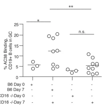
Fig. 2.5 Immunized CD16-/- mice have fewer Ag-specific B cells within the GC... 60
Fig. 2.6 Immunized CD16-/- mice have smaller GCs. ... 61
Fig. 2.7 Dendritic cells secrete BAFF in response to inhibit Ig secretion. ... 62
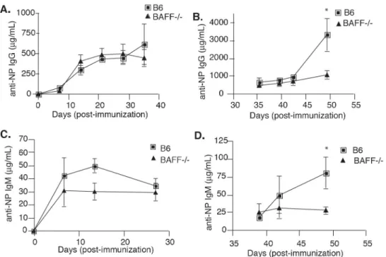
Fig. 2.8 BAFF-/- chimeras have defective secondary immune responses. ... 63
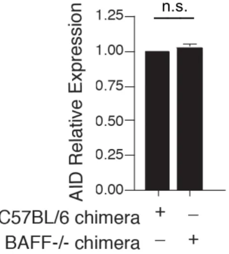
Fig. 2.9 BAFF-/- chimeras express comparable levels of AID. ... 64
Fig. 2.10 DC-derived BAFF restores GC response in immunized CD16-/- mice. ... 65
Fig. 2.11 Immunized BAFF-/- chimeras have fewer memory and GC B cells... 66
Fig. 2.12 Immunized CD16-/- mice and BAFF-/- chimeras have fewer TFH cells. ... 67
Fig. 2.13 DC-derived BAFF restores TFH population in CD16-/- mice. ... 69
Fig. 2.14 Immunized BAFF-/- chimeras also have reduced levels of Bcl-6. ... 70
Fig. 2.15 DC-derived BAFF restores Bcl-6 expression in CD16-/- mice. ... 71
Fig. 3. 1 Reduction of serum BAFF levels by BR3-Fc treatment in B6 mice. ... 89
Fig. 3. 2 Treatment with BR3-Fc results in fewer CD19+ B cells in B6 mice... 89
Fig. 3.3 BAFF is required for memory B cell reactivation... 91
Fig. 3.4 BAFF is required for memory B cell expansion. ... 92
Fig. 3. 5 BR3-Fc treated mice have fewer GC cells, 14 days post-boost. ... 93
Fig. 3. 6 BR3-Fc treated mice have fewer TFH cells 14 days post-boost... 94
Fig. 4.2 OVA-stimulated B6 B cells also secrete inhibitory factors. ... 112
Fig. 4.3 MZ B cells secrete factors that inhibit Ig secretion. ... 113
Fig. 4.4 Ag-stimulated B cells secrete TGF-β and CD40L ... 114
List of Abbreviations
Ah receptor – Aryl hydrocarbon receptor AID – activation induced deaminase
ADCC - antibody dependent cell cytoxicity ASC – antibody secreting cell
Ag - antigen
APC – antigen presenting cell BAFF – B cell activating Factor BMCA – B cell maturation antigen BCR - B cell Receptor
BR3 – BAFF Receptor
BMDC – bone marrow derived dendritic cells BTK – Bruton’s tyrosine kinase
CM – conditioned media DC – Dendritic cell FO – Follicular
FDC – follicular dendritic cell GC – germinal center
IC – immune complex Ig – immunoglobulin
IBDV – infectious bursal disease IL – interleukin
ITAM – immunoreceptor tyrosine-based activation motif ITIM – immunoreceptor tyrosine-based inhibition motif LCMV - Lymphocytic choriomeningitis
MF – macrophage MZ – marginal zone
NP – KLH – hydroxy-3-nitrophenylacetylkeyhole limpet hemocyanin OVA – ovalbamin
PNA - peanut agglutinin
PFCs - perfluorinated compounds PFOA - Perfluorooctanoic acid PFOS - Perfluorooctanesulfonic acid
RSV – respiratory syncytial virus s.c. - subcutaneous
SLE – systemic lupus erythematosus
TACI - transmembrane activator and calcium modulator and cyclophilin ligand interactor TCDD – 2, 3, 7, 8-Tetrachlorodibenzo-p-dioxin
TFH- T follicular helper
TCR – T cell receptor
TGF-b – transforming growth factor-beta Tg – transgenic
CHAPTER 1
Introduction
1.1 Germinal Centers are the Hallmark of Adaptive Immunity
T cell dependent adaptive immune responses generate high affinity, antigen-specific
antibodies that provide the host with long-lived protection against recurring infection. The ability of the immune system to generate such long-lasting responses is the basis for
successful vaccination programs (1). One of the most critical events in initiating the adaptive immune response is the formation of germinal centers (GCs). Germinal centers are transient structures, located within B cell follicles of secondary lymphoid organs, where
antigen-activated B cells, proliferate, undergo affinity maturation, and give rise to memory B cells, and long-lived plasma cells (2). Dysregulation of cell proliferation, mutation, and
differentiation within the germinal center can lead to various pathologies, including tumor development, autoimmunity, and immunodeficiency. Therefore, understanding the mechanisms that regulate the survival, proliferation, and differentiation of B cells within the
germinal center during T cell-dependent adaptive immune responses is of great importance.
A. Initiation of the germinal center response When naïve B cells bind antigen via the B cell receptor (BCR), they upregulate expression of the chemokine receptor, CCR7 and migrate to the outer T cell zones of secondary lymphoid organs, including the spleen and
helper T cells (3) and can now choose one of two cell fates. Specifically, these activated B cells can migrate into the extra-follicular space, under the influence of the G-protein coupled
receptor, EBI-2 (4, 5), and differentiate into short-lived antibody secreting cells. Most plasma cells generated during extra-follicular responses survive for approximately 3 days before undergoing apoptosis (6, 7). Alternatively, activated B cells can move into the B cell
follicle to establish germinal centers (8). The mechanisms responsible for this fate decision remain poorly understood, although several studies suggest that BCR affinity, BCR
engagement, and co-stimulatory signals from helper T cells might all be involved (9-13). The initial germinal centers that are formed are oligoclonal, and are colonized on average, by one to three activated B cells within each follicle (14). Germinal center B cells differ from
naïve B cells in many ways. These cells are much larger and can be identified by their expression of Fas, or CD95, GL-7, binding to peanut agglutinin (PNA), and loss of surface
IgD expression (15-17). In the initial phase of the GC, there is extensive proliferation, or clonal expansion, but very little variation in the BCR. During this time, dark and light zones appear, which are thought to be sites of clonal expansion and selection, respectively (18).
Intravital microscopy has shown that germinal center B cells can move between the dark and light zones (3). During clonal expansion, the chemokine receptor, CXCR4, is
downregulated, and germinal center B cells are no longer retained within the dark zone, by the chemokine, CXCL12 (3). CXCL13 produced by follicular dendritic cell (FDCs) facilitates the trafficking of germinal center B cells to the light zone. Thus, germinal center
including naïve follicular B cells that can enter the light zone (3), where they are tested for their ability to bind antigen by competing for surface antigen on FDCs.
B. Clonal Expansion, somatic hypermutation, and affinity maturation During the germinal center response, stromal FDCs and T cells generate the signals required for isotype switching, somatic hypermutation, memory B cell formation, and terminal differentiation
into plasma cells (20). The affinity of serum antibody for foreign antigen increases over time, due to somatic hypermutations in the B cell receptor. Most short-lived plasma cells in the
extra-follicular foci are relatively unmutated. In contrast, a high frequency of somatic hypermutations is evident in GC B cells (14, 21). These B cells express the enzyme, activation-induced deaminase (AID), which deaminates cytidine residues in the VDJ and
switch regions of the Ig gene, which leads to somatic hyper mutation and class switch recombination (22, 23). Clonal expansion of activated B cells followed by B cell receptor
diversification, results in either retention of cells in the lymphoid organ and the formation of a second germinal center, or exit from the germinal center and entry into the long-lived memory B cell compartment (20). Over time, the extra-follicular response wanes, while the
long-lived plasma cells and memory B cells begin to appear. Finally, during the contraction of a primary immune response, long-lived plasma cells migrate to the bone marrow, while
memory cells may enter into the periphery, as non-secreting, class-switched B cells. However a significant portion of the memory B cell pool remains within the secondary lymphoid organs, in close proximity to the marginal zone (24). The antibody variable regions in most, but not all of long-lived PCs and memory cells display a high degree of
high-affinity, antigen-experienced B cells that will enter into circulation until re-encounter with antigen.
C. Factors that govern germinal center formation and maintenance. Many factors govern the development and maintenance of the germinal center. Bcl-6 is highly expressed in germinal center B cells and is essential for their formation (27, 28). Bcl-6 functions by
binding to the regulatory regions of genes in GC B cells, ultimately inhibiting the DNA damage response and promoting rapid cell proliferation (29). Another target of Bcl-6 is
Prdm1, the gene encoding Blimp-1. Suppression of Blimp-1 expression by Bcl-6 prevents
plasma cell differentiation within the germinal center (30). There is a large body of evidence indicating that signals from the BCR, from FDCs, and from T follicular helper (TFH) cells all
contribute to germinal center B cell survival. BCR signals support the survival of centrocytes (B cells undergoing selection within the light zone), since GC B cells that lack
the BCR co-receptor, CD45, or the GTPase TC21, which links BCR signaling with PI3K activation, have increased rates of apoptosis (31). TFH cells also provide proliferation,
survival, and differentiation signals to GC B cells through CD40:CD40L interactions and the
cytokines IL-4 and IL-21. Although it is clear that CD40L is essential in mediating GC B cell survival, the cytokines and signals that are required for GC B cell differentiation into
either long-lived plasma cells or memory B cells remain undefined.
1.2 T follicular helper (TFH) cells
T follicular helper (TFH) cells are the specialized subset of CD4+ T cells that are
necessary for the initiation and GC maintenance, generation of memory B cells, and the
their constitutive expression of the transcription factor Bcl-6, as well as the expression of CXCR5, PD-1 and ICOS (32, 33). The transcriptional repressor Bcl-6 is essential for TFH
cell development, but it does not act alone in controlling TFH cell development. A network of
transcription factors is involved, including c-Maf, STAT3, Batf, and Bcl-6 all have roles in TFH cell differentiation and function (34, 35). It is postulated that Bcl-6 regulates TFH cell
differentiation by repressing alternative differentiation pathways, including those of the Th1,
Th2, or Th17 lineages (32). Early Bcl-6 up-regulation occurs in the first division of activated
CD4+ T cells, and is strongly induced by antigen presenting dendritic cells. This first wave of Bcl-6 expression is observed within the first three days following antigen-priming at the B and T cell border and in the inter-follicular regions of lymph nodes in both mice and humans
(36, 37). An additional wave of Bcl-6 expression coincides with CXCR5 induction and results in the development of a distinct BCL-6+CXCR5+ TFH cell population, independent of
cognate B cell interactions (38). In contrast to their induction, the maintenance of the TFH
cell population in the follicle is dependent on cognate B cell interactions (37, 38).
Once activated, TFH cells secrete IL-21, which has been shown to be the most potent
cytokine involved in regulating plasma cell differentiation in mice and humans (39, 40). IL-21 induction of plasma cell differentiation is STAT3 dependent (41), and involves the
upregulation of Blimp-1, the master regulator of plasma cells (42, 43). IL-21 is also important for optimal germinal center B cell proliferation. The expression of Bcl-6 in GC B cells is somewhat reduced in the absence of IL-21 (44, 45). In addition, IL-21 has been shown to be important for isotype switching to several IgG isotypes and it is suggested that
IL-21 is the master regulator of class switching to IgG1 (46). In addition to IL-21, TFH cells
PD-1 and BAFF, which ultimately compete with death signals induced by Fas-FasL interactions (35).
TFH cells are now defined as T cells that express the chemokine receptor CXCR5,
localize to the follicles, and are specialized in providing help to germinal center B cells. Although TFH cells are one of the first identified T cell subsets, the nature of their formation
and function has only recently begun to be defined. Many of the cell surface molecules expressed by these specialized cells are critical for their interactions with B cells during the
germinal center reaction. CXCR5 is a defining molecule for T cells, which enables them to home to the B cell follicle. TFH cells migrate in response to CXCL13, produced by FDCs
(35). The migration of TFH cells into the follicle allows for the critical B cell-dependent
phase of TFH cell differentiation to occur. Activated B cells express ICOSL, and the
expression of ICOSL by B cells is required for TFH cells. ICOS-mediated PI3 kinase
signaling is required for TFH cell differentiation, and for the production of IL-21 by TFH cells
(47). Global analysis of the gene expression profile in TFH cells has revealed that these cells
are of a totally separate lineage from Th1 and Th2 cells (48). For example, Bcl-6 is
preferentially expressed by TFH cells, but not by Th1 or Th2 cells. IL-21 is also preferentially
expressed by TFH cells, and plays a critical role in regulation immunoglobulin production and
germinal center formation (44, 49).
TFH cells provide germinal center B cells with survival and selection signals; therefore
limiting the numbers of these cells is critical to prevent inappropriate B cell responses and
the emergence of autoantibodies. Little is known about the regulatory mechanisms that control these TFH cells. A population of Foxp3+ follicular regulatory T cells have been
(50, 51). In response to immunization with T-dependent antigen, a potion of naïve regulatory T cells can express Bcl-6 (51). Expression of Bcl-6 allows them to express CXCR5 and
home to the B cells follicle and localize to the germinal center. These T follicular regulatory cells control the germinal center reaction by limiting the number of TFH cells that are formed
and by inhibiting the selection of non-antigen specific B cells, including those with
self-reactive B cell receptors (50).
Understanding mechanisms that regulate TFH cell differentiation and function is of
great important for improved vaccine design, since nearly all approved human vaccines function on the basis of protective T-dependent immune responses. In addition, TFH cells
play important roles in common autoimmune diseases, such as systemic lupus erythematosus
(SLE), since mice with expanded TFH populations and over-production of IL-21 develop
lupus-like autoimmune disease (52).
1.3 Formation and Maintenance of B cell memory
The pool of quiescent, non-immunoglobulin secreting B cells that are produced
during the germinal center reaction is largely composed of antigen specific, affinity-matured memory B cells. Some reported studies suggest that early memory B cell development can
occur independent of a germinal center response (44, 53), although how well these germ-line BCR-expressing memory B cells compete with post-germinal center B cells during recall responses remains to be determined. Secondary responses resulting from re-challenge of
memory B cells are faster, larger, and qualitatively different from primary responses (1). The ability to mount a recall response can be maintained for decades in humans and through the
Many factors, such as antigen and cytokine availability, can affect the differentiation and survival of memory B cells. The role of cognate antigen in memory B cell maintenance
has been heavily debated. It was originally shown that recall responses were attenuated over time after sorted memory B cells were transferred to naive recipients in the absence of antigen (54). Based on this study, in addition to others, it was proposed that antigen
containing immune complexes deposited on the surface of FDCs (via complement receptor and Fc receptors) was essential for memory cell maintenance (54, 55). However, more
recent studies by Shlomchik and colleagues have demonstrated that memory B cell survival and function is not dependent upon immune complex deposition on FDCs (56). Rajewsky and colleagues have also demonstrated that there is no requirement for continued exposure to
antigen to sustain memory B cells. Specifically, the BCR of memory B cells can be altered to recognize an antigen that has never been encountered by the host and memory B cells
would continue to persist (57). The basis of the controversy and conflicting data surrounding the factors required for memory B cell maintenance could result from the fact that there are multiple layers of B cell memory, with each layer consisting of multiple effector function.
Sophisticated studies by Dogan et. al have demonstrated that the memory B cell compartment can be composed of several layers, consisting of an antigen-independent layer located outside
of the B cell follicles, in the spleen, blood, and secondary lymphoid organs. It also consists of an antigen-dependent layer of proliferating centroblasts in GC-like structures that depend on the presence of antigen (58). The authors have shown that after challenge, the IgG+ memory
cells have an immediate effector and protective response (the hallmark of B cell memory). This subset seems to have little capacity to reinitiate a GC response. In contrast, the IgM+
mobilization and immediate isotype switching to IgG. In this model, the memory compartment is able to quickly neutralize the invading antigen, while continuing to replenish
the memory pool. Other studies have also confirmed a hierarchy of maturity with the memory B cell compartment in mice (59). These studies demonstrated that there is a spectrum of memory B cells, consisting of a progression from more naïve-like to more
memory-like properties.
Although we understand the basic features of the adaptive immune response, the
signals that are required for memory B cell formation and reactivation are still relatively unknown. The mechanisms that regulate the quality and quantity of the memory population are also unclear. To date, no single factor has been identified irrefutably as being essential
for memory formation. A recent study has hinted towards a role for IL-21 and BAFF in regulating memory B cell formation in humans (39). However, there are contrasting studies
in mice demonstrating that memory B cell survival is independent of BAFF (60).
1.4 The role of BAFF in adaptive immunity
The TNF family member, BAFF and its homologue, APRIL, are homotrimeric type II transmembrane proteins that are necessary for B cell homeostasis. It was previously believed
that only cells of a myeloid origin produced BAFF. It is now known that in addition, non-hematopoietic stromal cells can secrete BAFF and APRIL, which provide local niches to modulate the survival and function of B cells and plasma cells, especially in the bone marrow
(61-63). BAFF producing cells include monocytes, macrophages, neutrophils, activated T and B cells, FDCs, stromal cells, astrocytes, osteoclasts, and epithelial cells (64, 65). The
CD40L, as well as by the activation of Toll-like receptors (TLR4 and TLR9) (64, 65). Although it can be found in a membrane bound form, BAFF is usually produced in soluble
form as a result of furin cleavage (61, 65). In myeloid cells, the binding of immune complexes also increases BAFF processing through an Fc receptor-dependent mechanism (66, 67). Processed, soluble BAFF adopts a trimeric form, as seen with other TNF family
members; but it is the only member capable of further assembly as a 60-mer (65). BAFF has been shown to bind to three receptors, BCMA, BAFF-R (BR3), and TACI, all of which are
expressed on B cells. BCMA is preferentially expressed on plasma cells, plasmablasts and tonsillar germinal center B cells (65). TACI is expressed by all peripheral B cells, particularly marginal zone B cells and B-1 B cells (65). BR3 is the dominant BAFF receptor
expressed on all mature murine and human B cells. In mice, the expression of BAFF-R is initially low on newly formed immature B cells, but increases during B cell maturation.
BAFF-R is the key receptor that is responsible for BAFF-mediated B cell survival, as mice deficient in BAFF-R display a phenotype similar to that of BAFF deficient mice (61). BCMA-deficient mice display no abnormalities in immune function, aside from impaired
survival of long-lived plasma cells in the bone marrow (68). TACI, in contrast, has emerged as a negative regulator of B cell activation and expansion, since B cell numbers are increase
in TACI deficient mice. In addition, TACI deficient mice also eventually develop SLE-like disease and lymphoid cancers (69).
It has been well documented that BAFF has an important role in B cell homeostasis. For reasons that are not fully understood, this homeostasis requires BAFF that is produced by
therefore the generation of the mature B cell compartment in the spleen. This was demonstrated by an almost complete lack of follicular and marginal zone B cells and by a
block at the T1 cell stage in BAFF/BAFF-R deficient mice (71). In these mice, the B-1
compartment was unaffected, indicating that the development of this B cell subset is independent of BAFF and BAFF-R signaling. In contrast, transgenic mice over-expressing
BAFF display an increase in all B cell subsets (72). This suggests that all mature B cells express BAFF receptor on their surface and are capable of responding to BAFF. The
expanded B cell compartment in BAFF Tg mice also corresponded with the onset of autoimmunity and lupus-nephritis like disease (72). Recent studies have demonstrated a role for BAFF in plasma cell survival, however, memory B cell survival is independent of BAFF
(60, 73). There is no current literature addressing whether BAFF plays a role in either plasma cell or memory cell differentiation.
The role of BAFF in germinal center formation and maintenance has been controversial. Given that BAFF-/- mice have a deficit in mature B cells, one would predict that the ability to form germinal centers would be compromised. Surprisingly, BAFF and
BAFF-R deficient mice have the ability to form germinal centers subsequent to challenge with antigen. However, the kinetics of the germinal center reaction were altered in these
mice (74). Studies have shown that immunized BAFF-R deficient and BAFF deficient mice displayed normal numbers of germinal centers early on in the immune response; however, the germinal centers dissipated more rapidly than wild type mice (74). There was also an increase in frequency of smaller germinal centers and decline in larger germinal centers,
lack of antigen stimulation (74). These results suggest that BAFF receptor signaling is required for germinal center maintenance and that BAFF is also required for the formation of
the FDC reticulum and efficient antigen stimulation of B cells.
A. An Emerging Role for BAFF in Modulating T cell responses Although BAFF and its cognate receptors have a dominant role in B cell biology, it is now clear that either directly,
or indirectly, BAFF can also modulate T cell function in vitro and in vivo. Several in vitro studies have confirmed the role of BAFF as a co-stimulatory and survival factor for activated
T cells (75). In addition to providing survival signals, BAFF also impacts T cell activation. In the presence of suboptimal T cell receptor stimulation, BAFF enhances T cell proliferation and cytokine production (76-78). Neutralization of BAFF in vitro, using both TACI-Ig and
BAFF-R-Ig, resulted in decreased T cell activation. In addition, BAFF deficient mice show impaired T cell mediated graft rejection. These studies suggest that physiological levels of
BAFF are necessary for the generation of sufficient T cell responses. BAFF may also play a role in stimulating T cell function during T cell mediated pathogenesis. Studies by Mackay et al have shown that the over-expression of BAFF in BAFF Tg mice exacerbates the
severity of Th1-mediated delayed-type hypersensitivity responses, by enhancing T cell
proliferation and IFN-g production in lymph nodes (79). Although a role for BAFF has been
demonstrated in modulating Th1 responses, it remains to be determined whether BAFF is also
involved in regulating other T cell subsets, including T follicular helper (TFH) cells.
studies, BAFF levels have been correlated with increased disease activity and titers of pathogenic autoantibodies (84). These studies provide a strong case for the pathogenic role of
excess BAFF and APRIL in autoimmune disease.
The pathogenic role of BAFF in autoimmunity is further evidenced by the therapeutic benefits of BAFF neutralization. Studies have shown that lupus-prone mice treated with
TACI-Ig and BAFF-R Ig displayed less proteinuria, and as a result, prolonged survival. In
vivo BAFF antagonism has also provided protection in murine models of other autoimmune
diseases, including rheumatoid arthritis, multiple sclerosis, and Graves’ disease (63, 85-87). Neutralization using TACI-Ig and BAFF-R Ig is thought to have therapeutic effects, due to B cell depletion, similar to what has been observed with anti-CD20 immunotherapy
(Rituximab). Blocking BAFF may have some distinct advantages over B cell depletion using Rituximab. For example, BAFF receptor and CD20 expression overlap in many B cell
subsets, but they also differ in other populations, including plasma cells, which express BCMA, but not CD20 (84). B cell depletion using anti-CD20 therapy also results in elevated serum BAFF levels (88). Therefore, newly generated immature B cells could be
exposed to high levels of BAFF, which could cause another breakdown of immune tolerance and the resurgence of autoimmunity. Therefore the risk of another breach in immune
tolerance could be avoided by targeting BAFF in the treatment of SLE and other autoimmune disease.
Treatment of lupus-prone mice with TACI-Ig and BAFF-R Ig resulted in the
neutralization on memory B cell formation have not yet been addressed. Elucidating the role of BAFF in maintaining memory B cell function will be especially important in regulating B
cell memory formed to self-antigens, in the setting of autoimmune disease.
1.5 Systemic Lupus Erythematosus (SLE)
Systemic lupus erythematosus (SLE) is a complex, multi-organ autoimmune disease, affecting approximately 250,000 Americans, and is characterized by the production of autoantibodies to nuclear components (89-91). Alternating periods of flares and remissions
are associated with an increased burden of apoptotic cells, the formation and deposition of immune complexes, and subsequent inflammation (92). Although the etiology of SLE
remains unknown, multiple defects in immune regulation have been identified in lupus-prone mice and SLE patients. These include, complement deficiencies, T cell receptor (TCR) signaling abnormalities, and defective cytokine secretion (89, 92-94). These defects have
been shown to contribute to the onset and pathogenesis of SLE (95). It is likely that environmental factors act on genetically prone individuals to induce a breakdown in
tolerance mechanisms that regulate autoreactive lymphocytes. Some of these environmental factors include exposure to air pollutants and cigarette smoke, heavy metal poisoning (96), and previous viral infections, including Epstein Barr virus (97, 98). Hormonal influences
have also been implicated, with SLE primarily affecting women (10:1 ratio). Genetic differences and environmental factors may interact in the pathogenic processes and also
1.6 IgG Fc Receptors
IgG Fc receptors link innate and adaptive immune responses by their ability to
mediate cellular interactions with antigen-antibody immune complexes. Two general classes of IgG FcRs are now recognized by the presence of an activating ITAM motif within the
cytoplasm, and the inhibitory receptor, characterized by an ITIM sequence (99-102). These two classes of receptors function in concert and are usually co-expressed on the cell surface. The co-engagement of both the activating and inhibitory signaling pathways set the
appropriate thresholds, which ultimately balances protective and pathogenic effector responses after IgG immune complex engagement. Imbalances between activating and
inhibitory FcγR functions can contribute to autoimmunity and other pathologies in both mice
and humans (103).
A. ITAM-containing, Activating Fc Receptors There are three different activating FcγRs expressed on murine effector cells: FcγRI (CD64), FcγRIII (CD16), and the recently
described FcγRIV (also identified as CD16-2) (101). All three receptors generate signals
through the ITAM sequences found within a shared common γ chain subunit (FcγR).
Expression of the common γ chain is critical for the assembly of the activating Fc receptors
(100). Common gamma chain knockout (Fcγ-/-) mice were shown to have significant defects in antibody-dependent effector responses, including ADCC, phagocytosis of immune
complexes, and some inflammatory responses (104). In contrast, deletion of the individual activating Fc receptors showed less pronounced phenotypes, especially in responses involving IgG2a and IgG2b antibody isotypes (105). These activating Fc receptors are found
The only high-affinity Fcγ receptor in both humans and mice, FcγRI, binds IgG2a in mice, or
IgG1 and IgG3 in humans, with an affinity of 108 – 109 M-1. All other receptors have a
100-1000 fold lower affinity, and show broader specificities (107). IgG1 is the only isotype that is
consistently assigned to an individual activating Fc receptor, FcγRIII. The deletion of FcγRIII abrogates IgG1-mediated effector functions in many models of pathogenesis,
including arthritis, glomerulonephritis, IgG-dependent anaphylaxis, IgG-mediated hemolytic anemia, and immunothrombocytopenia (108-112). FcγRIV binds with intermediate affinity
to IgG2a and IgG2b in vitro (103). Studies have shown that even if several of activating Fc
receptors with the same isotype specificity are present on the same cell surface, only those Fc receptors will be engaged that show the optimal affinity for the respective isotype.
Therefore, IgG1 immune complexes will trigger only FcγRIII, since it is the only activating
Fc receptor that can bind IgG1 (106).
B. FcγRIIb, the single ITIM-containing, inhibitory Fc Receptor In contrast to the activating FcγRs, FcγRIIb (or CD32) contains an ITIM sequence that inhibits effector cell
responses (102). In addition to its expression on B cells, where it is the only IgG Fc receptor, FcγRIIb is also expressed on neutrophils, macrophages and mast cells, and is absent on T and
NK cells (101). FcγRIIb expression on the surface of B cells is critical in setting BCR signaling thresholds and B cell effector functions. Signaling within the ITIM motif of
FcγRIIb results in the recruitment of the phosphatases, SHIP and SHP-1, which prevents the
recruitment of kinases such as BTK or PLCγ to the cell membrane, thereby diminishing
events that are downstream of BCR activation such as intracellular calcium fluxes (107).
B cell responses, autoimmunity, and augmented IgG-mediated inflammation, demonstrating the inhibitory role of FcγRIIb in immune responses (113).
C. Fc receptors mediate important effector functions in immune cells Fcγ receptors play a significant role in vivo in maintaining peripheral tolerance, in augmenting T cells responses through antigen presentation, and in mediating antigen recognition and effector cell
activation. Although Fc receptors have many roles in linking innate and adaptive immunity, their role in promoting secondary adaptive immune responses has only recently began to
emerge. A recent study has shown that immune complex binding by Fc receptors enhances secondary antibody responses, although the precise mechanism by which this occurs is unknown (114). Immune complex binding by Fc receptors can induce many effector cell
functions, including cytokine production and phagocytic functions. It remains unclear how these effector functions impact secondary immunity.
1.7 The role of TGF-β in maintaining peripheral immune tolerance
The immune system has evolved to initiate robust responses to invading pathogens
while maintaining tolerance to self-antigens. Multiple mechanisms exist to ensure normal immunological function. The removal of self-reactive B and T cells during development
creates a repertoire within the periphery that will preferentially recognize and eliminate non-self-antigens (115-117). Although central tolerance mechanisms exist within the bone marrow and thymus to eliminate autoreactivity, there is clear evidence that self-reactive
immune cells can enter the periphery and secondary lymphoid organs. Therefore, peripheral tolerance mechanisms must also exist to limit the activation of autoreactive lymphocytes.
essential components of peripheral tolerance (118, 119). Transforming growth factor β
(TGF-β) is one such regulatory cytokine with a critical role in regulating immune responses to self.
TGF-βs are regulatory proteins with pleiotropic effects on cell proliferation,
differentiation, migration and survival, which affect many biological processes, including development, carcinogenesis, fibrosis, wound healing, and immune responses (120). The
generation and analysis of TGF-β deficient mice established the role of the cytokine in
inhibiting inflammation and autoimmunity, and has fostered an interest in this cytokine in immune regulation (121, 122). The immunoregulatory role of TGF-β was demonstrated in null mice. TGF-β-/- mice that are born, die shortly after weaning as a result of severe
inflammatory disease, with lymphocyte infiltration into multiple organs and autoimmune disease (122, 123). The identification of the receptors for TGF-β s and the Smad proteins, as
mediators of signaling, has also provided much needed information on the role of TGF-β s in regulating immune responses.
TGF-β is an important regulator of B cells activity, as demonstrated by the phenotype of mice with a B cell specific inhibition of TGF-β signaling (124). Secretion of TGF-β by
regulatory cells inhibits B cell proliferation, induces apoptosis of immature or resting B cells, BCR activation, and isotype switching to most IgG isotypes. Consistent with TGF-β
mediated inhibition of IgG class switching, conditional deletion of TGF-β signaling in B cells, in vivo, resulted in elevated serum Ig. These mice also developed enhanced IgG3
responses to a normally weak antigen (124). These studies implicate TGF-β in attenuating B
important regulator of B cell tolerance to self-antigens in vivo (124). In contrast to its inhibitory roles in immune function, TGF-β has a unique role in promoting switching to IgA
and IgG2b in murine B cells and IgA in human B cells, in vitro (124-126). Upon optimal
stimulation with antigen, and cytokine stimulation, the inhibitory activity of TGF-β subsides, and TGF-β can strongly promote IgA secretion. TGF-β mediated IgA class switching is
associated with increased transcription of alpha heavy chain transcripts. The role of TGF-β in IgA production is further demonstrated in mice with a condition deletion of TGF-β
signaling in B cells, where there is almost a complete absence of IgA within the serum (124). Thus, similar to T cells, TGF-β has both inhibitory and stimulatory effects on B cell function.
The adaptive immune response gives rise to long-lived humoral responses that protect
the host from recurring infection. Although dendritic cells indirectly promote adaptive immunity through antigen presentation, it remains unclear whether DCs actively shape these
1.8 References
1. Ahmed, R., and D. Gray. 1996. Immunological memory and protective immunity: understanding their relation. Science 272:54-60.
2. MacLennan, I. C. 1994. Germinal centers. Annu Rev Immunol 12:117-139.
3. Allen, C. D., T. Okada, and J. G. Cyster. 2007. Germinal-center organization and cellular dynamics. Immunity 27:190-202.
4. Gatto, D., D. Paus, A. Basten, C. R. Mackay, and R. Brink. 2009. Guidance of B cells by the orphan G protein-coupled receptor EBI2 shapes humoral immune responses. Immunity 31:259-269.
5. Pereira, J. P., L. M. Kelly, Y. Xu, and J. G. Cyster. 2009. EBI2 mediates B cell segregation between the outer and centre follicle. Nature 460:1122-1126.
6. Smith, K. G., T. D. Hewitson, G. J. Nossal, and D. M. Tarlinton. 1996. The phenotype and fate of the antibody-forming cells of the splenic foci. Eur J Immunol 26:444-448.
7. Ho, F., J. E. Lortan, I. C. MacLennan, and M. Khan. 1986. Distinct short-lived and long-lived antibody-producing cell populations. Eur J Immunol 16:1297-1301.
8. Jacob, J., G. Kelsoe, K. Rajewsky, and U. Weiss. 1991. Intraclonal generation of antibody mutants in germinal centres. Nature 354:389-392.
9. Vinuesa, C. G., M. A. Linterman, C. C. Goodnow, and K. L. Randall. T cells and follicular dendritic cells in germinal center B-cell formation and selection. Immunol Rev 237:72-89.
10. Shih, T. A., E. Meffre, M. Roederer, and M. C. Nussenzweig. 2002. Role of BCR affinity in T cell dependent antibody responses in vivo. Nat Immunol 3:570-575. 11. Paus, D., T. G. Phan, T. D. Chan, S. Gardam, A. Basten, and R. Brink. 2006. Antigen
recognition strength regulates the choice between extrafollicular plasma cell and germinal center B cell differentiation. J Exp Med 203:1081-1091.
12. Dal Porto, J. M., A. M. Haberman, M. J. Shlomchik, and G. Kelsoe. 1998. Antigen drives very low affinity B cells to become plasmacytes and enter germinal centers. J Immunol 161:5373-5381.
14. Jacob, J., R. Kassir, and G. Kelsoe. 1991. In situ studies of the primary immune response to (4-hydroxy-3-nitrophenyl)acetyl. I. The architecture and dynamics of responding cell populations. J Exp Med 173:1165-1175.
15. Yoshino, T., E. Kondo, L. Cao, K. Takahashi, K. Hayashi, S. Nomura, and T. Akagi. 1994. Inverse expression of bcl-2 protein and Fas antigen in lymphoblasts in peripheral lymph nodes and activated peripheral blood T and B lymphocytes. Blood 83:1856-1861.
16. Rose, M. L., M. S. Birbeck, V. J. Wallis, J. A. Forrester, and A. J. Davies. 1980. Peanut lectin binding properties of germinal centres of mouse lymphoid tissue. Nature 284:364-366.
17. Cervenak, L., A. Magyar, R. Boja, and G. Laszlo. 2001. Differential expression of GL7 activation antigen on bone marrow B cell subpopulations and peripheral B cells. Immunol Lett 78:89-96.
18. Nieuwenhuis, P., and D. Opstelten. 1984. Functional anatomy of germinal centers. Am J Anat 170:421-435.
19. Cyster, J. G. 2005. Chemokines, sphingosine-1-phosphate, and cell migration in secondary lymphoid organs. Annu Rev Immunol 23:127-159.
20. Cozine, C. L., K. L. Wolniak, and T. J. Waldschmidt. 2005. The primary germinal center response in mice. Curr Opin Immunol 17:298-302.
21. Berek, C., A. Berger, and M. Apel. 1991. Maturation of the immune response in germinal centers. Cell 67:1121-1129.
22. Pavri, R., and M. C. Nussenzweig. AID targeting in antibody diversity. Adv Immunol 110:1-26.
23. Neuberger, M. S., A. Lanoue, M. R. Ehrenstein, F. D. Batista, J. E. Sale, and G. T. Williams. 1999. Antibody diversification and selection in the mature B-cell compartment. Cold Spring Harb Symp Quant Biol 64:211-216.
24. Kurosaki, T., Y. Aiba, K. Kometani, S. Moriyama, and Y. Takahashi. Unique properties of memory B cells of different isotypes. Immunol Rev 237:104-116.
25. McHeyzer-Williams, L. J., L. P. Malherbe, and M. G. McHeyzer-Williams. 2006. Checkpoints in memory B-cell evolution. Immunol Rev 211:255-268.
27. Fukuda, T., T. Miki, T. Yoshida, M. Hatano, K. Ohashi, S. Hirosawa, and T. Tokuhisa. 1995. The murine BCL6 gene is induced in activated lymphocytes as an immediate early gene. Oncogene 11:1657-1663.
28. Fukuda, T., T. Yoshida, S. Okada, M. Hatano, T. Miki, K. Ishibashi, S. Okabe, H. Koseki, S. Hirosawa, M. Taniguchi, N. Miyasaka, and T. Tokuhisa. 1997. Disruption of the Bcl6 gene results in an impaired germinal center formation. J Exp Med 186:439-448.
29. Shaffer, A. L., X. Yu, Y. He, J. Boldrick, E. P. Chan, and L. M. Staudt. 2000. BCL-6 represses genes that function in lymphocyte differentiation, inflammation, and cell cycle control. Immunity 13:199-212.
30. Tunyaplin, C., A. L. Shaffer, C. D. Angelin-Duclos, X. Yu, L. M. Staudt, and K. L. Calame. 2004. Direct repression of prdm1 by Bcl-6 inhibits plasmacytic differentiation. J Immunol 173:1158-1165.
31. Vinuesa, C. G., I. Sanz, and M. C. Cook. 2009. Dysregulation of germinal centres in autoimmune disease. Nat Rev Immunol 9:845-857.
32. Nurieva, R. I., and Y. Chung. Understanding the development and function of T follicular helper cells. Cell Mol Immunol 7:190-197.
33. Yu, D., S. Rao, L. M. Tsai, S. K. Lee, Y. He, E. L. Sutcliffe, M. Srivastava, M. Linterman, L. Zheng, N. Simpson, J. I. Ellyard, I. A. Parish, C. S. Ma, Q. J. Li, C. R. Parish, C. R. Mackay, and C. G. Vinuesa. 2009. The transcriptional repressor Bcl-6 directs T follicular helper cell lineage commitment. Immunity 31:457-468.
34. Betz, B. C., K. L. Jordan-Williams, C. Wang, S. G. Kang, J. Liao, M. R. Logan, C. H. Kim, and E. J. Taparowsky. Batf coordinates multiple aspects of B and T cell function required for normal antibody responses. J Exp Med 207:933-942.
35. Crotty, S. Follicular helper CD4 T cells (TFH). Annu Rev Immunol 29:621-663. 36. Bentebibel, S. E., N. Schmitt, J. Banchereau, and H. Ueno. Human tonsil B-cell
lymphoma 6 (BCL6)-expressing CD4+ T-cell subset specialized for B-cell help outside germinal centers. Proc Natl Acad Sci U S A 108:E488-497.
37. Kerfoot, S. M., G. Yaari, J. R. Patel, K. L. Johnson, D. G. Gonzalez, S. H. Kleinstein, and A. M. Haberman. Germinal center B cell and T follicular helper cell development initiates in the interfollicular zone. Immunity 34:947-960.
38. Baumjohann, D., T. Okada, and K. M. Ansel. Cutting Edge: Distinct waves of BCL6 expression during T follicular helper cell development. J Immunol 187:2089-2092. 39. Ettinger, R., G. P. Sims, A. M. Fairhurst, R. Robbins, Y. S. da Silva, R. Spolski, W. J.
40. Ozaki, K., R. Spolski, R. Ettinger, H. P. Kim, G. Wang, C. F. Qi, P. Hwu, D. J. Shaffer, S. Akilesh, D. C. Roopenian, H. C. Morse, 3rd, P. E. Lipsky, and W. J. Leonard. 2004. Regulation of B cell differentiation and plasma cell generation by IL-21, a novel inducer of Blimp-1 and Bcl-6. J Immunol 173:5361-5371.
41. Avery, D. T., E. K. Deenick, C. S. Ma, S. Suryani, N. Simpson, G. Y. Chew, T. D. Chan, U. Palendira, J. Bustamante, S. Boisson-Dupuis, S. Choo, K. E. Bleasel, J. Peake, C. King, M. A. French, D. Engelhard, S. Al-Hajjar, S. Al-Muhsen, K. Magdorf, J. Roesler, P. D. Arkwright, P. Hissaria, D. S. Riminton, M. Wong, R. Brink, D. A. Fulcher, J. L. Casanova, M. C. Cook, and S. G. Tangye. B cell-intrinsic signaling through IL-21 receptor and STAT3 is required for establishing long-lived antibody responses in humans. J Exp Med 207:155-171.
42. Martins, G., and K. Calame. 2008. Regulation and functions of Blimp-1 in T and B lymphocytes. Annu Rev Immunol 26:133-169.
43. Savitsky, D., L. Cimmino, T. Kuo, G. A. Martins, and K. Calame. 2007. Multiple roles for Blimp-1 in B and T lymphocytes. Adv Exp Med Biol 596:9-30.
44. Linterman, M. A., L. Beaton, D. Yu, R. R. Ramiscal, M. Srivastava, J. J. Hogan, N. K. Verma, M. J. Smyth, R. J. Rigby, and C. G. Vinuesa. IL-21 acts directly on B cells to regulate Bcl-6 expression and germinal center responses. J Exp Med 207:353-363. 45. Zotos, D., J. M. Coquet, Y. Zhang, A. Light, K. D'Costa, A. Kallies, L. M. Corcoran,
D. I. Godfrey, K. M. Toellner, M. J. Smyth, S. L. Nutt, and D. M. Tarlinton. IL-21 regulates germinal center B cell differentiation and proliferation through a B cell-intrinsic mechanism. J Exp Med 207:365-378.
46. Ozaki, K., R. Spolski, C. G. Feng, C. F. Qi, J. Cheng, A. Sher, H. C. Morse, 3rd, C. Liu, P. L. Schwartzberg, and W. J. Leonard. 2002. A critical role for IL-21 in regulating immunoglobulin production. Science 298:1630-1634.
47. Rolf, J., S. E. Bell, D. Kovesdi, M. L. Janas, D. R. Soond, L. M. Webb, S. Santinelli, T. Saunders, B. Hebeis, N. Killeen, K. Okkenhaug, and M. Turner. Phosphoinositide 3-kinase activity in T cells regulates the magnitude of the germinal center reaction. J Immunol 185:4042-4052.
48. Chtanova, T., S. G. Tangye, R. Newton, N. Frank, M. R. Hodge, M. S. Rolph, and C. R. Mackay. 2004. T follicular helper cells express a distinctive transcriptional profile, reflecting their role as non-Th1/Th2 effector cells that provide help for B cells. J Immunol 173:68-78.
49. Spolski, R., and W. J. Leonard. IL-21 and T follicular helper cells. Int Immunol 22:7-12.
Smith, and C. G. Vinuesa. Foxp3+ follicular regulatory T cells control the germinal center response. Nat Med 17:975-982.
51. Chung, Y., S. Tanaka, F. Chu, R. I. Nurieva, G. J. Martinez, S. Rawal, Y. H. Wang, H. Lim, J. M. Reynolds, X. H. Zhou, H. M. Fan, Z. M. Liu, S. S. Neelapu, and C. Dong. Follicular regulatory T cells expressing Foxp3 and Bcl-6 suppress germinal center reactions. Nat Med 17:983-988.
52. Linterman, M. A., R. J. Rigby, R. K. Wong, D. Yu, R. Brink, J. L. Cannons, P. L. Schwartzberg, M. C. Cook, G. D. Walters, and C. G. Vinuesa. 2009. Follicular helper T cells are required for systemic autoimmunity. J Exp Med 206:561-576.
53. Toyama, H., S. Okada, M. Hatano, Y. Takahashi, N. Takeda, H. Ichii, T. Takemori, Y. Kuroda, and T. Tokuhisa. 2002. Memory B cells without somatic hypermutation are generated from Bcl6-deficient B cells. Immunity 17:329-339.
54. Gray, D., and H. Skarvall. 1988. B-cell memory is short-lived in the absence of antigen. Nature 336:70-73.
55. Barrington, R. A., O. Pozdnyakova, M. R. Zafari, C. D. Benjamin, and M. C. Carroll. 2002. B lymphocyte memory: role of stromal cell complement and FcγammaRIIB receptors. J Exp Med 196:1189-1199.
56. Anderson, S. M., L. G. Hannum, and M. J. Shlomchik. 2006. Memory B cell survival and function in the absence of secreted antibody and immune complexes on follicular dendritic cells. J Immunol 176:4515-4519.
57. Maruyama, M., K. P. Lam, and K. Rajewsky. 2000. Memory B-cell persistence is independent of persisting immunizing antigen. Nature 407:636-642.
58. Dogan, I., B. Bertocci, V. Vilmont, F. Delbos, J. Megret, S. Storck, C. A. Reynaud, and J. C. Weill. 2009. Multiple layers of B cell memory with different effector functions. Nat Immunol 10:1292-1299.
59. Tomayko, M. M., N. C. Steinel, S. M. Anderson, and M. J. Shlomchik. Cutting edge: Hierarchy of maturity of murine memory B cell subsets. J Immunol 185:7146-7150. 60. Benson, M. J., S. R. Dillon, E. Castigli, R. S. Geha, S. Xu, K. P. Lam, and R. J.
Noelle. 2008. Cutting edge: the dependence of plasma cells and independence of memory B cells on BAFF and APRIL. J Immunol 180:3655-3659.
61. Bossen, C., and P. Schneider. 2006. BAFF, APRIL and their receptors: structure, function and signaling. Semin Immunol 18:263-275.
63. Mackay, F., P. Schneider, P. Rennert, and J. Browning. 2003. BAFF AND APRIL: a tutorial on B cell survival. Annu Rev Immunol 21:231-264.
64. Kalled, S. L. 2006. Impact of the BAFF/BR3 axis on B cell survival, germinal center maintenance and antibody production. Semin Immunol 18:290-296.
65. Mackay, F., and P. Schneider. 2009. Cracking the BAFF code. Nat Rev Immunol 9:491-502.
66. Boule, M. W., C. Broughton, F. Mackay, S. Akira, A. Marshak-Rothstein, and I. R. Rifkin. 2004. Toll-like receptor 9-dependent and -independent dendritic cell activation by chromatin-immunoglobulin G complexes. J Exp Med 199:1631-1640. 67. Li, X., K. Su, C. Ji, A. J. Szalai, J. Wu, Y. Zhang, T. Zhou, R. P. Kimberly, and J. C.
Edberg. 2008. Immune opsonins modulate BLyS/BAFF release in a receptor-specific fashion. J Immunol 181:1012-1018.
68. O'Connor, B. P., V. S. Raman, L. D. Erickson, W. J. Cook, L. K. Weaver, C. Ahonen, L. L. Lin, G. T. Mantchev, R. J. Bram, and R. J. Noelle. 2004. BCMA is essential for the survival of long-lived bone marrow plasma cells. J Exp Med 199:91-98.
69. von Bulow, G. U., J. M. van Deursen, and R. J. Bram. 2001. Regulation of the T-independent humoral response by TACI. Immunity 14:573-582.
70. Gorelik, L., K. Gilbride, M. Dobles, S. L. Kalled, D. Zandman, and M. L. Scott. 2003. Normal B cell homeostasis requires B cell activation factor production by radiation-resistant cells. J Exp Med 198:937-945.
71. Rauch, M., R. Tussiwand, N. Bosco, and A. G. Rolink. 2009. Crucial role for BAFF-BAFF-R signaling in the survival and maintenance of mature B cells. PLoS One 4:e5456.
72. Mackay, F., S. A. Woodcock, P. Lawton, C. Ambrose, M. Baetscher, P. Schneider, J. Tschopp, and J. L. Browning. 1999. Mice transgenic for BAFF develop lymphocytic disorders along with autoimmune manifestations. J Exp Med 190:1697-1710.
73. Scholz, J. L., J. E. Crowley, M. M. Tomayko, N. Steinel, P. J. O'Neill, W. J. Quinn, 3rd, R. Goenka, J. P. Miller, Y. H. Cho, V. Long, C. Ward, T. S. Migone, M. J. Shlomchik, and M. P. Cancro. 2008. BLyS inhibition eliminates primary B cells but leaves natural and acquired humoral immunity intact. Proc Natl Acad Sci U S A 105:15517-15522.
75. Ye, Q., L. Wang, A. D. Wells, R. Tao, R. Han, A. Davidson, M. L. Scott, and W. W. Hancock. 2004. BAFF binding to T cell-expressed BAFF-R costimulates T cell proliferation and alloresponses. Eur J Immunol 34:2750-2759.
76. Huard, B., L. Arlettaz, C. Ambrose, V. Kindler, D. Mauri, E. Roosnek, J. Tschopp, P. Schneider, and L. E. French. 2004. BAFF production by antigen-presenting cells provides T cell co-stimulation. Int Immunol 16:467-475.
77. Huard, B., P. Schneider, D. Mauri, J. Tschopp, and L. E. French. 2001. T cell costimulation by the TNF ligand BAFF. J Immunol 167:6225-6231.
78. Ng, L. G., A. P. Sutherland, R. Newton, F. Qian, T. G. Cachero, M. L. Scott, J. S. Thompson, J. Wheway, T. Chtanova, J. Groom, I. J. Sutton, C. Xin, S. G. Tangye, S. L. Kalled, F. Mackay, and C. R. Mackay. 2004. B cell-activating factor belonging to the TNF family (BAFF)-R is the principal BAFF receptor facilitating BAFF costimulation of circulating T and B cells. J Immunol 173:807-817.
79. Sutherland, A. P., L. G. Ng, C. A. Fletcher, B. Shum, R. A. Newton, S. T. Grey, M. S. Rolph, F. Mackay, and C. R. Mackay. 2005. BAFF augments certain Th1-associated inflammatory responses. J Immunol 174:5537-5544.
80. Gross, J. A., J. Johnston, S. Mudri, R. Enselman, S. R. Dillon, K. Madden, W. Xu, J. Parrish-Novak, D. Foster, C. Lofton-Day, M. Moore, A. Littau, A. Grossman, H. Haugen, K. Foley, H. Blumberg, K. Harrison, W. Kindsvogel, and C. H. Clegg. 2000. TACI and BCMA are receptors for a TNF homologue implicated in B-cell autoimmune disease. Nature 404:995-999.
81. Zhang, M., K. H. Ko, Q. L. Lam, C. K. Lo, G. Srivastava, B. Zheng, Y. L. Lau, and L. Lu. 2005. Expression and function of TNF family member B cell-activating factor in the development of autoimmune arthritis. Int Immunol 17:1081-1092.
82. Zheng, Y., S. Gallucci, J. P. Gaughan, J. A. Gross, and M. Monestier. 2005. A role for B cell-activating factor of the TNF family in chemically induced autoimmunity. J Immunol 175:6163-6168.
83. Mackay, F., P. A. Silveira, and R. Brink. 2007. B cells and the BAFF/APRIL axis: fast-forward on autoimmunity and signaling. Curr Opin Immunol 19:327-336.
84. Stohl, W. 2005. BlySfulness does not equal blissfulness in systemic lupus erythematosus: a therapeutic role for BLyS antagonists. Curr Dir Autoimmun 8:289-304.
86. Huntington, N. D., R. Tomioka, C. Clavarino, A. M. Chow, D. Linares, P. Mana, J. Rossjohn, T. G. Cachero, F. Qian, S. L. Kalled, C. C. Bernard, and H. H. Reid. 2006. A BAFF antagonist suppresses experimental autoimmune encephalomyelitis by targeting cell-mediated and humoral immune responses. Int Immunol 18:1473-1485. 87. Wang, H., S. A. Marsters, T. Baker, B. Chan, W. P. Lee, L. Fu, D. Tumas, M. Yan,
V. M. Dixit, A. Ashkenazi, and I. S. Grewal. 2001. TACI-ligand interactions are required for T cell activation and collagen-induced arthritis in mice. Nat Immunol 2:632-637.
88. Lavie, F., C. Miceli-Richard, M. Ittah, J. Sellam, J. E. Gottenberg, and X. Mariette. 2007. Increase of B cell-activating factor of the TNF family (BAFF) after rituximab treatment: insights into a new regulating system of BAFF production. Ann Rheum Dis 66:700-703.
89. Rahman, A., and D. A. Isenberg. 2008. Systemic lupus erythematosus. N Engl J Med 358:929-939.
90. Lawrence, R. C., D. T. Felson, C. G. Helmick, L. M. Arnold, H. Choi, R. A. Deyo, S. Gabriel, R. Hirsch, M. C. Hochberg, G. G. Hunder, J. M. Jordan, J. N. Katz, H. M. Kremers, and F. Wolfe. 2008. Estimates of the prevalence of arthritis and other rheumatic conditions in the United States. Part II. Arthritis Rheum 58:26-35.
91. Helmick, C. G., D. T. Felson, R. C. Lawrence, S. Gabriel, R. Hirsch, C. K. Kwoh, M. H. Liang, H. M. Kremers, M. D. Mayes, P. A. Merkel, S. R. Pillemer, J. D. Reveille, and J. H. Stone. 2008. Estimates of the prevalence of arthritis and other rheumatic conditions in the United States. Part I. Arthritis Rheum 58:15-25.
92. Lipsky, P. E. 2001. Systemic lupus erythematosus: an autoimmune disease of B cell hyperactivity. Nat Immunol 2:764-766.
93. Ohl, K., and K. Tenbrock. Inflammatory cytokines in systemic lupus erythematosus. J Biomed Biotechnol 2011:432595.
94. Apostolidis, S. A., L. A. Lieberman, K. Kis-Toth, J. C. Crispin, and G. C. Tsokos. The dysregulation of cytokine networks in systemic lupus erythematosus. J Interferon Cytokine Res 31:769-779.
95. Mok, C. C., and C. S. Lau. 2003. Pathogenesis of systemic lupus erythematosus. J Clin Pathol 56:481-490.
96. Powell, J. J., J. Van de Water, and M. E. Gershwin. 1999. Evidence for the role of environmental agents in the initiation or progression of autoimmune conditions. Environmental health perspectives 107 Suppl 5:667-672.
98. Parks, C. G., G. S. Cooper, L. L. Hudson, M. A. Dooley, E. L. Treadwell, E. W. St Clair, G. S. Gilkeson, and J. P. Pandey. 2005. Association of Epstein-Barr virus with systemic lupus erythematosus: effect modification by race, age, and cytotoxic T lymphocyte-associated antigen 4 genotype. Arthritis Rheum 52:1148-1159.
99. Bolland, S., and J. V. Ravetch. 1999. Inhibitory pathways triggered by ITIM-containing receptors. Adv Immunol 72:149-177.
100. Daeron, M. 1997. Fc receptor biology. Annu Rev Immunol 15:203-234.
101. Ravetch, J. V., and S. Bolland. 2001. IgG Fc receptors. Annu Rev Immunol 19:275-290.
102. Ravetch, J. V., and J. P. Kinet. 1991. Fc receptors. Annu Rev Immunol 9:457-492. 103. Takai, T. 2002. Roles of Fc receptors in autoimmunity. Nat Rev Immunol 2:580-592. 104. Takai, T. 2005. Fc receptors and their role in immune regulation and autoimmunity. J
Clin Immunol 25:1-18.
105. Takai, T. 1997. [Immunoregulatory functions of Fc receptors]. Seikagaku 69:394-409.
106. Nimmerjahn, F., and J. V. Ravetch. 2006. Fcγamma receptors: old friends and new family members. Immunity 24:19-28.
107. Nimmerjahn, F., and J. V. Ravetch. 2008. Fcγamma receptors as regulators of immune responses. Nat Rev Immunol 8:34-47.
108. Bruhns, P., A. Samuelsson, J. W. Pollard, and J. V. Ravetch. 2003. Colony-stimulating factor-1-dependent macrophages are responsible for IVIG protection in antibody-induced autoimmune disease. Immunity 18:573-581.
109. Fossati-Jimack, L., A. Ioan-Facsinay, L. Reininger, Y. Chicheportiche, N. Watanabe, T. Saito, F. M. Hofhuis, J. E. Gessner, C. Schiller, R. E. Schmidt, T. Honjo, J. S. Verbeek, and S. Izui. 2000. Markedly different pathogenicity of four immunoglobulin G isotype-switch variants of an antierythrocyte autoantibody is based on their capacity to interact in vivo with the low-affinity Fcγamma receptor III. J Exp Med 191:1293-1302.
110. Hazenbos, W. L., J. E. Gessner, F. M. Hofhuis, H. Kuipers, D. Meyer, I. A. Heijnen, R. E. Schmidt, M. Sandor, P. J. Capel, M. Daeron, J. G. van de Winkel, and J. S. Verbeek. 1996. Impaired IgG-dependent anaphylaxis and Arthus reaction in Fc gamma RIII (CD16) deficient mice. Immunity 5:181-188.
den Berg, and J. S. Verbeek. 1998. Murine IgG1 complexes trigger immune effector functions predominantly via Fc gamma RIII (CD16). J Immunol 161:3026-3032. 112. Nimmerjahn, F., and J. V. Ravetch. Antibody-mediated modulation of immune
responses. Immunol Rev 236:265-275.
113. Bolland, S., and J. V. Ravetch. 2000. Spontaneous autoimmune disease in Fc(gamma)RIIB-deficient mice results from strain-specific epistasis. Immunity 13:277-285.
114. Goins, C. L., C. P. Chappell, R. Shashidharamurthy, P. Selvaraj, and J. Jacob. Immune complex-mediated enhancement of secondary antibody responses. J Immunol 184:6293-6298.
115. Cornall, R. J., C. C. Goodnow, and J. G. Cyster. 1995. The regulation of self-reactive B cells. Curr Opin Immunol 7:804-811.
116. Goodnow, C. C., J. G. Cyster, S. B. Hartley, S. E. Bell, M. P. Cooke, J. I. Healy, S. Akkaraju, J. C. Rathmell, S. L. Pogue, and K. P. Shokat. 1995. Self-tolerance checkpoints in B lymphocyte development. Adv Immunol 59:279-368.
117. Kappler, J. W., N. Roehm, and P. Marrack. 1987. T cell tolerance by clonal elimination in the thymus. Cell 49:273-280.
118. Fehervari, Z., and S. Sakaguchi. 2004. Development and function of CD25+CD4+ regulatory T cells. Curr Opin Immunol 16:203-208.
119. Zhang, X., D. N. Koldzic, L. Izikson, J. Reddy, R. F. Nazareno, S. Sakaguchi, V. K. Kuchroo, and H. L. Weiner. 2004. IL-10 is involved in the suppression of experimental autoimmune encephalomyelitis by CD25+CD4+ regulatory T cells. Int Immunol 16:249-256.
120. Blobe, G. C., W. P. Schiemann, and H. F. Lodish. 2000. Role of transforming growth factor beta in human disease. N Engl J Med 342:1350-1358.
121. Kulkarni, A. B., C. G. Huh, D. Becker, A. Geiser, M. Lyght, K. C. Flanders, A. B. Roberts, M. B. Sporn, J. M. Ward, and S. Karlsson. 1993. Transforming growth factor beta 1 null mutation in mice causes excessive inflammatory response and early death. Proc Natl Acad Sci U S A 90:770-774.
122. Shull, M. M., I. Ormsby, A. B. Kier, S. Pawlowski, R. J. Diebold, M. Yin, R. Allen, C. Sidman, G. Proetzel, D. Calvin, and et al. 1992. Targeted disruption of the mouse transforming growth factor-beta 1 gene results in multifocal inflammatory disease. Nature 359:693-699.
124. Cazac, B. B., and J. Roes. 2000. TGF-β eta receptor controls B cell responsiveness and induction of IgA in vivo. Immunity 13:443-451.
125. Kriegel, M. A., M. O. Li, S. Sanjabi, Y. Y. Wan, and R. A. Flavell. 2006. Transforming growth factor-beta: recent advances on its role in immune tolerance. Curr Rheumatol Rep 8:138-144.
CHAPTER 2
CD16-mediated BAFF production promotes Bcl-6 expression
2.1 Introduction
The success of adaptive immune responses requires appropriate interactions between
antigen specific B and T cells leading to germinal center (GC) responses (1, 2). The specialized microenvironment provides sites for rapid expansion and mutation of
antigen-specific B cell clones, antibody isotype switching, and finally commitment to the plasma cell fate or memory B cell pool (3-5). The formation of GCs and the emergence of memory B cells are subsequent to the production of antibody induced by T-independent formation of
extrafollicular foci (2, 6), suggesting that early antibody responses may play a role in early GC events.
The affinity matured memory B cells that emerge from the GC require T cell help. Recent studies have identified CD4+ follicular helper T cells (TFH) as critical effectors that
provide help to B cells during the GC response (7, 8). The formation of TFH cells is
dependent on the expression of Bcl-6 and the attenuation of Blimp-1, which inhibits the differentiation of other TH subsets (7, 9). TFH cells are distinguished from other CD4+
subsets in that they express CXCR5, ICOS, PD-1, and IL-21. TFH cells are important in