Defects in lysosomal maturation facilitate the
activation of innate sensors in systemic
lupus erythematosus
Andrew J. Monteith
a,b, SunAh Kang
a, Eric Scott
a, Kai Hillman
a, Zenon Rajfur
c,d, Ken Jacobson
c,e, M. Joseph Costello
c,
and Barbara J. Vilen
a,e,1aDepartment of Microbiology & Immunology, University of North Carolina, Chapel Hill, NC 27599;bCurriculum in Biochemistry & Biophysics, University of
North Carolina, Chapel Hill, NC 27599;cDepartment of Cell Biology & Physiology, University of North Carolina, Chapel Hill, NC 27599;dDepartment of
Physics, Astronomy, and Applied Computer Science, Institute of Physics, Jagiellonian University, 31-007, Krakow, Poland; andeLineberger Comprehensive
Cancer Center, University of North Carolina, Chapel Hill, NC 27599
Edited by Jason G. Cyster, University of California, San Francisco, CA, and approved March 1, 2016 (received for review July 15, 2015)
Defects in clearing apoptotic debris disrupt tissue and immunolog-ical homeostasis, leading to autoimmune and inflammatory diseases. Herein, we report that macrophages from lupus-prone MRL/lprmice have impaired lysosomal maturation, resulting in heightened ROS production and attenuated lysosomal acidification. Impaired lyso-somal maturation diminishes the ability of lysosomes to degrade apoptotic debris contained within IgG–immune complexes (IgG-ICs) and promotes recycling and the accumulation of nuclear self-anti-gens at the membrane 72 h after internalization. Diminished degra-dation of IgG-ICs prolongs the intracellular residency of nucleic acids, leading to the activation of Toll-like receptors. It also promotes phag-osomal membrane permeabilization, allowing dsDNA and IgG to leak into the cytosol and activate AIM2 and TRIM21. Collectively, these events promote the accumulation of nuclear antigens and activate innate sensors that drive IFNαproduction and heightened cell death. These data identify a previously unidentified defect in lysosomal maturation that provides a mechanism for the chronic activation of intracellular innate sensors in systemic lupus erythematosus.
systemic lupus erythematosus
|
Fc gamma receptors|
lysosomal acidification|
AIM2|
TRIM21T
he disposal of apoptotic debris is initiated by membrane
changes that facilitate the binding of IgM antibodies, acute
phase proteins (C-reactive protein, CRP), and other serum
op-sonins to enhance phagocytosis (1, 2). The disposal of apoptotic
debris is crucial to immune homeostasis because the accumulation
of apoptotic debris (3
–
5), and the formation of immune complexes
(ICs) (6), have long been associated with systemic lupus
erythe-matosus (SLE). Similarly, impaired clearance of apoptotic bodies
in mice lacking scavenger receptors and complement proteins
in-duces spontaneous autoimmunity (7, 8). The idea that accumulated
apoptotic bodies contribute to SLE is further supported in human
studies describing polymorphisms or decreased expression of
scav-enger receptors, increased expression of FcγRs, or deficiencies in
complement (9
–
14). Despite these findings, it remains unclear
whether macrophages (MFs) harbor intrinsic defects that impaired
apoptotic cell clearance (15, 16).
Apoptotic debris bound by IgG autoantibodies forms immune
complexes [henceforth referred to as IgG
–
immune complexes
(IgG-ICs)] that heighten autoantibody production by chronically
stimulating autoreactive B-cell receptors (BCRs) and/or Toll-like
receptors (TLRs) upon delivery of nucleic acids to the endosome
(17, 18). IgG-ICs also bind FcγRs on myeloid cells stimulating
IFNα
(19) and BAFF (20) secretion. In addition, activation of
cy-tosolic sensors contributes to the pathology of SLE. Polymorphisms
in the sensor that recognizes cytoplasmic IgG (tripartite motif
containing 21; TRIM21; ref. 21), and heightened expression of
TRIM21 (22) and its regulated genes (22, 23), have been identified
in SLE patients. Further, two cytosolic sensors that recognize
dsDNA (p202 and absent in melanoma 2; AIM2) have been
im-plicated in type 1 interferon (IFN) production in murine lupus (24,
25). The involvement of other cytosolic sensors, including NLRP3/
NLRP1 (26, 27) and STING (28, 29), have been more
controver-sial. Despite the mounting evidence implicating cell debris in the
activation of innate sensors, a mechanism explaining how IgG-ICs
gain access to the cytosol and chronically activate intracellular
re-ceptors/sensors has never been resolved.
Herein, we show that lupus-prone MFs fail to fully mature
lysosomes, causing diminished lysosomal acidification and the
inability to degrade phagocytosed IgG-ICs. As a result, intact
IgG-ICs recycle back to the cell membrane, promoting the
ac-cumulation of surface-bound nuclear antigens. The prolonged
residency of intracellular IgG-ICs in the phagolysosome leads to
membrane permeabilization, allowing dsDNA and IgG to leak
into the cytosol and activate AIM2 and TRIM21. Furthermore,
accumulation of undegraded nucleic acids inside the phagolysosome
leads to the activation of TLR7 and TLR9. The combined
activa-tion of these receptors heightens cell death through inflammasome
formation and leads to IFNα
secretion.
Results
IgG-ICs Accumulate in Multiple Murine Models of Autoimmunity.
The
accumulation of apoptotic debris has been identified in
autoim-mune diseases other than SLE, including apoptotic beta cells in
diabetes (30), and apoptotic synoviocytes in rheumatoid arthritis
(31). We find that MFs from lupus-prone mice (MRL/
lpr
and
Significance
Activation of innate sensors by self-antigen contributes to autoimmunity, although how intracellular sensors are chroni-cally exposed to self-antigen has remained unknown. Here, we identify a previously unidentified defect in which lupus-prone macrophages fail to mature the lysosome, promoting the accu-mulation of apoptotic debris-containing IgG–immune complexes (IgG-ICs). Interestingly, macrophages from other autoimmune diseases accumulate IgG-ICs, indicating that lysosomal defects may underlie multiple autoimmune diseases. Furthermore, the prolonged intracellular residency chronically activates Toll-like receptors and permeabilizes the phagolysosomal membrane, allowing activation of cytosolic sensors. These findings identify lysosomal maturation as a unique defect in MRL/lpr mice that impacts multiple events known to underlie SLE, including pathogenic cytokine secretion.
Author contributions: A.J.M., S.K., E.S., and B.J.V. designed research; A.J.M., S.K., E.S., K.H., Z.R., and M.J.C. performed research; Z.R., K.A.J., and M.J.C. contributed new reagents/analytic tools; A.J.M., S.K., and E.S. analyzed data; and A.J.M. and B.J.V. wrote the paper.
The authors declare no conflict of interest.
This article is a PNAS Direct Submission.
1To whom correspondence should be addressed. Email: [email protected].
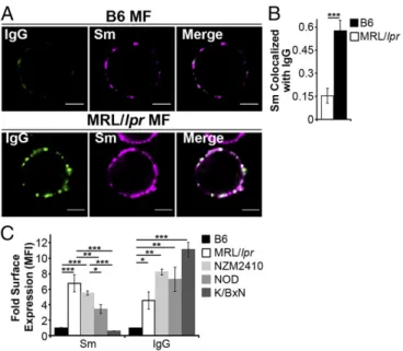
NZM2410) accumulate high levels of FcγR-bound IgG-ICs
(Fig. 1) (32). Similarly, SLE patients experiencing active disease
accumulate nuclear antigens on peripheral blood mononuclear
cells (32). Therefore, we assessed whether the accumulation of
IgG-ICs occurs in other autoimmune models by quantifying
the levels of surface IgG and nuclear antigen on MFs from
murine models of diabetes (NOD) and rheumatoid arthritis
(K/BxN). We found that the levels of surface IgG on MFs from
NOD and K/BxN mice with active disease were elevated (Fig.
1
C
) and had a punctate staining pattern (
Fig. S1
) similar to
MRL/
lpr
MFs, suggesting aggregated, rather than monomeric, IgG.
Surface Sm levels were slightly elevated on NOD MFs, but absent
on MFs from K/BxN mice, raising the possibility that in other
autoimmune diseases, MFs accumulate aggregated surface IgG
that might be part of immune complexes containing
disease-spe-cific antigens. Because NOD, MRL/
lpr
, and NZM2410 mice are
genetically unrelated, these findings also suggest that multiple
distinct genetic defects could lead to the accumulation of immune
complexes on the cell surface. Thus, understanding the
mech-anism underlying the accumulation of IgG-ICs on the surface
of MRL/
lpr
MFs could elucidate a fundamental defect in
autoimmunity.
Lupus-Prone MFs Phagocytose IgG-ICs but Exhibit Defective Phagolysosome Maturation.
In SLE, the accumulation of
apopto-tic debris has been attributed to heightened cell death, impaired
clearance of cell debris, decreased complement, and increased IgG
levels (3
–
5, 33
–
36). Whether lupus-prone MFs have intrinsic
fects contributing to impaired clearance of apoptotic debris is
de-bated because no mechanism has been defined (15, 16). To identify
how IgG-ICs accumulate, we formed ICs using anti-nucleosome
(PL2.3, IgG2a) bound to apoptotic blebs. The use of apoptotic
blebs and autoantibody allows B6 and MRL/
lpr
MFs to internalize
physiologically relevant IgG-ICs, and a means to compare
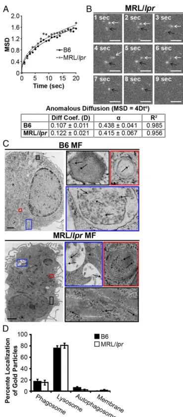
phago-cytosis, intracellular trafficking, and degradation in real time. Using
total internal reflection fluorescence (TIRF) microscopy, we found
B6 and MRL/
lpr
MFs had comparable diffusion coefficients before
internalization and internalized IgG-ICs at similar rates (Fig. 2
A
and
B
). Therefore, the accumulation of IgG-ICs on the surface of
MRL/
lpr
MFs is not the result of impaired phagocytosis.
Another possible explanation for accumulation of IgG-ICs on
MRL/
lpr
MFs is improper trafficking to lysosomes. However,
cryo-electron microscopy showed that
∼
80% of gold-labeled IgG-ICs
reached lysosomal structures in B6 and MRL/
lpr
MFs within 2 h of
phagocytosis (Fig. 2
C
and
D
). Therefore, intracellular trafficking is
not impaired.
Impaired lysosomal degradation could promote membrane
ac-cumulation of IgG-ICs after internalization and trafficking to
ly-sosomal structures. Lysosomes contain hydrolytic enzymes that
degrade cargo entering the cell through multiple receptors, including
FcγRs (37). Activation of lysosomal enzymes requires the
termina-tion of reactive oxygen species (ROS) productermina-tion and activatermina-tion of
the vacuolar H
+-ATPase (V-ATPase) to achieve a pH
≤
5 (38). To
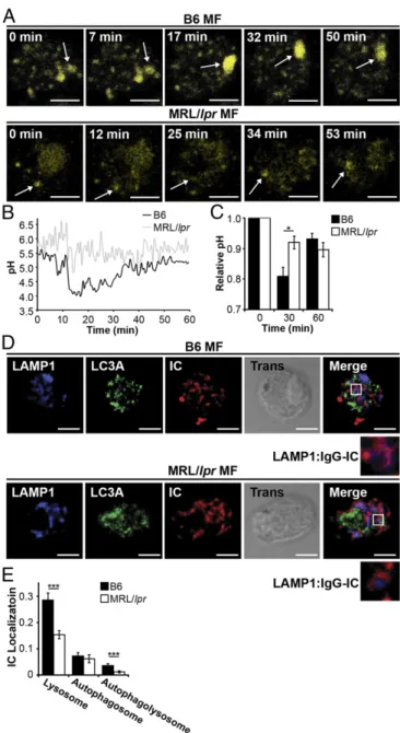
determine the pH of maturing phagosomes, we introduced an
acidotropic ratiometric dye during phagocytosis of IgG-ICs. In B6
MFs, real-time two-photon microscopy identified vesicular fusion
events, resulting in large acidic structures (pH
≤
4.5) (Fig. 3
A
and
B
). In MRL/
lpr
MFs, large acidic structures were rarely evident and
vesicles failed to sustain a pH below 5.5. To analyze larger numbers
of MFs, we used ratiometric flow cytometry to quantify the relative
pH of the population. Within 30 min of exposure to IgG-ICs, B6
MFs reduced vesicular pH by 20% then deacidified within 1 h (Fig.
3
C
). Conversely, MRL/
lpr
MFs showed an 8% drop in pH.
Con-current with the inability to fully acidify lysosomes, MRL/
lpr
MFs
exhibited heightened and prolonged production of ROS (
Fig. S2
).
These results demonstrate that MRL/
lpr
MFs are functionally
im-paired in lysosomal acidification, but the impairment is not absolute
as seen in lysosomal storage disorders. These findings are consistent
with the idea that antigen processing and MHC presentation remain
partially intact in lupus-prone mice (39, 40).
The impaired acidification and heightened ROS production
suggests that MRL/
lpr
MFs are not properly maturing the
phagolysosome, thus preventing the degradation of IgG-ICs.
Maturation of the phagolysosome and autophagosome requires
membrane stabilization, achieved through the recruitment of
lyso-some-associated membrane proteins (LAMPs) and light chain 3
(LC3). To assess whether the phagolysosome fully matures, we
quantified the levels of LAMP-1 and LC3A to distinguish
autopha-gosomes (LC3A
+, LAMP-1
−) and autophagolysosomes (LC3A
+,
LAMP-1
+) from lysosomes (LC3A
−, LAMP1
+). Using confocal
mi-croscopy, we found that MRL/
lpr
MFs showed a twofold reduction in
the association of IgG-ICs with LC3A
−, LAMP-1
+structures (Fig. 3
D
and
E
). Because IgG-ICs arrive at lysosomal structures (Fig. 2
C
and
D
), the reduced association of LAMP-1 with vesicles containing
IgG-ICs demonstrates that the impaired acidification of the
lyso-somes is consistent with defective phagolysosome maturation.
Lupus-Prone MFs Recycle IgG-ICs to the Cell Membrane.
Damaged
membrane proteins traffic to the lysosome for degradation;
how-ever, impaired degradation promotes their recycling back to the
cell membrane (41, 42). We hypothesized that a similar mechanism
might promote recycling and the accumulation of FcγR-bound
IgG-ICs because they also are targeted to lysosomal structures
following activation (37). To determine whether impaired
lyso-somal degradation promotes accumulation of surface nuclear
antigens, we cocultured MFs with fluorophore-conjugated
IgG-ICs and monitored their localization over time. Greater than
75% of the MFs phagocytosed IgG-ICs and both B6 and MRL/
lpr
MFs bound similar levels of IgG-ICs (Fig. 4
A
and
B
). Bound
IgG-ICs were rapidly phagocytosed and evident within vesicular
compartments at 24 h. By 72 h, MFs from the MRL/
lpr
mice
recycled the IgG-ICs back to the cell membrane, whereas B6
MFs retained them within the cell (Fig. 4
A
and
B
and
Fig. S3
).
The IgG-ICs appeared to remain intact and bound by FcγRs,
because both the antibody and apoptotic debris colocalized on
the surface of the cell (
Fig. S4
), and the levels of surface FcγRI
Fig. 1. Autoimmune-prone MFs accumulate IgG-ICs on the cell membrane.
(AandB) CD11b+cells were purified from spleen and analyzed for surface Sm and IgG by confocal imaging. (Scale bars: 2.5μm.) Data represent two experiments, two mice, 8–10 cells. A representative image of a MF from the indicated mice (A) and quantification of IgG colocalization with Sm for all cells imaged (B) is presented. (C) Flow cytometry analysis of surface IgG and Sm on splenic MFs (CD11b+, CD11c−) relative to B6 IgG [mean fluorescence intensity (MFI) range=1.81–7.64] or B6 Sm (MFI range=1.02–7.55); four experiments, 4–8 mice (B6≥12 wk, MRL/lpr≥12 wk, NZM2410=12–22 wk, NOD≥26 wk; blood glucose levels>500 mg/dL, K/BxN≥13 wk; clinical score=10). Error bars indicate SEM. Studentttest or ANOVA, *P≤0.05, **P≤0.01, ***P≤0.001.
IMMUNOLO
GY
AND
INFLAMMA
TION
PNAS
and IgG on MRL/
lpr
MFs increased proportionately (32).
Overall, these findings support a model wherein impaired
mat-uration of the lysosome in MRL/
lpr
MFs diminishes degradation
of IgG-ICs, inducing their recycling back to the membrane.
The levels of nuclear antigen on MFs from MRL/
lpr
mice lacking
FcγRI (FcγRI
−/−MRL/
lpr
) are decreased 60% compared with cells
from FcγRI
+/+MRL/lpr mice, and they remain disease-free (32),
implicating the accumulation of IgG-ICs on FcγRI in SLE. Despite
FcγRI
−/−MRL/
lpr
MFs binding 60% fewer IgG-ICs (Fig. 4
A
and
B
), they remain impaired in lysosomal acidification and recycle
internalized IgG-ICs. Therefore, FcγRI is not the only receptor that
recycles ICs, and loss of FcγRI decreases the amount of
IgG-ICs that are internalized.
To assess whether recycling is unique to ICs containing apoptotic
debris, we bound anti-LPS (IgG2b) to gentamicin-killed
Escherichia
coli
(
E. coli
-ICs). Although MRL/
lpr
MFs phagocytosed fewer
E
.
coli
-ICs (Fig. 4
C
), 72 h following phagocytosis, the levels of LPS on
the cell surface increased threefold compared with those on B6
MFs (Fig. 4
D
). These findings indicate that
E. coli
-ICs also fail to
be degraded by MRL/
lpr
MFs. To assess whether all ICs were
recycled in MRL/
lpr
MFs, we cultured MFs with ICs formed by
binding TNP
20KLH to anti-TNP IgG2a (TNP-ICs). Surprisingly,
both MRL/
lpr
and B6 MFs phagocytosed and degraded the
TNP-ICs (Fig. 4
E
,
Right
), unlike IgG-ICs (Fig. 4
E
,
Left
). These data
demonstrate that MRL/
lpr
MFs degrade some IgG-ICs, and that
recycling is not unique to ICs containing apoptotic debris.
To assess whether impaired lysosomal acidification is sufficient
to induce recycling of IgG-ICs, we inhibited lysosome function in
B6 MFs and assessed whether this induced IgG-ICs to recycle.
Concanamycin A prevents acidification and degradation of
phago-cytosed cargo by specifically inhibiting the lysosomal V-ATPase. B6
MFs treated with concanamycin A recycled IgG-ICs at levels similar
as MRL/
lpr
MFs (Fig. 4
F
). Thus, diminished lysosomal acidification
is sufficient to promote recycling and accumulation of IgG-ICs.
Impaired Lysosomal Maturation Permeabilizes the Phagolysosomal Membrane Allowing dsDNA and IgG To Leak into the Cytosol.
Stud-ies of microbial pathogens have shown that some intracellular
bacteria prevent phagosomal maturation, resulting in bacterial
antigens accessing the cytosol (43, 44). In a similar manner,
impaired lysosomal maturation in MRL/
lpr
MFs might allow
antigens from IgG-ICs to gain access to the cytosol and activate
innate sensors. Therefore, we selected two cytosolic sensors
(AIM2 and TRIM21) that recognize different components from
IgG-ICs (dsDNA and IgG) to determine whether the inability to
mature the lysosome permeabilizes the phagolysosome, allowing
antigens to leak into the cytosol. To assess whether nuclear
anti-gens gained access to the cytosol, we stimulated B6 and MRL/
lpr
MFs with IgG-ICs containing fluorescent dsDNA.
Immunopre-cipitation of AIM2 from B6 and MRL/
lpr
MFs showed equal
levels of AIM2 protein (Fig. 5
A
). Despite equal amounts of protein,
MRL/
lpr
MFs had a 2.5-fold increase in the amount of dsDNA
bound to AIM2 compared with B6 (Fig. 5
B
). In contrast, AIM2
from FcγRI
−/−MRL/
lpr
MFs bound the same level of dsDNA as
B6. These findings suggest that although FcγRI
−/−MRL/
lpr
MFs
recycle IgG-ICs (Fig. 4
A
and
B
), they leak fewer antigens into the
cytosol, possibily as a consequence of decreased internalization of
IgG-ICs, or because FcγR-specific signals are necessary to
per-meabilize the phagolysosome.
AIM2 initiates inflammasome formation by recruiting
procas-pase-1 through the linker molecule ASC resulting in the cleavage
and activation of caspase-1 (24). To assess whether phagocytosis of
IgG-ICs by MFs induces inflammasome formation, we quantified
active caspase-1, and enumerated cytosolic ASC foci (Fig. 5
C
and
Fig. 2. MRL/lprMFs phagocytose and traffic IgG-ICs to lysosomal structures.
(AandB) Bone marrow-derived macrophages (BMMFs) were cultured with Alexa488-labeled IgG-ICs and examined over time by TIRF microscopy; four experiments, four mice, cells= 9–12,n =47–48. The mean squared dis-placement (MSD) was calculated by tracking the disdis-placement of IgG-ICs on the surface of the cell until the IgG-IC left the imaging plane (phagocytosis). (A) Each point is the average MSD of 4–7 IgG-ICs from two to four cells in the imaging plane. (B) A representative time series of IgG-ICs on the surface of a MRL/lprMF (arrows) is presented. (CandD) BMMFs were cultured with gold-labeled IgG-ICs for 2 h and examined by cryo-electron microscopy; two ex-periments, two mice, 12–15 cells. A representative image of a MF from the indicated mice (C) and the subcellular localization of IgG-ICs in each cell
and quantified for all cells imaged (D) is presented. IgG-ICs were found in phagosomes (single membrane, electron light), lysosomes (single membrane, electron dense), autophagosomes (double membrane), and on the cell membrane. Error bars=SEM. Studentttest, *P≤0.05, **P≤0.01, ***P≤
D
). In the resting state, the number of MRL/
lpr
MFs containing ASC
foci was consistently elevated compared B6 and FcγRI
−/−MRL/
lpr
MFs, but this difference was not statistically significant. After
culturing the MFs for 4 h with IgG-ICs,
∼
40% of the MRL/
lpr
MFs exhibited ASC foci compared with 20% in B6 and FcγRI
−/−MRL/
lpr
MFs (Fig. 5
D
). This finding was consistent with diminished
binding of dsDNA to AIM2 in B6 and FcγRI
−/−MRL/
lpr
MFs (Fig. 5
A
and
B
). In MRL/
lpr
MFs, heightened formation of ASC foci
co-incided with a 4.5-fold increase in active caspase-1 compared with
either B6 or FcγRI
−/−MRL/
lpr
MFs (Fig. 5
E
). Similarly, ex vivo
splenic myeloid cells from MRL/
lpr
mice exhibited a twofold increase
in active caspase-1 compared with B6 (Fig. 5
F
). Inflammasome
for-mation was the consequence of lysosomal dysfunction because B6
MFs treated with concanamycin A had high levels of ASC foci and
caspase-1 activation (Fig. 5
D
and
E
). Thus, using dsDNA binding to
AIM2 as a sentry for phagolysosomal membrane permeabilization,
we found that impaired lysosomal degradation of IgG-ICs allows
nuclear antigens to leak into the cytosol. These antigens bind AIM2
coincident with the activation of caspase-1 and the formation of
inflammasomes. Although it would be ideal to assess the contribution
of AIM2 in caspase-1 activation and inflammasome formation in
MRL/
lpr
MFs, the heightened ROS production, heterogeneity of the
ligands contained in the IgG-ICs, and the number of DNA-binding
cytosolic sensors make knockdown of any single sensor unlikely
to have an effect.
To corroborate the idea that antigens leak into the cytosol as a
consequence of diminished phagolysosomal maturation and
mem-brane permeabization, we assessed whether IgG from exogenous
IgG-ICs activated TRIM21, a cytosolic sensor with high affinity for
IgG (45). We found that 4 h after coculture with IgG-ICs, MRL/
lpr
MFs showed a twofold increase compared with B6 MFs in
fluo-rophore-tagged IgG bound to TRIM21 (Fig. 6
A
and
B
). The level
of TRIM21-bound IgG in FcγRI
−/−MRL/
lpr
MFs was not different
from B6, indicating that similar to nuclear antigen, diminished
in-ternalization of IgG-ICs reduces the amount of IgG reaching the
cytosol. Similarly, coculture of MRL/
lpr
MFs with IgG-ICs
in-creased the levels of IgH/IgL bound by TRIM21 (Fig. 6
A
). We do
not believe that the heightened levels of IgG bound to TRIM21 are
a consequence of the 8.8-fold increase in TRIM21 protein levels in
MRL/
lpr
MFs because FcγRI
−/−MRL/
lpr
MFs also exhibited
heightened levels of TRIM21 (Fig. 6
A
and
C
), and their levels of
IgG bound to TRIM21 were not different from B6 (Fig. 6
A
and
B
).
Thus, elevated TRIM21 may contribute to disease pathology, but it
alone is insufficient unless diminished lysosomal maturation
pro-vides heightened levels of IgG ligand.
TRIM21 is an E3 ligase that possesses two unique functions.
First, it inhibits type 1 IFN production by diminishing interferon
regulatory factor (IRF) protein levels through ubiquitination and
proteasomal degradation. Second, the binding of IgG to TRIM21
stabilizes IRF proteins and activates NF-κB (46, 47), heightening
TLR activation and type 1 IFN production. To assess whether
TRIM21 was activated, we cocultured B6 and MRL/
lpr
MFs with
IgG-ICs and quantified the nuclear translocation of p65 during
NF-κB activation. Resting MRL/
lpr
MFs exhibited slightly elevated
nuclear p65 levels; however, this difference was not statistically
sig-nificant. After coculture with IgG-ICs, nuclear translocation of p65
was increased 2.5-fold (Fig. 6
D
and
E
). Loss of FcγRI in MRL/
lpr
MFs restored nuclear p65 levels to those in B6 MFs. Further, p65
nuclear translocation was the consequence of failed lysosomal
acidification because concanamycin A-treated B6 MFs stimulated
with IgG-ICs induced a 2.5-fold increase in nuclear p65 (Fig. 6
D
and
E
). It was possible that NF-κB activation in response to IgG-ICs
resulted from the binding of apoptotic debris to intracellular TLRs.
To assess this possibility, we cocultured MRL/
lpr
MFs with apoptotic
blebs lacking IgG. Like IgG-ICs, apoptotic blebs recycled in MRL/
lpr
MF (
Fig. S5
) possibly because they are opsonized by CRP and enter
cells via FcγRI (48). Despite recycling, apoptotic blebs lacking IgG
did not translocate p65 to the nucleus, indicating that IgG was
re-sponsible for NF-κB activation (Fig. 6
D
and
E
). To determine
whether NF-κB activation in MRL/
lpr
MFs was the consequence of
TRIM21, we diminished intracellular TRIM21 levels by using
shRNA. Targeting TRIM21 in B6 and MRL/
lpr
bone
marrow-de-rived macrophages (BMMFs) stably reduced TRIM21 levels
com-pared with transduction with a nontargeting control (Fig. 6
F
). In
MRL/
lpr
MFs where TRIM21 levels were diminished, phagocytosis
of IgG-ICs reduced the nuclear p65 levels 3.4-fold, levels comparable
to B6. In contrast, cells transduced with the nontargeting shRNA did
not change nuclear p65 levels (Fig. 6
G
and
H
). Thus, the inability to
mature the phagolysosome allows IgG from ICs to leak into the
cytosol and activate TRIM21.
Fig. 3. MRL/lprMFs fail to mature the lysosome. (AandB) BMMFs were
stimulated with IgG-ICs, and the pH of the maturing phagosomes were assessed by ratiometric two-photon microscopy; three experiments, three mice, 10–25 cells. A representative time series of a MF from the indicated mice (A) with the quantified pH of a single phagosomal acidification event (arrow) (B) is presented. Ratiometric flow cytometry was used to quantify the relative phagosomal pH across the entire population of MFs; six exper-iments, 6–8 mice (C). (DandE) Colocalization of IgG-ICs with LAMP-1 and/or LC3A was assessed by confocal imaging; three experiments, three mice, 36– 39 cells. A representative image of a MF from the indicated mice (D) and quantification of IgG-IC colocalization with indicated compartments for all cells imaged (E) is presented. Error bars=SEM. Studentttest, *P≤0.05, **P≤
0.01, ***P≤0.001. (Scale bars: 5μm.)
IMMUNOLO
GY
AND
INFLAMMA
TION
PNAS
The activation of TRIM21 by IgG promotes its monoubiquitination,
which acts as a docking site for E2 Ube2N/Ube2V2, inducing
TRIM polyubiquitination and proteasomal degradation (49, 50).
To assess TRIM21 activity in vivo, we quantified the levels of
ubiquitinated TRIM21 in ex vivo splenic myeloid cells from B6 and
MRL/
lpr
mice. Consistent with TRIM21 activity, we found that
splenic myeloid cells from MRL/
lpr
mice had 85-fold more
mono-ubiquitinated TRIM21 compared with ex vivo B6 myeloid cells.
Polyubiquitination was not evident (Fig. 6
I
and
J
). IgG was
nec-essary for TRIM21 ubiquitination because ex vivo myeloid cells
from age-matched AID
−/−MRL/
lpr
mice (lack IgG) had
signifi-cantly less ubiquitinated TRIM21 (13.5-fold vs. 85-fold) compared
with B6 myeloid cells. Acute activation of TRIM21 stabilizes IRF3,
whereas chronic activation increases IRF7 levels by activating
NF-κB (46, 47). In ex vivo myeloid cells from MRL/
lpr
mice, we found
that ubiquitinated TRIM21 was coincident with nuclear
trans-location of p65 and heightened levels of IRF7 protein, but not
with heightened IRF3 (Fig. 6
K
). The finding that IRF7 protein
levels are selectively increased suggests that TRIM21 activation in
myeloid cells from MRL/
lpr
mice is not an acute event, but
in-stead, induced by chronic activation through IgG.
Impaired Lysosomal Maturation Results in Heightened Intracellular TLR Activation.
Diminished lysosomal maturation allows IgG
and nuclear antigens to leak into the cytosol and activate innate
sensors. However, a large fraction of IgG-ICs remains inside the
phagosome and is capable of activating TLRs. Coupled with
heightened IRF7 levels from activated TRIM21 (Fig. 6
H
),
phagosomal TLR ligands could heighten IFNα
secretion through
the activation of TLR7 and TLR9 (TLR7/9). To assess whether
the prolonged residency of nuclear antigens within the
phagolysosome activates intracellular TLRs, we quantified
interleukin-1 receptor-associated kinase 1 (IRAK1) levels in MFs
24 h after exposure to IgG-ICs. This time point was sufficient for
B6 MFs to degrade the ICs, but was before recycling of
IgG-ICs in MRL/
lpr
MFs (Fig. 4
A
and
B
). We found that MRL/
lpr
MFs exposed to IgG-ICs showed a 2.8-fold decrease in IRAK1
levels, consistent with TLR activation (Fig. 7
A
). Chloroquine, an
acidotropic molecule that binds double- and single-stranded
nucleotides and sterically hinders their binding to TLRs (51),
restored IRAK1 levels supporting that TLR7/9 are activated in
MFs that fail to degrade IgG-ICs. Further, impairing lysosomal
acidification with concanamycin A prevented the degradation of
IgG-ICs in B6 MFs (Fig. 4
F
) and reduced IRAK1 levels to those
found in MRL/
lpr
MFs (Fig. 7
A
). Therefore, the impaired
lyso-somal acidification in MRL/
lpr
MFs is sufficient to heighten TLR
activation in the presence of IgG-ICs.
Formation of the TLR
–
MyD88
–
IRAK1 complex downstream
of TLR7/9 promotes phosphorylation and nuclear translocation
of IRF7, resulting in the production of type 1 IFN (52). To assess
whether impaired degradation of IgG-ICs promotes nuclear
translocation of IRF7, we cocultured MRL/
lpr
MFs with IgG-ICs
and found they exhibited a twofold increase in nuclear IRF7
levels (Fig. 7
B
and
C
) that was sustained for 24 h. IgG-ICs did
not localize all IRF proteins to the nucleus as IRF3 remained
cytoplasmic (Fig. 7
C
). Reducing the lysosomal burden through
loss of FcγRI also reduced the nuclear translocation of IRF7 in
MRL/
lpr
MFs to levels comparable to B6. These findings were
not an artifact of BMMFs because ex vivo myeloid cells from
MRL/
lpr
mice also showed a twofold increase in nuclear IRF7,
Fig. 4. IgG-ICs recycle and accumulate on the cell membrane of MRL/lpr
MFs. (AandB) BMMFs from the indicated mice were cultured with IgG-ICs and examined over time by confocal imaging; four experiments, 3–4 mice, 17–23 cells. A representative image of a MF from the indicated mice (A) and quantification of IgG-IC colocalization with the cell membrane for all cells imaged (B) is presented. BMMFs were cultured with GFP-expressingE. coli-ICs for 1 h. At indicated time points relative levels (to B6+E. coli-IC) of
whereas nuclear IRF3 was not elevated (Fig. 7
D
). Collectively,
the data support a model wherein impaired lysosomal maturation
prolongs phagolysosomal residency of IgG-ICs, facilitating chronic
intracellular activation of IRAK-coupled TLRs.
The inability to degrade IgG-ICs resulted in increased levels of
IRF7 and heightened intracellular TLR activation. Combined, these
events could elevate IFNα
secretion (52). To assess this possibility,
we cultured MRL/
lpr
MFs with IgG-ICs (12, 24 h) and found they
secreted two- and threefold more IFNα
(Fig. 7
E
). The production of
IFNα
was a consequence of FcγRI-mediated internalization because
IFNα
levels secreted by FcγRI
−/−MRL/
lpr
MFs were comparable
to B6. It was also, in part, a consequence of TLR7 and/or
TLR9 because MRL/
lpr
MFs deficient in TLR7 and TLR9
(TLR7
−/−xTLR9
−/−/MRL/
lpr
) secreted 3.2-fold less IFNα
com-pared with MRL/
lpr
MFs. The data indicate that FcγRI is
re-quired for IFNα
secretion by MRL/
lpr
MFs, and that activation
of TLR7 and/or TLR9 contributes to
∼
70% of the IFNα
pro-duced. These findings are consistent with a model wherein
di-minished lysosomal maturation promotes the accumulation of
IgG-ICs in the phagolysosome of MRL/
lpr
MFs activating TLR7
and/or TLR9 and contributing to IFNα
secretion.
Discussion
The mechanism underlying the accumulation of IgG-ICs in the
periphery of SLE patients has been highly debated. A number of
observational studies showed that MFs have intrinsic defects in the
phagocytosis of latex beads, apoptotic cells, IgG-ICs, bacteria, and
yeast (10, 34, 35, 53). Other studies found that phagocytosis is intact,
but the ability to degrade the internalized cargo is impaired (35, 54,
55). We find that lupus-prone MRL/
lpr
MFs phagocytose and traffic
IgG-ICs to lysosomal structures, but that the lysosomal structures
were unable to mature and acidify. As a result, the IgG-ICs were
not degraded and recycled back to the cell membrane.
Hemato-poietic cells from SLE patients have high levels of IgG and nuclear
antigens (32), and they express LAMP1/2 on the cell surface (56).
These findings support the idea that the lysosomal compartment
has trafficked to the membrane and are consistent with our data
showing that MFs from lupus-prone mice accumulated high levels
of nuclear antigens (Fig. 1) as a consequence of the recycling of
undegraded ICs from the lysosome (Fig. 4). Interestingly
IgG-ICs, apoptotic blebs, and
E. coli
-ICs recycled to the cell membrane,
whereas TNP-ICs were degraded, suggesting that the size and/or
content of the cargo impairs the lysosome. Alternatively, the
op-sonin coating the incoming apoptotic debris could impact lysosomal
maturation. Combined, these findings could explain the variability
of previous studies aimed at identifying defects in SLE monocytes.
Spontaneous SLE has been linked to alterations in expression
(57) and activation of TLR7/9 (17, 18), dysregulation of the
cy-tosolic sensors p202 and AIM2 (24, 25), and polymorphisms (21)
and heightened expression in TRIM21 (22). We now define that
defective degradation of FcγR-bound cargo in the lysosome is a
critical upstream event that overburdens the phagolysosome.
Prolonged intracellular residency of IgG-ICs promoted the
ac-tivation of TLRs and the permeabilization of the phagolysosomal
membrane, allowing IgG and nuclear antigen to access the cytosol
and activate innate sensors. Other enzymes critical in degrading
nuclear antigens independent of lysosomes including RNase H2
(58), DNase I (59, 60), and DNase III (TREX1) (61) have been
implicated in SLE and may operate in concert with impaired
ly-sosomal maturation to promote autoimmunity. Collectively, our
findings identify the events underlying the accumulation of nuclear
antigens and activation innate sensors that promote autoantibody,
IFNα, and heightened apoptosis in SLE.
Although this study focuses on defining how nuclear antigens
accumulate on MFs, it is important to keep in mind that nuclear
antigen accumulates on DCs and B and T cells (32). Thus, it is
possible that other cells may harbor defects in lysosomal
matu-ration, contributing other disease manifestations. For example,
diminished lysosomal maturation in plasmacytoid dendritic cells
could heighten secretion of IFNα
(19), whereas the same defect in
MFs and neutrophils may heighten cell death (62). Further, the
presence of nuclear self-antigens on the cell surface could provide
a source of high avidity antigen that renews BCR signaling and
ac-tivates autoreactive B cells (63), facilitates uptake of TLR ligands
by BCR-mediated endocytosis (17), and positions autoreactive B
cells to further differentiate into memory cells if T-help is
available (64). Thus, the same overarching lysosomal defect may
contribute to the activation of multiple cell types in SLE.
Overlapping autoimmune diseases are common in patients
di-agnosed with SLE including diabetes (65), rheumatoid arthritis (66),
and Sjögren
’
s syndrome (67). Recent GWAS studies have identified
common genetic polymorphisms including the major
histocompat-ibility complex, TNFAIP3, PTPN22 (68), STAT4 (69), and CD40
(70) that span multiple autoimmune diseases, although their
func-tional role in breaking tolerance is unknown. Our finding that
punctate IgG accumulates on MFs from multiple murine models of
autoimmunity including SLE, diabetes, and rheumatoid arthritis is
interesting because it might reflect that aggregated FcγR/IgG-ICs
are common to multiple autoimmune diseases. Further, the IgG did
not colocalize with high levels of nuclear antigens, suggesting that
the antigens contained in the IgG-ICs might be disease-specific,
thus activating the immune system in different ways. Therefore, the
accumulation of punctate IgG on the surface of MFs from NOD
and K/BxN mice raises the possibility that impaired lysosomal
maturation underlies other autoimmune diseases.
Fig. 5. Impaired lysosomal maturation allows dsDNA to leak into the cytosol and
activate AIM2. BMMFs were stimulated for 4 h with Hoechst-labeled IgG-ICs (A andB). (A) A representative blot of immunoprecipitated AIM2 (5–8×10⁶cells) is
shown. (B) Relative levels (to B6 untreated) of fluorescent DNA isolated from the AIM2 immunoprecipitation were quantified by using a fluorescent plate reader; four experiments, four mice. (C–E) BMMFs were stimulated for 4 h with IgG-ICs±
concanamycin A (20 ng/mL) and then examined for the formation of ASC foci and caspase-1 activation by confocal imaging. A representative image of a MF, with and without ASC foci is presented (C). (Scale bars: 5μm.) The percentage of cells with ASC foci for each experiment (D) and relative levels (to B6 untreated) of caspase-1 activation (E) was quantified for all cells imaged; 10 experiments, 2–10 mice, 63–364 cells. (F) Relative levels (to B6) of caspase-1 activation was measured in splenic myeloid cells (CD11b+) by flow cytometry; four experiments, 5–9 mice (≥17 wk; active disease confirmed with kidney H&E). Error bars=SEM. Studentt test or ANOVA, nd>0.05, *P≤0.05, **P≤0.01, ***P≤0.001.
IMMUNOLO
GY
AND
INFLAMMA
TION
PNAS
Materials and Methods
Mice.C57BL/6 (B6) and MRL/MpJ-Tnfrs6lpr/J (MRL/lpr; JAX mice Stock 000485)
colonies were maintained in an accredited animal facility at University of North Carolina at Chapel Hill. Experimental methods were approved by the University of North Carolina’s Institutional Animal Care and Use Committee (IACUC). NZM2410 mice (71) were obtained from Gary Gilkeson (mice were 12–22 wk of age; Medical University of South Carolina, Charleston, SC), AID−/−MRL/lprmice (72) from Marilyn Diaz, National Institute of Environmental
Health Sciences (NIEHS), Research Triangle Park, NC, NOD mice from Roland Tisch (blood glucose levels>500 mg/dL; University of North Carolina), K/BxN mice (73) from Christophe Benoist (clinical score=10, ankle thickening=3.85– 3.95 mm; Harvard Medical School, Boston), and C57BL/6-Tg(UBC-GFP)30Scha/J (GFP-expressing) mice (74) from Bill Goldman, University of North Carolina. We generated FcγRI−/−MRL/lprmice by backcrossing FcγRI−/−/C57BL/6 mice to
MRL/lprmice for 10 generations. TLR7−/−/MRL/lprand TLR9−/−/MRL/lprmice
were reconstituted from sperm (75) and then intercrossed to generate TLR7−/−×TLR9−/−/MRL/lprmice.
Reagents.Antibodies specific for LAMP1 and CD11b were purchased from BD
Biosciences; LC3A from Cell Signaling; goat rabbit IgG and rabbit anti-goat IgG from Molecular Probes; AIM2, ASC, TRIM21, p65, IRAK1, IRF3, and IRF7 from Santa Cruz Biotechnologies; anti-IgG from Jackson ImmunoResearch; and anti-LPS (Escherichia coli J5) from Thermo Scientific. Concanamycin A and chloroquine diphosphate salt were purchased from Sigma-Aldrich, Immuno-gold conjugate EM streptavidin from BB International, and TNP20KLH from
Biosearch Technologies. Antibodies specific to Smith (Sm; 2.12.3), nucleosome (PL2-3), CD16/32 (2.4G2), and TNP (Hy1.2) were purified from hybridoma culture supernatant by using protein G-Sepharose (GE Healthcare) then left unlabeled or conjugated with Alexa Fluor according to the manufacturer’s instructions (Molecular Probes). Fluorescent molecules LysoSensor, dihydrorhodamine 123, and CellMask were purchased from Molecular Probes, and FAM-FLICA caspase-1 assay kit from ImmunoChemistry Technologies. LI-COR blocking buffer, IRDye680-, and IRDy800-conjugated antibodies (anti-rabbit, anti-mouse, anti-goat) were purchased from LI-COR Biosciences. To differentiate BMMFs, we generated L929 cell (European Collection of Cell Cultures) supernatant by plating 2.5×10⁵cells
into a T150 flask with 50 mL of D10 media [DMEM with 10% (vol/vol) FBS, 1 mM sodium pyruvate, 50μg/mL gentamicin, 100μg/mL Pen/Strep, 2 mML-glutamine, 50 nMβmercaptoethanol (β-ME)]. After 12 d, supernatant was collected and filtered.
Splenocyte Isolation.Spleens from mice were ground between frosted glass
slides and filtered through a 70-μm filter to create a single-cell suspension in PBS. Pelleted cells were resuspended in 2 mL of NH4Cl2(0.154 M) for 7 min at
room temperature, and then filtered through a 40-μm filter into PBS. Cells were pelleted, then resuspended at 108cells/mL and used for experiments.
Alternatively, CD11b+splenocytes were purified per manufacturer’s instruc-tions (Stemcell Technologies).
BMMF Cultures.Single-cell suspensions of bone marrow were prepared from
the tibias and femurs of mice. Mononuclear cells were isolated by using Lympholyte Separation Medium (CEDARLANE Laboratories), plated in a 60-mm Petri dish with 6 mL of MF differentiation media [D10 media with 10% (vol/vol) L-cell supernatant], and cultured overnight (37 °C, 5% CO2). Nonadherent cells
were plated into nontissue culture-treated 100-mm Petri dishes (0.75–1 mL cells per Petri dish) with 7 mL of fresh MF differentiation media. To promote MF differentiation, cells were incubated for 6 d (37 °C, 5% CO2) with an
ad-ditional 5 mL of MF differentiation media being added on day 4. The resulting BMMFs were removed from the dish by washing with ice cold PBS. BMMF cultures were 98% CD11b+, I-Alo, and B7.2lo.
Formation of Immune Complexes.
IgG-ICs.Single-cell suspensions of thymocytes were prepared from 5- to 8-wk mice, irradiated (600 rads), and cultured 16–18 h in 10 mL of PBS (37 °C, 5% CO2). Apoptotic thymocytes were centrifuged for 5 min (350×g), and the
supernatant containing apoptotic debris was incubated with autoantibodies (2.12.3 or PL2-3) on ice for 30 min (6.67μg of Ab/1 mL of supernatant). IgG-ICs were pelleted (160,000×g) at 4 °C for 45 min and resuspended in 250μL of R10 media [RPMI with 10% (vol/vol) FBS, 1 mM sodium pyruvate, 50μg/mL genta-micin, 100μg/mL Pen/Strep, 2 mML-glutamine, 50 nMβ-ME].
Fig. 6. Impaired lysosomal maturation allows IgG to
leak into the cytosol and activate TRIM21. BMMFs were stimulated with fluorescently tagged IgG-ICs (4 h), and TRIM21 was immunoprecipitated (5–8×
106cells) (A–C). (A) Representative blots of TRIM21
and IgH/IgL chains is shown. (B) Relative levels (to B6 untreated) of fluorescent IgG isolated from TRIM21 immunoprecipitates; seven experiments, seven mice. (C) Relative levels (to B6 untreated) of immunopre-cipitated TRIM21 was quantified by densitometry; three experiments, three mice. (Dand E) Nuclear translocation of p65 subunit of NF-κB was quantified in BMMFs stimulated for 4 h with IgG-ICs or apo-ptotic debris (lacking IgG),±concanamycin A (20 ng/mL); six experiments, 3–6 mice, 16–57 cells. Representa-tive image of p65 nuclear localization in MFs from the indicated mice (D) and quantification of p65 colocalization with the nucleus of all cells imaged (E) is presented. (F–H) BMMFs were transduced with lentivirus expressing indicated shRNA. Relative levels of TRIM21 (to B6+nontargeting; MFI=2.57–7.25) in transduced cells (GFP+) were quantified by flow cytometry; three experiments, three mice (F), and nuclear translocation of the p65 subunit of NF-κB was quantified in transduced cells (GFP+) stimulated for 4 h with IgG-ICs; two experiments, two mice, 20– 22 cells (GandH). Localization of p65 in MFs from the indicated mice (G) and quantification of p65 colocalization with the nucleus (H) is presented. (Iand J) Ubiquitin was immunoprecipitated from splenic myeloid cells (CD11b+; 35 ×106 cells) and
immunoblotted for TRIM21; two experiments, 4–5 mice (≥17 wk). A representative blot of TRIM21 fol-lowing immunoprecipitation of ubiquitin (I) and relative levels (to B6) of ubiquitinated TRIM21 was
TNP-ICs.TNP20-KLH was incubated with anti-TNP antibody (Hy1.2) on ice for
30 min (30μg of Ab/1μg of TNP-KLH). TNP-ICs were pelleted (160,000×g) at 4 °C for 45 min and resuspended in 200μL of R10 media (as above). E. coli-ICs.GFP-expressingE. coli(76) was incubated with anti-LPS at room temperature for 2 h (1.5μg/6.25×106E. coli) in the presence of gentamicin
(10μg/1 mL).E. coli-ICs were cultured with BMMFs [multiplicity of infection (MOI)=25].
Fluorescent Microscopy.All confocal microscopy used a Zeiss 710 confocal
microscope with a 63×1.4 N.A. (oil) PLAN APO lens and Zeiss Zen software. All two-photon microscopy used an Olympus FlouView FV1000MPE multi-photon microscope with a 25×1.05 N.A. (water) XLPlan N lens and Olympus FluoView software. Cells were randomly selected from many fields of view across the coverslip. Excluded cells were clumped (5–15% of the cells), un-healthy (5–15% of the cells, determined by Hoechst staining), or improperly stained (<2% of the cells). Data were analyzed by using ImageJ. IgG-IC/apoptotic bleb localization. BMMFs were cultured in the presence of Alexa488-labeled IgG-ICs or GFP-expressing apoptotic debris in R10 media (as above). After 2 h, the media was aspirated and cells were cultured in fresh R10 media. CellMask and Hoechst 33342 were introduced to BMMFs 15 min before fixation at indicated time points. Cells were fixed in 2% (vol/vol) para-formaldehyde in PBS and transferred to 4 °C for 15 min. Cells were resuspended in FluorSave and loaded onto coverslips for imaging. The membrane locali-zation of IgG-ICs or apoptotic debris was quantified by calculating the Man-der’s coefficient of colocalization (colocalized pixels/total fluorescent pixels). Two-photon microscopy/pH quantification.Two hours before imaging, BMMFs were incubated (37 °C, 5% CO2) on a glass bottom Petri dish (MatTek)
in rhodamine-free R10 media (as above). Immediately after adding 40μL of IgG-ICs and LysoSensor (2 mg/mL), cells were imaged for 1 h. The dye was excited by using two-photon excitation (710 nm), and emissions at 420–460 nm and 495–540 nm were quantified. The ratio of the emission channels were used to determine the pH of the vesicles by using a stan-dard curve generated by exciting LysoSensor with medium of varying pH. ASC localization/caspase-1 activation.BMMFs were cultured with FAM-FLICA caspase-1 (5μM) for 20 min before indicated time points. At the indicated time points, cells were washed, fixed with 2% (vol/vol) paraformaldehyde in PBS, and incubated at 4 °C for 15 min. Cells were blocked in 2.4G2 for 30 min at 4 °C in FACS media [2% (vol/vol) FBS, 0.02% NaN3in PBS] then stained
with an anti-ASC and Hoechst 33342 (1μg/mL) in permeabilization buffer (PBS with 0.05% Saponin and 0.5% BSA) for 30 min at 4 °C. Cells were washed, stained with goat anti-rabbit IgG-Alexa 647 in permeabilization buffer for 30 min at 4 °C, washed, costained with anti-CD11b in FACS media for 30 min at 4 °C, washed, resuspended in FluorSave, and loaded onto coverslips for imaging. Cells with ASC foci were counted and expressed as a percentage of the total cells. Total caspase-1 activation per cell was quan-tified (background fluorescence subtracted) and normalized by cell area. LAMP1/LC3A, IRF, and p65 localization.BMMFs, or splenic myeloid cells (CD11b+) purified by positive selection, were prepared as described for ASC localiza-tion/caspase-1 activation except cells were stained for LAMP1 and LC3A, IRF3, IRF7, or p65. The nuclear localization of IRF or p65 was quantified by calculating the Mander’s coefficient of colocalization (colocalized pixels/ total fluorescent pixels). Total IRF protein in myeloid cells was quantified by determining the total fluorescent pixels (normalized to cell area). Ex vivo surface stain. Ex vivo splenic cells were fixed in 2% (vol/vol) para-formaldehyde in PBS and incubated for 15 min at 4 °C. Cells were blocked in 2.4G2 for 30 min at 4 °C in FACs media (as above), washed, stained with anti-Sm (2.12.3) and IgG in FACS media for 30 min at 4 °C, washed, stained with anti-CD11b in FACS media for 30 min at 4 °C, and washed. Cells were resuspended in FluorSave and loaded onto coverslips for microscopic imaging. The membrane localization of IgG with Sm was quantified by calculating the Mander’s co-efficient of colocalization (colocalized pixels/total fluorescent pixels).
Flow Cytometry.All flow cytometry used an 18-color Becton Dickinson LSR II
Flow cytometer, and data were acquired by using Becton Dickinson FACSDiva 8.0.1 software.
Recycling of IgG-IC and TNP-IC using flow.BMMFs were incubated (37 °C, 5% CO2)
with 40μL of Alexa488-labeled IgG-ICs or TNP-ICs in R10 media (as above). To quantify surface-bound ICs at 0 h, phagocytic uptake was impaired by culturing with IgG-ICs on ice for 2 h. This protocol was sufficient to allow the ICs to bind to the surface of the cell but not be phagocytosed. For all other time points, cells were incubated (37 °C, 5% CO2) for 2 h, then media was replaced to
remove all unbound ICs. At indicated time points, cells were blocked in 2.4G2 for 30 min on ice in FACS media (as above), washed, and split into two samples. One sample was incubated with an anti-Alexa488 antibody (quenches Alexa488 fluorescence), whereas the other sample was left in FACS media for
Fig. 7. Impaired lysosomal maturation promotes intracellular TLR
activa-tion and IFNαsecretion. (A) BMMFs (1–1.5×10⁶cells) were stimulated for 24 h
with IgG-ICs±hydroxychloroquine (50μg/mL) or±concanamycin A (20 ng/ mL); five experiments, 3–5 mice. IRAK1 was immunoblotted from whole cell lysates and relative levels (to B6 untreated) of IRAK1 were quantified by densitometry. A representative IRAK1 blot is shown. (BandC) BMMFs were stimulated with IgG-ICs (4 h), and the nuclear translocation of IRF7 or IRF3 was quantified by confocal microscopy; four experiments, 3–4 mice, 20–42 cells. (Scale bars: 5μm.) A representative image of a MF from the indicated mice (B) and quantification of IRF colocalization with the nucleus of all cells imaged (C) is presented. (D) Splenic myeloid cells (CD11b+) were analyzed for nuclear IRF7 and IRF3 by confocal microscopy; two experiments, two mice (≥17 wk; active disease confirmed with kidney H&E), 10–28 cells. Supernatants from IgG-IC stimulated BMMFs were collected at the indicated time points and cocultured with WISH cells. Relative levels (to B6 0 h) of IFIT message as an indirect measure of IFNαwere quantified by RT-PCR (E); three experiments, three mice. Error bars=SEM. Studentttest or ANOVA, nd>0.05, *P≤0.05, **P≤0.01, ***P≤0.001.
IMMUNOLO
GY
AND
INFLAMMA
TION
PNAS
30 min on ice. Both samples were washed and fixed with 2% paraformaldehyde and incubated at 4 °C for 15 min. Surface-bound IgG-ICs were calculated by subtracting the mean fluorescence intensity (MFI) of the quenched sample (in-tracellular IgG-ICs) from the MFI of the unquenched sample (total IgG-ICs). Values were normalized to B6 0 h.
E. coli-IC recycling flow.BMMFs were incubated (37 °C, 5% CO2) withE. coli-ICs
(MOI=25) in R10 media (as above). Cells were incubated (37 °C, 5% CO2) for
1 h, then media was replaced to remove all unbound ICs. At indicated time points, cells were blocked in 2.4G2 for 30 min on ice in FACS media (as above), washed, and incubated with Alexa647-labeled anti-LPS in FACS media for 30 min on ice. Samples were washed and fixed with 2% paraformaldehyde, and then incubated at 4 °C for 15 min. Cells were resuspended in FACS media, and the levels of phagocytosedE. coli-ICs and surface LPS were quantified by flow cytometry. MFI values for phagocytosedE. coli-ICs were normalized to B6+E. coli-ICs. MFI for surface LPS was normalized to an isotype control. Ratiometric flow cytometry.BMMFs were incubated (37 °C, 5% CO2) for 2 h
before the addition of 40μL of IgG-ICs in R10 media (as above). To quantify a cell that has not acidified, concanamycin A (20 ng/mL) was introduced to one sample from each cell type 2 h before addition of IgG-ICs and left on the cells throughout the experiment. IgG-ICs and LysoSensor (2 mg/mL) were in-troduced for 30 min, aspirated, and replaced with fresh rhodamine-free R10 media. Cells were incubated until indicated time points and analyzed by flow cytomualifiedetry. The dye was excited with a UV laser (355 nm). Rel-ative pH was quantified by ratioing the MFIs from the emission channels (450/20 nm, 585/42 nm) (normalized to concanamycin A control).
Ex vivo surface stain.Ex vivo splenic cells were prepared as described for“Ex vivo surface stain” in the“Fluorescent Microscopy” section. Cells were resuspended in FACS media, and the MFI of the surface Sm and IgG were determined by flow cytometry and normalized to an isotype control. Ex vivo caspase-1 activation.Ex vivo splenic cells were cultured with FAM-FLICA caspase-1 (5 μM) for 20 min before fixation. Cells were fixed in 2% para-formaldehyde and incubated for 15 min at 4 °C. Cells were washed, stained with anti-CD11b in FACS media for 30 min at 4 °C, and washed. Cells were resus-pended in FACS media, and the MFI of the surface Sm was determined by flow cytometry and normalized to an isotype control. Values are normalized to B6. TRIM21 flow cytometry.Following viral transduction, BMMFs were fixed with 2% (vol/vol) paraformaldehyde in PBS, then incubated at 4 °C for 15 min. Cells were blocked in 2.4G2 for 30 min at 4 °C in FACS media (as above), washed, and stained with anti-TRIM21 in permeabilization buffer (as above) for 30 min at 4 °C. Cells were washed, stained with rabbit anti-goat IgG-Alexa 647 in permeabilization buffer for 30 min at 4 °C, washed, and resuspended in FACS media. The MFI of the TRIM21 were determined by flow cytometry and normal-ized to an isotype control.
Cryo-Electron Microscopy.BMMFs were incubated with 40μL of gold-labeled
IgG-ICs for 2 h (37 °C, 5% CO2). Suspensions of MFs were loaded into gold
planchettes (model 16706897; well size 1.2 mm×200μm), placed in high pres-sure-freeze (HPF) holders, and torqued to make a tight seal. Each sample was placed in a HPF chamber where the pressure was increased with cyclohexane to
∼2,000 bars just milliseconds before a blast of liquid nitrogen cooled the as-sembly at approximately 18,000 °C/sec by using a Leica EM PACT HPF. The pressure and cooling curves were recorded and examined after each run to ensure consistency. Frozen samples were transferred to liquid nitrogen for storage. Samples were then transferred to vials containing 2% osmium tetroxide in dry acetone cooled in liquid nitrogen. The vials were transferred cold to a chamber at−90 °C in a Leica EM AFS freeze substitution device, where samples remained for 72 h. Samples were warmed automatically by using a program that increased the temperature to−20 °C at 4 °C/h, held at−20 °C for 10 h, then warmed at 4 °C/h to 20 °C. The warmed fixed samples were processed for transmission electron microscopy (TEM) by washing with fresh acetone and replacing the acetone with propylene oxide, embedding in epon (EMS EMbed-812), and hardening at 60 °C. Thin sections, 60–70 nm thick, were cut with a diamond knife on a Leica Ultracut UTC and stained with uranyl acetate and lead citrate. Stained thin sections were examined with an FEI Tecnai T12 G2 TEM at 80 kV by using a Gatan 794 digital camera and Gatan Digital Montage software to prepare up to 5×5 montages of selected MFs imaged at 6,000–30,000×.
Immunoprecipitation and Western Blot.Lysates were prepared by the addition
of lysis buffer containing 1% CHAPS, 150 mM NaCl, 10 mM Tris (pH 7.5), 2 mM sodium orthovanadate, 1 mM PMSF, 0.4 mM EDTA, 10 mM NaF, and 1μg/mL
each of aprotinin, leupeptin, andα1-antitrypsin (in water) to cell pellets. Lysates were held on ice for 10 min followed by the removal of particulate material by centrifugation at 12,000×gfor 10 min at 4 °C.
Antibodies used in the immunoprecipitations were conjugated to cyanogen bromide-activated Sepharose 4B according to manufacturer’s in-structions (Amersham Pharmacia Biotech). Approximately 2μg of precipitating antibodies was incubated with 1.5×10⁶cell equivalents of cleared lysate for
1 h at 4 °C. Immunoprecipitates were washed twice with lysis buffer, resus-pended in reducing SDS/PAGE sample buffer, and fractionated by 10% SDS/ PAGE. Separated proteins were transferred to Immobilon-FL membranes. Membranes were blocked in LI-COR Blocking Buffer, incubated with the var-ious immunoblotting Abs followed by the appropriate fluorophore-conju-gated secondary Abs. Immunoreactive proteins were detected by using a LI-COR Odyssey infrared imaging system with Odyssey 3.0 software.
Plate Reader.All analysis used a Tecan M200 fluorescence plate reader.
ROS assay.BMMFs were incubated (37 °C, 5% CO2) for 2 h before the addition of
40μL of IgG-ICs in R10 media (as above) in an opaque 96-well plate. Thirty minutes before indicated time points, dihydrorhodamine 123 (3μg/mL) was added to each well. At the indicated time points, cells were washed, fixed with 2% para-formaldehyde, and transferred to 4 °C for 15 min. Wells were analyzed in FACS media (as above). Each sample had anN-acetylcysteine–treated control (1 mM). Fluorescent readings were normalized toN-acetylcysteine–treated B6 MFs. Immunoprecipitation assay.All beads from immunoprecipitation sample were added to a single well in an opaque 96-well plate in PBS. Wells were analyzed for Hoechst-labeled dsDNA (AIM2 immunoprecipitation) and Alexa647-labeled IgG (TRIM21 immunoprecipitation). All fluorescent readings were normalized to the background fluorescence of beads alone.
BMMF Transduction.BMMFs were prepared as described above. On day 3, cells
in a 24-well low cluster plate were transduced with lentiviral particles (SMARTvector Lentiviral mouse TRIM21 mCMV-TurboGFP shRNA; MOI=40; polybrene=5μg/mL) according to manufacturer’s instructions (Dharmacon). Separate cells were transduced with SMARTvector Nontargeting mCMV-TurboGFP control particles. On days 5 and 6, culture medium was replen-ished with an additional 750μL of macrophage differentiation media. Stably transduced cells expressed GFP by day 6 (8–20% of the culture).
WISH Cell IFNαAssay.WISH cells (product no. CCL-25; American Type Culture
Collection) were grown in minimum essential medium supplemented with
L-glutamine (2 mM), Hepes (20 mM), penicillin (100 units/mL), streptomycin (100μg/mL), and 10% (vol/vol) FBS (37 °C, 5% CO2). To measure IFNαlevels,
we quantified mRNA of IFN-regulated genes as described (77). WISH cells were plated (0.5×10⁵cells per 0.1 mL) in 96-well flat bottom plates and
cultured with media alone, recombinant mouse IFNα(100 units/mL; Bio-Source International), or BMMF supernatant (200μL) for 24 h (37 °C, 5% CO2). WISH cells were lysed, RNA extracted (RNeasy Mini Kit; Qiagen), and
cDNA prepared from 500 ng of RNA (iScript c-DNA Synthesis Kit; Bio-Rad Laboratories). The cDNA obtained from each sample was diluted 1:60, and 2μL was amplified in a 20-μL real-time quantitative PCR by using 10 mM forward and reverse primers and the 2×iQ SYBR Green Supermix (Roche Laboratories).
Statistics.Error bars represent the SEM. For the TIRF assay, error bars
rep-resent the variation between individual IgG-ICs on the surface of the cell. For other microscopy-based experiments, the error bars represent the variation between cells. In all other assays, the error bars represent the variation between mice. AllPvalues were calculated by using an unpairedttest (two groups) or an ANOVA (groups≥3). Significance is indicated on the graphs (“nd”P>0.05, *P≤0.05, **P≤0.01, ***P≤0.001).
ACKNOWLEDGMENTS.We thank Drs. Roland Tisch and Nick Spidale for
NOD mice, Dr. Gary Gilkeson for NZM2410 mice, Dr. Christophe Benoist for K/BxN mice, Drs. Edward Miao and Bill Goldman for the GFP-E. coliand B6/GFP mice, Flow Cytometry Core (NIH Grant NCI P30CA016086), the Microscopy Services Laboratory (NIH Grant CA 16086-26) for their sup-port, and Robert Currin at the University of North Carolina Olympus Cen-ter for assistance with two-photon imaging. This work was supported by NIH Grants R01AI070984, R21AI105613, and R21AR064951 and a grant from the Alliance for Lupus Research. A.J.M. was supported by NIH Grant 5T32AI07273.
1. Elkon K, Casali P (2008) Nature and functions of autoantibodies.Nat Clin Pract Rheumatol4(9):491–498.
2. Poon IK, Lucas CD, Rossi AG, Ravichandran KS (2014) Apoptotic cell clearance: Basic biology and therapeutic potential.Nat Rev Immunol14(3):166–180.
3. Hepburn AL, et al. (2007) In vivo evidence for apoptosis in the bone marrow in sys-temic lupus erythematosus.Ann Rheum Dis66(8):1106–1109.