Cover Page
The handle
http://hdl.handle.net/1887/28521
holds various files of this Leiden University
dissertation
Author
:
Katsanos, Spyridon
Title
: Outcomes of transcatheter aortic valve implantation
Outcomes of transcatheter aortic valve implantation
The studies described in this thesis were performed at the Department of Cardiology of Leiden University Medical Center, Leiden, The Netherlands
Cover: Spyridon Katsanos
Lay-out and print: Optima Grafische Communicatie, Rotterdam, The Netherlands
ISBN: 978-94-6169-583-3
Copyright © Spyridon Katsanos, Leiden, The Netherlands. All rights reserved. No part of this book may be reproduced or transmitted, in any form or by any means, without permission of the author.
OUTCOMES OF TRANSCATHETER AORTIC
VALVE IMPLANTATION
Proefschrift
ter verkrijging van de graad van doctor aan de Universiteit Leiden, op gezag van Rector Magnificus
prof.mr.C.J.J.M Stolker, volgens besluit van het College voor Promoties te verdedigen op donderdag 4 september 2014 klokke 11.15 uur
door
Spyridon Katsanos
Promotores
Prof.dr. Jeroen J Bax Dr. Victoria Delgado
Overige Leden
Dr. N. Ajmone Marsan Dr. E.R. Holman Prof.dr.J.H. Reiber Prof.dr.B.P. Lelieveldt
Contents
Chapter 1 Introduction 9
Part I: Multi-detector row computed tomography to plan transcatheter aortic valve implantation and to evaluate the results
Chapter 2 Multi-detector row computed tomography parameters associated with paravalvular regurgitation after transcatheter aortic valve implantation.
Am J Cardiol. 2013 Dec 1;112(11):1800-6
21
Chapter 3 Multi-detector row computed tomography after transcatheter aortic valve implantation: insights into new onset rhythm conduction disorders.
Submitted
35
Chapter 4 Position of Edwards SAPIEN transcatheter valve in the aortic root in relation with the coronary ostia: implications for percutaneous coronary interventions
Submitted
51
Chapter 5 Pericardial effusion following transcatheter aortic valve implantation: echocardiography and multi-detector computed tomography evaluation.
Submitted
65
Part II: Transcatheter aortic valve implantation in specific subpopulations
Chapter 6 Impact of valvuloarterial impedance on 2-year outcome of patients undergoing transcatheter aortic valve implantation. J Am Soc Echocardiogr. 2013 Jul;26(7):691-8.
79
Chapter 7 Quantitative analysis of changes in mitral regurgitation after transcatheter aortic valve implantation.
Submitted
97
Chapter 8 Fate of transcatheter valve-in-valve implantation and redo cardiac surgery for failing bioprosthetic valves in patients with high operative risk
Submitted
Summary and conclusions 125
Samenvatting en conclusive 131
List of publications 137
Curriculum vitae 141
Chapter 1
TRANSCATHETER AORTIC VALVE IMPLANTATION: CURRENT STATUS ANd CHALLENgES TO IMPROVE OUTCOMES
Degenerative severe aortic valve stenosis is a frequent valvular disease affecting 3% of patients aged>75 years and its prevalence is expected to increase.(1) Elderly patients with symptomatic severe aortic stenosis have poor outcFome if medically treated, whereas surgical aortic valve replacement reduces the 1-year mortality rates significantly.(2,3) However, at least 30% of symptomatic severe aortic stenosis patients are considered of excessive surgical risk and are not referred to or are denied for surgical treatment. (4) Balloon aortic valvulotomy is associated with limited clinical improvement, does not show any prognostic improvement and is associated with high rate of recurrence of aortic stenosis (80% at 1 year follow up).(5,6) Transcatheter aortic valve implantation (TAVI) has been an important therapeutic breakthrough for patients with symptomatic severe aortic stenosis and contraindications for surgical aortic valve replacement.
The first-in-human TAVI was successfully performed in 2002, in a critically ill patient with severe aortic stenosis in whom previous balloon valvulotomy had failed.(7) The prosthesis device and implantation technique were further developed and the results of the cohort B Placement of Aortic TraNscathetER Valves (PARTNER) trial demonstrated that TAVI is a safe and effective treatment for patients with symptomatic severe aortic stenosis and contraindications for surgery, improving the outcome of these patients compared with patients who were conventionally treated (medically or with balloon aortic valvulotomy): 1 year mortality 30.7% vs. 50.7%, respectively.(8) Subsequently, the Food and Drug Ad-ministration approved TAVI as an alternative to surgical aortic valve replacement for non-operable patients. The results of the cohort A PARTNER trial, randomizing patients with severe aortic stenosis and high operative risk to TAVI or surgical aortic valve replacement demonstrated that TAVI was also safe in this subgroup of patients and led to comparable outcomes at follow-up (1-year mortality: 24.2% with TAVI vs. 26.8% with surgical treat-ment).(9) The results of these randomized trials and the numerous national registries have established TAVI as a safe and feasible alternative for patients with symptomatic severe aortic stenosis who are non-surgical candidates or have high surgical risk. Furthermore, TAVI is currently included in the 2012 European Society of Cardiology guidelines for the management of patients with symptomatic severe aortic stenosis and contraindications or high-risk for surgery, with class IB and IIa B indications, respectively.(10)
and pacemaker implantation compared with surgically treated patients.(12) A number of additional complications of this relatively new procedure that may affect the clinical course of patients were also acknowledged: perioperative myocardial infarction, acute kidney injury, pericardial effusion, vascular and bleeding complications.(13)
The patient characteristics are one of the main determinants of the risk of procedural complications. The rate of the observed complications may also differ between
catheter aortic valve manufacturers. Currently, the balloon-expandable Edwards SAPIEN (Edwards SAPIEN or SAPIEN XT, Edwards Lifescience, Irvine, CA) and the self-expandable CoreValve (CoreValve system, Medtronic, Minneapolis, MN) are widely commercially available although a plethora of new designs have been clinically studied (Table 1).(14)
The Edwards SAPIEN valve can be implanted both through transfemoral and trans-apical access whereas the CoreValve system is implanted mainly through transarterial access (transfemoral, transsubclavian, transaxillary or direct transaortic). The design of the frames has undergone several modifications in order to optimize its deployment in the aortic root and avoid related complications. The optimal recommended deployment of the frame is not easy to achieve and a shallow or deep implantation of the valve in the left ventricular outflow tract may be observed, which may increase the risk of acute coronary ostia occlusion, paravalvular regurgitation or prosthesis migration.(15) In addi-tion, it is acknowledged that some complications may be expected more frequently in specific transcatheter valve designs. For example, in patients treated with the CoreValve device, pacemaker implantation is more frequent compared with patients treated with the Edwards SAPIEN valve.(14)
It is becoming apparent that in order to optimize the management of TAVI candidates there should be an emphasis on careful selection of patients that will benefit most from this procedure, in combination with an effort to minimize procedural complications that influence their post-operative clinical course. Consequently, understanding the patho-physiology of TAVI complications and defining the outcome of specific high risk groups may be of clinical importance.
EMERgINg ROLE OF MULTI-dETECTOR ROw COMPUTEd TOMOgRAPHy TO PREdICT OUTCOMES IN PATIENTS UNdERgOINg TRANSCATHETER AORTIC VALVE IMPLANTATION
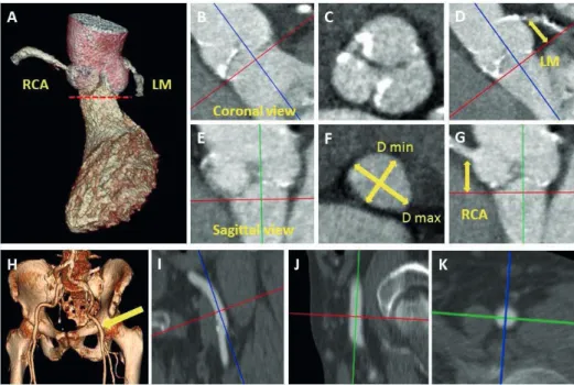
Multi-detector row computed tomography (MDCT) is an important imaging technique to evaluate patients with symptomatic severe aortic stenosis who are candidates for TAVI. The superb spatial resolution of this imaging technique permits accurate sizing of the aortic annulus, key to select the most appropriate transcatheter valve size. Studies have shown that the choice of valve size based on MDCT measurements, as opposed to echocardiography or angiography measurements, has led to less postoperative paraval-vular AR and therefore MDCT is emerging as the ‘’gold standard’’ method for valve sizing in patients undergoing TAVI (Figure 1). (16)
system), assisting the decision for the appropriate the procedural access (transfemoral, transarterial or transapical) (Figure 1).(17)
The use of post-operative MDCT has also shed light into many procedural related complications in TAVI patients. Optimal expansion of the frames has been evaluated with MDCT and interestingly under-expanded frames may be found in 8% of patients which has been related with significant paravalvular AR and prosthesis migration.(18) The pathophysiology of paravalvular AR has also been investigated with post-operative MDCT.(17) Moreover, post-operative MDCT studies have shown that deep implantation of the frame in the left ventricular outflow tract may be responsible for new conduction disorders after TAVI.(19) In a few patients with perioperative coronary ostia occlusion successfully treated with immediate percutaneous coronary artery intervention MDCT has also revealed the possibility of direct impingement of the coronary ostia by the frame.(20)
Figure 1. Multi-detector row computed tomography may give critical information in patients
PREdICTION OF OUTCOME IN SPECIFIC POPULATIONS
In real-world clinical practice, patients with symptomatic severe aortic stenosis that do not strictly fulfil the inclusion criteria of PARTNER trial (cohort A and B) may receive a so called ‘’off–label” treatment with TAVI. These patients are deemed at excessive surgical risk and TAVI may be a last resource treatment.
Indeed, TAVI may be a successful alternative treatment, with acceptable rate of in-hospital and long-term mortality rates, in patients with pure native aortic valve re-gurgitation deemed inoperable.(21) Moreover, patients with concomitant severe aortic stenosis and severe mitral regurgitation may receive TAVI treatment, although generally these cases were excluded from large randomized trials.(22) TAVI may reduce concomi-tant mitral regurgitation in this group. However, it remains unclear how to identify the patients that will show an improvement in mitral regurgitation after TAVI.(22)
Registries have also shown low complication rates and acceptable survival for high-risk patients with failing bioprosthesis treated with transcatheter valve-in-valve.(23) However, so far there has not been a direct comparison with a similar group of high risk patients undergoing surgical treatment (Figure 2).
Figure 2. Transcatheter aortic valve implantation may have an “off- label” use for failing bioprtosthetic valves. Successful implantation of a 23 mm Edwards SAPIEN valve in a failing 23 mm Carpentier Edwards PERIMOUNT aortic bioprosthesis.
ObjECTIVES ANd OUTLINE OF THE THESIS
The objective of this thesis was to investigate the role of MDCT to predict outcomes in patients undergoing TAVI and also to focus on the outcome of specific populations undergoing this procedure.
Addition-ally, the deployment of the frame in relation to the coronary ostia will be systemati-cally studied with post-operative MDCT and its implications for percutaneous coronary interventions at follow-up will be carefully addressed (Chapter 4). The combination of pre- and post-procedural MDCT images in addition to echocardiography measurements may also help us better identify the prevalence of late pericardial effusion in patients treated with TAVI (Chapter 5). Patients undergoing transfemoral TAVI do not experience pleuro-pericardial surgical trauma and they are expected to develop less frequently late pericardial effusion as compared with patients treated with transapical TAVI.
REFERENCES
1. Lindroos M, Kupari M, Heikkila J, et al. Prevalence of aortic valve abnormalities in the elderly: an echocardiographic study of a random population sample. J Am Coll Cardiol 1993; 21: 1220-5. 2. Kojodjojo P, Gohil N, Barker D, Y et al. Outcomes of elderly patients aged 80 and over with
symp-tomatic, severe aortic stenosis: impact of patient’s choice of refusing aortic valve replacement on survival. QJM: monthly journal of the Association of Physicians 2008; 101: 567-73.
3. Vasques F, Messori A, Lucenteforte E, et al. Immediate and late outcome of patients aged 80 years and older undergoing isolated aortic valve replacement: a systematic review and meta-analysis of 48 studies. Am Heart J 2012; 163: 477-85.
4. Iung B, Cachier A, Baron G, et al. Decision-making in elderly patients with severe aortic stenosis: why are so many denied surgery? Eur Heart J 2005; 26: 2714-20.
5. O’Neill WW. Predictors of long-term survival after percutaneous aortic valvuloplasty: report of the Mansfield Scientific Balloon Aortic Valvuloplasty Registry. J Am Coll Cardiol 1991; 17: 193-8. 6. Letac B, Cribier A, Koning R, et al. Results of percutaneous transluminal valvuloplasty in 218 adults
with valvular aortic stenosis. Am J Cardiol 1988; 62: 598-605.
7. Cribier A, Eltchaninoff H, Bash A, et al. Percutaneous transcatheter implantation of an aortic valve prosthesis for calcific aortic stenosis: first human case description. Circulation 2002; 106: 3006-8. 8. Leon MB, Smith CR, Mack M, et al. Transcatheter aortic-valve implantation for aortic stenosis in
patients who cannot undergo surgery. N Engl J Med 2010; 363: 1597-607.
9. Smith CR, Leon MB, Mack MJ, et al. Transcatheter versus surgical aortic-valve replacement in high-risk patients. N Engl J Med 2011; 364: 2187-98.
10. Vahanian A, Alfieri O, Andreotti F, et al. Guidelines on the management of valvular heart disease (version 2012). Eur Heart J 2012; 33: 2451-96.
11. Kodali SK, Williams MR, Smith CR, et al. Two-year outcomes after transcatheter or surgical aortic-valve replacement. N Engl J Med 2012; 366: 1686-95.
12. Bagur R, Rodes-Cabau J, Gurvitch R, et al. Need for permanent pacemaker as a complication of transcatheter aortic valve implantation and surgical aortic valve replacement in elderly patients with severe aortic stenosis and similar baseline electrocardiographic findings. JACC Cardiovasc Interv 2012; 5: 540-51.
13. Kappetein AP, Head SJ, Genereux P, et al. Updated standardized endpoint definitions for trans-catheter aortic valve implantation: the Valve Academic Research Consortium-2 consensus docu-ment. EuroIntervention 2012; 8: 782-95.
14. Khatri PJ, Webb JG, Rodes-Cabau J, et al. Adverse effects associated with transcatheter aortic valve implantation: a meta-analysis of contemporary studies. Ann Intern Med 2013; 158: 35-46. 15. Dvir D, Lavi I, Eltchaninoff H, et al. Multicenter Evaluation of Edwards SAPIEN Positioning
Dur-ing Transcatheter Aortic Valve Implantation With Correlates for Device Movement DurDur-ing Final Deployment. JACC Cardiovasc Interv 2012; 5: 563-70.
16. Binder RK, Webb JG, Willson AB, et al. The impact of integration of a multidetector computed tomography annulus area sizing algorithm on outcomes of transcatheter aortic valve replace-ment: a prospective, multicenter, controlled trial. J Am Coll Cardiol 2013; 62: 431-8.
18. Willson AB, Webb JG, Gurvitch R, et al. Structural integrity of balloon-expandable stents after transcatheter aortic valve replacement: assessment by multidetector computed tomography. JACC Cardiovasc Interv 2012; 5: 525-32.
19. Binder RK, Webb JG, Toggweiler S, et al. Impact of post-implant SAPIEN XT geometry and position on conduction disturbances, hemodynamic performance, and paravalvular regurgitation. JACC Cardiovasc Interv 2013; 6: 462-8.
20. Nazeri I, Abdi S, Mandegar MH, et al. How should I treat acute left main coronary obstruction after transapical aortic valve implantation? EuroIntervention 2013; 9: 761-4
21. Roy DA, Schaefer U, Guetta V, et al. Transcatheter aortic valve implantation for pure severe native aortic valve regurgitation. J Am Coll Cardiol 2013; 61: 1577-84.
22. Toggweiler S, Boone RH, Rodes-Cabau J, et al. Transcatheter aortic valve replacement: outcomes of patients with moderate or severe mitral regurgitation. J Am Coll Cardiol 2012; 59: 2068-74. 23. Dvir D, Webb J, Brecker S, et al. Transcatheter Aortic Valve Replacement for Degenerative
Part I
Chapter 2
Multidetector Row Computed
Tomography Parameters Associated
With Paravalvular Regurgitation After
Transcatheter Aortic Valve Implantation
Spyridon Katsanos, See Hooi Ewe, Philippe Debonnaire, Frank van der Kley, Arend de Weger, Meindert Palmen, Arthur JHA Scholte, Martin J Schalij, Jeroen J Bax, Nina Ajmone Marsan, Victoria Delgado
AbSTRACT
background: Multidetector row computed tomography (MDCT) assessment of aortic annulus dimensions and frame position and deployment have been associated with paravalvular aortic regurgitation (PAVR) after transcatheter aortic valve implantation (TAVI). The present evaluation investigated the (pre- and post-procedure) MDCT asso-ciates of PAVR≥2+. Methods: In total, 123 patients referred for TAVI underwent clinical evaluation, transthoracic echocardiography and pre- and post-TAVI MDCT. Pre-TAVI MDCT measurements of the aortic annular dimensions and post-TAVI MDCT evaluation of the position and deployment of the prosthesis in the native annulus were performed.
INTROdUCTION
Transcatheter aortic valve implantation (TAVI) is an established alternative for patients with severe aortic stenosis and high operative risk mortality or contraindications for surgical aortic valve replacement.(1-3) Paravalvular aortic regurgitation (PAVR) remains still as one of the main concerns of this therapy since the prognostic implications of PAVR are not negligible and data from the PARTNER cohort B trial have shown a two-fold increased mortality among patients with mild or more PAVR compared with patients showing none or trace PAVR.(4)
Determinants of PAVR are still debated. Of particular importance is the measurement of aortic valve annular dimensions with 3-dimensional imaging techniques such as multidetector row computed tomography (MDCT) since relative undersizing of the transcatheter prosthesis has been related to increased incidence of PAVR.(5-7) In ad-dition, position and deployment of the prosthesis have been suggested as relevant underlying mechanisms of PAVR after TAVI.(7-9) However, few studies have demon-strated the relevance of post-procedural MDCT to identify the determinants of PAVR after TAVI. Accordingly, the present study aimed to identify the MDCT-derived pre- and post-procedural parameters independently associated with significant PAVR after TAVI.
METHOdS
TAVI was performed at the hybrid operating room under general anesthesia. Fluo-roscopy was the mainstay imaging technique to guide the procedure assisted by trans-esophageal echocardiography (iE33, Philips Medical System, Andover, MA, USA). A 23-, 26- or 29-mm Edwards Sapien and Sapien XT valve (Edwards Lifesciences, Irvine, CA) was implanted based on the dimensions of the aortic annulus. The transfemoral approach was the preferred delivery technique whereas the transapical approach was performed in patients with non-suitable peripheral artery anatomy or in patients in whom a 29-mm valve was implanted.(12) During rapid right ventricular pacing, aortic valve balloon dilatation was performed and subsequently the balloon-expandable prosthesis valve was deployed.(12) The presence of significant PAVR was evaluated with transesophageal echocardiography and re-ballooning of the prosthesis or valve-in-valve were performed as bail-out techniques to reduce aortic regurgitation severity. Patients who underwent a valve-in-valve procedure were excluded from further analysis.
A commercialy available ultrasound system (Vivid 7, E9, General Electric Horten, Norway) was used for pre- and post-TAVI TTE. The pre-procedural evaluation included the assessment of the valve morphology at the parasternal short-axis view, and the left ventricular outflow tract (LVOT) diameter was measured at the parasternal long-axis view.(13) The peak and mean transaortic pressure gradients were assessed in the apical long-axis or 5-chamber views and the aortic valve area was calculated with the continuity equation.(13) Aortic stenosis was considered severe if aortic valve area was <1.0 cm2 and/or the transaortic mean gradient was ≥40 mmHg.(14) LV end-diastolic and
end-systolic volumes were calculated with the Simpson’s method and LV ejection frac-tion was derived.(15)
In order to evaluate the presence of PAVR after TAVI, color-flow Doppler echocar-diography was performed after optimization of Nyquist limit and gain settings. PAVR was evaluated on multiple echocardiographic views and conventional criteria such as the vena contracta width, the ratio of the regurgitant jet width to the LVOT diameter, pressure half-time and the proportion of the circumference of the sewing ring occupied by the regurgitant jet were used to estimate the PAVR (0 absent, 1+ trace or mild, 2+ mild-to-moderate, 3+ moderate-to-severe and 4+ severe).(16, 17) PAVR ≥2+ at the first post-operative month was considered significant.
kV, 120 kV or 135 kV (based on body mass index of the patients), respectively. Unless contraindicated, patients received beta-blockers if their heart rate was ≥70 beats per minute. All scans were performed during mid-inspiratory breath-hold and 80-90 mL of non-ionic contrast (Iomeron 400, Bracco, Milan, Italy) was injected into the antecubital vein. Subsequently, data sets were reconstructed and off-line post-processing of MDCT images was performed on dedicated workstations (Vitrea2, Vital Images, Minneapolis, Minnesota, USA).
Diastolic and systolic images of the aortic root at the respective 75% and 30-40% of RR interval were selected. By aligning the three orthogonal multiplanar reformation planes, the double-oblique transversal plane that bisects the aortic annulus beneath the hinge points of the aortic cusps was obtained. At this level, the minimum and maximum aortic annulus diameters and the annulus area were measured. From the orthogonal sagittal and coronal views, the aortic annulus diameters were also measured as previously de-scribed.(18) In addition, from the non-contrast enhanced images, the calcium Agatston score of the aortic valve and landing zone was calculated. On 1-month follow-up MDCT scans, the prosthesis deployment and position in relation to aortic root were evaluated. (8) Particularly, the distance between the lower rim of the prosthesis frame in the LVOT and the native aortic annulus at the level of the left coronary cusp (LCC) was measured (Figure 1). Moreover, the distance between the upper rim of the valve frame and the right and left coronary ostia was evaluated. Additionally the prosthesis deployment was visualized at the double-oblique transverse plane of the aortic annulus. At this level, the area of the deployed prosthetic valve was assessed by planimetry and additionally the maximal and minimal diameters of the prosthetic valve frame were measured.
Following previous studies, an eccentricity index, calculated as [1-(minimum prosthe-sis diameter /maximum prostheprosthe-sis diameter)] ≥0.1 defined a noncircular deployment of the prosthesis.(8) Moreover shallow or deep implantation of the frame were evaluated and defined as depth of the frame in the LVOT<2 mm or >8 mm from the level of the hinge point of the LCC, respectively (Figure 1).
Additionally the difference between the MDCT derived coronal and maximal diam-eters of the aortic annulus and the nominal diameter of the implanted prosthesis were calculated. Furthermore, the difference between the MDCT derived aortic annular area and the nominal area of the implanted prosthesis was also assessed. Among several pre- and post-TAVI MDCT parameters, the determinants of PAVR were evaluated.
significant PAVR (≥2+) at 1-month follow-up. Continuous variables were compared with the unpaired Student’s t-test if normally distributed or the Mann-Whitney test other-wise. Categorical variables were also compared with the χ2 test or Fisher’s exact test, as
appropriate. Receiver operating characteristic curve analyses were performed to assess the accuracy of several MDCT parameters to predict the presence of PAVR≥2+ and the cut-offs values for each variable were obtained from the highest sum of sensitivity and specificity. Binary logistic regression analysis was performed to evaluate independent determinants of PAVR≥2+ and the estimated odds ratios (OR) and the 95% confidence intervals (CI) were calculated. Variables with a p<0.1 in the univariate analysis were included in the multivariate model. A two-sided p<0.05 was considered statistically significant.
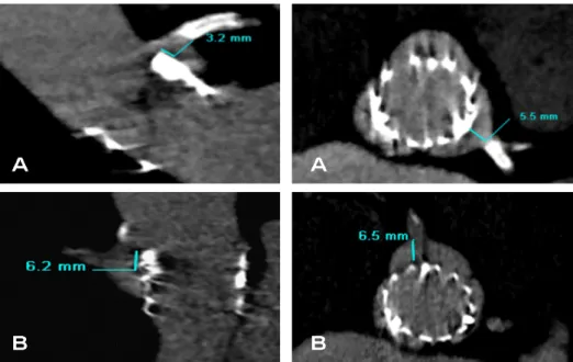
Figure 1. Examples of optimal (6.7 mm), shallow (0 mm) and deep (10.9 mm) deployment of prosthesis in
the left ventricular outflow tract, leading to trivial, mild and trivial paravalvular regurgitation at 1 month fol-low up, respectively. Shalfol-low deployment was considered <2 mm and deep deployment >8 mm distance from the level of the left coronary cusp in the in the left ventricular outflow tract.
L A
L
A LA
A o
A o
A o L
V
L
V L
RESULTS
A total of 123 patients (81±7 years, 49% male) with symptomatic severe aortic stenosis treated with TAVI and complete evaluation including pre- and post-TAVI MDCT were evaluated. The baseline characteristics of the patients are listed in Table 1.
Repeated balloon-dilatation of the prosthesis was performed in 15 (12.2%) patients. In 10 (66%) patients PAVR significantly reduced (<2+) after this intervention. At 1-month follow-up, all patients underwent TTE. In 30(24.4%) patients, no PAVR was observed whereas in respective 68 (55.3%), 22 (17.9%) and 3 (2.4%) patients, trivial-to-mild, mild-to-moderate and moderate-to-severe PAVR were documented. Therefore, significant PAVR≥2+ was observed in 25 (20.3%) patients. In 5 of these patients a repeat balloon dilatation was performed.
Table 1. Baseline characteristics of overall population and patients with and without significant
paravalvu-lar aortic regurgitation at follow-up
Variable Overall
(n = 123)
Paravalvular aortic regurgitation<2+
(n = 98)
Paravalvular aortic regurgitation ≥2+
(n = 25)
p-value
Age (years) 81±7 81±7 80±7 0.446
Men 60 (49%) 50(51%) 10 (40%) 0.325
Body surface area (m2) 1.7±0.3 1.7±0.3 1.7±0.4 0.998
Hypertension 47 (38%) 38 (39%) 9 (36%) 0.799
Diabetes melitus 36 (29%) 30 (30%) 6 (24%) 0.517 Peripheral vascular disease 23 (19%) 21 (21%) 2 (8%) 0.158
Smoking 28 (22%) 23 (23%) 5 (20%) 0.796
Coronary artery disease 87 (71%) 70 (71%) 17 (68%) 0.737 New York Heart Association
functional class III-IV
75 (61%) 62 (63%) 13 (52%) 0.303
Pacemaker 12 (10%) 10 (10%) 2 (8%) 1.000
Atrial fibrilation 29 (24%) 21 (21%) 8 (32%) 0.266 Medications Beta-blockers Diuretics Statins Calcium antagonists 71 (58%) 76 (62%) 74 (60%) 36 (29%) 56(57%) 59(60%) 62(63%) 29(29%) 15(60%) 17(68%) 12(48%) 7(28%) 0.796 0.474 0.164 0.876 Logistic EuroSCORE 23.4 ±14.1 23.4±14.3 21.6±13.6 0.567 Aortic valve area (cm2) 0.73±0.18 0.72±19 0.78±0.15 0.108
Mean transaortic valve gradient (mmHg)
42±15 42±15 39±13 0.421
Left ventricular end-systolic volume (ml)
60±40 60±40 62±39 0.771
Left ventricular end-diastolic volume (ml)
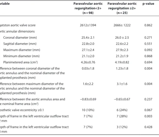
Patients with PAVR<2+ and patients with PAVR≥2+ had comparable Agatston aortic valve scores and similar aortic valve annular diameters and area measured at pre-procedural MDCT scans (Table 2). However, the difference between the aortic annulus diameter measured in the coronal plane and the nominal diameter of the implanted prosthesis was significantly larger in patients with PAVR≥2+, compared with patients without significant PAVR. Similarly, the difference between the maximum aortic annulus diameter measured at the double-oblique transverse cross-sectional plane and the nominal diameter of the implanted prosthesis was significantly larger in patients with PAVR≥2+ compared with patients without significant PAVR. In contrast, there were no significant differences between groups in terms of difference between aortic annular area and nominal frame area (Table 2).
Based on post-TAVI MDCT data, the percentage of eccentrically deployed valves was higher among patients with PAVR≥2+ compared with patients without significant PAVR, although this difference was not statistically significant. In terms of positioning of the transcatheter aortic valve, patients with PAVR≥2+ showed more often a shallow posi-tioning of the frame in the LVOT (<2mm) compared to patients without significant PAVR. In contrast, a deep positioning of the frame (>8mm) in the LVOT was not significantly different between groups (Table 2).
At multivariate analysis, the difference between the MDCT derived maximum diam-eter of the aortic annulus and the nominal diamdiam-eter of the implanted prosthesis and depth of frame in LVOT <2mm were independently associated with PAVR≥2+ at 1 month follow-up (Table 3). Receiver operating characteristic curves were performed to evaluate the accuracy of MDCT measurements to predict the occurrence of PAVR≥2+ (Figure 2).
Interestingly, implanting a transcatheter aortic valve which nominal diameter was undersized ≥2 mm relative to the maximum aortic annulus diameter measured on pre-procedural MDCT had 72% sensitivity and 61.2% specificity to predict PAVR≥2+ (Figure 2).
Table 1. Baseline characteristics of overall population and patients with and without significant
paravalvu-lar aortic regurgitation at follow-up (continued)
Variable Overall
(n = 123)
Paravalvular aortic regurgitation<2+
(n = 98)
Paravalvular aortic regurgitation ≥2+
(n = 25)
p-value
Left ventricular ejection fraction (%)
50±13 50±13 48±13 0.510
Transcatheter aortic valve implantation approach 0.799 Transfemoral Transapical 47 (38%) 76 (62%) 38 (36%) 60 (64%) 9 (32%) 16 (68%) Edwards SAPIEN valve, n (%)
Table 2. Pre- and post-procedural multidetector row computed tomography parameters in patients with and without significant paravalvular aortic regurgitation at follow up
Variable Paravalvular aortic
regurgitation<2+ (n = 98)
Paravalvular aortic regurgitation ≥2+
(n = 25)
p-value
Agatston aortic valve score 2612±1394 2666± 1222 0.862 Aortic annular dimensions
Coronal diameter (mm) 25.4± 2.1 26.0 ± 2.5 0.271 Sagittal diameter (mm) 22.8±2.0 22.6±2.2 0.551 Maximum diameter (mm) 27.1±2.4 27.9±2.3 0.092 Minimum diameter (mm) 21.1±2.0 21.2±1.9 0.868 Planimetered area (cm2) 4.26±0.76 4.19±0.82 0.694
Difference between coronal diameter of the aortic annulus and the nominal diameter of the implanted prosthesis (mm)
0.03±1.8 1.23±1.8 0.004
Difference between maximum diameter of the aortic annulus and the nominal diameter of the implanted prosthesis (mm)
1.6±2.2 3.1±1.6 0.004
Difference between the aortic annulus area and the nominal frame area (cm2)
−0.83±0.69 −0.65±0.67 0.237
Prosthetic valve eccentricity ≥0.1 10 (10%) 6 (24%) 0.067 Depth of frame in the left ventricular outflow tract
<2 mm
7 (7%) 7 (28%) 0.003
Depth of frame in the left ventricular outflow tract >8 mm
7 (7%) 3 (12%) 0.428
Table 3. Univariate and multivariate binary logistic regression analyses
Variable Univariate analysis Multivariable analysis Odds ratio (95%
confidence interval)
p-value Odds ratio (95% confidence
interval)
p-value
Agatston aortic valve score 1.000 (1.000-1.000) 0.860 …. …. Aortic annular maximum diameter 1.177(0.973-1.424) 0.094 0.718(0.498-1.035) 0.076 Difference between maximum diameter
of the aortic annulus and the nominal diameter of the implanted prosthesis
1.398(1.122-1.744) 0.003 1.912(1.257-2.908) 0.002
Prosthetic valve eccentricity ≥0.1 2.779(0.900-8.577) 0.075 2.724(0.690-10.761) 0.153 Depth of frame in the left ventricular
outflow tract <2 mm
5.056(1.580-16.180) 0.006 4.865(1.331-17.786) 0.017
Depth of frame in the left ventricular outflow tract >8 mm
dISCUSSION
The present study indicates that a large difference between the maximal annulus di-ameter and the nominal didi-ameter of the prosthesis, as well as a shallow position of the frame in the LVOT, are independently associated with significant PAVR after TAVI. MDCT is a valuable imaging technique to understand the underlying mechanisms of PAVR after TAVI.
Several studies have consistently shown that aortic annulus dimensions are strongly associated with PAVR after TAVI.(5-7)Therefore, accurate measurement of aortic annulus dimensions is crucial to select the most appropriate prosthesis size and optimize the outcomes of TAVI. Accumulating evidence shows that 3-dimensional imaging techniques such as MDCT are the most accurate methods to size the aortic valve annulus.(5-7) How-ever, the golden standard measurement of the aortic annulus to select the most appro-priate prosthesis size has not been established to date. Mean aortic annulus diameter
Figure 2. Receiver operating characteristic curve analyses of multidetector row computed tomography
- related measurements of the aortic annulus to predict the occurrence of significant postoperative para-valvular aortic regurgitation.
(derived from the average of the minimum and maximum diameters), area-derived diameter, coronal and sagittal diameters have been proposed to select the transcatheter valve size.(5-7) Importantly, the nominal diameter of the available transcatheter valves should be taken into consideration to estimate the grade of over- or undersizing relative to the aortic annulus once the valve is implanted. A significant oversizing of the prosthe-sis will minimize the risk of significant PAVR at the expense of increasing the potential risk of aortic annulus rupture whereas a significant prosthesis undersizing will increase the risk of significant PAVR and, less frequent, prosthesis migration. Jilaihawi et al dem-onstrated that the difference between the maximum diameter of the aortic annulus as assessed with MDCT and the nominal diameter of the implanted prosthesis had the best accuracy to predict significant PAVR after TAVI.(6) Similarly, Willson et al showed that patients with a nominal transcatheter valve area <10% larger than the cross-sectional area of the native aortic annulus (less oversized) had significantly higher incidence of PAVR as compared to patients with a difference >10% (more oversized) (19.1% vs. 0%; odds ratio 18.4, p<0.01).(7) Furthermore, Hayashida and coworkers demonstrated that the use of MDCT to measure the aortic valve annulus resulted in lower incidence of PAVR after TAVI as compared with 2-dimensional transesophageal echocardiography. (5 )The ratio between the nominal diameter of the transcatheter valve and the mean diameter derived from the cross-sectional area of the aortic valve annulus measured with MDCT was strongly associated with the presence of significant PAVR after TAVI (hazard ratio 0.36 per each 0.1 increase; 95% confidence interval 0.17-0.77). (5) In the present study the difference between the maximum diameter of the aortic annulus assessed with MDCT and the nominal diameter of the implanted prosthesis predicted best the presence of significant PAVR after TAVI. Therefore, estimation of the degree of prosthesis oversizing seems to be an important parameter to minimize the incidence of significant PAVR. However, this parameter should be further confirmed in prospective studies.
In addition, calcification of the aortic valve has been related to the presence of signifi-cant PAVR.(19-21) However, it remains unclear the relative merits of the total amount of valve calcification or the (asymmetrical) location of calcification.(19, 20) In the present study, there were no differences in the amount of valve calcification (as quantified with the Agatston valve score) between the two groups of patients.
Furthermore, the depth of the deployed valve in the LVOT as assessed with left ven-triculography has been related to PAVR in patients treated with self-expandable pros-theses.(24) Sherif et al demonstrated that the optimal position of the self-expandable prosthesis was approximately 10 mm deep into the LVOT (as measured from the an-nular hinge point of the non-coronary cusp).(24) A depth >10 mm may leave part of the prosthesis frame uncovered by the sealing skirt into the LVOT and subsequently the blood may regurgitate through the frame struts. In contrast, a shallow position of the prosthesis may be associated with malapposition of the frame into the annulus that leads to subsequent PAVR. In the present evaluation a shallow position of the frame was significantly associated with the presence of significant PAVR at follow-up. These results therefore confirm previous studies underscoring the relevance of accurate positioning of the prosthesis frame.
REFERENCES
1. Genereux P, Head SJ, Van Mieghem NM, et al. Clinical outcomes after transcatheter aortic valve replacement using valve academic research consortium definitions: a weighted meta-analysis of 3,519 patients from 16 studies.J Am Coll Cardiol 2012;59:2317-2326.
2. Leon MB, Smith CR, Mack M,et al. Transcatheter aortic-valve implantation for aortic stenosis in patients who cannot undergo surgery. N Engl J Med 2010;363:1597-1607.
3. Smith CR, Leon MB, Mack MJ,et al. Transcatheter versus surgical aortic-valve replacement in high-risk patients. N Engl J Med 2011;364:2187-2198.
4. Kodali SK, Williams MR, Smith CR,et al. Two-year outcomes after transcatheter or surgical aortic-valve replacement. N Engl J Med 2012;366:1686-1695.
5. Hayashida K, Bouvier E, Lefevre T,et al. Impact of CT-guided valve sizing on post-procedural aortic regurgitation in transcatheter aortic valve implantation. EuroIntervention 2012;8:546-555. 6. Jilaihawi H, Kashif M, Fontana G,et al. Cross-sectional computed tomographic assessment
im-proves accuracy of aortic annular sizing for transcatheter aortic valve replacement and reduces the incidence of paravalvular aortic regurgitation. J Am Coll Cardiol 2012;59:1275-1286.
7. Willson AB, Webb JG, LaBounty TM,et al. 3-dimensional aortic annular assessment by multide-tector computed tomography predicts moderate or severe paravalvular regurgitation after transcatheter aortic valve replacement: a multicenter retrospective analysis. J Am Coll Cardiol 2012;59:1287-1294.
8. Delgado V, Ng AC, van de Veire N,et al . Transcatheter aortic valve implantation: role of multi-detector row computed tomography to evaluate prosthesis positioning and deployment in relation to valve function. Eur Heart J 2010;31:1114-1123.
9. Schultz CJ, Tzikas A, Moelker A, et al . Correlates on MSCT of paravalvular aortic regurgitation after transcatheter aortic valve implantation using the medtronic CoreValve prosthesis. Catheter Cardiovasc Interv 2011;78:446-455.
10. Vahanian A, Alfieri O, Al-Attar N,et al . Transcatheter valve implantation for patients with aortic stenosis: a position statement from the European Association of Cardio-Thoracic Surgery (EACTS) and the European Society of Cardiology (ESC), in collaboration with the European Association of Percutaneous Cardiovascular Interventions (EAPCI). Eur Heart J 2008;29:1463-1470.
11. Roques F, Michel P, Goldstone AR,et al. The logistic EuroSCORE. Eur Heart J 2003;24:881-882. 12. Rodes-Cabau J, Dumont E, De LaRochelliere R,et al. Feasibility and initial results of
percutane-ous aortic valve implantation including selection of the transfemoral or transapical approach in patients with severe aortic stenosis. Am J Cardiol 2008;102:1240-1246.
13. Baumgartner H, Hung J, Bermejo J,et al. Echocardiographic assessment of valve stenosis: EAE/ASE recommendations for clinical practice. Eur J Echocardiogr 2009;10:1-25.
14. Vahanian A, Alfieri O, Andreotti F,et al. Guidelines on the management of valvular heart disease (version 2012): The Joint Task Force on the Management of Valvular Heart Disease of the European Society of Cardiology (ESC) and the European Association for Cardio-Thoracic Surgery (EACTS). Eur Heart J 2012;33:2451-2496.
15. Lang RM, Bierig M, Devereux RB, et al. Recommendations for chamber quantification. Eur J Echo-cardiogr 2006;7:79-108.
17. Detaint D, Lepage L, Himbert D, et al. Determinants of significant paravalvular regurgitation after transcatheter aortic valve: implantation impact of device and annulus discongruence. JACC Cardiovasc Interv 2009;2:821-827.
18. Tops L, Wood D, Delgado V,et al. Noninvasive Evaluation of the Aortic Root With Multislice Com-puted Tomography Implications for Transcatheter Aortic Valve Replacement. J Am Coll Cardiol Img 2008;1:321-330.
19. Colli A, D’Amico R, Kempfert J, et al. Transesophageal echocardiographic scoring for transcatheter aortic valve implantation: impact of aortic cusp calcification on postoperative aortic regurgita-tion. J Thorac Cardiovasc Surg 2011;142:1229-1235.
20. Ewe SH, Ng AC, Schuijf JD, et al. Location and severity of aortic valve calcium and implications for aortic regurgitation after transcatheter aortic valve implantation. Am J Cardiol 2011;108:1470-1477. 21. Haensig M, Lehmkuhl L, Rastan AJ, et al. Aortic valve calcium scoring is a predictor of significant
paravalvular aortic insufficiency in transapical-aortic valve implantation. Eur J Cardiothorac Surg 2012;41:1234-1240.
22. Schultz CJ, Weustink A, Piazza N, et al. Geometry and degree of apposition of the CoreValve Re-Valving system with multislice computed tomography after implantation in patients with aortic stenosis. J Am Coll Cardiol 2009;54:911-918.
23. Jilaihawi H, Chin D, Spyt T, et al. Prosthesis-patient mismatch after transcatheter aortic valve implantation with the Medtronic-Corevalve bioprosthesis. Eur Heart J 2010;31:857-864.
24. Sherif MA, Abdel-Wahab M, Stocker B,et al. Anatomic and procedural predictors of paravalvular aortic regurgitation after implantation of the Medtronic CoreValve bioprosthesis. J Am Coll Cardiol 2010;56:1623-1629.
25. Willson AB, Webb JG, Gurvitch R, et al. Structural integrity of balloon-expandable stents after transcatheter aortic valve replacement: assessment by multidetector computed tomography. JACC Cardiovasc Interv 2012;5:525-532.
26. Athappan G, Patvardhan E, Tuzcu EM, et al. Incidence, predictors, and outcomes of aortic regur-gitation after transcatheter aortic valve replacement: meta-analysis and systematic review of literature. J Am Coll Cardiol 2013;61:1585-1595.
Chapter 3
Multi-detector row computed tomography
after transcatheter aortic valve implantation:
insights into new onset rhythm
conduction disorders
Spyridon Katsanos, Philippe van Rosendael, Vasileios Kamperidis, Frank van der Kley, Emer Joyce, Philippe Debonnaire, Ioannis Karalis, Jeroen J Bax, Nina Ajmone Marsan, Victoria Delgado
AbSTRACT
background: New onset rhythm conduction disorders are frequent after transcatheter aortic valve implantation (TAVI). Multi-detector row computed tomography (MDCT) may help elucidate the pathophysiology of rhythm conduction disorders in patients treated with the Edwards SAPIEN valve. Methods: A total of 94 patients (age 81±7 years, men 48%) treated with TAVI with the Edwards SAPIEN valve and undergoing a pre- and post-TAVI MDCT were included. Patients with pre-existent right or left bundle branch block (LBBB) and permanent pacemakers were excluded. Position and deployment of the transcatheter frame into the aortic root was evaluated at post-TAVI MDCT. Pacemaker implantation or new onset LBBB at 1 month follow-up was the combined endpoint.
INTROdUCTION
New onset persistent left bundle branch block (LBBB) after transcatheter aortic valve implantation (TAVI) has been described in 10-19% of patients receiving an Edwards SAPIEN prosthesis (Edwards Lifesciences, Irvine, CA) and in 28-57% of patients receiving a CoreValve system (Medtronic, Minneapolis, MN).(1-4) Likewise, the need for permanent pacemaker implantation is higher among recipients of the CoreValve system (23-33%) compared with recipients of the Edwards SAPIEN valve (2.5-11.5%).(3-6) Besides the dif-ferent design characteristics of these two devices, which may be associated with the differences in incident new onset LBBB or pacemaker implantation, baseline QRS com-plex duration and deep implantation of the prosthesis into the left ventricular outflow tract (LVOT) have been consistently associated with these complications after TAVI.(2,4) Particularly, the implantation depth may vary considerably with the CoreValve system which has a longer frame than the Edwards SAPIEN valve. The series that have observed the association between implantation depth into the LVOT and the development of new onset LBBB and need for permanent pacemaker have assessed this parameter based on aortograms performed immediately after valve deployment.(2,4) The higher spatial resolution of multi-detector row computed tomography (MDCT) allows more accurate evaluation of the spatial relationships of the implanted prosthesis into the aortic root and may help elucidate the pathophysiology of the development of new onset LBBB and need for permanent pacemaker in patients undergoing TAVI. The present study evaluated the MDCT associates of new onset LBBB and need for pacemaker in patients treated with TAVI.
METHOdS
Patient population
are routinely performed. All clinical information on demographics, ECG, and imaging techniques are digitally stored in the departmental database (EPD vision version 8.3.3.6; Leiden, The Netherlands) and can be retrospectively analyzed. For this study, baseline clinical data in combination with pre- and post-TAVI MDCT parameters were related to the combined endpoint: need for pacemaker implantation or new onset persistent LBBB at 1 month follow-up. For this retrospective analysis, the Institutional Review Board waived the need for patient written informed consent.
ECg analysis
All ECGs were retrospectively reviewed at 3 time points: baseline (pre-TAVI), during hos-pitalization and at 1 month follow-up. The presence of RBBB and LBBB was diagnosed according to current recommendations.(8) Duration of the PR interval and QRS complex were automatically calculated with dedicated software (Ziemens/ Dräger, Mega Care ECG Management System, Lubeck, Germany).
Multi-detector row computed tomography
A 64- or a 320-detector row computed tomography scanner was used for pre-operative and post-operative scanning of the patients. When the Aquilion 64 system (Toshiba Medical Systems, Otawara, Japan) was used, data were acquired with a collimation of 64 x 0.5 mm and a gantry rotation time of 400 ms (tube current was 300-400 mA, voltage was 120 kV or 135 kV) and when the AcquilionONE system was used, data were acquired with a collimation of 320 x 0.5 mm (gantry rotation time of 350 ms, tube current and voltage set at 400-580 mA and 100 kV, 120 kV or 135 kV according to patients body mass index).
According to the acquisition protocol, patients with heart rate ≥70 beats per minute received beta-blockers unless contraindicated. A volume of 80-90 mL of non-ionic con-trast (Iomeron 400, Bracco, Milan, Italy) was used according to patients’ body surface area. Scans were acquired in mid-inspiratory breath-hold and data was digitally stored.
between the rim of the frame in the LVOT and the native aortic annulus was measured (Figure 1).(10)
The ratio (effective planimetered prosthesis area - MDCT derived aortic annulus area)/MDCT derived aortic annulus area was calculated from pre- and post-TAVI MDCT measurements as a parameter of prosthesis expansion. Prosthesis overexpansion was considered significant when the area of the expanded frame was >15% larger than the native aortic annulus valve area.(9,11)
Statistical analysis
A package of SPSS software version 20, (SPSS Inc., Chicago, IL, USA) was used for all statistical analyses. Continuous variables were considered normally or not normally distributed based on visual inspection of the histograms and were presented as mean and standard deviation or median and inter-quartile range, respectively. Categorical variables were presented as number and frequencies. Patients were categorized accord-ing to the need for new pacemaker implantation or the induction of new onset LBBB (patients with combined endpoint vs. patients free of combined endpoint). Continuous variables were compared with the unpaired Student’s t-test if normally distributed or
Figure 1. Multi-detector row computed tomography can define implantation depth of the frame in the
left ventricular outflow tract. The red line illustrates the level of the native aortic annulus. The distance between the native aortic annulus and the lower rim of the frame in the left ventricular outflow tract can be measured. There was an induction of a new onset left bundle branch block in patients (A) and (B) at one month follow-up and a new pacemaker was implanted in patient (C) at the 5th postoperative day. LVOT: left
the Mann-Whitney test otherwise. Categorical variables were compared with the χ2 test
or Fisher’s exact test, as indicated. Binary logistic regression analysis was used for the evaluation of the occurrence of the combined endpoint at 1 month follow-up and the estimated odds ratios and the 95% confidence intervals were calculated. Variables with a p < 0.1 in the univariable model were included in the multivariable analysis. A two-sided p < 0.05 was considered statistically significant.
RESULTS
Patient characteristics
From an initial cohort of 161 patients in whom a pre- and post-procedural MDCT was available, 21 patients with right bundle branch block (RBBB), 24 patients with LBBB and 22 patients with permanent pacemakers at baseline were excluded. Baseline clinical and echocardiographic characteristics of the remaining 94 patients (81±7 years old, 48% men) who were finally included in the current analysis are outlined in Table 1. The mean logistic Euroscore was 20.0±11.7%. Fifty-six (60%) patients had NYHA II-IV heart failure symptoms. Overall, 38 (40%) patients were treated through a transfemoral approach and the remainder 56 (60%) through a transapical approach. The size of the implanted vales was 23 mm in 28 (30%) patients, 26 mm in 62 (66%) and 29 mm in 4 (4%). In 9 (10%) patients, reballooning of the implanted prosthesis was performed to minimize paraval-vular aortic regurgitation. Eighty patients were in sinus rhythm and the remaining 14 patients had atrial fibrillation. In patients in sinus rhythm, the PR interval was 179±24 ms. In the overall population, the mean QRS duration was 98±10 ms and 7 (7%) patients had left axis deviation.
Table 1. Baseline characteristics of patients
Clinical data n = 94
Age (years) 81±7
Male, n (%) 45(48)
Body surface area (m2) 1.71±0.31
Creatinine (μmol/L) 87(70-101)
Hypertension, n (%) 40(43)
Diabetes, n (%) 28(30)
Smoking, n (%) 22(23)
Coronary artery disease, n (%) 65(70)
CABG, n (%) 24(26)
Conduction abnormalities
Compared to baseline ECG, there was a significant increase in QRS duration in the pre-discharge ECG (from 98±10 ms to 112±22 ms, p < 0.000) but not in PR interval (from 179 ±29 ms to 180±41 ms, p = 0.878). At this time point, new onset left axis deviation in the ECG was observed in 7 (7%) patients and 11 (11%) patients developed new onset RBBB. New onset LBBB developed in 15 (16%) patients pre-discharge. Moreover, 1 (1%) patient
Table 1. Baseline characteristics of patients (continued)
Clinical data n = 94
Medication
Beta-blockers 53(56)
Diuretics 55(58)
Statins 57(60)
Calcium channel blockers 29(31)
Logistic Euroscore (%) 20.0±11.7
Procedural data
Transfemoral, n (%) 38(40)
Transapical, n (%) 56(60)
Balloon post-dilatation, n (%) 9(10)
Edwards SAPIEN valve
23 mm, n (%) 28(30)
26 mm, n (%) 62(66)
29 mm, n (%) 4(4)
Echocardiography data
Aortic valve area (cm/m2) 0.72±0.19
Intra-ventricular septum thickness (cm) 1.4±0.2
Mean transaortic gradient (mmHg) 43±17
Peak transaortic gradient (mmHg) 70±25
Left ventricular ejection fraction (%) 51±12 MdCT data
Agatston score of the aortic valve and LVOT (Hounsfield units) 2927±1643 LVOT ‘landing zone’ calcification (grade 1-4) 2(2-3) baseline ECg data
Heart rate (beats/min) 72 ± 12
Atrial fibrillation, n (%) 14(15)
PR interval duration (ms) 179±24
QRS duration (ms) 98±10
Left axis deviation, n (%) 7(7)
underwent a dual-chamber pacemaker implantation 5 days after TAVI due to persistent 3rd grade atrio-ventricular block (Figure 1).
Compared to pre-discharge ECG, there were no significant changes in QRS complex or PR interval duration at 1 month follow-up (from 112±22 ms to 111±23 ms, p = 0.461 and from 180±41 ms to 179 ±36 ms, p = 0.781, respectively). In the repeat surface ECG at 1 month there were 7 (7%) patients with left axis deviation, 10 (10%) patients with RBBB and 14 (14%) patients with LBBB. New onset inhospital RBBB and LBBB resolved in 1 and 2 patients at 1 month follow-up, respectively. Moreover 1 patient developed a new onset LBBB post-discharge at 1 month follow-up. Consequently, the combined endpoint of the study at 1 month follow-up was met in 15 (16%) patients.
Post- TAVI MdCT
Post-TAVI MDCT scans showed that the deployed prosthesis was overexpanded by >15% of the native annulus area in 18 (19%) patients. Interestingly, Edwards SAPIEN valves expanded to their nominal area in only 2 patients whereas in the majority of patients frames were underexpanded. In the post-TAVI scan the mean depth of the deployed frame in the LVOT was 4.2±2.4 mm.
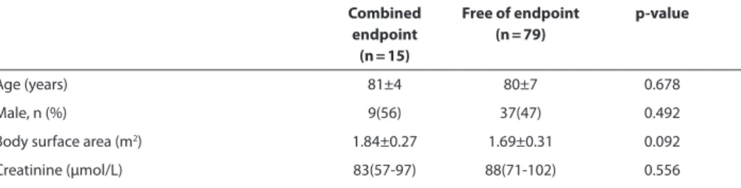
MdCT parameters associated with need for permanent pacemaker implantation and new onset Lbbb
The study endpoint (need for permanent pacemaker implantation and/or new onset persistent LBBB) was observed in 15 (16%) patients. As shown in Table 2, the proportion of Edwards SAPIEN valves that were overexpanded by >15% of the native aortic annulus as assessed with MDCT was higher in patients who met the study endpoint compared to patients free of the combined endpoint (6 (40%) vs. 12 (15%), p = 0.025). Also the frame was implanted deeper in the LVOT of patients that required a new pacemaker or devel-oped new LBBB compared to patients free of the combined endpoint (5.7±2.7 mm vs. 4.0±2.3 mm, p = 0.014, respectively) (Table 2). Binary logistic regression analysis showed that overexpansion of the valve by >15% of the native aortic annulus (OR 5.277, 95% CI
Table 2. Comparison between patients with new onset LBBB or need for pacemaker implantation at 1
month follow-up vs. patients free of endpoint.
Combined endpoint
(n = 15)
Free of endpoint (n = 79)
p-value
Age (years) 81±4 80±7 0.678
Male, n (%) 9(56) 37(47) 0.492
Body surface area (m2) 1.84±0.27 1.69±0.31 0.092
Table 2. Comparison between patients with new onset LBBB or need for pacemaker implantation at 1 month follow-up vs. patients free of endpoint. (continued)
Combined endpoint
(n = 15)
Free of endpoint (n = 79)
p-value
Hypertension, n (%) 9(60) 31(39) 0.136
Diabetes, n (%) 4(25) 24(30) 0.667
Smoking, n (%) 3(20) 19(24) 0.734
Coronary artery disease, n (%) 11(73) 54(68) 0.702
CABG, n (%) 5(31) 19(24) 0.648
NYHA functional class III-IV, n (%) 9(60) 47(59) 0.917 Medication
Beta-blockers 8(47) 45(57) 0.609
Diuretics 8(53) 47(59) 0.675
Statins 10(40) 47(59) 0.602
Calcium channel blockers 6(40) 23(29) 0.403 Logistic Euroscore (%) 22.7±9.4 19.4±12.1 0.332
Transfemoral, n (%) 6(38) 32(41) 0.832
Transapical, n (%) 9(60) 47(59) 0.971
Balloon post-dilatation, n (%) 2(13) 7(8) 0.865 Edwards SAPIEN valve
23 mm, n (%) 2(13) 26(33) 0.129
26 mm, n (%) 13(86) 49(62) 0.065
29 mm, n (%) 0(0) 4(5) 0.373
Echocardiography data
Aortic valve area (cm/m2) 0.76±0.16 0.71±0.19 0.324
Intra-ventricular septum thickness (cm) 1.4±0.3 1.4±0.2 0.915 Mean transaortic gradient (mmHg) 45±21 43±16 0.634 Peak transaortic gradient (mmHg) 76±33 69±24 0.339 Left ventricular ejection fraction (%) 51±12 52±12 0.818 MdCT data
Agatston score of the aortic valve and LVOT (Hounsfield units)
3026±1765 2909±1633 0.814
LVOT ‘landing zone’ calcification (grade 1-4) 2(2-3) 2(2-3) 0.440 baseline ECg data
Heart rate (beats/min) 73±13 71±11 0.446
Atrial fibrillation, n (%) 3(19) 11(14) 0.629 PR interval duration (ms) 188±39 177±21 0.203
QRS duration (ms) 97±10 98±10 0.630
Left axis deviation, n (%) 7(46%) 0 (0%) 0.231
1.398-19.917, p = 0.014) and depth of frame into the LVOT (OR 1.401, 95% CI 1.083-1.811, p = 0.010) were independently associated to the study endpoint (Table 3).
dISCUSSION
The present evaluation expands on the pathophysiological determinants of new onset persistent LBBB or need for pacemaker implantation in patients treated with TAVI using the balloon-expandable Edwards SAPIEN prosthesis. With the use of MDCT post-TAVI, the present evaluation demonstrated that overexpansion of the frame by >15% of the native annulus area and deep implantation of the prosthesis into the LVOT were independently related to the presence of new onset persistent LBBB and pacemaker implantation.
Table 3. Univariate and multivariate analysis of clinical, electrocardiogram and MDCT parameters related to
new onset persistent LBBB or need for pacemaker implantation at 1 month follow-up.
Variables Univariate Multivariate
Odds Ratio (95% Confidence
Interval)
p-value Odds Ratio (95% Confidence
Interval)
p-value
Age (years) 1.018 (0.938-1.104) 0.675 Male, n (%) 1.297 (0.429-3.923) 0.645 Creatinine (μmol/L) 0.994 (0.976-1.013) 0.548 Diabetes, n (%) 0.833 (0.241-2.882) 0.833 CAGB, n (%) 1.447 (0.439-4.774) 0.544 NYHA functional class III-IV, n (%) 1.200 (0.364-3.957) 0.765 Beta-blockers, n (%) 0.795 (0.285-2.614) 0.863 Transfemoral, n (%) 0.979 (0.317-3.020) 0.971 Balloon post dilatation, n (%) 1.582 (0.295-8.480) 0.592 Aortic valve area (per cm/m2) 4.284 (0.241-76.231) 0.322
Intra-ventricular septum (per cm) 0.988 (0.797-1.225) 0.891 Left ventricular ejection fraction (%) 0.995 (0.953-1.038) 0.816 Agatston score (per 100 units) 0.999 (0.956-1.044) 0.959 LVOT ‘landing zone’ calcification
(grade 1-4)
0.791 (0.437-1.430) 0.437
Overexpansion >15% of native annulus area, n (%)
3.722 (1.119-12.382) 0.032 5.277 (1.398-19.919) 0.014
Depth of frame in LVOT (per mm) 1.320 (1.045-1.667) 0.020 1.401 (1.083-1.811) 0.010 Atrial fibrillation, n (%) 1.545 (0.375-6.371) 0.547
PR duration (per ms) 1.015 (0.992-1.040) 0.208 QRS duration (per ms) 0.987 (0.935-1.041) 0.626
Incidence of new onset Lbbb and need for pacemaker implantation after TAVI with the Edwards SAPIEN valve
The incidence of new conduction abnormalities in TAVI patients varies widely across the several trials.(1-3,12-22) Pre-existent conduction abnormalities, type of implanted prosthesis and timing and duration of new conduction abnormalities (early vs. late after TAVI2(2,5,17,18,23) and transitory versus persistent abnormalities(2,17,24)) are the
main confounder factors underlying the disparate incidences. Few trials and registries have documented the incidence of new onset persistent conduction abnormalities and need for pacemaker implantation after TAVI in patients without preexistent conduction abnormalities.(1-3) The PARTNER trial and the continued access registry, including 1157 patients without preexistent conduction abnormalities or pacemaker, reported an inci-dence of new onset of LBBB of 10.5% before hospital discharge.(1) Urena et al reported an incidence of 30.2% in 202 patients undergoing TAVI with the Edwards SAPIEN valve. (2) Using the self-expandable CoreValve system, Testa et al reported an incidence of new onset LBBB of 27.4%.(3) However, at mid follow-up, the prevalence of persistent LBBB reduced in all series: data from the PARTNER trials and the continued access registry showed a prevalence of persistent LBBB of 7.8% and 8.5% at 30 days and 6-12 months follow-up, respectively, whereas in the series by Urena et al LBBB resolved in 37.7% and 57.3% of patients at hospital discharge and 6-12 months follow-up, respectively.(1,2) Among patients treated with the CoreValve system, Testa and colleagues reported no change in LBBB prevalence at 1 month follow-up.(3) The incidence of new onset persis-tent LBBB in the present study (n = 14 patients, 15%) was comparable to previous studies including patients treated with the Edwards SAPIEN valve.
The incidence of new pacemaker implantation after TAVI has been reported in 9.2% to 42% among patients treated with a CoreValve system and in 2.5% to 11.5% in patients treated with the Edwards SAPIEN valve.(12,13,17-22,25) Preexistent RBBB or LBBB have been associated with increased risk of pacemaker implantation with complete atrioventricular block and symptomatic bradycardia being the main reasons for pacemaker implanta-tion.(4,5,18,19) In the present study, patients with preexistent conduction abnormalities were excluded which would explain the low incidence of new pacemaker implantation. Complete atrioventricular block was the indication for pacemaker implantation.
MdCT associates of new onset Lbbb and need for pacemaker implantation after TAVI
con-duction abnormalities, data from the PARTNER trials and the continued access registry showed that prior coronary artery bypass grafting was associated with increased risk of new onset LBBB(1) while Urena et al demonstrated larger baseline QRS duration and deep implantation of balloon-expandable prosthesis into the LVOT as determinants of this endpoint.(2) A low implantation of the prosthesis frame into the LVOT was also iden-tified by Testa and coworkers as determinant of persistent LBBB in patients treated with a self-expandable valve.(3) The implantation depth of the device has been assessed in previous studies with aortography performed during the TAVI procedure.(23) Few studies have evaluated with MDCT the position of the deployed frame into the LVOT and have correlated it with the occurrence of conduction disturbances.(24,27) Binder et al showed that the implantation depth of the Edwards SAPIEN frame was significantly lower into the LVOT in 4 patients developing new LBBB compared to patients without LBBB (5.5±2.9 mm vs.3.4±2.9 mm).(24) Similarly, Caudron et al reported a mean implantation depth of 4.1± 2.6 mm in 7 patients who developed new conduction abnormalities post-TAVI compared to 2.0±2.4 mm in patients without new onset conduction disturbances.(27) The cardiac conduction system penetrates the membranous septum from right to left, and continues its course in the LVOT, giving rise to the left bundle branch which is at the base of a triangle defined by the right and non-coronary aortic cusps. A direct compres-sion of the valve frame on the cardiac tissue in proximity to the conduction system may be the key factor to induce conduction disorders in TAVI patients.
Limitations
Some limitations should be acknowledged. The study was retrospective and it was per-formed in a single centre. Moreover, only Edwards SAPIEN and SAPIEN XT valves were used and results may not be reproducible with other commercially available valves.
CONCLUSIONS
Overexpansion of the Edwards SAPIEN frame by >15% of the native annulus area and low implantation in the LVOT were independently related to new onset of persistent LBBB and need for pacemaker implantation.
Acknowledgments
None.
Conflict of interest statement
REFERENCES
1. Nazif TM, Williams MR, Hahn RT, et al. Clinical implications of new-onset left bundle branch block after transcatheter aortic valve replacement: analysis of the PARTNER experience. Eur Heart J 2013 in press
2. Urena M, Mok M, Serra V, et al. Predictive factors and long-term clinical consequences of persis-tent left bundle branch block following transcatheter aortic valve implantation with a balloon-expandable valve. J Am Coll Cardiol 2012; 60: 1743-52.
3. Testa L, Latib A, De Marco F, et al. Clinical impact of persistent left bundle-branch block after transcatheter aortic valve implantation with CoreValve Revalving System. Circulation 2013; 127: 1300-7.
4. Khawaja MZ, Rajani R, Cook A, et al. Permanent pacemaker insertion after CoreValve transcatheter aortic valve implantation: incidence and contributing factors (the UK CoreValve Collaborative). Circulation 2011; 123: 951-60.
5. Bagur R, Rodes-Cabau J, Gurvitch R, et al. Need for permanent pacemaker as a complication of transcatheter aortic valve implantation and surgical aortic valve replacement in elderly patients with severe aortic stenosis and similar baseline electrocardiographic findings. JACC Cardiovasc Interv 2012; 5: 540-51.
6. Kodali SK, Williams MR, et al. Two-year outcomes after transcatheter or surgical aortic-valve replacement. N Engl J Med 2012; 366: 1686-95.
7. Ewe SH, Delgado V, Ng AC, et al. Outcomes after transcatheter aortic valve implantation: trans-femoral versus transapical approach. Ann Thorac Surg 2011; 92: 1244-51.
8. Surawicz B, Childers R, Deal BJ, et al. AHA/ACCF/HRS recommendations for the standardization and interpretation of the electrocardiogram: part III: intraventricular conduction disturbances: a scientific statement from the American Heart Association Electrocardiography and Arrhythmias Committee, Council on Clinical Cardiology; the American College of Cardiology Foundation; and the Heart Rhythm Society: endorsed by the International Society for Computerized Electrocardi-ology. Circulation 2009; 119: 235-40.
9. Barbanti M, Yang TH, Rodes Cabau J, et al. Anatomical and procedural features associated with aortic root rupture during balloon-expandable transcatheter aortic valve replacement. Circula-tion 2013; 128: 244-53.
10. Katsanos S, Ewe SH, Debonnaire P, et al. Multidetector Row Computed Tomography Parameters Associated With Paravalvular Regurgitation After Transcatheter Aortic Valve Implantation. Am J Cardiol 2013; 112: 1800-6.
11. Blanke P, Reinohl J, Schlensak C, et al. Prosthesis oversizing in balloon-expandable transcatheter aortic valve implantation is associated with contained rupture of the aortic root. Circulation Cardiovascular interventions 2012; 5: 540-8.
12. Leon MB, Smith CR, Mack M, et al. Transcatheter aortic-valve implantation for aortic stenosis in patients who cannot undergo surgery. N Engl J Med 2010; 363: 1597-607.
13. Smith CR, Leon MB, Mack MJ, et al. Transcatheter versus surgical aortic-valve replacement in high-risk patients. N Engl J Med 2011; 364(23): 2187-98.
14. Thomas M, Schymik G, Walther T, Himbert D, Lefevre T, Treede H, et al. Thirty-day results of the SAPIEN aortic Bioprosthesis European Outcome (SOURCE) Registry: A European registry of trans-catheter aortic valve implantation using the Edwards SAPIEN valve. Circulation 2010; 122: 62-9. 15. Mack MJ, Brennan JM, Brindis R, et al. Outcomes following transcatheter aortic valve replacement