325
THE ANTIHEMOSTATIC PROFILE OF VITAMIN C: MECHANISMS THAT UNDERLIE
THE TECHNICAL APPLICATION OF A PHYSIOLOGICAL MOLECULE
Plínio C. Sathler1,2,*, André L. Lourenço¹, Max S. Saito1, Ana P. G. Arêas1, Carlos R. Rodrigues2, Lúcio M. Cabral2, Helena C. Castro1 and Hye C. Kang1*
1Universidade Federal Fluminense, Hospital Universitário Antônio Pedro, Programa de Pós-Graduação Patologia, Centro,
CEP 24033-900, Niterói, RJ, Brazil
2Universidade Federal do Rio de Janeiro, Faculdade de Farmácia, Cidade Universitária, CEP 21541-590, Rio de Janeiro, RJ,
Brazil
*Corresponding authors: [email protected]; [email protected]; [email protected]
Received: April 13, 2015; Revised: July 8, 2015; Accepted: July 9, 2015; Published online: March 23, 2016
Abstract: The potential of antioxidants as tools for decreasing the incidence of diseases, including cardiovascular events, is of growing interest. Some antioxidants (e.g. vitamin E and acetyl-salicylic acid) have been described as effective on cardiovascular diseases with mechanisms that differ from other scavenging agents. Currently, vitamin C is used to open occluded long-term central venous catheters, which avoids the process of reinserting a new one and injuring the patient. In this work, we investigated the vitamin C antihemostatic profile by evaluating its effects on the coagulation process. We used different assays, including prothrombin time (PT), activated partial thromboplastin time (aPTT), thrombin time (TT) and ancrod time tests. We also examined the overall pH disturbance caused by vitamin C at different concentrations and its effect on the thrombin-initiated fibrin polymerization assay. Our results revealed a significant anticoagulant activity of vitamin C at high plasma concentrations (surpassing the normal 100 μmol/L ratio) in a cell-independent mechanism. Our results suggest that vitamin C may affect blood coagulation by a direct impairment of fibrin assembly and further formation of a cohesive clot microstructure. This study supports the literature that points to the antihemostatic ability of antioxidant agents, and clarifies the mechanism of vitamin C in opening occluded long-term central venous catheters.
Key words: Vitamin C; ascorbic acid; antioxidant; blood; coagulation
INTRODUCTION
Vitamin C is a water-soluble antioxidant and an en-zyme cofactor that can be found in many biological systems of different origin [1-3]. Most plants and animals synthesize ascorbic acid, with the exception of apes and humans that cannot synthesize this vita-min due to a lack of gulonolactone oxidase. Currently the human population obtains this molecule mainly through fruits, vegetables and pharmaceutical supple-ments [4]. International guidelines, such as the United States Department of Agriculture and National Cancer Institute publications, recommend the ingestion of an average of five fruits daily (200-300 mg of ingested vitamin C, depending on the fruit consumed) [5].
time of thrombus formation, decrease platelet aggre-gation rates and superoxide anion generation [13,14].
The influence of antioxidants in hemostasis has been known for decades. This knowledge extended the use of vitamin K-dependent carboxylase antago-nists (vitamin E) to influence fibrinogen acetylation [15]. They affect fibrin clot structure and its suscep-tibility to lysis (e.g. a high-dose treatment with acetyl salicylic acid) [15-17].
Macedo and Kang reported the clinical usage for vitamin C in flow-recovery from occluded long-term central venous catheters, instead of streptokinase or tissue plasminogen activator, establishing its applica-tion as an ongoing practice in hospitals for over 13 years [18]. These results revealed the influence of high doses of vitamin C on the fibrinolytic process, however, no studies have clarified its role in blood coagulation and/or its direct influence in the plasma phase of clot formation. In this work, we analyzed the effects of vitamin C in several cell-independent in vitro assays, with the aim of examining the direct influence of this molecule at high-concentrations in the plasma phase of clot formation.
MATERIALS AND METHODS
In order to evaluate the vitamin C anticoagulant file, we performed different assays, including pro-thrombin time (PT), activated partial thromboplastin time (aPTT), thrombin time (TT) and ancrod time tests. Different concentrations of vitamin C (0.033, 0.16, 0.3, 1.16 and 3.33 mg/mL) were pre-incubated (0.10 min and 30 min) in citrated plasma pools (n=6) from donors of the Antônio Pedro University Hospital (HUAP) in compliance to the protocol approved by the Human Research Ethics Committee of the Federal Fluminense University Medicine School (ID number: 177/11). All tests were performed using the coagulom-eter Coab Lab IV – Labor Lab®. We assessed the over-all pH disturbance ratio for each sample of vitamin C at different concentrations during the assays using a pH meter [19]. Minimal disturbance was observed for all samples, which presented pH values ranging from
7 to 7.4. Further, we performed a thrombin-initiated fibrin polymerization assay to assess the role of vita-min C in fibrin network formation.
Activated partial thromboplastin time assay (APTT)
Activated partial thromboplastin time was measured as previously described by Martinichen-Herrero [20] to evaluate the clotting of human plasma initi-ated through the intrinsic coagulation pathway. The reactant (aPTT-EA, TECO GmgH®, Germany) and plasma samples were preheated at 37ºC and plasma was incubated with vitamin C at different concentra-tions (0.033-3.3 mg/mL) for 2 min. Then, the reactant was added to the preheated plasma and incubated for 2 min at 37ºC. Finally, 0.025 M calcium chloride (100 µL) was added to the sample to trigger the coagulation cascade, and the clot formation time was assessed. Prothrombin time assay (PT)
Prothrombin time (PT) was used to evaluate human plasma clotting initiated through the extrinsic co-agulation pathway. As for the APTT, the reactant for PT (TeClot, TECO GmgH®, Germany) and plasma samples were preheated at 37ºC and plasma was pre-incubated with vitamin C at different concentrations (0.033-3.3mg/mL) for 2 min. Further, PBS (100 µL) was added to the preheated plasma and incubated for 2 min at 37ºC. Finally, 100 µL of the reactant were added to the sample to trigger the coagulation cas-cade, and clot formation time was monitored [20]. Thrombin time assay (TT)
Thrombin-initiated fibrin polymerization (TIFP) assay
The TIFP assay was performed as described in the lit-erature [21,22] to evaluate the formation of thrombin-catalyzed-fibrin clots. Briefly, 5 μL of bovine thrombin was incubated in 10 mM HEPES buffer with differ-ent concdiffer-entrations of vitamin C in a final volume of 100 μL at pH 7.4. After 20 min at 37°C, the rate of fibrin polymerization was measured at 405 nm us-ing a microplate reader (Thermoplate TP-Reader®) to evaluate fibrin network formation by global turbidity values. The polymerization reaction was triggered by the addition of 50 µL of bovine fibrinogen (4 mg/mL) (Sigma Aldrich, Code: T4648).
Ancrod time
Ancrod is a thrombin-like serine protease that dif-fers from thrombin by releasing only fibrinopeptide A instead of both fibrinopeptide A and B from cleav-ing fibrinogen. It does not activate factor XIII, which results in an unstable clot due to a poor fibrin net-work. In order to evaluate the influence of vitamin C in the ancrod-catalyzed fibrin clot formation, 200 µL of plasma were preheated at 37°C for 2 min contain-ing 3 µL of vitamin C at the same concentrations as used in the thrombin time assays (0.033-3.3mg/mL). Then, 100 µL of preheated ancrod (0.33 mg/mL) were added to the sample to trigger reaction and onset of fibrin clot formation [23].
Statistical analysis
All statistical analyses are expressed by representative means±SD of three independent experiments ana-lyzed by ANOVA, followed by Tukey’s multiple com-parison test, using SPSS 14.0® for Windows. P<0.05 was considered statistically significant.
RESULTS
Activated partial thromboplastin time (APTT)
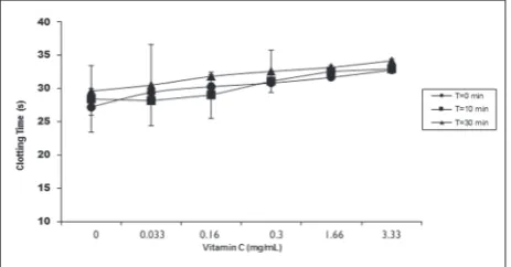
According to the experimental data, vitamin C has no significant influence on the intrinsic pathway of
the coagulation cascade, even at high concentration levels or high incubation periods (Fig. 1). Therefore, a potential vitamin C inhibitory coagulation mechanism does not affect plasmatic proteins from the blood co-agulation cascade’s intrinsic pathway. These data sug-gest that the direct effect of vitamin C on thrombus formation must occur either uniquely in vivo or affect the coagulation cascade from the extrinsic or common pathways as suggested by Wannamethee et al. [14]. Prothrombin time (PT)
The prothrombin time (PT) data showed a slight in-crease in prothrombin time when increasing the vi-tamin C concentration in plasma. This effect has no relation to the pre-incubation time (Fig. 2). These data pointed to the influence of vitamin C in clot formation from a very specific mechanism that significantly
af-Fig. 1. Comparison of the activated partial thromboplastin time from human plasma at different concentrations of vitamin C (0.033-3.33 mg/mL) and pre-incubation times (T = 0.10 and 30 min).
fects the extrinsic pathway on the coagulation cascade at concentrations above 1.66 mg/mL, when triggered by thromboplastin.
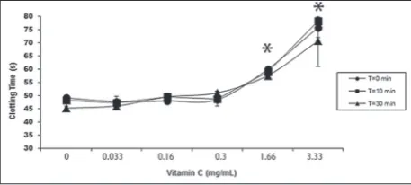
Thrombin time (TT)
Thrombin time assay revealed a dose-dependent activ-ity of vitamin C in the common pathway of the coagu-lation cascade, with statistical significance observed at 1.66 and 3.33 mg/mL final concentrations (Fig. 3). Different concentrations of vitamin C influenced the thrombin time assay. Our results also revealed that the incubation time affected the inhibitory activity of vitamin C, pointing to a kinetic-like behavior. Thrombin-initiated fibrin polymerization
In order to evaluate the direct anticoagulant activ-ity of vitamin C and its influence on fibrin network formation, we performed thrombin-initiated fibrin polymerization assays. Incubation with 3.33 mg/mL of vitamin C decreased the global fibrin polymeriza-tion promoted by thrombin in an ascorbate-free sam-ple at 37ºC (Fig. 3). These data corroborate the data of Zbikowska et al. [24] in which ascorbate reduced the fibrin polymerization from gamma-irradiated fibrinogen, while reducing the incorporation of car-bonyl groups to the fibrinogen molecule. This leads to an effective protection of fibrinogen from gamma irradiation-induced oxidation. However, our results suggest that the fibrinogen oxidation generated by thrombin may be impaired by the natural antioxidant profile of ascorbate.
Ancrod time (AT)
To analyze the antioxidant influence of vitamin C in clot formation, we measured the clot formation time catalyzed by ancrod as an alternative clot-promoting agent to thrombin. Our results showed that vitamin C still affects the overall clot formation even when using ancrod, a thrombin-like enzyme from a venom-ous snake. This result implies that vitamin C does not have a molecular specificity for thrombin but rather directly affects the normal fibrin assembly. These
re-sults are similar to thrombin time data, with statistical significance observed for ancrod clotting time also at the highest concentrations of vitamin C tested (1.66 mg/mL and 3.33 mg/mL) (Fig. 4). This supported the potential of vitamin C to impair fibrin clot formation despite of its cleavage origin.
DISCUSSION
In this work, we evaluated the anticoagulant profile of vitamin C using different coagulation assays. Our results revealed that vitamin C directly affects clot formation, probably through a molecular mechanism that impairs the assembly of a cohesive fibrin network. According to our data, the mechanism is independent
of the clot-promoting agent as vitamin C affected both thrombin- and ancrod-catalyzed assays. These data suggest that vitamin C promotes the impairment over clot formation, possibly due to the poor assembling of fibrin protofibrils and further fiber network formation.
Overall, our data is similar to that of Therani et al. [17], who studied the influence of high antioxidant-agent doses in normal fibrin clot formation. Ascorbate is the physiological form of vitamin C at pH 7.4 and displays an important chemical role as a donor antioxi-dant agent [25,26]. Our results suggest that ascorbate may affect fibrin network assemblage and final micro-structure through a direct impairment of protofibrils and fiber formation. These results are reinforced by previous data from Macedo and Kang [18] that de-scribed a reduction in protofibrils and fiber formation on clots originating from blood treated with vitamin C. The literature indicated that coagulation clots stay under intense lysis conditions [27]. Thus, the effect of vitamin C on fibrin network microstructure observed herein led to a fragile final product that was suscepti-ble to lyses, similar to that described by Therani et al. [17] who studied acetylsalicylic acid. Our data sup-port the positive effect of vitamin C in cardiovascular diseases such as thrombosis described previously [12]. Wannamethee et al. [14] reported that a fragile fibrin network would improve lysis after the activation of fibrinolytic proteins, such as tissue plasminogen activator/plasmin, where thrombus formation time would be considerably reduced along with superox-ide generation. In such a context, according to the literature, platelet aggregation could be affected by ascorbate through the interaction of fibrinogen with the integrin glycoprotein complex present in platelet glycoprotein IIB/IIIA [28].
In this work, we observed a distinct influence of vitamin C on clot formation in a dose-dependent manner that relies on other parameters, such as in-cubation time (but without affecting the pH even at high concentrations). According to our data, the an-ticoagulant activity of vitamin C does not rely on the establishment of an acidic medium that could lead to acidosis and further promote endogenous fibrinolytic
events in vivo, as suggested previouly [29]. We propose an anticoagulant mechanism based on the antioxi-dant potential of the active agent, where the substrate cleavage of the E-portion of fibrinogen by thrombin removes the N-terminal 16 and 14 peptides known as fibrinopeptides A (FPA) and B (FPB), respectively (Fig. 5). This removal would lead to the release of carboxyl groups in the E nodule (E) in fibrin, which are necessary for the interaction with the D nodule (D) of adjacent fibrin monomers. Furthermore, the release of FPA would promote protofibril formation, while the release of FPB would promote lateral aggre-gation of preexisting protofibrils, fiber formation and a cohesive fibrin network. Therefore, ascorbate (C), the soluble form of vitamin C in plasma, would donate a hydrogen atom at pH 7.4 to the oxidizing radical of (E) to produce the resonance-stabilized tricarbonyl ascorbate free-radical while thermodynamically
pairing the interaction between E and D terminal resi-dues of fibrin monomers and their further linkage by factor XIII transglutaminase [30]. This might prevent the formation of covalent bonds that are important in promoting a final cohesive fibrin network structure, leading to an unstable clot that is more susceptible to physiological lysis (Fig. 5).
CONCLUSIONS
Our results suggest a direct influence of vitamin C in the final pathway of clot formation. Its anticoagulant profile probably relies on its antioxidant potential, which impairs the physiological assembly of fibrin monomers and thus, the formation of a cohesive net-work of fibrin fibers. Unlike the mechanisms observed for other antioxidants, such as vitamin E (through vi-tamin K antagonism) and acetylsalicylic acid (through fibrinogen acetylation), vitamin C might prevent fi-brin assembly by direct impairment of the interac-tion between the E and D terminal residues of fibrin monomers at pH 7.4, while forming the resonance-stabilized tricarbonyl ascorbate free-radical from its stable ascorbateform. This work lends support to the proposed antihemostatic ability of this antioxidant, and to its potential use against thrombotic situations and thrombogenic conditions.
Acknowledgments: This work was supported by the Brazilian agencies FAPERJ and CNPq. Fellowships granted by UFF, CAPES, CNPq and FAPERJ are gratefully acknowledged.
Authors’ contributions: Plínio Cunha Sathlerwrote the protocol, and the first draft of the manuscript. André Luiz Lourenço. Ana
Paula Gentile Arêasand Max Seidy Saitoperformed the statistical
and the experimental analysis. Carlos Rangel Rodrigues, Lúcio Mendes Cabral, Helena Carla Castro managed the analyses of the study and the literature searches. Hye Chung Kang designed the study. All authors read and approved the final manuscript. Conflict of interest disclosure: No potential conflicts of interest relevant to this article were reported.
Ethical approval: This study was submitted to the Research Ethics Committee of the Faculty of Medicine of the University Hospital Antônio Pedro / HUAP Fluminense Federal University, and ap-proved according to Resolution 196/96 of the Ministry of Health (CMM/HUAP, REGISTRATION ID 177/11).
REFERENCES
1. Su Z-Y, Shu L, Khor TO, Lee JH, Fuentes F, Kong A-NT. A perspective on dietary phytochemicals and cancer chemo-prevention: oxidative stress, nrf2, and epigenomics. Top Curr Chem. 2013;329:133-62.
2. Benzie IFF, Wachtel-Galor S. Increasing the antioxidant con-tent of food: a personal view on whether this is possible or desirable. Int J Food Sci Nutr. 2012;63(Suppl 1):62-70. 3. Li S, Xue J, Shi J, Yin H, Zhang Z. Combinatorial
admin-istration of insulin and vitamin C alleviates the cerebral vasospasm after experimental subarachnoid hemorrhage in rabbit. BMC Neurosci. 2011;12:77.
4. Kannoujia DK, Kumar S, Nahar P. Covalent immobilization of ascorbate oxidase onto polycarbonate strip for L-ascorbic acid detection. J Biosci Bioeng. 2012;114(4):402-4. 5. Padayatty SJ, Katz A, Wang Y, Eck P, Kwon O, Lee J-H, Chen
S, Corpe C, Dutta A, Dutta SK, Levine M. Vitamin C as an antioxidant: evaluation of its role in disease prevention. J Am Coll Nutr. 2003;22(1):18-35.
6. Roberts LJ, Traber MG, Frei B. Vitamins E and C in the pre-vention of cardiovascular disease and cancer in men. Free Radic Biol Med. 2009;46(11):1558.
7. Azizova OA, Piryazev AP, Aseychev AV, Shvachko AG. Oxi-dative modification of fibrinogen inhibits its transformation into fibrin under the effect of thrombin. Bull Exp Biol Med. 2009;147(2):201-3.
8. Das S, Raychaudhuri U, Falchi M, Bertelli A, Braga PC, Das DK. Cardioprotective properties of raw and cooked eggplant (Solanum melongena L). Food Funct. 2011;2(7):395-9. 9. Frombaum M, Le Clanche S, Bonnefont-Rousselot D,
Borderie D. Antioxidant effects of resveratrol and other stil-bene derivatives on oxidative stress and NO bioavailability: Potential benefits to cardiovascular diseases. Biochimie. 2012;94(2):269-76.
10. Vinson JA, Cai Y. Nuts, especially walnuts, have both antioxi-dant quantity and efficacy and exhibit significant potential health benefits. Food Funct. 2012;3(2):134-40.
11. Obrenovich ME, Li Y, Parvathaneni K, Yendluri BB, Palacios HH, Leszek J, et al. Antioxidants in health, disease and aging. CNS Neurol Disord Drug Targets. 2011;10(2):192-207. 12. Loots D, Oosthuizen W, Pieters M, Spies C, Vorster HH.
Foodstate vitamin C complex may beneficially affect hae-mostasis and fibrin network structure in hyperlipidaemic patients. Blood Coagul Fibrinolysis Int J Haemost Thromb. 2004;15(8):677-85.
13. Mehta J, Li D, Mehta JL. Vitamins C and E prolong time to arterial thrombosis in rats. J Nutr. 1999;129(1):109-12. 14. Wannamethee SG, Lowe GDO, Rumley A, Bruckdorfer KR,
Whincup PH. Associations of vitamin C status, fruit and veg-etable intakes, and markers of inflammation and hemostasis. Am J Clin Nutr. 2006;83(3):567-74; quiz 726-7.
16. Ajjan RA, Standeven KF, Khanbhai M, Phoenix F, Gersh KC, Weisel JW, Kearney MT, Ariëns RA, Grant PJ. Effects of aspirin on clot structure and fibrinolysis using a novel in vitro cellular system. Arterioscler Thromb Vasc Biol. 2009;29(5):712-7.
17. Tehrani S, Antovic A, Mobarrez F, Mageed K, Lins P-E, Adam-son U, Wallén HN, Jörneskog G. High-dose aspirin is required to influence plasma fibrin network structure in patients with type 1 diabetes. Diabetes Care. 2012;35(2):404-8.
18. Macedo AA. In vitro action of vitamin C in the fibrinolytic process. Rev Bras Hematol E Hemoter. 2009;31(6):484-484. 19. Candeiro GT de M, Correia FC, Duarte MAH,
Ribeiro-Siqueira DC, Gavini G. Evaluation of radiopacity, pH, release of calcium ions, and flow of a bioceramic root canal sealer. J Endod. 2012;38(6):842-5.
20. Martinichen-Herrero JC, Carbonero ER, Sassaki GL, Gorin PAJ, Iacomini M. Anticoagulant and antithrombotic activi-ties of a chemically sulfated galactoglucomannan obtained from the lichen Cladonia ibitipocae. Int J Biol Macromol. 2005;35(1-2):97-102.
21. Bijak M, Bobrowski M, Borowiecka M, Podsędek A, Golański J, Nowak P. Anticoagulant effect of polyphenols-rich extracts from black chokeberry and grape seeds. Fito-terapia. 2011;82(6):811-7.
22. Kumar KR, Vennila R, Kanchana S, Arumugam M, Balasub-ramaniam T. Fibrinogenolytic and anticoagulant activities in the tissue covering the stingers of marine stingrays Dasyatis sephen and Aetobatis narinari. J Thromb Thrombolysis. 2011;31(4):464-71.
23. Bain BJ, Bates I, Laffan MA, Lewis SM. Dacie and Lewis Practical Haematology: Expert Consult: Online and Print. Edição: 11. Churchill Livingstone; 2011. 668 p.
24. Zbikowska HM, Nowak P, Wachowicz B. The role of ascor-bate and histidine in fibrinogen protection against changes following exposure to a sterilizing dose of gamma-irradi-ation. Blood Coagul Fibrinolysis Int J Haemost Thromb. 2007;18(7):669-76.
25. Codoñer-Franch P, Valls-Bellés V, Arilla-Codoñer A, Alonso-Iglesias E. Oxidant mechanisms in childhood obe-sity: the link between inflammation and oxidative stress. Transl Res J Lab Clin Med. 2011;158(6):369-84.
26. Du J, Cullen JJ, Buettner GR. Ascorbic acid: chemistry, biol-ogy and the treatment of cancer. Biochim Biophys Acta. 2012;1826(2):443-57.
27. Collet J-P, Lesty C, Montalescot G, Weisel JW. Dynamic changes of fibrin architecture during fibrin formation and intrinsic fibrinolysis of fibrin-rich clots. J Biol Chem. 2003;278(24):21331-5.
28. Nowak P, Wachowicz B. Peroxynitrite-mediated modifica-tion of fibrinogen affects platelet aggregamodifica-tion and adhesion. Platelets. 2002;13(5-6):293-9.
29. Sørensen B, Tang M, Larsen OH, Laursen PN, Fenger-Eriksen C, Rea CJ. The role of fibrinogen: a new paradigm in the treatment of coagulopathic bleeding. Thromb Res. 2011;128(Suppl 1):S13-6.