© 2019 by the Serbian Biological Society How to cite this article: Kozarski M, Klaus A, Jakovljević D, Todorović N, Wan- 253 Mohtar WAAQI, Nikšić M. Ganoderma lucidumas a cosmeceutical: Antiradical
potential and inhibitory effect on hyperpigmentation and skin extracellular matrix degradation enzymes. Arch Biol Sci. 2019;71(2):253-64.
Ganoderma lucidum
as a cosmeceutical: antiradical potential and inhibitory effect on
hyperpigmentation and skin extracellular matrix degradation enzymes
Maja Kozarski1,*, Anita Klaus2, Dragica Jakovljević3, Nina Todorović3, Wan Abd Al Qadr Imad Wan-Mohtar4 and Miomir Nikšić2
1Department of Chemistry and Biochemistry, Institute of Food Technology and Biochemistry, Faculty of Agriculture,
University of Belgrade, Nemanjina 6, 11080 Belgrade, Serbia
2Department of Industrial Microbiology, Institute of Food Technology and Biochemistry, Faculty of Agriculture, University
of Belgrade, Nemanjina 6, 11080 Belgrade, Serbia
3Institute of Chemistry, Technology and Metallurgy, University of Belgrade, Njegoševa 12, 11001 Belgrade, Serbia
4Functional Omics and Bioprocess Development Laboratory, Institute of Biological Sciences, Faculty of Science, University of
Malaya, 50603 Kuala Lumpur, Malaysia
*Corresponding author: [email protected]
Received: December 17, 2018; Revised: January 30, 2019; Accepted: January 31, 2019; Published online: March 5, 2019
Abstract: Three different polysaccharide extracts of a wild European source of the mushroom Ganoderma lucidum were screened for their free radical-blocking potential, which could strengthen the skin’s barrier function, and provide a skin-lightening effect via potential inhibition of tyrosinase. The anti-collagenase and anti-elastase activities, which can help to restore skin elasticity and tensile strength, were also evaluated for the three extracts. Carbohydrates were the most abundant components of the extracts, followed by smaller quantities of proteins, phenols and flavonoids. The glucan fraction rep-resented between 48% and 61% of carbohydrate content in all three extracts. None of the analyzed extracts showed overt toxicity to spontaneously-transformed immortal human keratinocytes (HaCaT) at concentrations up to 2 mg/mL, and displayed superior scavenging ability on 2,2'-azinobis(3-ethylbenzothiazoline-6-sulfonic acid (ABTS) and 1,1-diphenyl-2-picrylhydrazyl (DPPH) radicals. The hot water crude polysaccharide extract (HWCP) and partially purified fraction (HWPP) were found to be effective inhibitors of lipid peroxidation (LPx), with an almost two-fold increased inhibition of
LPx compared with ascorbic acid (EC50=1.65±0.08 mg/mL), a common additive in cosmeceutical formulations and used
at mg levels. Among the investigated extracts, HWCP showed the strongest inhibition potential on tyrosinase and skin
extracellular matrix (ECM) degradation enzymes. These diverse functionalities indicate that G. lucidum may represent a
promising source of natural cosmeceutical ingredients.
Keywords: Ganoderma lucidum; cosmeceutical; maturate skin; oxidative stress; polysaccharide extracts
Abbreviations and acronyms: 2,2'-azinobis(3-ethylbenzothiazoline-6-sulfonic acid (ABTS); bovine serum albumin (BSA);
catechin equivalents (CEs); dimethyl sulfoxide (DMSO); heavy water (D2O); 1,1-diphenyl-2-picrylhydrazyl (DPPH);
2,2-dimethyl-2-silapentane-5-sulfonic acid sodium salt (DSS); effective concentrations of each extract that are required to
show 50% antioxidant properties (EC50); extracellular matrix (ECM); ethylenediaminetetraacetic acid (EDTA);
epigallo-catechin gallate (EGCG); N-(3-[2-furylacryloyl)-Leu-Gly-Pro-Ala (FALGPA); ferric-reducing antioxidant power (FRAP); Fourier-transform infrared spectroscopy (FTIR); gallic acid equivalents (GAE); spontaneously transformed immortal human keratinocytes (HaCaT); hydrogen atom transfer (HAT); hot alkali polysaccharide extract (HWAP); hot water crude poly-saccharide extract (HWCP); hot water polypoly-saccharide extract purified by dialysis (HWPP); 50% inhibitory concentration
(IC50); dihydroxyphenylalanine (L-DOPA); lipid peroxidation (LPx); N-methoxysuccinyl-Ala-Ala-Pro-Val-p-nitroanilide
INTRODUCTION
Oxidative stress induced by reactive oxygen species (ROS) plays an important role in the process of human skin aging [1,2]. The skin is an interface between the body and the environment and is constantly exposed to atmospheric oxygen, ultraviolet (UV) irradiation, pollutants and xenobiotics. Oxidative damage caused by these exogenous sources can impair skin structure and function, leading to the phenotypic features of extrinsic aging [1]. In addition, excessive consumptionof alco-hol, improper diet, physical inactivity and mechanical stress can contribute to oxidative damage of skin [3-5].
The endogenous defense systems against ROS are often insufficient to combat oxidative processes in mature skin. ROS that are not neutralized can target biomolecules and lead to cellular dysfunction or death and accelerated aging [6]. Furthermore, ROS can induce the overexpression of MMPs, which increase collagen fragmentation and can downregulate TGF-β and promote collagen synthesis [7]. ROS can also increase elastase activity and trigger wrinkle forma-tion by cleaving elastin and further degradaforma-tion of collagen fibrils in the ECM [5,7]; it may also be an essential causative factor for hyperpigmentation or even carcinogenic processes in the skin [8].
Ganoderma lucidum (Curtis) P. Karst. (lingzhi) is a
medicinal mushroom with a wide variety of bioactive components, possessing nutraceutical and pharmaco-logical benefits [9]. Popularly known as the “mushroom of immortality”, G. lucidum has been widely used in China for medicinal purposes for more than 2000 years [10]. Historically, lingzhi was grown in the wild and consumed only by wealthy individuals. Nowadays, large quantities are cultivated for production at an industrial level and are widely consumed [10]. G. lucidum is used in cancer treatment as an alternative to or alongside modern medicine to enhance the immune response, alleviate the side effects of chemotherapy and protect cellular DNA by increasing cellular antioxidant capacity [11]. Moreover, it has been used in the prevention of hypertension and hypercholesterolemia, treatment of diabetes, maintenance of gut health and stimulation of probiotics [9,10]. The use of bioactive extracts from mushrooms in the design of cosmeceutical formula-tions for topical application is receiving increasing attention [12-14]. Modern trends in the cosmetics
industry prioritize ingredients or extracts from natural sources with nontoxic effects and the ability to delay the aging process [12,14].
The objective of the present study was to investi-gate the potential therapeutic effect on mature skin of polysaccharide extracts from the fruiting bodies of a wild European source of G. lucidum. These effects are expected to influence oxidative stress via the potential antiradical activity of the extracts and inhibitory ef-fect on tyrosinase and enzymes responsible for ECM degradation.
MATERIALS AND METHODS
Reagents and chemicals
DPPH, ABTS, MTT, porcine pancreatic elastase type I [EC 3.4.21.36], (N-methoxysuccinyl)-Ala-Ala-Pro-Val-chloromethyl ketone, MAAPVN, collagenase from
Clostridium histolyticum [EC 3.4.24.3], tyrosinase from
Mushroom material
Fresh wild-growing fruiting bodies of mushroom G.
lucidum were collected from the forest/park Košutnjak
near Belgrade, Republic of Serbia. After collection, carpophores were identified according to the methods of classical herbarium taxonomy to confirm the correct species[15]. The representative voucher specimens were deposited at the herbarium of the Department for Industrial Microbiology at the Faculty of Agriculture-University of Belgrade (under reference number GLK-12) together with their mycelial cultures, and stored at 4°C in the culture collection for mushrooms until further use. Samples were brush-cleaned, air-dried at 40°C to a constant mass and ground into fine powder prior to extraction and analysis.
Preparation of extracts
Crude polysaccharide extracts were prepared by hot water extraction of 100 g of powdered sample [16]. After centrifugation, the resulting pellets were washed with ethanol 70% (v/v), dried under a vacuum and stored in the cold as hot water-extracted crude poly-saccharides, HWCP. A portion of the HWCP sample was dialyzed against Milli-Q water (MQ) for 24 h at room temperature to remove residual small molecules as polyphenols, peptides and polysaccharides <8-10 kDa. After centrifugation, high molecular weight polysaccharides were ethanol precipitated and vacuum dried. This material was partly purified and designated HWPP. Alkali-soluble polysaccharides were extracted from the original hot water filter cake by autoclaving in 2 L of 1 M NaOH solution[16]. This extract was designated HWAP.
Chemical composition of extracts
To explain the differences observed, we measured: (i) the total polysaccharide content of the extract using the phenol-sulfuric acid method [17], with results expressed as g of Glc equivalents per 100 g of dry weight (DW) of the extract; (ii) the protein content by the Bradford method [18], with results expressed as g of bovine serum albumin (BSA) equivalents per 100 g of DW of the extract; (iii) the total phenolic content using a Fast Blue BB (FBBB) method [19], with
the content of total phenols expressed as gallic acid equivalents (GAE) per 100 g of DW of the extract; (iv) the flavonoid content [19], with results expressed as g of catechin equivalents (CEs) per 100 g of extract; (v) the content of total α- and β-glucans, after prior partial hydrolysis, using the Mushroom and Yeast β-glucan Assay Procedure K-YBGL 07/11.
FT-IR spectroscopy
FT-IR spectra were obtained using a Fourier transform infrared spectrophotometer (Thermo-Nicolet Model 6700, Thermo Scientific, USA), equipped with a Smart Orbit (Diamond) ATR accessory and OMNIC 7.3 software, in the 4000-400 cm-1 range at a resolution of 4 cm-1 in transmission mode.
Analysis of monosaccharide composition
TLC and NMR spectroscopy were used to determine the monosaccharide composition. Each polysaccharide extract was hydrolyzed separately with 2 M TFA at 121°C. After hydrolysis, the final residue was dissolved in 2 mL of MQ and used for further analysis. TLC was run on silica gel H plates [16]. Authentic sugars were: GlcA, GalA, Glc, Gal, Man, L-Fuc, D-Xyl, D-Ara, and L-Rha. NMR spectra were measured on a Bruker AVANCE III 500 spectrometer (500.26 MHz for 1H nuclei), using a 5 mm broad-band probe head at 25°C in D2O with DSS as the internal standard [20]. Signals were assigned according to the web-based version of the computer program CASPER (Widmalm Research Group, Stockholm, Sweden) [21].
Evaluation of radical-blocking capacities
Enzyme inhibition activities Assay of tyrosinase activity
Tyrosinase activity [23] was determined in a 96-well plate using an absorbance microplate reader (ELx808, BioTek Instruments, Inc., USA) controlled by Gen5TM Software to measure absorbance at 475 nm. An aliquot of the extract (40 µL) in 0.067 M of phosphate buffer (pH 6.8) containing 5% DMSO was incubated with 80 µL of phosphate buffer (pH 6.8) and 40 µL of tyrosi-nase (46 units/mL) at 23°C for 10 min. Next, 40 µL of 2.5 mM L-DOPA in 0.067 M of phosphate buffer (pH 6.8) was added. Extracts were analyzed at the concen-tration range of 0.04-5 mg/mL, using kojic acid as a reference. The anti-tyrosinase activities of the extracts were expressed as IC50 values, which were calculated using linear regression analyses as the concentration of extract required for 50% inhibition in vitro.
Assay of elastase activity
An extract of appropriate concentration (50 µL) in 0.2 M tris-HCl buffer (pH 7.6) containing 5% DMSO was incubated with 100 μL of tris-HCl buffer (pH 7.6) and 25 μL of elastase (0.3 units/mL) at room temperature for 20 min, after which 25 μL of the substrate MAAPVN (10 mM) was added and the plates incubated for a further 40 min at 25°C [24]. Absorbance was read at 405 nm. Extracts were analyzed at a concentration range of 0.04-5 mg/mL. (N-methoxysuccinyl)-Ala-Ala-Pro-Val-chloromethyl ketone was used as a reference. Anti-elastase activities were expressed as IC50 values.
Assay of collagenase activity
Extracts of appropriate concentration (0.04-5 mg/ mL) were dissolved in 50 mM of Tricine buffer (pH 7.5 with 0.4 M NaCl, 10 mM CaCl2) containing 5% DMSO [25]. Next, 20 µL of collagenase (0.16 units/ mL) was allowed to react with 40 µL of each extract and 70 µL of Tricine buffer in the dark at 37°C for 20 min [25]. After preincubation, 40 μL of 0.8 mM FALGPA were added to each well and the microplate was further incubated in the dark at 37°C for 30 min. Absorbance was measured at 340 nm. EGCG was used as a positive control, and anti-collagenase activities were expressed as IC50 values.
Cytotoxicity analysis
Stock solutions of polysaccharideextracts were pre-pared in DMSO [19]. HaCaT cells were cultured as a monolayer in nutrient medium at 37oC in 5% CO
2 and a humidified air atmosphere. HaCaT (5,000 c/w) cells were seeded into 96-well microtiter plates and incubated for 20 h before different concentrations of investigated extracts were added to the wells in the range 0.0625-2 mg/mL. Nutrient medium alone was added to the cells in the control wells. Corresponding concentrations of extracts in nutrient medium but without cells were used as blanks. Cell survival was determined by MTT testing [26], 24 h after the investigated extracts were added. The data were expressed as the concentration of sample required to decrease cell viability by 50% (IC50) compared with the controls.
Statistical analysis
All experiments were carried out in triplicate and ex-pressed as the mean±standard deviation (SD). Statistical analyses were performed with the Statistica 8.0 software package (StatSoft Inc., Tulsa, OK), using one-way analysis of variance (ANOVA) for all collected data. Differences between the means for each treatment were determined using Duncan’s multiple range tests (P<0.05). The cor-relation coefficients (r) between antioxidant activities and polysaccharide fractions were determined.
RESULTS
Extraction yields
Extraction yields of HWCP, HWPP, and HWAP from
G. lucidum were 1.8±0.4, 0.4±0.1 and 6.2±0.7 g/100 g
Chemical composition of G. lucidum polysaccha-ride extracts
The chemical compositions of G. lucidum extracts are presented in Table 1. Carbohydrates were the most abundant compounds, followed by proteins, phenols and flavonoids. HWPP yielded the highest content of total carbohydrates (82.7±2.8 g/100 g) with molecular masses greater than 8-10 kDa after dialysis. HWAP had a significantly lower carbohydrate content (27.9±1.3 g/100 g DW) compared with HWCP and HWPP. The glucan fraction comprised between 46% and 61% of the carbohydrate content in all three extracts. Given the powdery nature of the material, the glucan fraction could be readily extracted and isolated; it consisted almost exclusively of β-glucans.
Phenols were found in considerable quantities in all extracts. The highest content was detected in HWCP (Table 1). The phenol content in HWPP was statistically different and approximately 43% lower than that in HWCP. Dialysis and ethanol reprecipitation were insufficient for the removal of small polyphe-nols from HWPP, indicating possible formation of polysaccharide-polyphenol conjugates. The flavonoid content of the tested samples was ranked in the order of HWCP>HWAP>HWPP, being about 75%, 83% and 69% of the total phenolics, respectively.
Proteins were present in all extracts (Table 1). Deproteinization of the polysaccharide fractions was not achieved with thermal treatment (autoclaving at 120°C for 2 h). Dialysis was also insufficient for the removal of all proteins in HWPP, resulting in a 38% reduction in the total protein content of HWCP.
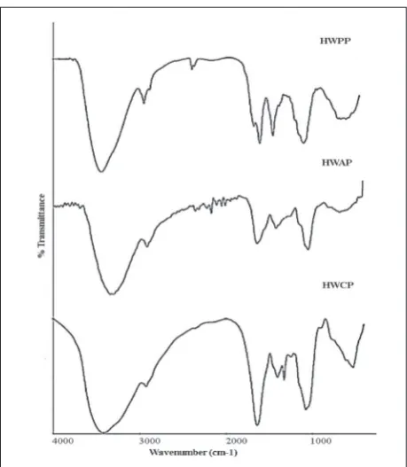
FT-IR spectroscopy
The FT-IR spectra of polysaccharide extracts HWCP, HWPP, and HWAP isolated from G.lucidum showed
typical patterns of polysaccharides with similar struc-tural characteristics (Fig. 1). The FT-IR spectrum of HWCP showed a wide intense absorption band at 3000 cm−1 to 3500 cm−1, characteristic for -OH stretching vibration related to inter- and intramolecular hy-drogen bonds with a peak at 2950-2850 cm−1, which corresponded to CH2 stretching [27,28]. The peaks at 1200-1000 cm−1 were related to C-O absorption and ring vibrations, and included a prominent sharp peak at 1079 cm−1, characteristic for stretching of C-O and C-C bonds in beta-linked glucose residues; a peak at 1055 cm−1 corresponded to C-O-C stretching [20,27,28]. The weak absorptions at 890 cm−1 and 850 cm−1 were specific for β- and α-glycosidic linkages, respectively [20,27,28]. The presence of residual proteins can be attributed to bands at 1642 cm-1, 1455 cm-1 and 1403
Table 1. Chemical composition ofpolysaccharide extracts derived from the fruiting body of a wild European source of G. lucidum.
Extract polysaccharidesTotal Total Glucan contentα β Total proteins Total phenolics flavonoidsTotal
HWCP 40.7±2.2b 23.4±1.1b 1.8±0.3b 21.6±0.8b 3.9±0.3a 4.5±0.2a 3.4±0.2a
HWPP 82.7±2.8a 39.4±0.7a 3.2±0.6a 36.2±1.2a 2.4±0.1b 2.6±0.1c 1.8±0.2c
HWAP 27.9±1.3c 13.9±1.0c 1.7±0.5b 12.2±0.7c 1.0±0.1c 3.0±0.1b 2.5±0.1b
All values are expressed per dry weight of extract (g/100 g). Data are the mean±SD (n=3). Within the same column, means followed by different letters are significantly different at P<0.05.
cm-1 [20]. The characteristic N-H vibration at 3400 cm-1 was overlapped by stretching of the O-H vibration at 3000-3500 cm-1 [20]. The frequency at 1642 cm-1 also corresponded to the bending vibration O-H of adsorbed water molecules. Additionally, the peak at 1642 cm-1 was due to overlap with absorptions of C=C and C=O stretching vibrations, indicative of aromatics [20]. Vibrations between 1410 cm-1 and 1310 cm-1 were indicative of O-H groups of phenolic compounds, as was a band at 1403 cm-1, which was related to aliphatic groups originating from the aromatic pigments [20].
The FT-IR spectrum of the HWPP sample showed the same characteristic absorption bands as the HWCP sample, except that absorption at 1642 cm-1 related to the presence of the protein was somewhat reduced when compared with the FT-IR spectrum of HWCP, indicating that a certain amount of protein molecules had been removed by dialysis. However, the presence of bands for amide I at 1642 cm-1 and amide II at 1568 cm-1 were indicative of the presence of some residual protein in this polysaccharide extract. Absorptions that were related to the frequency of phenolic compounds were also altered to some extent.
The FT-IR spectrum of HWAP retained similar structural features, with absorptions related to the polysaccharide structure as well as to the content of proteins and phenolic compounds, although the intensity of some absorptions was altered. The peak at 1080 cm-1 characteristic for β-glycosidic bonds was reduced, probably due to alkaline degradation of polysaccharide molecules during extraction [29]. In addition, a characteristic weak peak at 890 cm−1 cor-responding to β-glycosidic linkages had disappeared, indicating that hot alkaline treatment of polysaccharide caused degradation, especially at the basic chain [16].
The absence of a characteristic absorption band related to ester-carbonyl stretching absorption at 1740
cm−1 in the FT-IR spectra [20] of all three samples indicated that the investigated polysaccharide extracts did not contain uronic acid in their structure, which was also confirmed by TLC.
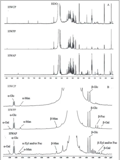
Monosaccharide composition
All three polysaccharide extracts from G. lucidum
showed D-Glc as the predominant component after total acid hydrolysis, with smaller amounts of D-Gal and
Table 2. Quantitative ratio of the monosaccharide components after acid hydrolysis in samples HWCP, HWPP and HWAP according
to their 1H-NMR spectra.
Extract Monosaccharide α/β % Aromatic
compounds compounds***Aliphatic
Glc Gal Man Fuc and/or Xyl **
HWCP 1*/1.6 - 0.17/- - 35 +
HWPP 1/1.5 0.13/0.13 0.22/0.1 -/0.13 5 +
HWAP 1/1.6 0.1/0.25 0.23/0.22 0.16/0.25** - ++
* H(1)-integral of α-Glc is calibrated to 1. ** Signals overlapped; assignation uncertain. *** Many overlapping signals; the presence notified.
Fig. 2. A – The full 1H-NMR spectra of HWCP, HWPP and HWAP
upon acid hydrolysis (the signals labeled with asterisk originate from 2,2-dimethyl-2-silapentane-5-sulfonic acid sodium salt-DSS).
traces of D-Man and D-Fuc and/or D-Xyl, on the basis of TLC data, by comparison of their chromatographic mobility with monosaccharide standards. No uronic acid was found in the thin-layer chromatogram when a solvent for the separation of acidic components was used (chromatogram not shown).
The 1H-NMR spectra of HWCP, HWPP and HWAP extracts after acid hydrolysis were relatively similar (Fig. 2A). The monosaccharide components were identified and quantified based on their H-1 signals (Table 2, Fig. 2B). According to these data, D-Glc was the main constituent, with -anomer dominating in all three samples. Among the other monosaccharides, weak signals of Man, Fuc, and/or Xyl and D-Gal were also present.
G. lucidum polysaccharide extracts as anti-aging
agents
Anti-radical potential
The results regarding the anti-radical activity of G.
lu-cidum polysaccharide extracts are presented in Table 3.
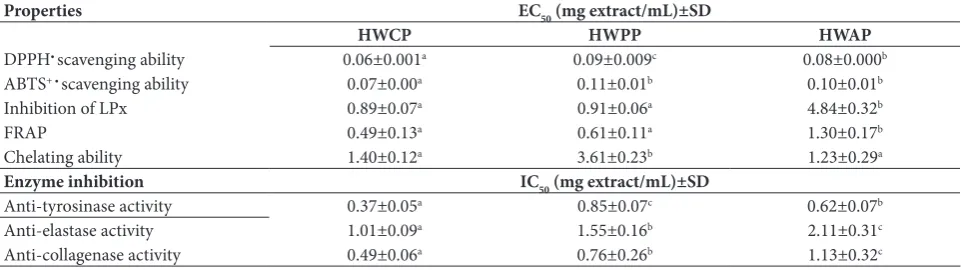
The higher the anti-radical capacity, the lower the value of EC50. The mean values of EC50 indicated that all ex-tracts of G. lucidum were potent antioxidants. EC50 val-ues for thescavenging activities on DPPH• of the tested samples were in the order of: HWCP<HWAP<HWPP, and for the prevention on the generation of ABTS+• they were: HWCP<HWPP≈HWAP. Furthermore, the extracts showed comparable values of EC50 with ascorbic acid (0.08±0.001 mg/mL) in scavenging
ac-tivities of DPPH• and prevention of ABTS+• generation (0.06±0.000 mg/mL). HWCP and HWPP were potent inhibitors of LPx, exerting an almost two-fold higher potential for LPx inhibition compared to ascorbic acid (1.65±0.08 mg/mL), a common additive in cosmeceuti-cal formulations and used at mg levels. There was no significant difference in EC50 values for the reducing power ofHWCP and HWPP. Ascorbic acid showed a lower EC50 value in the FRAP assay (0.056±0.007 mg/ mL) and higher reducing power than the analyzed ex-tracts.Likewise, no significant difference at P<0.05 was found between the EC50 values of HWCP and HWAP with respect to chelating ability. For comparison, the chelator EDTA, widely used in the cosmetics industry, showed a higher activity (EC50<0.04 mg/mL).
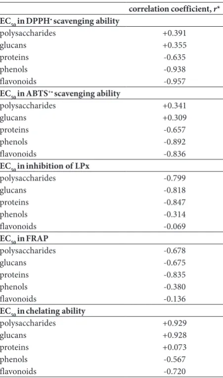
Correlation between the anti-radical potential and components of polysaccharide extracts
Regression analysis revealed a highly significant cor-relation between the EC50 of scavenging activities on DPPH• and prevention of ABTS+• generation and total phenols and flavonoids, (Table 4). A decrease in EC50 value correlated with higher phenol and flavonoid contents. A significant correlation was also observed for total proteins. In contrast, regression analyses revealed a significant role for the carbohydrate components of the extracts in the inhibition of LPx in the linoleic acid model system and FRAP assay. A highly significant cor-relation was observed for total protein in both assays. A highly significant correlation was also observed between the chelating ability and the flavonoid content (Table 4).
Table 3. EC50 and IC50values of polysaccharide extracts from G. lucidum in radical-blocking properties and enzyme inhibition.
Properties EC50 (mg extract/mL)±SD
HWCP HWPP HWAP
DPPH• scavenging ability 0.06±0.001a 0.09±0.009c 0.08±0.000b
ABTS+ • scavenging ability 0.07±0.00a 0.11±0.01b 0.10±0.01b
Inhibition of LPx 0.89±0.07a 0.91±0.06a 4.84±0.32b
FRAP 0.49±0.13a 0.61±0.11a 1.30±0.17b
Chelating ability 1.40±0.12a 3.61±0.23b 1.23±0.29a
Enzyme inhibition IC50 (mg extract/mL)±SD
Anti-tyrosinase activity 0.37±0.05a 0.85±0.07c 0.62±0.07b
Anti-elastase activity 1.01±0.09a 1.55±0.16b 2.11±0.31c
Anti-collagenase activity 0.49±0.06a 0.76±0.26b 1.13±0.32c
EC50 value: the effective concentrations of each mushroom extract providing 50% of antioxidant activity or 0.5 of absorbance in the reducing power
assay; IC50 value: concentration of extract required for 50% inhibition in vitro. Each value is expressed as the mean±SD (n=3). Means with different
Enzyme inhibition activities
The inhibitory activities for tyrosinase, elastase and collagenase exhibited by HWCP, HWPP, and HWAP are presented in Table 3, and they show that all ex-tracts had the ability to inhibit these enzymes. HWCP displayed the strongest inhibitory activity among the investigated extracts and exhibited a moderate potential when compared with standard inhibitors. The anti-tyrosinase IC50 value of kojic acid, which is currently used in topical dermatological products, was 0.079±0.004 mg/mL, while an IC50 value of 0.23±0.05 mg/mL was obtained for
(N-methoxysuccinyl)-Ala-Ala-Pro-Val-chloromethyl ketone, a common irreversible inhibitor of human leukocyte and neutrophil elastase that was found to be an effective inhibitor of porcine elastase in the present study. The IC50 value of EGCG, which has anti-collagenase activity, was 0.068±0.03 mg/mL. This compound is an established ingredient in dermocosmeticsto prevent skin aging and maintain skin balance.
Cytotoxicity analysis
G. lucidum extracts were subjected to an in vitro
cyto-toxicity assay in HaCaT cells (Fig. 3). After incubation for 24 h, HWCP, HWPP and HWAP were found to be nontoxic to cells at concentrations up to 2 mg/mL. High levels of cell viability (more than 80%) were observed, meaning that IC50 values could not be determined for the extracts. Furthermore, HWCP and HWPP induced significant proliferation of up to 50% in HaCaT cells at the concentration range 0.125-0.250 mg/mL (P<0.05), as compared with the control.
DISCUSSION
A major barrier to the acceptance of natural products in the pharmaceutical and cosmetic industries is their complexity. However, this complexity can bring sig-nificant advantages. For example, certain components of natural products can reduce the cytotoxicity of the whole product, and interactions between different biologically active components can be responsible for
Table 4. Correlation coefficient (r) between total polysaccharide, glucan, protein, phenol and flavonoid content vs. EC50 values in antioxidant activities of HWCP, HWPP and HWAP.
correlation coefficient, r* EC50 inDPPH• scavenging ability
polysaccharides +0.391
glucans +0.355
proteins -0.635
phenols -0.938
flavonoids -0.957
EC50 inABTS+• scavenging ability
polysaccharides +0.341
glucans +0.309
proteins -0.657
phenols -0.892
flavonoids -0.836
EC50 ininhibition of LPx
polysaccharides -0.799
glucans -0.818
proteins -0.847
phenols -0.314
flavonoids -0.069
EC50 inFRAP
polysaccharides -0.678
glucans -0.675
proteins -0.835
phenols -0.380
flavonoids -0.136
EC50 inchelating ability
polysaccharides +0.929
glucans +0.928
proteins +0.073
phenols -0.567
flavonoids -0.720
r* – all values are statistically significant (P<0.05); for absolute values of r, 0-0.19 is regarded as a ‘very weak’, 0.2-0.39 as a ‘weak’, 0.40-0.59 as a ‘moderate’, 0.6-0.79 as a ‘strong’, and 0.8-1 as a ‘very strong’ correlation.
their in vivo effects [30]. Moreover, synthetic skincare products containing active ingredients may cause adverse reactions such as allergic contact dermatitis, irritant contact dermatitis, phototoxic, photoallergic reactions, or even infections [31]. The demand for multifunctional products is also driving innovation in the cosmetic industry, as price-conscious consum-ers are opting for products that can provide sufficient vitality and skin protection.
The results of the present research highlight the potential capacity of polysaccharide fractions derived from the fruiting bodies of a wild European source of
G. lucidum: for free radical blocking, which may help
to strengthen the skin’s barrier; for a skin-lightening effect as inhibitors of tyrosinase, and for the anti-collagenase and anti-elastase activities to help restore skin elasticity and tensile strength.
Moreover, no obvious toxicity was observed in HaCaT keratinocytes, which represent the first line of defense of the body against the outside environment [32]. This is the first report of the preventive effect of
G. lucidum originating from Serbia in the potential
development of cosmeceutical formulations and the slowing of skin aging.
The presence of different bioactive molecules from the classes of polysaccharides, proteins and phenolic and flavonoid compounds in the HWCP, HWPP and HWAP fractions may correlate with their observed bioactivities.
Carbohydrates were the most abundant compounds in the extracts. A significant and strong correlation was observed between polysaccharides and the pre-vention of LPx in a linoleic acid model system. The ROS-scavenging mechanism of polysaccharides may be similar to that of phenol compounds, i.e. via H atom transfer (HAT) reactions [33]. However, HAT reactions are more likely to occur in neutral polysaccharides, which were also observed in the investigated fractions, while electron transfer (ET) typically occurs in acidic polysaccharides [33]. Additionally, chain-breaking/free radical scavengers can act via secondary or preventive mechanisms, which may include inhibition of lipid hy-droperoxide breakdown to unwanted volatile products, regeneration of primary antioxidants, singlet oxygen quenching and deactivation of metals [6].
All three polysaccharide extracts were shown to be good sources of β-glucans. β-glucans, despite their considerable molecular weight, are known to enter the
stratum corneum and epidermis, penetrating deep into
the dermis [34]. β-glucans do not directly enter the cell, but penetrate skin through the intercellular space. It has been suggested that β-glucans form a thin film above the stratum corneum and epidermis to promote moisturization [34]. Within the dermis, they can stimu-late collagen synthesis through direct interaction with fibroblasts and through indirect cytokine-mediated interaction with macrophages [34]. Collagen synthesis is one possible mechanism by which the elasticity of the skin is enhanced.
Polyphenols were present in all investigated ex-tracts, even after purification procedures to remove molecules with molecular masses lower than 8-10 kDa. Natural polyphenolic compounds are the most abundant antioxidants, and they exert their antioxi-dant effect by quenching free radical species and/or promoting the endogenous antioxidant capacity [6]. The ABTS and DPPH assays are typically classified as SET reactions, but these two indicator radicals may in fact be neutralized either by direct reduction via electron transfers or by radical quenching via the HAT mechanism [35]. Furthermore, polyphenolics can modulate the activity of nuclear factor kappa B (NF-κB) or sirtuin 1 (SIRT1), thus exerting protective effects [6,36]. SIRT1 acts as a “rescue gene”, capable of repairing damage caused by the action of free radicals and of preventing the premature death of cells [6]. SIRT1 in the epidermis regulates cell migration, redox response, inflammation, epidermis re-epithelialization, and granulation formation, it and plays a crucial role in wound repair [36]. It is an important regulator of the keratinocyte differentiation pathway and a potential regulator of skin aging [37].
and carbonyl moiety, whenever present [39]. The use of natural chelators such as flavonoids is preferred to the use synthetic counterparts, due to reduced toxicity [40]. Likewise, natural flavonoids have a photoprotec-tion potential because of their ability to absorb UV light and to act as direct and indirect antioxidants as well as anti-inflammatory and immunomodulatory agents, thus providing an exciting platform for the development of photoprotection [41].
It has been confirmed that mushroom extracts are a promising source of natural phenolic and flavonoid compounds [42-45]. Hot water extracts of medicinal mushrooms can be a source of bioactive flavonoids that could not be isolated at lower temperatures. Thus, daidzein was identified in Trametes versicolor hot water extract [45]. Also, amentoflavone and catechin were isolated in two-fold greater concentrations, as compared with the ethanol extract of the same mushroom [45].
Dialysis and thermal treatment (autoclaving at 120°C) were insufficient to remove all proteins from the extracts. Proteins can provide protection against harmful ROS, as many amino acids possess a strong antioxidant potential due to the presence of polar R groups [46]. Bioactive peptides might be effective in-hibitors of enzymes that are involved in the turnover of ECM proteins and melanin production [47].
All three extracts showed no toxicity to keratino-cytes in vitro. Our previous report confirmed a lack of toxicity up to 10 mg/mL of the commercial water extract obtained from an industrial strain of G.
lu-cidum (Fujian, China) [48]. Furthermore, this extract
stimulated the in vitro proliferation of HTR-8/SVneo cells, which are essential for normal placentation, establishment of pregnancy, and maintenance of fetal growth in humans [49].
Finally, the results of the present study strongly support the existing scientific data on the use of in-gredients from natural sources as anti-aging agents and their application in the cosmeceutical industry. The polysaccharide extracts of G. lucidum may rep-resent promising alternative raw materials for use in cosmetic products. However, these findings require further verification in clinical trials to examine the stability, skin permeation, and efficacy of the final cosmeceutical product.
Acknowledgments: The study was supported by the Serbian
Ministry of Science and Technological Development, Project Numbers III 46001, III 46010 and III 43004.
Author contributions: MK participated in all the experiments, wrote the draft and performed the literature search; AK par-ticipated in all the experiments and reviewed the manuscript; DJ performed the chemical analysis, FTIR spectroscopy and reviewed the manuscript; NT performed the NMR analysis and reviewed the manuscript; WAAQIWM performed the literature search and critically reviewed the manuscript; MN supervised all phases of the research and reviewed the manuscript. All authors read and approved the final manuscript.
Conflicts of interest disclosure: The authors have no conflicts of interest to declare.
REFERENCES
1. Rinnerthaler M, Bischof J, Streubel MK, Trost A, Richter K. Oxidative stress in aging human skin. Biomolecules. 2015;5(2):545-89.
2. Haydont V, Bernard BA, Fortunel NO. Age-related evolu-tions of the dermis: Clinical signs, fibroblast and extracel-lular matrix dynamics. Mech Ageing Dev. 2019;177:150-6. 3. Poljšak B, Dahmane R. Free radicals and extrinsic skin
aging. Dermatol Res Pract. 2012;2012:135206.
4. Kruk J, Duchnik E. Oxidative stress and skin diseases: possible role of physical activity. Asian Pac J Cancer Prev. 2014;15(2):561-8.
5. Naidoo K, Birch-Machin MA. Oxidative Stress and Ageing: The influence of environmental pollution, sunlight and diet on skin. Cosmetics.2017;4:4.
6. Kozarski M, Klaus A, Jakovljevic D, Todorovic N, Vunduk J, Petrovic P, Niksic M, Vrvic MM, Van Griensven L. Antioxi-dants of edible mushrooms. Molecules. 2015;20(10):19489-525.
7. Mukherjee PK, Maity N, Nema NK, Sarkar BK. Bioactive compounds from natural resources against skin aging. Phytomedicine. 2011;19(1):64-73.
8. Diehl C. Melanocytes and Oxidative Stress. J Pigment Dis-ord. 2014;1:127.
9. Ahmad MF. Ganoderma lucidum: Persuasive biologically active constituents and their health endorsement. Biomed Pharmacother. 2018;107:507-19.
10. Bishop KS, Kao CHJ, Xu Y, Glucina MP, Paterson RRM, Ferguson LR. From 2000 years of Ganoderma lucidum to recent developments in nutraceuticals. Phytochemistry. 2015;114:56-65.
11. Wasser SP. Reishi or Ling Zhi (Ganoderma lucidum). In: Wasser SP, Coates P, Blackman M, Cragg G, Levine M, Moss J, White J, editors. Encyclopedia of Dietary Supplements. New York: Marcel Dekker; 2005. p. 680-90.
12. Hyde KD, Bahkali AH, Moslem MA. Fungi-an unusual source for cosmetics. Fungal Divers. 2010;43(1):1-9. 13. Taofiq O, González-Paramás AM, Martins A, Barreiro MF,
cos-metics, cosmeceuticals and nutricosmetics. Ind Crops Prod.
2016;90:38-48.
14. Wu Y, Choi MH, Li J, Yang H, Shin HJ. Mushroom cosmet-ics: The present and future. Cosmetics. 2016;3(3):22. 15. Phillips R. Mushrooms and other fungi of Great Britain &
Europe. New York: Macmillan; 1979.
16. Klaus A, Kozarski M, Niksic M, Jakovljevic D, Todorovic N, Van Griensven LJLD. Antioxidative activities and chemi-cal characterization of polysaccharides extracted from the basidiomycete Schizophyllum commune. LWT-Food Sci Technol. 2011;44(10):2005-11.
17. Dubois M, Gilles KA, Hamilton JK, Reders PA, Smith F. Col-orimetric method for determination of sugars and related substances. Anal Chem.1956;28(3):350-6.
18. Bradford MM. A refined and sensitive method for the quantitation of microgram quantities of protein using the principle of protein-dye binding. Anal Biochem. 1976;72(1-2):248-55.
19. Kozarski M, Klaus A, Jakovljevic D, Todorovic N, Niksic M, Vrvic MM, Van Griensven LJLD. Dietary polysaccharide extracts of Agaricus brasiliensis fruiting bodies: chemical characterization and bioactivities at different levels of puri-fication. Food Res Int. 2014;64:53-64.
20. Kozarski M, Klaus A, Niksic M, Vrvic MM, Todorovic N, Jakovljevic D. Van Griensven LJLD. Antioxidative activities and chemical characterization of polysaccharide extracts from the widely used mushrooms Ganoderma applanatum,
Ganoderma lucidum, Lentinus edodes and Trametes versi-color. J Food Comp Anal. 2012;26:144-53.
21. Jansson PE, Stenutz R, Widmalm G. Sequence determination of oligosaccharides and regular polysaccharides using NMR spectroscopy and a novel Web-based version of the com-puter program CASPER. Carbohyd Res. 2006;341(8):1003-10.
22. Wei L, Zhang W, Yin L, Yan F, Xu Y, Chen F. Extraction opti-mization of total triterpenoids from Jatropha curcas leaves using response surface methodology and evaluations of their antimicrobial and antioxidant capacities. Electron J Biotech-nol. 2015;18(2):88-95.
23. Masuda T, Yamashita D, Takeda Y, Yonemori S. Screening for tyrosinase inhibitors among extracts of seashore plants and identification of potent inhibitors from Garcinia subel-liptica. Biosci Biotechnol Biochem. 2005;69(1):197-201. 24. Azmi N, Hashim P, Hashim DM, Halimoon N, Majid
NMN. Anti-elastase, anti-tyrosinase and matrix metallo-proteinase-1 inhibitory activity of earthworm extracts as potential new anti-aging agent. Asian Pac J Trop Biomed. 2014;4(Suppl 1):S348-52.
25. Van Wart HE, Steinbrink DR. A continuous spectrophoto-metric assay for Clostridium histolyticum collagenase. Anal Biochem. 1981;113(2):356-65.
26. Mosmann T. 1983. Rapid colorimetric assay for cellular growth and survival: application to proliferation and cyto-toxicity assays. J Immunol Methods. 1983;65(1-2):55-63. 27. Karaman M, Janjušević Lj, Jakovljević D, Šibul F, Pejin B.
Anti-hydroxyl radical activity, redox potential and anti-AChE activity of Amanita strobiliformis polysaccharide extract. Nat Prod Res. 2018; 10.1080/14786419.2017.1422183
28. Pejin B, Tešanović K, Jakovljević D, Kaišarević S, Šibul F, Rašeta M, Karaman M. The polysaccharide extracts from the fungi Coprinus comatus and Coprinellus truncorum
do exhibit AChE inhibitory activity. Nat Prod Res. 2017; 10.1080/14786419.2017.1405417
29. Wang ZB, Pei JJ, Ma HL, Cai PF, Yan JK. Effect of extrac-tion media on preliminary characterizaextrac-tions and antioxidant activities of Phellinus linteus polysaccharides. Carbohydr Polym. 2014;109:49 -55.
30. Silva D. Ganoderma lucidum in cancer research. Leuk Res. 2006;30(7):767-8.
31. Kerdudo A, Burger P, Merck F, Dingas A, Rolland Y, Michel T, Fernandez X. Development of a natural ingredient-Natu-ral preservative: A case study. C R Chimie. 2016;19:1077-89. 32. Yang S, Sun Y, Geng Z, Ma K, Sun X, Fu X. Abnormalities
in the basement membrane structure promote basal kera-tinocytes in the epidermis of hypertrophic scars to adopt a proliferative phenotype. Int J Mol Med. 2016;37(5):1263-73. 33. Kishk YFM, Al-Sayed HMA. Free-radical scavenging and
antioxidative activities of some polysaccharides in emul-sions. LWT- Food Sci Technol. 2007;40:270-7.
34. Pillai R, Redmond M, Roding J. Anti-wrinkle therapy: sig-nificant new findings in the non-invasive cosmetic treat-ment of skin wrinkles with beta-glucan. IFSCC Magazine.
2005;8(1):1-6.
35. Prior R., Wu X, Schaich K. Standardized methods for the determination of antioxidant capacity and phenolics in foods and dietary supplements. J Agric Food Chem. 2005;53(10):4290-302.
36. Aioi A. Sirtuins in wound healing. Trends Immunother. 2017;1(3):114-20.
37. Blander G, Bhimavarapu A, Mammone T, Maes D, Ellis-ton K, Reich C, Matsui MS, Guarente L, Loureiro JJ. SIRT1 promotes differentiation of normal human keratinocytes. J Invest Dermatol. 2009;129(1):41-9.
38. Rawlings ND, Barrett AJ, Thomas PD, Huang X, Bateman A, Finn RD. The MEROPS database of proteolytic enzymes, their substrates and inhibitors in 2017 and a comparison with peptidases in the PANTHER database. Nucleic Acids Res. 2018;46(D1):D624-32.
39. Cherrak SA, Mokhtari-Soulimane N, Berroukeche F, Bensenane B, Cherbonnel A, Merzouk H. Elhabiri M. In Vitro antioxidant versus metal ion chelating properties of flavonoids: a structure-activity investigation. PLoSOne. 2016;11(10):e0165575.
40. Lanigan RS, Yamarik TA. Final report on the safety assess-ment of EDTA, calcium disodium EDTA, diammonium EDTA, dipotassium EDTA, disodium EDTA, TEA-EDTA, tetrasodium EDTA, tripotassium EDTA, trisodium EDTA, HEDTA, and trisodium HEDTA. Int JToxicol.
2002;21(Suppl 2):95-142.
41. Saewan N, Jimtaisong A. Photoprotection of natural flavo-noids. J Appl Pharm Sci. 2013;3(09):129-41.
43. Janjušević Lj, Pejin B, Kaišarević S, Gorjanović S, Pastor F, Tešanović K, Karaman M. Trametes versicolor ethanol extract, a promising candidate for health-promoting food supplement. Nat Prod Res. 2018;32(8):963-7.
44. Tešanović K, Pejin B, Šibul F, Matavulj M, Rašeta M, Janjušević Lj, Karaman M. A comparative overview of anti-oxidative properties and phenolic profiles of different fungal origins: fruiting bodies and submerged cultures of Copri-nus comatus and Coprinellus truncorum J Food Sci Technol. 2017;54(2):430-8.
45. Janjušević Lj, Karaman M, Šibul F, Tommonaro G, Iodice C, Jakovljević D, Pejin B. The lignicolous fungus Trametes versicolor (L.) Lloyd (1920): a promising natural source of
antiradical and AChE inhibitory agents. J Enzyme Inhib Med Chem. 2017;32(1):355-62.
46. Marcuse R. Antioxidative effect of amino-acids. Nature. 1960;186:886-7.
47. Schagen SK. Topical peptide treatments with effective anti-aging results. Cosmetics. 2017;4:16.
48. Klaus A, Kozarski M, Nikšić M, Jakovljević D, Todorović N, Stefanoska I, Van Griensven LJLD. The edible mushroom
Laetiporus sulphureus as potential source of natural antioxi-dants. Int J Food Sci Nutr. 2013;64:599-610.