Address for correspondence Dr. Firdous Jahan, Senior Resident
Department of Dermatology, Venereology & Leprology
RIMS, Ranchi, India
Email: [email protected]
Original Article
Spectrum of mucocutaneous manifestations of
diabetes mellitus in Jharkhand
Introduction
Diabetes is fast gaining the status of a potential epidemic in India with more than 62 million diabetic individuals currently diagnosed with the disease.1,2 The population in India has an increased susceptibility to diabetes mellitus.3 According to Wild et al.4 the prevalence of
diabetes is predicted to double globally from 171 million in 2000 to 366 million in 2030 with a maximum increase in India. The International Diabetes Federation (IDF) estimates the total number of diabetic subjects to be around 40.9 million in India and this is further set to rise to 69.9 million by the year 2025.5
Once regarded as a single disease entity, diabetes is now seen as a heterogeneous group of diseases, characterized by a state of chronic hyperglycemia, resulting from a diversity of etiologies, environmental and genetic, acting jointly.6 Approximately one third of all diabetics
develop skin lesions during the disease course.7
The skin is affected by the acute metabolic derangements and the chronic degenerative complications of diabetes. Although the mechanism for many diabetes-associated skin conditions remains unknown, the pathogenesis of others is linked to abnormal carbohydrate metabolism, other altered metabolic pathways, atherosclerosis, microangiopathy, neuron degeneration, and impaired host mechanisms.8
Autoimmune skin lesions are more common in type 1 diabetes while non-infectious involvement of the skin is more prevalent in type 2 diabetes. Skin manifestations usually
Firdous Jahan, Pooja Choubey, Ankur Ghosh, Shyam Sundar Chaudhary, Prabhat Kumar Department of Dermatology, Venereology & Leprosy, RIMS, Ranchi
Abstract
Objective To study the prevalence and the pattern of mucocutaneous manifestations among diabetic patients to aid in better management of diabetic skin diseases.Methods Three hundred consecutive patients with the diagnosis of diabetes mellitus (DM) and seeking treatment for skin lesions in the OPD of Department of Dermatology in Rajendra Institute of Medical Sciences, Ranchi were included in the study.
Results Diabetic patients accounted for 7.2% of Dermatology OPD attendance with a male preponderance (M:F=1.4:1). The common skin disorders for which patients sought treatment were: superficial fungal infections (24%), acrochordons (17.7%), xerosis (13.7%) and bacterial infections (7.4%).
Conclusion Skin problems are quite common among diabetic population. Most of the dermatoses were infectious in nature. The early detection of mucocutaneous manifestations in DM is of utmost importance to be able to avoid and manage the complications and prevent disability.
Key words
appear during the course of the disease in patients known to have diabetes, but they may also be the first presenting sign of diabetes or may precede diagnosis by several years.9 This
study was designed to analyze the prevalence and pattern of mucocutaneous manifestations among diabetic patients in Jharkhand.
Methods
The study was conducted in the Departments of Dermatology, Venereology & Leprosy, in a tertiary care centre in Jharkhand, over a period of one year from August 2016 to July 2017. Three hundred consecutive patients with the diagnosis of diabetes mellitus and having mucocutaneous lesions constituted the study population. The mucocutaneous lesion for which the patient was seeking treatment was taken into
consideration. Gestational diabetes, HIV,
malignancies, those on dialysis,
immunosuppressive drugs and those not consenting to participate in study were excluded. The clinical details regarding age, sex, occupation, type and duration of diabetes mellitus, family history and treatment modalities were noted.
Complete physical examination along with local examination of lesions, complete blood picture, fasting and postprandial blood sugar, HbA1c were done in all cases. Serum lipid profile, serum creatinine, and fundus examination were
done to detect complications. Relevant
microbiological and histopathological
investigations to confirm the diagnosis were carried out.
Results
Among the 300 diabetics studied, 176 were males and 124 were females with a male: female ratio 1.4:1. Among 300 patients selected in the
Table 1 Age distribution of diabetic patients studied (n=300).
Age group (years) N (%)
0 – 10 0 (0)
11 – 20 06 (2)
21 – 30 12 (4)
31 – 40 63 (21)
41 – 50 99 (33)
51 – 60 90 (30)
61 – 70 18 (6)
71 – 80 12 (4)
6th decades of life. The youngest patient in the study group was 19 years old and the oldest patient was 75 years of age (Table 1).
Out of 300 patients, 58% were Hindus, 34% the Muslims and rest 8% were Christians. Among 176 males; 45.2% were office workers, 28.5% labourers, 16.7% businessmen and 9.5% students. Among 124 females; 62% were housewives, 20.6% office workers, 13.7% labourers and 3.4% students. 111 patients (37%) belonged to the rural region while 189 patients (63%) belonged to the urban regions.
Table 2: Common mucocutaneous manifestations observed in diabetics (n=300).
Disease Males Females Total Percentage(%)
Cutaneous infections and infestations Superficial fungal infections
Candidal infections Dermatophytic infections
Pityriasis versicolor
18 14 9
13 13 5
31 27 14
10.3 9 4.7 Bacterial infections
Folliculitis Furuncle Cellulitis Carbuncle
7 6 1 1
4 2 1 0
11 8 2 1
3.7 2.7 0.7 0.3 Viral infections
Herpes zoster 3 5 8 2.7
Warts 3 1 4 1.3
Varicella 1 0 1 0.7
Parasitic infestations
Scabies 3 6 9 3.0
Pediculosis 2 0 2 0.7
Cutaneous changes that may be associatedwith neurovascular changes
Diabetic foot 10 4 14 4.7
Progrssive pigmented purpura 5 1 6 2
Diabetic dermopathy 1 0 1 0.3
Cutaneous changes that may be associated with DM
Acrochordons 31 22 53 17.7
Xerosis 27 14 41 13.7
Xanthelasma 3 8 11 3.7
Vitiligo 3 4 7 2.3
Psoriasis 1 1 2 0.7
Miscellaneous
Eczemas 12 7 19 6.3
IGH 5 1 6 2
Cherry angioma 2 2 4 1.3
Seborrhoeic keratosis 2 0 2 0.7
Total 176 124 300
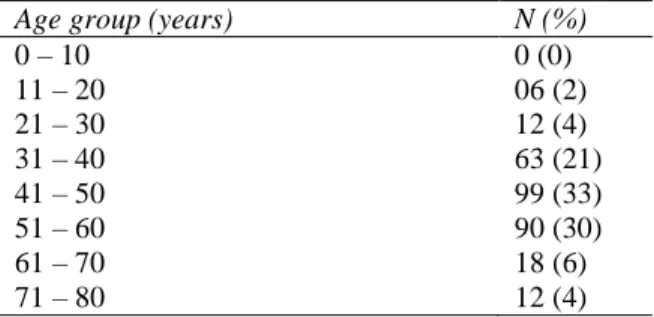
Figure 1 Different clinical types of candidiasis 6
11 7
8
0 2 4 6 8 10 12
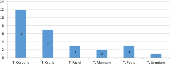
Figure 2 Different clinical types of Dermatophytic infections
Figure 3 Diabetic foot ulcers
Figure 4 Extensive xanthelasma
(32, 10.7%), retinopathy (13, 4.3%) and nephropathy (9, 3.3%). Out of 300 patients, 12% patients were taking human insulin, 60% were on oral sulfonylureas alone and 28% both oral sulfonylureas and biguanides.
Various mucocutaneous manifestations observed in diabetics are shown in Table 2. Different types of infections and infestations were commonest finding seen 118 (39.3%) patients.
Amongst fungal infections, candidiasis (Figure 1) was the most frequent fungal infection
pityriasis versicolor. 7.4% patients presented with bacterial infections. Folliculitis and furuncles were the common bacterial infections. Viral infections and infestations were relatively less frequent (Table 2). Noninfectious manifestations included acrochordons (53, 17.7%), xerosis (41, 13.7%), eczema (19, 6.3%), diabetic foot ulcers (14, 4.7%) [Figure 3], xanthelasma (11, 3.7%) [Figure 4] and vitiligo (7, 2.3%). Other rare conditions are shown in
Table 2.
12
7
3 2 3
1 0
2 4 6 8 10 12 14
Discussion
Cutaneous signs of DM are extremely valuable to the clinician. They generally appear after the primary disease has developed, but may signal or appear coincidentally with its onset, or even precede diabetes by many years.10 Cutaneous
manifestations of diabetes are classified into
four categories: skin lesions with a strong-to weak association with diabetes (necrobiosis lipoidica, diabetic dermopathy, diabetic bullae, yellow skin, eruptive xanthomas, perforating disorders, acanthosis nigricans, oral leucoplakia, lichen planus); infections (bacterial, fungal); cutaneous manifestations of diabetic complications (microangiopathy, macroangiopathy, neuropathy); and skin reactions to diabetic treatment (sulfonylureas or insulin).11
In our study common skin disorders associated with diabetes were acrochordons (17.7%), xerosis (13.7%), candidal infection (10.3%), dermatophytic infections (9%), eczemas (6.3%), acanthosis (5.3%), diabetic foot (4.7%), pityriasis versicolor (4.7%), xanthelasma (3.7%) and vitiligo (2.3%).
In this study, skin tags (acrochordons) were the most common manifestations reported in 17.7% of the patients whereas other studies reported 3.7%,12 32%,10 and 40.9%.13 High insulin levels
stimulate keratinocyte proliferation, resulting in the growth of these lesions, and acrochordons may be a cutaneous marker for impaired carbohydrate metabolism.14
Xerosis was second most common disorder associated with diabetes seen in 13.7% in our study. Goyal et al.10 and Shahzad et al.14 reported
xerosis in 44% and 36.9%, respectively. In DM, dry skin occurs due to impaired skin barrier function and hypohidrosis, which may lower the
threshold for itching.15 The chronic
hyperglycemic condition causes marked decrease in stratum corneum hydration proportional to the disease duration, leading to xerosis.
It is well-known that diabetic patients are susceptible to infections, probably due to hyperglycemia and defects in polymorphonuclear leukocyte function. Among fungal infections, candidal infection was most commonly reported (10.3%) followed by dermatophytic infection (9%) and pityriasis versicolor (4.7%), while among bacterial infections, folliculitis (3.7%) and furuncle (2.7%) were mainly reported. Goyal et al.10
reported that fungal infections were seen in 16% of the patients (9% had candidiasis and 7% had dermatophytosis) while bacterial infections were seen in 15% of the patients. Shahzad et al.14
reported fungal and bacterial infections in 28.1% and 5%, respectively. Viral (herpes zoster, wart) and parasitic infections (scabies, pediculosis) in diabetic patients were found in 13 and 11 cases, respectively. Vahora et al.16
reported viral and parasitic infestations in 3% and 2.3% cases, respectively. As shown previously, cutaneous infections work as an important marker to diagnose DM.
Acanthosis was observed in 5.3% of the patients while Sanad et al.17 reported 3% and Goyal et
al.10 reported 8% cases. Acanthosis nigricans is
believed to evolve from a complex mechanism ultimately resulting in the interaction between excess insulin and insulin-like growth factor-1 receptor present on keratinocytes and fibroblasts. This interaction stimulates epidermal cell proliferation, leading to the clinical manifestation of hyperkeratosis and acanthosis.18
foot ulcers during their lifetime.19 Although
accurate figures are difficult to obtain for the prevalence of diabetic foot ulcers, the prevalence of this complication ranges from 4%-27%.20-22
The pathophysiology of diabetic foot ulcers has neuropathic, vascular, and immune system components, which all show a base relationship with the hyperglycemic state of diabetes.23,24
Xanthelasma was reported in 3.7% of the patients. Goyal et al.10 and Sanad et al.17 reported
a figure of 10% in their studies. Diabetic patients often suffer from high lipid (cholesterol and triglycerides) levels in the blood. This causes fat to be deposited in the skin and presents as xanthomas or xanthelasma.
Vitiligo was reported in 2.3% of our patients while Vahora et al.16 reported 3.33% and Ahmed
et al.12 reported 5.7% vitiligo patients. Vitiligo
and diabetes may have a causal relationship and both are associated with autoimmunity. Familial hereditary tendencies occur in both diseases. There are neuropathic complications in diabetes, and in vitiligo, a dermatodermal variety occurs with evidence of degenerated nerve endings. In diabetes, the products of oxidative stress, free radical generation, and release of various growth factors may be cytotoxic, affecting melanogenesis.25
In this study, the prevalence of skin manifestations was higher in type II than in type I diabetic patients, especially cutaneous infections, and as the duration of diabetes increased, the likelihood of developing skin manifestations also increased.
Conclusion
In conclusion, this study sheds light on the importance of the early detection and the understanding of the pathogenesis of skin
properly manage the complications and prevent disability. Patient with multiple skin manifestations must be evaluated for DM. The frequency of cutaneous infections, as well as, diabetes-related dermatoses was higher among the diabetics than among nondiabetics. A good glycemic control definitely reduces the incidence and severity of cutaneous disorders. Long-term effects of DM on the microcirculation and on dermal collagen eventually result in skin disorders in almost all the diabetic patients. Thus, dermatologists play an important role in reducing the dermatologic morbidity, improvement of quality of life, and management strategy.
References
1. Joshi SR, Parikh RM. India-diabetes capital of the world: now heading towards hypertension. J Assoc Physicians India. 2007;55:323-4.
2. Kumar A, Goel MK, Jain RB, Khanna P, Chaudhary V. India towards diabetes control: Key issues. Australas Med J. 2013;6:524-31.
3. Park K, editor. Preventive medicine in obstetrics, paediatrics and geriatrics. In: Park K, editor. Park’s Textbook of Preventive and Social Medicine. 24th edn. Jabalpur: M/S Banarsidas Bhanot Publishers; 2017. P. 412.
4. Wild S, Roglic G, Green A, Sicree R, King H. Global prevalence of diabetes- estimates for the year 2000 and projections for 2030. Diabetes Care. 2004;27:1047-53.
5. Sicree R, Shaw J, Zimmet P. Diabetes and impaired glucose tolerance. In: Gan D, editor. Diabetes Atlas. International Diabetes federation. 3rd ed. Brussels, Belgium: International Diabetes Federation; 2006. P. 15-103.
6. WHO (1980). Techn. Rep. Ser., No. 646. 7. Gkogkolou P, Bohm M. Skin disorders in
diabetes mellitus. J Dtsch Dermatol Ges. 2014;12:847-64. doi:10.1111/ddg.12424 8. Bhat YJ, Gupta V, Kudyar RP. Cutaneous
in Endocrine, Metabolic, Nutritional and Deposition Diseases. Berlin: Springer Science; 2010.
10. Goyal A, Raina S, Kaushal SS, Mahajan V, Sharma NL. Pattern of cutaneous manifestations in diabetes mellitus. Indian J Dermatol. 2010;55:39-41.
11. Romano G, Moretti G, Di Benedetto A, Giofre C, Di Cesare E Russo G et al. Skin lesions in diabetes mellitus: Prevalence and clinical correlations. Diabetes Res Clin Pract. 1998;39:101-6.
12. Ahmed K, Muhammad Z, Qayum I. Prevalence of cutaneous manifestation of diabetes mellitus. J Ayyub Med Coll Abbottabad. 2009;21:76-9.
13. Shahzad M, Al Robaee A, Al Shobaili HA, Alzolibani AA, Al Marshood AA, Al Moteri B et al. Skin manifestations in diabetic patients attending a diabetic clinic in the Qassim region, Saudi Arabia. Med Princ Pract. 2011;20:137-41.
14. Rasi A, Soltani-Arabshahi R, Shahbazi N. Skin tag as a cutaneous marker for impaired carbohydrate metabolism: A case-control study. Int J Dermatol. 2007;46:1155-9. 15. Yamaoka H, Saski H, Yamasaki H, Oqawa
K, Ohta T, Furuta H et al. Truncal pruritus of unknown origin may be a symptom of diabetic polyneuropathy. Diabetes Care. 2010;33:150-5.
16. Vahora R, Thakkar S, Marfatia Y. Skin, a mirror reflecting diabetes mellitus: A longitudinal study in a tertiary care hospital in Gujarat. Indian J Endocr Metab. 2013;17:659-64.
17. Sanad EM, El Fangary MM, Sorour NE, El Nemisy NM. Skin manifestations in Egyptian diabetic patients: a case series study. Egypt J Dermatol Venereol. 2013;33:56-62.
18. Levy L, Zeichner JA. Dermatologic manifestation of diabetes. J Diabetes. 2012;4:68-76.
19. Leone S, Pascale R, Vitale M, Esposito S. Epidemiology of diabetic foot. Infez Med. 2012;20 Suppl 1:8-13.
20. Richard JL, Schuldiner S. Epidemiology of diabetic foot problems. Rev Med Interne. 2008;29 Suppl 2:S222-S230.
21. Nather A, Bee CS, Huak CY, Chew JL, Lin CB, Neo S et al. Epidemiology of diabetic foot problems and predictive factors for limb loss. J Diabetes Complications. 2008;22 :77-82.
22. Bakri FG, Allan AH, Khader YS, Younes NA, Ajlouni KM. Prevalence of diabetic foot ulcer and its associated risk factors among diabetic patients in Jordan. J Med J. 2012;46:118-25.
23. Clayton W Jr, Elasy TA. A review of the pathophysiology, classification and treatment of foot ulcers in diabetic patients. Clinical Diabetes. 2009;27:52-8.
24. Wolf G. New insights into the pathophysiology of diabetic nephropathy: from haemodynamics to molecular pathology. Eur J Clin Invest. 2004;34 :785-96.