R E V I E W A R T I C L E
Concomitant mandibular hypo-hyperdontia: Report of two
rarest cases with the literature review
N. B. Nagaveni, Meghna Bajaj, Kirthiga Muthusamy, P. Poornima, Suryakanth M. Pai, V. V. Subba Reddy
Department of Pedodontics and Preventive Dentistry, College of Dental Sciences, Davangere, Karnataka, India
Abstract
Concomitant occurrence of both hypodontia (congenital tooth agenesis) and hyperdontia (supernumerary tooth) in the same dental arch is an extremely rare dental anomaly. Literature search shows very few cases of this anomalous condition with all cases depicting the unilateral presence of supernumerary tooth. Therefore, the intention of the current article is to report two cases of concomitant occurrence of mandibular both hypo-hyperdontia. In that one case exhibited bilateral occurrence of mesiodens teeth in the midline of mandible with associated agenesis of permanent both central incisors and taurodontism in permanent molars, which is not published so far. The article also provides comprehensive literature review on this rarest clinical entity.
Keywords: Mandibular mesiodens, supernumerary tooth, tooth agenesis
Correspondence
Dr. N.B. Nagaveni, Department of Pedodontics and Preventive Dentistry, College of Dental Sciences, Davangere, Karnataka, India. Email: [email protected]
Received 16 December 2014; Accepted 18 January 2015
doi: 10.15713/ins.ijcdmr.22
How to cite the article:
N. B. Nagaveni, Meghna Bajaj, Kirthiga Muthusamy, P. Poornima, Suryakanth M. Pai, V. V. Subba Reddy, “Concomitant mandibular hypo-hyperdontia: Report of two rarest cases with literature review”, Int J Contemp Dent Med Rev, Vol. 2014, Article ID: 091214, 2014. doi: 10.15713/ins.ijcdmr.22
Introduction
Thorough clinical knowledge pertaining to the tooth development and chronology of eruption is necessary to understand the occurrence of various dental anomalies. Supernumerary tooth (hyperdontia) is an extra tooth compared with the normal tooth number count and these can develop as single, multiple, can be seen unilateral or bilateral and be found in either maxillary arch or mandibular or both.[1]
The frequency of occurrence of these teeth is 8.2-10 times more in the maxilla compared to mandible. In maxilla, the pre-maxillary region involving the incisor area (90%) is the frequently aff ected part. Other regions seen with a supernumerary tooth in decreasing order are distomolars (mandibular or maxillary fourth molars), premolars and lateral incisors.[2] The existence of extra teeth in the mandibular arch is extremely rare and very few cases have been reported [Table 1].[3-27]
Missing teeth due to tooth agenesis also known as hypodontia is one more developmental defect in the normal number of teeth and most commonly involving the permanent teeth in children.[28] Congenital agenesis of mandibular central incisors is a rare fi nding, and few cases have been reported as given in Table 2.[7-34]
The term “concomitant hypo-hyperdontia (CHH)” is used to state the occurrence of both diff erent clinical entities within the
same patient. This condition is also called as “oligopleiodontia” as suggested by Nathanail.[35] Later, Gibson[27] used the new term only “hypohyperdontia” to overcome the familiarity with the term “concomitant.”
The reported prevalence of hypohyperdontia is between 0.002% and 3.1%, and most of these were of CHH involving both the arches.[27] Males were more commonly aff ected with this condition. However, there is no particular gender predominance associated with this condition. This dental numerical variation has been reported more commonly in a permanent dentition compared with the primary dentition.[27]
Case Reports
Case report 1
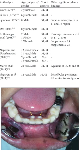
A 9-year-old male patient accompanied by his father, visited to the Department of Pedodontics and Preventive Dentistry complaining of decayed tooth in lower left back region and grossly decayed tooth in upper right and left back region of jaw along with loosening of teeth in lower front region of jaw, with no history of pain associated with any of the mentioned fi ndings. Medical history and family history were unremarkable. There was not history of consanguineous marriage. History of any trauma, infections or systemic disorders was not found. Intra-oral examination revealed that the patient was in mixed dentition period. Oral hygiene was adequate and there were over retained deciduous mandibular central incisors (FDI 71 and 81) [Figure 1] and mandibular permanent both central incisors (FDI 31 and 41) appeared clinically missing [Figure 2].
On radiographic examination, the panoramic radiograph revealed that conical shaped teeth were present without complete root formation and with no periapical changes; congenital agenesis of permanent right and left central incisors was also confi rmed [Figure 3]. Taurodontism was evident in all four permanent fi rst molars of the patient. Complete physical examination and clinical investigations were carried out to rule out any syndromic features.
Based on the appearance and size the teeth were diagnosed as bilateral mandibular supernumerary teeth. Other fi ndings were normal considering the patient’s age. Based on history, clinical and radiographic examinations diagnosis of concomitant Figure 1: Intraoral photograph showing over retained primary central incisors and lingually erupting conical shaped supernumerary teeth (arrows)
Table 1: Reported cases of mandibular mesiodens in the literature Author Year Cases reported
De Jonge[3] 1965 Mandibular mesiodens
De Jonge[4] 1966 Partially erupted mandibular mesiodens
Boer[5] 1968 Partially erupted mandibular mesiodens
Furman and Williams[6] 1970 Mandibular lateral incisor
Spyropoulos et al.[7] 1979 Mandibular incisor
Ranta[8] 1983 Supplemental 72, 32
Pilley[9] 1989 Mandibular mesiodens
Macpherson[10] 1991 Supernumerary teeth in 83, 82, 72 region
Peyrano and Zmener[11] 1995 Mandibular mesiodens
Chow and O’Donnell[12] 1997 Supernumerary teeth in 32 and 42 region
Tanaka et al.[13] 1998 Bilateral mandibular mesiodens
Heathcote[14] 1999 Bilateral mandibular mesiodens
Sharma[15] 2001 Mandibular mesiodens
Cassia et al.[16] 2004 Mandibular mesiodens
Oncag et al.[17] 2005 Mandibular mesiodens
Alencar et al.[18] 2005 Mandibular mesiodens
Yokose et al.[19] 2006 Two cases: Mandibular mesiodens
Cho[20] 2006 Two cases: Supplemental mandibular mesiodens
Das et al.[21] 2006 Partial anodontia and mandibular mesiodens
Cho[22] 2006 Mandibular mesiodens
Zengin et al.[23] 2007 Mandibular mesiodens
Schmuckli et al.[24] 2010 Mandibular mesiodens
Nagaveni et al.[25] 2010 Mandibular mesiodens
Bargale and Kiran[26] 2011 Mandibular mesiodens
Naganahalli et al.[27] 2013 Supplemental mesiodens
Table 2: Documented cases of agenesis of mandibular central incisors
Author/year Age (in years)/ gender
Tooth agenesis
Other signifi cant dental fi ndings
Low (1977)[28] 7 year/Male 31, 41
Gibson (1979)[29] 6 year/Female 31, 41
-Symons (1992)[30] 8/Male 31, 41 Supernumerary teeth in 11 and 13 region
Das (2006)[18] 8 year/Female 31, 41
-Anthonappa et al. (2008)[31]
7/Male 11/Male 12/Female
31, 41 32 31, 41
Two supernumerary teeth in 11, 21 area
Supplemental 15 Supplemental 23
Nagaveni and Umashankara (2009)[32]
12 year/Female 11 year/Male 9 year/Female 13 year/Female
31, 41 31, 41 31,41 31,41
-Marya et al. (2011)[33]
20 year/Male 31, 41 Agenesis of 18, 28 and 48
Nagaveni et al. (2011)[34]
mandibular hypo-hyperdontia was made. The patient’s parents were informed about the condition. Esthetics was given priority and was advised rehabilitation for the anterior teeth with composite resin once it erupts completely.
Complete oral prophylaxis along with extraction of over retained mandibular deciduous central incisors (FDI 1 and 81) was done in fi rst appointment and also extraction of 54 and 64 (FDI tooth notation) was done in next appointment followed by space maintainer; pulpectomy, followed by stainless steel crown in relation to 75 was done in consecutive appointments and the patient has been kept under regular follow-up.
Case report 2
A 13-year-old female patient visited to the Department of College of Dental Sciences, with the chief complaint of labially placed upper front teeth. The medical history was not signifi cant, and the general health was found normal. On intra-oral examination, patient exhibited complete permanent dentition (excluding second and third molars), with the presence of two supernumerary teeth in the mandibular anterior region exactly in the midline. On careful examination, we also noticed the absence of permanent mandibular central incisors [Figure 4]. The Orthopantomograph showed the agenesis of mandibular two central incisors (both right and left) and the presence of a supernumerary tooth in the midline that was of conical shape with fully developed root [Figure 5]. The patient was advised oral prophylaxis and orthodontic treatment for the anterior teeth proclination. As the patient did not show any concern for the mandibular mesiodens, no treatment was done.
Discussion
According to the detailed survey done by Gibson, in 1979,[29] hypo-hyperdontia can be divided into pre-maxillary, maxillary, mandibular and bi-maxillary hypo-hyperdontia based on the site of occurrence. Among these, co-existence of hypo-hyperdontia in the mandibular anterior region is rarely seen clinical phenomenon. This is most commonly observed in the Figure 2: Photograph showing bilateral conical shaped mesiodens
aft er extraction of primary incisors (arrows)
Figure 3: Orthopantomograph illustrating congenital agenesis of permanent mandibular both central incisors, bilateral mesiodens and taurodontism in permanent molars
Table 3: Reported cases of CMHH
Case no. Author/year Age (in years)/gender Hypodontia Hyperdontia
1. Low (1977)[28] 7/Male 31, 41 Unilateral mesiodens
2. Gibson (1979)[29] 8/Female 31, 41 Unilateral mesiodens
2. Das et al. (2006)[18] 8/Female 31, 41 Unilateral mesiodens
3. Raghavan (2009)[35] 8.5/Female 31, 41 Unilateral mesiodens
4. Nuvvula et al. (2010)[36] 15/Female 31, 41 Unilateral mesiodens
5. Nayak et al. (2010)[37] 15.5/Male 31, 41 Unilateral mesiodens
6. Venkataraghavan et al. (2011)[38] 9/Female 31, 41 Unilateral mesiodens
7. Verma et al. (2012)[39] 9/Male 31, 41 Unilateral mesiodens
8. Marya et al. (2012)[33] 20/Male 31, 41 Unilateral mesiodens
9. Nirmala et al. (2013)[40] 10.9/Male 9.2/Male 8.5/Female
41 41 41
Unilateral mesiodens Unilateral mesiodens Unilateral mesiodens
10. Present authors (2014) 9/Male
13/Female
31, 41 31, 41
Bilateral mesiodens Unilateral mesiodens
permanent dentition than the primary dentition.[29] The fi rst report on this rarity was given by Low in 1977.[28] After this, only eight clinical reports are available in the literature. All cases are reported in the mixed dentition except Marya et al.[33] case, which is reported in a permanent dentition. Recently, Nirmala et al.[40] reported three cases of CHH in Indian patients. Our two cases found in mixed dentition. However, our fi rst case exhibited an incomplete root formation in the mesiodentes. This fi nding is also reported in a case published by Das et al.[29] This case is unique as it shows bilateral presence of supernumerary tooth in association with bilateral agenesis of permanent central incisors and taurodontism involving all permanent molars.
There is no exact etiology for CHH and it is not clearly mentioned in the literature. However, environmental and genetic factors have been suspected to explain the occurrence of these anomalies.[31] A recent study from Poland[38] suggests that CHH is a rare phenomenon in association with predominant hypodontia. In addition, CHH has been found in patients with Dubowitz syndrome, Ellis–van Creveld syndrome, Down syndrome, G/BBB syndrome, Marfan syndrome, cleft lip and palate and fucosidosis.[18,20] However, all published cases on the hypo-hyperdontia involving mandibular anterior region including the present cases does not show any signifi cant
systemic abnormality or syndrome.[19,28,36-40] This suggests that these cases are sporadic and may be due to a specifi c gene mutation that may be enabling the hypodontia of novel dentition and encouraging odontogenesis of the supernumerary teeth.
Occurrence of supernumerary tooth in the mandibular anterior segment is a rare entity. Primosch[41] described two types of mesiodens as supplemental and rudimentary. Supplemental mesiodens resemble normal tooth and are also named as incisiform. Rudimentary mesiodens are again categorized as three types based on morphology as conical, tuberculate and molariform. Conical mesiodens are generally solitary, have completely formed roots and are usually seen palatal to maxillary central incisors. However, Nagaveni et al. in 2010, reported a unique case of multilobed mesiodens in the maxillary arch.[42] Ribbons[43] reported that the most of the supernumerary teeth seen in the mandibular anterior part are of supplemental type (2%). The same fi nding was also showed in the cases published by Marya et al.[33] and Nirmala et al.[40] However, the maximum frequency of occurrence of mesiodens in the mandibular front region reported belong to conical shape (56%). The patients described in the current report too exhibited conical shaped mesiodens. Moreover, the fi rst case is diff erent from other previous reported cases as it shows bilateral midline supernumerary teeth in association with missing both central incisors.
There is a tendency for dental agenesis to occur more unilaterally than bilaterally. Literature review shows few cases of bilateral agenesis of mandibular central incisors than unilateral cases [Table 2].[28,34] In 2009, Nagaveni and Umashankara,[32] reported four cases of bilateral agenesis of permanent mandibular central incisors in Indian patients.
Many authors have researched the association between
taurodontism and hypodontia. Seow and Lai[44] reported
34.8% of patients showed association between hypodontia and taurodontism. Whereas two Indian reports[33,40] shows association between taurodontism and CHH. Similarly, our fi rst case too, along with hypohyperdonita exhibited taurodontism of both permanent and primary molars. Contrarily, reports published by Marya et al.[33] and Nuvvula et al.[37] shows agenesis of maxillary and mandibular third molars. Unfortunately, this fi nding cannot be compared with the present as well as all other previous reports because the age in other published cases varies from young to old age (5-20 years). Apart from this, most recently double tooth and dens evaginatus have been reported in association with CHH.[13,45]
The diagnosis of CHH is critical sometimes especially in a growing child. In the fi rst case presented here, at fi rst clinical examination, it was diffi cult to diagnose the condition as the tooth appeared entirely diff erent in size and shape from normal appearance of mandibular central incisor. It created dilemma whether the tooth is a normal central incisor or a conical mesiodens with agenesis of permanent incisors. Careful clinical examination in addition to thorough radiographic evaluation helped us to diagnose fi nally as a mesiodens tooth not the central incisor. Moreover, literature evidence revealed no data on the Figure 4: Photograph showing conical shaped mesiodens with
missing permanent both central incisors
unusual appearance, i.e., microdontic or distinct conical shape of the mandibular central incisors, except for mesiodens tooth.
CHH in the mandibular anterior region can interfere with dental and facial aesthetics by causing a shift in the midline and hence requires correct treatment. The management of CHH is quite challenging as it requires a multidisciplinary team approach and treatment depends on the various stages of developing dentition and varies from individual to individual.[19,28,36-40] Therefore, careful treatment planning is highly essential in order to deal with immediate, as well as long-term complications.
Although literature shows no standardized treatment protocols, in the current scenario, extraction of mesiodens and closing the space with fi xed orthodontics, no treatment, extraction of mesiodens, followed by placing a Maryland bridge as short-term appliance, composite build up to restore as incisor and implants in the future are the suggested rehabilitation options in the literature. In the fi rst case no treatment was carried out as mesiodentes were still in erupting stage.[33,40] The possible treatment options were explained to the parents and patient is still under follow-up. In the second patient, parents did not show any concern for the mesiodens. Therefore, no treatment was carried out.
Conclusion
• The occurrence of both hypodontia (agenesis of bilateral permanent mandibular central incisors) and bilateral midline supernumerary tooth in the same dental arch especially in the mandibular anterior segment is an uncommon and extremely rare dental anomaly
• Careful clinical examination along with detailed radiographic evaluation enhances the diagnosis of this condition, which ultimately helps to provide appropriate treatment for an individual.
References
1. Gündüz K, Celenk P, Zengin Z, Sümer P. Mesiodens: a radiographic study in children. J Oral Sci 2008;50:287-91. 2. Scheiner MA, Sampson WJ. Supernumerary teeth: a review of
the literature and four case reports. Aust Dent J 1997;42:160-5. 3. de Jonge TE. Th e mandibular mesiodens. Ned Tijdschr
Tandheelkd 1965;72:95-101.
4. de Jonge TE. Th e mandibular mesiodens. SSO Schweiz Monatsschr Zahnheilkd 1966;76:14-9.
5. Boer JG. A mandibular mesiodens. Ned Tijdschr Tandheelkd 1968;75:258-63.
6. Furman RL, Williams JJ. A supernumerary mandibular lateral incisor. Oral Surg Oral Med Oral Pathol 1970;29:395.
7. Spyropoulos ND, Patsakas AJ, Angelopoulos AP. Simultaneous presence of partial anodontia and supernumerary teeth. Oral Surg Oral Med Oral Pathol 1979;48:53-6.
8. Ranta R. Premature mineralization of permanent canines associated with aplasia of their primary predecessors: report of four cases. ASDC J Dent Child 1983;50:274-7.
9. Pilley JR. An unusual mandibular supernumerary. Dent Update 1989;16:215-6.
10. Macpherson DW. Dental anomalies in fucosidosis. Br Dent J 1991;170:408-10.
11. Peyrano A, Zmener O. Endodontic management of mandibular lateral incisor fused with supernumerary tooth. Endod Dent Traumatol 1995;11:196-8.
12. Chow KM, O’Donnell D. Concomitant occurrence of hypodontia and supernumerary teeth in a patient with Down syndrome. Spec Care Dentist 1997;17:54-7.
13. Tanaka S, Murakami Y, Fukami M, Nakano K, Fujisawa S, Miyoshi S. A rare case of bilateral supernumerary teeth in the mandibular incisors. Br Dent J 1998;185:386-8.
14. Heathcote R. Supernumaries in the mandibular incisors. Br Dent J 1999;186:602.
15. Sharma SA. Mandibular midline supernumerary tooth: a case report. J Indian Soc Pedod Prev Dent 2001;19:143-4.
16. Cassia A, El-Toum S, Feki A, Megarbane A. Five mandibular incisors: an autosomal recessive trait? Br Dent J 2004;197:307-9.
17. Alencar M, Duarte D, Cury P, Bönecker M. Lower mesiodens: report of an unusual case. J Clin Pediatr Dent 2005;29:353-5. 18. Onçag O, Candan U, Arikan F. Comprehensive therapy of a
fusion between a mandibular lateral incisor and supernumerary tooth: case report. Int Dent J 2005;55:213-6.
19. Yokose T, Sakamoto T, Sueishi K, Yatabe K, Tsujino K, Kubo S, et al. Two cases with supernumerary teeth in lower incisor region. Bull Tokyo Dent Coll 2006;47:19-23.
20. Cho SY. Supplemental mandibular permanent incisor teeth: Report of two cases. Prim Dent Care 2006;13:76-8.
21. Das G, Sarkar S, Bhattacharya B, Saha N. Coexistent partial anodontia and supernumerary tooth in the mandibular arch: a rare case. J Indian Soc Pedod Prev Dent 2006;24 Suppl 1:S33-4. 22. Cho SY. Six mandibular permanent incisors: report of a case.
Gen Dent 2006;54:428-30.
23. Zengin AZ. Mandibular mesiodens. Br Dent J 2007;202:644. 24. Schmuckli R, Lipowsky C, Peltomäki T. Prevalence and
morphology of supernumerary teeth in the population of a Swiss community. Short communication. Schweiz Monatsschr Zahnmed 2010;120:987-93.
25. Nagaveni NB, Sreedevi B, Praveen BS, Praveen Reddy B, Vidyullatha BG, Umashankara KV. Survey of mesiodens and its characteristics in 2500 children of Davangere city, India. Eur J Paediatr Dent 2010;11:185-8.
26. Bargale SD, Kiran SD. Non-syndromic occurrence of true generalized microdontia with mandibular mesiodens - A rare case. Head Face Med 2011;7:19.
27. Naganahalli M, Honnappa A, Chaitanya NC. Supplemental mandibular mesiodens: a diagnostic challenge. J Clin Diagn Res 2013;7:3077-8.
28. Low T. Hypodontia and supernumerary tooth: report of a case and its management. Br J Orthod 1977;4:187-90.
29. Gibson AC. Concomitant hypo-hyperodontia. Br J Orthod 1979;6:101-5.
30. Symons AL. Ectopic eruption of a maxillary canine following trauma. Endod Dent Traumatol 1992;8:255-8.
31. Anthonappa RP, Lee CK, Yiu CK, King NM. Hypohyperdontia: literature review and report of seven cases. Oral Surg Oral Med Oral Pathol Oral Radiol Endod 2008;106:e24-30.
33. Marya CM, Sharma G, Parashar VP, Dahiya V, Gupta A. Mandibular midline supernumerary tooth associated with agenesis of permanent central incisors: a diagnostic conundrum. Stomatologija 2012;14:65-8.
34. Nagaveni NB, Radhika NB, Umashankara KV, Satisha TS. Concomitant occurrence of canine transmigration and symmetrical agenesis of incisors – A case report. Bangladesh J Med Sci 2011;10:133-6.
35. Raghavan VH. Mandibular mesiodens with agenesis of central incisors – A rare association. Niger Dent J 2009;17:27-8.
36. Nuvvula S, Kiranmayi M, Shilpa G, Nirmala SV. Hypohyperdontia: Agenesis of three third molars and mandibular centrals associated with midline supernumerary tooth in mandible. Contemp Clin Dent 2010;1:136-41.
37. Nayak AG, Chhaparwal Y, Pai KM, Lele AS. Non-syndromic hypo-hyperdontia of the permanent dentition with involvement of the mandibular anterior region: a rare occurrence. Rev Clin Pesqui Odontol 2010;6:281-4.
38. Venkataraghavan K, Muralikrishnan B, Anantharaj A. Mandibular mesiodens with agenesis of central incisors
(hypohyperdontia): A case report and review. Int J Contemp Dent 2011;2:26-30.
39. Verma KG, Verma P, Rishi S. Case report: a rare occurrence of non-syndromic hypo-hyperdontia in the mandibular anterior region. Eur Arch Paediatr Dent 2012;13:47-9.
40. Nirmala SV, Sandeep C, Nuvvula S, Mallineni SK. Mandibular hypo-hyperdontia: A report of three cases. J Int Soc Prev Community Dent 2013;3:92-6.
41. Primosch RE. Anterior supernumerary teeth – assessment and surgical intervention in children. Pediatr Dent 1981;3:204-15. 42. Nagaveni NB, Umashankara KV, Sreedevi, Reddy BP, Radhika
NB, Satisha TS. Multi-lobed mesiodens with a palatal talon cusp: a rare case report. Braz Dent J 2010;21:375-8.
43. Ribbons JW. An incisiform supernumerary in an unusual site. A case report. Br Dent J 1970;129:174.
44. Seow WK, Lai PY. Association of taurodontism with hypodontia: a controlled study. Pediatr Dent 1989;11:214-9.