Journal of Global Pharma Technology
Available Online at:
www.jgpt.co.in
RESEARCH ARTICLE
Determination of Clarithromycin and its Applications in
Pharmaceutical Samples Using New Selective Electrodes
Sarra A. Abrahem
1*, Suhad A. Ibrahim1, Abdul kader S. Abdul kader
21. Dept. of Chemistry, College of Science at Al-Nahrain University in Baghdad, Iraq.
2. Dept. of Physics, College of Science at Mustansiriyah University in Baghdad, Iraq.
*Corresponding Author: Sarra A. Abrahem
Abstract
Two clarithromycin electrodes were constructed using clarithromycin-methyl orange as an ion pair complex in a Polyvinyl-chloride (PVC) matrix. These electrodes were plasticized using two plasticizers, Acetophenone (AP) and Di-butyl phosphate (DBP).Clarithromycin electrodes, a and b, gave slopes of 53.95 and 51.50mV/decade, and linear which ranged from (10-4-10-1, and 6×10⁻⁴-10-1 M) respectively. Electrode (a) relied on using a plasticizer (DBP) of a slope 53.95mV/decade, and a correlation coefficient 0.9996, with a detection limit of 5x10⁻⁵M in a lifetime of 26 days. The proposed electrode showed reproducibility and a good stability. It was therefore employed for determining clarithromycin within pharmaceutical samples. The measurements of interferences in the availability of (Na+, Cu+2, K+, Mn+2, Al+3, Fe+3, gelatin and sucrose) were also studied for selectivity coefficient determination using the separation method and the mixed method.
Keywords: Clarithromycin, Methyl orange, Ion selective electrode.
Introduction
Clarithromycin (CR), 6-o-methyl–
erythromycin or 4-[(2,
6-Dideoxy-3-Cmethyl-3-O-methyl-L-ribohexopyranosyl)
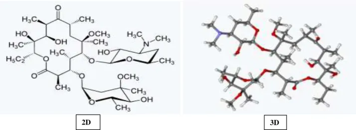
oxy]-14-ethyl-12, 13-dihydroxy-7-methoxy-3, 5, 7, 9, 11, 13-hexamethyl-6-[3, 4, 6-trideoxy-3-(di-methyl amino)-Dxylohexopyranosyl] oxy] oxa-cyclo-tetra-decane-2, 10-dione, which has the empirical formula (C38H69NO13), shown in Figure (1), is a white crystalline powder which of a molecular weight of 748 g/mole, slightly soluble in methanol, soluble in methylene chloride and in acetone, and practically insoluble in water[1].
It is a semi synthetic macrolide antibiotic and it has a good antimicrobial activity against various gram-negative and gram-positive organisms. CR is commonly used to treat Mycoplasmas, Chlamydia species, Rickettsia as well as Haemophilus influenzae [2, 3]. Researchers developed various analytical
methods for the determination of
Clarithromycin in biological samples and formulations, such as High-Performance Liquid Chromatography (HPLC) method [4, 5] and spectrophotometry methods [6,7,8].
In pharmaceutical analysis, applications of ion selective electrodes are still of interest to researchers [9] for these sensors have the advantages of reasonable selectivity, simple operation and design, low cost, fast response, and applicability to colored solutions and turbid [10,11].
Figure 1: Structure formula of Clarithromycin
Experimental Part Equipment
In Lab 740 with terminal 740-WTW which is a digital pH/ion meter, made in Germany, was employed for all pH and potentiometric measurements. Sartorius Handy 4digits Analytical Balance, Hotplate Stirrer type LMS-1003, Daihan Lab tech, pH combination electrodes (SenTix® 82 WTW) made in Germany, Saturated Calomel reference electrode and a Silver - silver chloride wire were also a part of the equipment used in this research.
Reagents and Solutions
Using Claricide tablets, Clarithromycin was extracted relying on the procedure mentioned in the literature [12].These tablets which contains 500 mg of clarithromycin were made in Turkey and were obtained from Bilim pharmaceuticals. Methyl orange (M.O) with a molecular weight of 327.33 g•mol-1, Sodium tetra-phenylborate (NaTPB), and PVC with a slightly high molecular weight, were purchased from Fluka. AP and DOP were purchased from Fluka AG and were made in Switzerland, Tetrahydrofuran (E. Merck), Acetonitrile from Sigma-Aldrich. The rest of solvents and chemicals were obtained from BDH and were of an analytical reagent grade.
-0.1 M stock solutions of KCl, NaCl,
Cu(NO3)2.3H2O, Fe2(SO4)3.9H2O,
AlCl3.6H2O, MnSO4, as well as sucrose and gelatin, were prepared by dissolving 0.3722, 0.2922, 1.2077, 2.8100, 1.2066, 0.7550, 1.7115 and 1.5000 g in distilled water (50 mL), respectively.-0.01 M of Methyl orange (M.O) standard solution was created by dissolving 0.3273 g of pure (M.O) in distilled water, a
0.1 M standard solution of Clarithromycin was prepared by dissolving 3.74 g in water and acetonitrile.
- Using the same solvent, (10-7 - 10-1 M) working solutions of CR were created using a serial-appropriate dilution from the stock solution.
Procedure
Ion-pair Preparation
The ion pair was prepared by mixing 0.01 M solution of Methyl Orange (M.O) dissolved in distilled water with an equimolar CR solution dissolved in acetonitrile and water in a proportion of (1:3). After adding some concentrated drops of hydrochloride acid, a precipitate formed immediately. It took 24 hr for the precipitate to form.
Preparation of Membrane and
Construction of ISE:
A mixture of 0.0400 g of ion pair, 0.1700 g of PVC powder, and 0.3600 g of plasticizer, were dissolved in THF (5 ml) and were stirred to obtain a clear viscous solution. A cut membrane was attached to a tube made of polyethylene in an electrode formation and was attached to the last part of a tube made of glass. Ag/ AgCl wire electrode was used as an interior reference electrode. This electrode was then attached to a double junction Ag/AgCl electrode which was used as an external reference electrode. [13]
Potential Measurements
An electrochemical cell can be presented as the following:
Ag/AgCl/internal filling
solution//membrane//test solution/SCE
standard analyte solutions of (10-7-10-1 M) were used, each electrode constructed a calibration curve. These curves were prepared by plotting concentration (M) log scale versus the potential E using Microsoft office Excel 2010.
Pharmaceutical Samples Preparation
The powder content of 10 tablets containing 500 mg CR was weighted accurately.
Their weight was found to be 5.8692 g. A 0.0878 g of this powder was dissolved in 250 mL acetonitrile and was then filtered. Distilled water was added to the solution until it reached 1 L. The resultant solution is 1×10⁻⁴ M.
Calculation of Selectivity Coefficient
For the coefficient of selectivity
measurement, a method of separate solution [14] was employed and calculated according to the equation below:
Log Kpot A, B = (EB–EA) / S+ (1–zA/zB) logaA (eq.1)
zA, zB are charge numbers; EA, EB are the potentials; and aA is the activities for the primary A ion at aA = aB.
The mixed method (Fixed interference method) [15, 16] was used for the selectivity coefficients which were measured using the equation below:
Kpot A, B=aA/ (aB) zA/zB (eq.2)
Results and Discussion
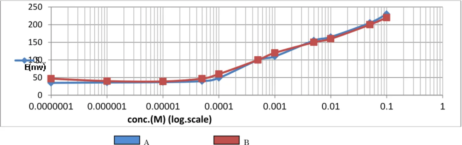
Two ion-selective electrodes of clarithromycin (CR) (a and b) which were based on using CR, M.O, and two plasticizers (DBP and AP) and a PVC matrix were examined respectively. Sensor (a) displayed a linear response which ranged between 10-4 and 10-1 M (CR) and a Nernstian slope of 53.95mV/ decade. The limit of detection at the lower end 5×10⁻⁵M was calculated at the intersection of the extrapolated segments of two linear parts of CR curve. The electrode (a) displayed a slope value due to a strong mixing of (DBP & PVC) can be attributed to the compatibility of DBP used in the electro-active compound in the composition. A typical plot for the calibration curves of electrodes (a & b) based on the two plasticizers AP and DBP shown in the Figure (2).
A B
Figure 2: Calibration curves of clarithromycin ion-selective electrodes using DBP and AP plasticizer
The electrodes which relied on (AP & DBP), (a and b) membranes, gave slopes’ values of (53.95 and 51.50 mV/ decade), correlation coefficients of 0.9996, 0.9989 respectively.
The linear range of electrodes (a & b) were 10-4-10-1 and 6×10⁻⁴-10-1M with detection limits of 5×10⁻⁵ and 2×10⁻⁵M, respectively. The results are shown in Table (1).
Table 1: the parameters for two clarithromycin electrodes
Electrode Slope (mV/Decade)
Correlation coefficient
(r)
Linear concentration
range (M)
Detection limit (M)
Time of Response (seconds) Lifetime (day) 1×10⁻²(M) 1×10⁻³(M) 1×10⁻⁴ (M)
a CR+DBP+
M.O
53.95 0.9996 10-4-10-1 5×10⁻⁵ 24 14 8 26
b
CR +AP+ M.O 51.50 0.9989 6×10⁻⁴-10-1 2×10⁻⁵ 29 17 10 15 0
50 100 150 200 250
0.0000001 0.000001 0.00001 0.0001 0.001 0.01 0.1 1
S…
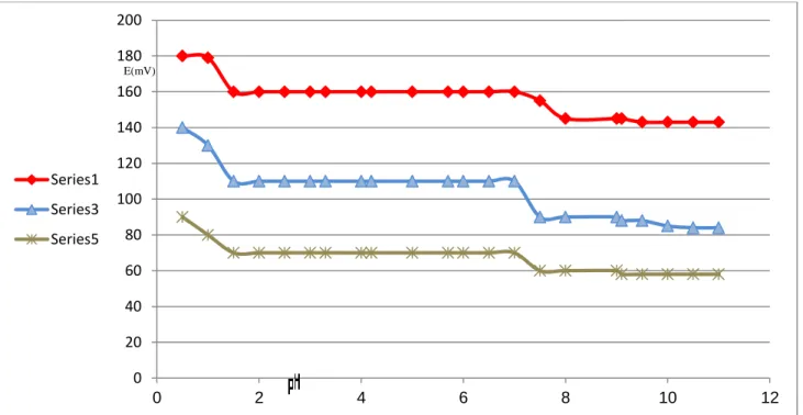
Effect of PH
The pH effect on the potentials of the electrode for CR selective membrane of electrode (a) was studied and analyzed through the measurement of the (E) of the
CR solutions cell at three concentrations (0.01, 0.001, 0.0001) M where the pH range was between (0.5 to 11.0). The pH was adjusted by adding proper amounts of sodium hydroxide solution and/or hydrochloric acid. See Figure (3).
Figure 3: Effect of pH on the potential (E) of the electrode (a) at the concentrations of 0.01, 0.001 and 0.0001M
In high acidity or in pH values less than 1.5, the response obtained from the electrode has irregularly increased, and this response could be because of the response to H+ activities by the electrode, in addition to analyte ions.
Whereas, alkaline solution in which pH is above 7, the electrode response decreased. This response can be the result of the decrease in CR solubility. [14] See Table (2) or the ranges of the working pH.
Table 2: The ranges of the working pH for CR (a) electrode. for (0.01, 0.001, 0.0001) M of CR Electrode Composition of electrode (a) pH range
0.01(M) 0.001 (M) 0.0001 (M)
(a) CR+M.O+DBP 1.5-7.7 1.9-7.6 1.9-7.3
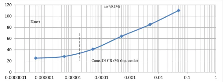
Studies of Interference
To examine the selectivity of membrane (a) selective electrode in relation to CR and with regard to different interfering ions, a separate solution method was used relying on to equation (1), and mixed solution method
relying on equation (2). The selectivity coefficients’ values according to the separate method and the mixed method are listed in Table (3). Figure (4) shows the curve of the calibration of the FIM (fixed interfering method) of CR selective electrode (a) for (Mn+2).
Table 3: Values of Kpot A, B using electrode (a) according to FIM and separate method
Separate method Mixed method
Interfering
ions Log K
potA,B KpotA,B KpotA,B
Gelatin -1.225 5.96×10⁻² 4.01×10⁻⁴
Sucrose -1.123 8.33×10⁻² 6.02×10⁻⁴
Na+ -1.473 3.37×10⁻² 1.01×10⁻²
Fe+3 -3.150 7.10×10⁻⁴ 1.33×10⁻⁴
Al+3 -3.232 5.82×10⁻⁴ 2.64×10⁻⁵
Cu+2 -2.833 1.51×10⁻³ 1.98×10⁻⁴
Mn+2 -2.029 9.28×10⁻³ 6.69×10⁻⁵
0 20 40 60 80 100 120 140 160 180 200
0 2 4 6 8 10 12
Series1
Series3
Series5
Figure 4: Curve of Calibration of FIM clarithromycin selective electrode (a).Where a Mn+2=5*10-2 M, a CR=1.5*10-5M
Sample Analysis
For the determination of clarithromycin, four potentiometric techniques were used. These
include the following: The direct method and
SAM (the standard addition method) using the following equation:
CU=CS /10 ΔE/S [1+ (VU /VS)]-(VU / VS)
Where CU, CS, is the concentrations, VU and VS represent the volumes. U stands for unknown and s is standard solution. MSA (The multiple standard additions) relied on the below equation:
CU = VS × CS / VU
Where CU and CS are the concentration of unknown and standard, respectively, VS stands for the volume added of standard solution. The method was carried out as shown in Figure (5).
Figure 5: Calibration curve of antilog (E/S) against the volume added of standard (10-2 M) for determining 20mL CR solution 10-3 M using (MSA)
The recovery (Re %), relative standard
deviation (RSD %), and relative error (Er %) for each method were calculated. The results are shown in Table (4) below.
Table 4: Analysis of CR using ISE (a) according potentiometric techniques by
Methods Conc.(M) Found(M) RSD*% Re% Er% S X ts √N
Direct method* 1.000×10⁻⁴ 0.993×10⁻⁴ 0.469% 99.3% 0.7% 4.832×10⁻⁷ 0.993×10⁻⁴ 0.600×10⁻⁶
Multi SAM* 1.000×10⁻³ 1.007×10⁻³ --- 100.7% -0.7% --- ---
SAM* 1.000×10⁻³ 1.011×10⁻³ 0.763% 101. 1% -1.1% 7.622×10⁻⁶ 1.011×10⁻³ 0.931×10⁻⁵ 0
20 40 60 80 100 120
0.0000001 0.000001 0.00001 0.0001 0.001 0.01 0.1 1
0 50 100 150 200 250
-3 -2 -1 0 1 2 3
Mn+2(0.1M)
E(mv)
Conc. Of CR (M) (log. scale)
The plotting of the volume of the five additions of the standard clarithromycin versus antilog (E/S) showed the concentration in comparison with the calibration curve of the working range for MSA which was
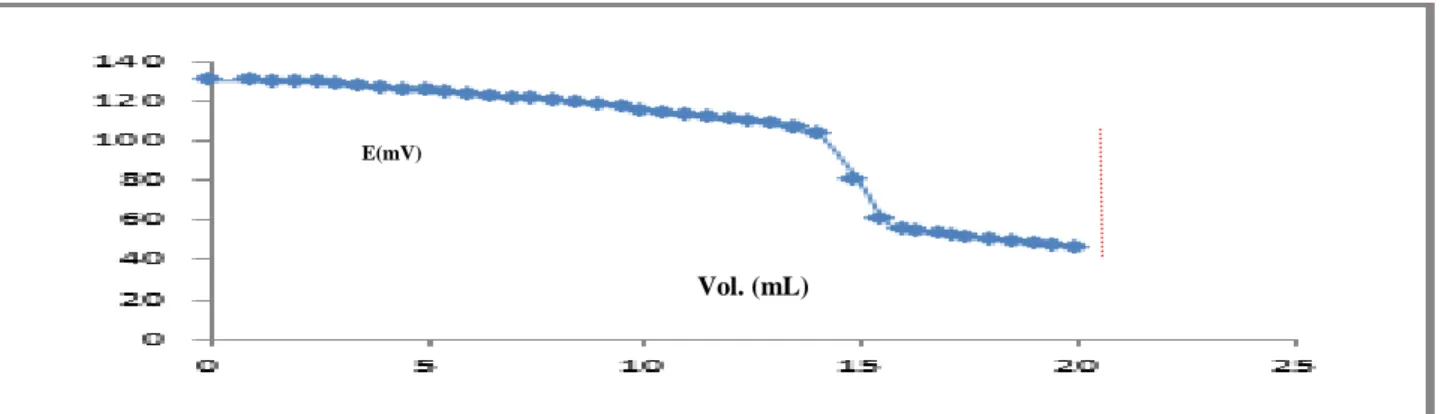
employed in the potentiometric titration method for the determination of CR concentration. Figure (6) below shows a typical titration plot is shown in.
Figure 6: Titration curve of electrode (a) for 15 mL sample solution 10-2 M clarithromycin with 10-2 1M using a titrant solution (TPB)
The recovery (Re %), relative standard
deviation (RSD %), and relative error (Er %) for the titration method were calculated. See table (5) for the results.
Table 5: CR analysis using ISE (a) according to titration method Method used Concentration
(M)
Found(M) RSD*% Re% Er% S X ts √N
Titration method** 1.000×10⁻² 0.990×10⁻² 0.5% 99.0% 1% --- ---
Electrode (a) proved to be useful for clarithromycin potentiometric determination in pharmaceutical preparations. The data obtained for pharmaceutical samples are listed in Table (6) below.
Table 6: Analyses of CR in pharmaceutical samples
Factors Direct method * SAM* Multi SAM * Titration method *** Concentration
(M) 1.000×10⁻³ 1.000 × 10⁻³ 1.000 × 10⁻³ 1.000×10⁻³
Found (M) 0.995×10⁻³ 0.999 × 10⁻³ 1.003 × 10⁻³ 0.998× 10⁻³
RSD * % 0.670% 0.909% - - - 0.920 %
Re % 99.5% 99.9% 100.3% 99.8 %
Er % 0.5% 0.1% -0.3% 0.2 %
S 6.682× 10⁻⁶ 9.069× 10⁻⁶ - - - - - -
X ts √N 0.995×10⁻³ 0.830×10⁻⁵ 1.001×10⁻³ 0.113×10⁻⁴ - - - - - - RSD***% for n=2, t=12.7
RSD**% for n=3, t=4.3 RSD*% for n=5, t=2.7
Conclusions
Sensitive polymer electrodes (a, b) were prepared for the purpose of clarithromycin determination using the ionic complex (clarithromycin-methyl orange) to act as an active substance with PVC in order to form the membrane. These two electrodes gave slopes of (53.95 and 51.50m V/decade) and a linear concentration which ranged between (10-1 -10-4) and (10-1-6 *10-4). The best
electrode for the determination of
clarithromycin in pharmaceuticals was (a)
which used dibutyl phosphate as a plasticizer and gave a slope of 53.95, a correlation coefficient of 0.9996 and a detection limit of 5×10⁻⁵ and lifetime of 26 days with repeatability and good stability. Selectivity coefficient of the ion-selective electrode (the proposed membrane a) towards CR was also examined and studied in this research using a separate solution and a mixed solution method with the following materials and ions, sucrose, Al + 3, Fe +3, Cu + 2, K +, Mn + 2, Na +, and gelatin.
Vol. (mL)
References
1. British pharmacopoeia (2014) “Medicinal
and Pharmaceutical Substances”;
Published by The Stationery Office on behalf of the Medicines and Healthcare products Regulatory Agency (MHRA), 1(2): 1406-1407.
2. K Isadore, FS Michael, BW Roderick
(1998) "Analysis of macrolide antibiotics", Chromatogr. A, 812 (2): 255-286.
3. HD Langtry, RN Brogden (1997)
"Clarithromycin. A review of its efficacy in the treatment of respiratory tract infections in immunocompetent patients", Drugs, 53 (1): 973-1004.
4. A Hossein, A Abolhassan (2005) "Sensitive determination of clarithromycin in human
plasma by high-performance liquid
chromatography with spectrophotometric detection", J. Chrom. B, 817 (2): 193-197. 5. W Li, H Jia, K Zhao (2007) "Determination
of clarithromycin in rat plasma by
HPLC-UV method with pre-column
derivatization", Talanta, 71 (3): 385-390.
6. S Jasmin, MJ Rasul, M Suraya (2008)
"Extractive Spectrophotometric Methods for Determination of Clarithromycin in
Pharmaceutical Formulations Using
Bromothymol Blue and Cresol Red", J. Chin. Chem. Soc., 55 (3): 1107-1112.
7. W Mohamed, R Mohamed, E Manal, F
Muna (2007) "Spectrophotometric
Determination of Four Macrolide
Antibiotics in Pharmaceutical
Formulations and Biological Fluids via Binary Complex Formation with Eosin and Spectrophotometry", AOAC Int., 90 (6): 1579-1589.
8. Y Srinivasa, KP Chowdary, JV
Seshagiriao (2003) "A new
spectrophotometric method for
determination of clarithromycin in
tablets", Int. J. Chem. Sci., 3 (1): 225-226, 2003.
9. Sulekh Chandra, Kusum Sharma, Adarsh
Kumar (2013) “Mg(II) Selective PVC Membrane Electrode Based on Methyl Phenyl Semicarbazone as an Ionophore”; Journal of Chemistry, Article ID 189464, 7.
10.J Koryta (1977) “Theory and applications of ion-selective electrodes part II”, Anal. Chem. Acta, 91(1): 1-85.
11.Craggs A, Moody GJ, Thomas JDR (1974)
“PVC matrix membrane ion-selective electrodes. Construction and laboratory experiments”; Chem. Educ., 8: 541-544. 12.British pharmacopoeia (2009) "Specific
Monographs", Published by The Stationery Office on behalf of the Medicines and Healthcare products Regulatory Agency (MHRA), 3: 8359.
13.Umezawa Y, Umezawa K, Sato H (1995)
“Selectivity Coefficients for Ion Selective Electrodes'', Pure Appl. Chem., 67: 508-518.
14.M Rachidi, J Elharti, K Digua, Y Cherrah,
A Bouklouze (2007) "New Polymeric Membrane Electrode for Azithromycin Determination", Analytical Letters, 40 (1): 53-66.
15.Susan S, Mohammad T, Hossein N (2006)
Wiley Inter science Journal, 96 (1-2): 65-74.
16.Najwa IA, Abdul-Muhsin A, Moen IA,
Nabil SN (2005) “Construction and Characterization of Indium Liquid Ion Selective Electrodes Based on Crown Ethers in a PVC Matrix Membrane”; Turk. J. Chem., 29: 687-696.