THE INFLUENCES OF ECCENTRIC HAMSTRING STRENGTH AND STIFFNESS ON JUMP LANDING BIOMECHANICS
Derek Richard Dewig
A thesis submitted to the faculty at the University of North Carolina at Chapel Hill in partial fulfillment of the requirements for the degree of Master of Arts in Athletic Training in the
Department of Exercise and Sport Science in the College of Arts and Sciences.
Chapel Hill 2016
©2016
ABSTRACT
Derek Richard Dewig: The Influences of Eccentric Hamstring Strength and Stiffness on Jump Landing Biomechanics
(Under the direction of Troy Blackburn)
Anterior Cruciate Ligament (ACL) injury risk can be assessed during landing tasks. Greater hamstrings stiffness is associated with better landing biomechanics, but is difficult to assess in the clinical setting. Because eccentric hamstring strength can easily be assessed in the clinical setting, the purpose of this study was to examine relationships between eccentric hamstring strength, hamstring stiffness, and landing biomechanics.
Eccentric hamstring strength, hamstring stiffness, and lower extremity kinematic and kinetic during double- and single-leg landing tasks were assessed in 34 healthy, physically active participants. Correlations between eccentric strength and each biomechanical outcome were evaluated via Pearson correlations.
TABLE OF CONTENTS
LIST OF TABLES... vi
LIST OF FIGURES ... vii
CHAPTER I: INTRODUCTION ... 1
Research Questions ... 4
CHAPTER II: LITERATURE REVIEW ... 5
Prevalence and Consequences ... 5
ACL Loading Mechanisms ... 6
ACL Injury Risk Factors ... 8
Hamstring Function ...10
Muscle Stiffness ...11
Eccentric Strength ...14
Landing Biomechanics ...15
Conclusion ...16
CHAPTER III: METHODOLOGY ...17
Participants ...17
Procedures ...17
Hamstring Stiffness Assessment ...17
Landing Biomechanics Assessment ...20
Hamstring Strength Assessment ...22
CHAPTER IV: RESULTS ...24
Double Leg Landing vs Strength ...24
Single Leg Landing vs Strength ...25
Stiffness vs Strength ...27
CHAPTER V: DISCUSSION ...28
Conclusion ...32
LIST OF TABLES
Table 1- Double Leg Landing Kinetics vs Strength ...24
Table 2- Double Leg Landing Kinematics vs Strength ...25
Table 3- Single Leg Landing Kinetics vs Strength ...26
Table 4- Single Leg Landing Kinematics vs Strength ...26
LIST OF FIGURES
Chapter I: Introduction
Anterior cruciate ligament (ACL) injury is prevalent in athletics and has become a focal point for present research. A rising trend in ACL injury rate and reconstruction rate supports the need for continued research. The number of annual ACL reconstruction (ACL-R) increased from 86,687 in 1994 to 129,836 in 2006.1 Moreover, a greater incidence of
osteoarthritis (OA) has been found in ACL injured patients. At 10 to 20 years following injury, approximately 50% of those with a diagnosed anterior cruciate ligament or meniscus tear develop OA with associated pain and functional impairment regardless of surgical intervention or conservative treatment.2 Shelbourne et al.3 reported that 39% of
ACL-R patients with normal extension and flexion range of motion and 53% patients with restricted flexion or extension range of motion displayed radiographic evidence of OA at follow-up (>5 years following ACL-R). ACL injury also incurs a large economic burden; $7.6 billion annual lifetime burden for ACL-R and $17.7 billion when treated non-surgically.4
Psychological factors can play a critical, and oftentimes demoralizing, role in ACL injury and rehabilitation. Ardern et al.5 reported 60% of participants did not return to preinjury
sport or recreation activity at follow-up following ACL-R. Fear of reinjury is reported as one of the key psychological factors limiting return to activity.5, 6 Long-term disability,
Biomechanical factors that load the ACL include anterior tibial translation (ATT), axial rotation, valgus moment, and anterior tibial shear force (ATSF).7-10 Landing with the
knee in a more extended position also increases the load placed on the ACL due to two factors. First, the ACL is increasingly oriented vertically as the knee approaches full extension, thus making ACL loading attributable to anterior loading increasingly shear in nature.11 Second, the quadriceps is capable of rupturing the cadaveric ACL, and its moment
arm and ability to produce anterior tibial shear force increase as the knee approaches full extension.9, 12 Given the orientation of the ACL in all three cardinal planes of motion, these
factors likely have a summative effect on ACL loading in vivo. Cadaver data indicate that when combined with ATSF, varus and valgus loading and tibial internal rotation produce summative ACL loading that exceeds the load resulting from each mechanism in isolation.8
Hamstring activity can limit these biomechanical stresses, potentially limiting the load placed on the ACL. Li et al.9 demonstrated that simulated hamstring activity decreased
ATT and internal tibal rotation. Similarly, Withrow et al.13 found that simulated eccentric
hamstring activity reduced ACL strain. Stiffness of the hamstring musculature appears to be an important factor influencing its ability to minimize ACL loading mechanisms.
Stiffness refers to the ratio of change in force to change in length, and quantifies a muscle’s resistance to lengthening.14 The hamstrings function as dynamic stabilizers and act to resist
ATT, ATSF, valgus loading and internal rotation.9, 10, 15, 16 During ATT, the hamstrings
lengthen, producing tensile force that resists further lengthening.17 By virtue of their
controlled joint perturbation.17 Similarly, individuals with greater hamstring stiffness
display smaller peak knee valgus moments and peak anterior tibial shear force during dynamic landing tasks.18 These findings suggest that greater hamstring stiffness may
reduce ACL loading.
Muscle stiffness can be altered via training and rehabilitation. It is a modifiable factor that could be included in ACL injury prevention programs. Several modes of exercise have been demonstrated to enhance musculotendinous stiffness including eccentric19,
plyometric18, 20, isometric, and isotonic21. Specific to the hamstrings, Blackburn and
Norcross21 noted increases in hamstring muscle stiffness following isometric and isotonic
intervention training. Unfortunately, musculotendinous stiffness cannot be plausibly measured in the clinical setting. Identifying a clinically-based measurement of hamstring stiffness may be beneficial for identifying individuals with insufficient stiffness as part of pre-participation screenings and for tracking recovery of hamstring stiffness throughout the rehabilitation process following ACL injury. The “gold standard” measurement of musculotendinous stiffness is the oscillatory method, but this technique is laboratory-intensive, and is not clinically feasible. Conversely, strength can be easily measured in the clinic, but isometric hamstring strength is not correlated with musculotendinous stiffness and does not differ between individuals with high and low ATT.17 The lack of association
perturbation of the knee, the hamstrings lengthen and provide resistance to anterior shear force, valgus loading, and transverse plane motion.9, 15-18 This muscle lengthening is similar
to that of an eccentric action. The ability of muscle to absorb energy during an eccentric action can be used to brake a movement and serves to protect less compliant elements (e.g., bone, cartilage, ligament) of the musculoskeletal system from damage due to high-impact forces and repetitive low- level forces.22 Functionally, eccentric hamstring strength may be
a more important factor in providing dynamic joint stability than isometric or concentric strength. However, the relationships between hamstring eccentric strength, hamstring stiffness, and ACL loading mechanisms have yet to be evaluated.
As musculotendinous stiffness is modifiable and is associated with lesser ACL loading during dynamic tasks, including mechanisms to enhance hamstring stiffness may be important additions to ACL injury prevention programs and rehabilitation following ACL-R. Importantly, eccentric strength can be measured in a clinical setting and could be a valuable measure when assessing ACLR rehabilitation progress or when implementing a preventative program. Therefore the purpose of this study is to assess the relationship between eccentric hamstring strength, hamstring stiffness, and landing biomechanics linked to ACL injury.
Research Questions
1. What is the relationship between eccentric hamstring strength and jump landing biomechanics?
H1: Greater eccentric hamstring strength will be associated with lesser ATSF,
greater peak knee flexion angle, and lesser internal knee varus moment.
2. What is the relationship between eccentric hamstring strength and muscle stiffness? H2: Greater eccentric hamstring strength will be associated with greater
Chapter II: Literature Review
Prevalence and Consequences
Anterior cruciate ligament (ACL) injury is prevalent in athletics and has become a focal point for present research. A rising trend in ACL injury rate and reconstruction rate supports the need for continued research. The number of annual ACL reconstructions (ACL-R) increased from 86,687 in 1994 to 129,836 in 2006.1 Hootman et al.23 reported a
1.3% increase in ACL injury rate from 1989 to 2004 in NCAA athletes. Between ages 15-45 years, the ACL injury rate is 1 per every 1,750 persons.24 The NCAA’s Injury Surveillance
System uses athlete-exposure as a unit of risk defined as one athlete participating in one practice or game where he or she is exposed to the possibility of an athletic injury.25, 26
Arendt et al.25 reported data as injuries per 1000 athlete-exposures. The injury rate in
women’s soccer (0.31) was more than double that of men (0.13). Similarly, the injury rate in women’s basketball (0.29) was more than four times that of the men’s game (0.07).25 In
men’s soccer there was 1 ACL injury every 385 activity sessions while women’s soccer tallied 1 ACL injury every 161 sessions. Men’s basketball produced 1 ACL injury every 952 activity sessions whereas women’s basketball had 1 ACL injury every 247 activity
sessions.25
Shelbourne et al.3 reported that 39% of ACL-R patients with normal extension and flexion
range of motion and 53% patients with restricted flexion or extension range of motion displayed radiographic evidence of OA at follow-up (>5 years following ACL-R). Of the 200,000 US annual ACL tears, if all of these individuals were treated with ACL-R and
rehabilitation, 118,000 of these patients would develop radiographic OA over their lifetime with 31,600 becoming symptomatic and 25,800 needing total knee arthroplasty.4 In
comparison, if all were treated only with rehabilitation rather than ACL-R, 140,000 would develop radiographic OA with 38,000 becoming symptomatic and 30,800 needing total knee arthroplasty.4
ACL injury also incurs a large economic burden; $7.6 billion annual lifetime burden for ACL-R and $17.7 billion when treated non-surgically.4 The mean lifetime cost for a
typical patient undergoing ACL-R is $38,121 compared to $88,538 for non-surgical
rehabilitation.4 Psychological factors can play a critical, and oftentimes demoralizing, role
in ACL injury and rehabilitation. Ardern et al.5 reported that 60% of participants did not
return to preinjury sport or recreation activity following ACL-R. Fear of reinjury is reported as one of the key psychological factors limiting return to activity.5, 6 Long-term disability,
psychological limitations, and economic challenges suggest intervention is necessary to prevent initial injury.
ACL Loading Mechanisms
The ACL is a collagenous structure that originates on the medial wall of the lateral femoral condyle and crosses anteromedially to its insertion on the anterior aspect of the tibial articular surface.27 Primarily it functions as a static stabilizer of the knee to resist
rotary movements.7-10, 27 Secondarily it resists valgus and varus movements in all degrees
of knee flexion.7, 27
Knee positioning plays a critical role in ACL loading, and various studies have described the relationship between this positioning and ACL strain. Markolf et al.8
demonstrated various knee positions and their effect on ACL strain in cadaveric knees. When varus or valgus moment was applied with an anterior force, the two loading states generally were additive in terms of generating ACL force.8 Similarly, internal rotation
torque increased ACL force in all flexion angles, and when combined with a valgus moment, ACL load increased from 10 to 70 degrees of knee flexion.8 ACL loading is greatly influenced
by the sagittal plane position, with greater loading resulting from a more extended position. Markolf et al. reported that ACL force was roughly equal to the applied anterior tibial force near 30 degrees of knee flexion while full knee extension resulted in an increase of ACL force to ~150% of applied force.8 Internal tibial rotation torque combined with
anterior tibial force increased ACL force for knee flexion angles less than 20 degrees while decreasing ACL force for angles greater than 40 degrees.8 External tibial rotation torque
produced a similar effect, increasing ACL force in knee flexion angles less than 10 degrees and decreasing ACL force in knee flexion angles greater than 10 degrees.8 Georgoulis28 and
Yoo29 reported greater tibial rotation and ATT in ACL deficient knees, thus highlighting the
importance of the ACL in restricting these joint motions. These findings support that ACL loading and injury are multifactorial.
active musculature31 Although active surrounding musculature decreases overall knee
laxity, variable contraction or relaxation can produce additive or subtractive load on the ACL. Li et al.9 evaluated the effect of simulated hamstring and quadriceps force on the ACL
in cadaveric knees. Generally, the quadriceps produced a loading effect on the ACL and the hamstrings produced a protective effect. With an isolated 200 N quadriceps load, ATT increased from full extension to 30 degrees of knee flexion and then decreased in flexion angles greater than 30 degrees.9 With the addition of an 80 N antagonistic hamstring load,
ATT was significantly reduced at all knee flexion angles except at 0 and 15 degrees.9
Similarly, lateral tibial translation was found to respond in an analogous manner as ATT with a 200 N isolated quadriceps load and with the addition of an 80 N hamstring load.9
Internal tibial rotation was observed at all flexion angles with an isolated 200 N quadriceps load. Internal tibial rotation increased from full extension to 30 degrees of knee flexion and then decreased gradually.9 With an additive 80 N hamstring force, internal tibial rotation
was significantly reduced between 30 and 120 degrees of flexion.9 Maximum ATT occurred
at 30 degrees of knee flexion with an isolated quadriceps load. ACL Injury Risk Factors
ACL injury risk is multifactorial. Anatomic, environmental, and biomechanical factors play a role in ACL loading and injury risk. Anatomical and environmental factors typically are non-modifiable. Females are at greater risk for ACL injury than males, especially in sports that involve cutting, deceleration, and/or jumping.10, 15, 24, 25, 27
Environmental factors such as playing surface may also factor into ACL injury risk. Orchard et al.35 reported that natural grass type with a lower thatch layer might be protective of the
ACL. Olsen et al.36 found that women had higher instances of ACL injury on artificial courts
than on wooden courts. Similarly, Lambson et al.37 found that higher torsional resistance
cleats increased ACL injury risk. Sport type and level of competition are also
environmental factors that may play a role in ACL injury. Generally, female sports produce higher instances of ACL injury with women’s soccer, gymnastics, lacrosse, and basketball being the top threats.23 Men’s football produced the highest total number of ACL injuries in
a 16-year NCAA sport sample.23
Biomechanical risk factors are potentially modifiable through rehabilitation and training, thus they are potential targets from injury prevention efforts and have received the greatest attention in the research literature. As noted previously, knee positioning and muscle activity play critical roles in ACL injury and prevention. The majority of ACL injuries are produced by a noncontact mechanism.32, 38 Boden et al.10 describe a
non-contact mechanism of injury incorporating various limb malalignments during a jump landing. Described as ‘the position of no return’, biomechanical concerns include landing with an extended knee and hip, knee valgus, internal tibial rotation, and a pronated foot.10
Hewett and Meyer30, 39 also described poor trunk control, lateral trunk motion with the
body shifted over the weight bearing leg, high knee abduction moment, and medial knee collapse as a mechanism of injury to the ACL. Hewett et al.30 also prospectively found a
ground reaction force (GRF) were observed in ACL-injured subjects.30 Furthermore, peak
external knee valgus moment prospectively predicted ACL injury risk, and individuals who injured their ACL displayed peak external knee-valgus moments 2.5 times greater than those of uninjured subjects.30 DeMorat et al.12 found that aggressive quadriceps loading of
4500 N over one second was sufficient to rupture the ACL in cadaveric knees. Given the influence of these factors on ACL loading and injury risk, proper neuromuscular control and proprioception are important for functional joint stability and a lack thereof
predisposes ACL injury.24
Hamstring Function
The hamstring muscle group is biarticular and consists of three separate but
synergistic muscles. With their common origin at the ischial tuberosity, the biceps femoris is the most lateral hamstring muscle and inserts on the fibular head. The
semimembranosus and semitendinosus are the two medial hamstrings and insert on the medial condyle of the tibia and the pes anserinus, respectively. As a group, the hamstrings flex the knee and extend the hip. The medial hamstrings can produce internal rotation of the tibia whereas the biceps femoris can produce external tibial rotation.40 The hamstrings
can limit ACL loading by resisting ATT and anterior shear force.15 During ATT, the
hamstrings are lengthened, producing tensile force that resists further lengthening.17 The
hamstrings can also limit knee valgus moment by these same mechanisms, as the medial hamstrings are lengthened with valgus motion due to their attachments on the medial side of the shank segment. Claiborne et al.41 reported that subjects with greater hamstring
strength demonstrated lesser knee valgus motion. These authors suggested that
al.16 found that the hamstrings muscles act as dynamic stabilizers that exert a posterior
force on the proximal tibia, thus protecting the ACL by decreasing ATT and internal tibial rotation. Withrow et al.13 reported similar results with simulated joint loading and
eccentric hamstring force in cadaveric knees. Increasing eccentric hamstring force also reduced peak ACL strain.13 Claiborne et al.41 also found that concentric and eccentric
strength of the knee flexors (along with knee extension, hip internal rotation and hip abduction) may contribute more than other actions when predicting motion of the knee in the frontal plane. Co-contraction of the quadriceps and hamstrings occurred to stabilize frontal plane knee movement.41 Zhang et al.42 found that voluntary contraction of the
medial hamstrings reduced valgus moment at the knee. Dhaher et al.43 also found that
valgus perturbation of the knee resulted in reflex activation of all the muscles spanning the joint. Besier et al.40 found that selective activation of the biceps femoris and
semimembranosus was apparent during preplanned cutting tasks indicating their ability to protect the knee. During preplanned cutting tasks, the medial muscle group was more selectively active during sidestepping tasks.40 Likewise, Boden et al.10 found that weight
training programs that preferentially strengthen the hamstrings over the quadriceps may diminish the incidence of noncontact ACL injuries. Conversely, in athletes with above-average hamstring flexibility, the ability for this muscle group to protect the hamstrings may be diminished.10 These findings demonstrate the hamstrings’ ability to limit ACL
loading.
Muscle Stiffness
Muscle stiffness (stiffness (k)= Δ force/Δ length) provides an estimate of the
reflects passive and active contributions to a muscle ability to resist lengthening. Greater muscle extensibility may predispose an individual to insufficient passive and active stiffness, therefore potentially limiting the dynamic restraint capabilities about a joint.14
Hamstring musculotendinous stiffness (MTS) is greater in males than in females.15, 44 MTS
is related to cross-sectional area, with larger muscles possessing greater stiffness.15, 44
Blackburn et al.44 found that hamstring MTS was significantly and positively correlated to
tendon stiffness, cross-sectional area, and fascicle length, thus implicating influences of muscle architecture on stiffness of the muscle-tendon unit. Muscle strength and muscle stiffness are seemingly related properties, as they both incorporate force production. However, Blackburn et al.17 found that isometric hamstring strength is not correlated with
muscle stiffness. Furthermore, while hamstring stiffness differed between individuals who displayed high vs. low ATT during a controlled joint perturbation, hamstring strength did not. Similarly, Hannah and Folland45 did not find a correlation between maximum
voluntary force for an isometric contraction and muscle-tendon unit stiffness in the
quadriceps and hamstrings. However, isometric contraction likely doesn’t reflect the status of the hamstrings during dynamic activities in which ACL injury is likely to occur (i.e. lengthening). As such, eccentric hamstring strength may be a better indicator of hamstring stiffness and joint stability, but the relationship between eccentric strength and muscle stiffness has yet to be determined.
Since the anatomical orientation of the hamstrings allows them to influence knee loading in the sagittal, transverse, and frontal planes, greater hamstring MTS may provide a protective mechanism for the ACL. Conversely, insufficient hamstring stiffness may
females15, 44, potentially contributing to the greater risk of ACL injury in females. McNair et
al.46 discovered that in ACL deficient subjects, those with greater hamstring stiffness
reported higher functional ability. It was also noted that an increase in hamstring muscle activity caused an increase in hamstring stiffness. Additionally, Blackburn et al.17, 18 found
that greater hamstring stiffness was associated with lesser ATT during controlled joint perturbations. Furthermore, individuals with greater hamstring stiffness demonstrate smaller peak knee valgus and extension moments and greater knee flexion at the instants of peak anterior tibial shear force,18 suggesting lesser ACL loading.
Muscle stiffness is a modifiable factor that can be enhanced with training. Burgess et al.47 found that isometric and plyometric training both increased medial gastrocnemius
stiffness. This increase in stiffness may be partially caused by an increase in cross-sectional area. Similarly, Blackburn et al.21 found that hamstring stiffness can be increased with
isometric training. Kubo et al.48 also reported an increase in stiffness of the patella tendon
following static training, and Pousson et al.19 found that eccentric training of the elbow
flexors increased muscle stiffness. Animal studies have also reported similar phenomena. Kovanen et al.49 found that endurance training increased the passive stiffness of slow
twitch muscle fibers of both the rectus femoris and soleus muscles in rats. Similarly, Goubel and Marini50 found that endurance training increased muscle stiffness in rats.
injury risk. Though isometric muscle strength is not correlated with muscle stiffness, it is unclear whether eccentric strength is correlated with muscle stiffness. If a significant correlation can be made between eccentric strength and muscle stiffness, this may provide a valuable and feasible measurement technique to evaluate hamstring stiffness clinically. Eccentric Strength
Eccentric muscle actions occur when the force exerted by working muscle is less than that of an external resistance. This phenomenon is characterized by muscle
lengthening, despite actin-myosin cross bridge formation.51 Eccentric actions are
characterized by the ability to achieve high muscle forces and perhaps require unique control strategies by the central nervous system.22 The stretch-shorten cycle involves an
initial eccentric action (typically a small-amplitude stretch at a moderate-to-fast velocity) that is followed immediately by a concentric contraction. This movement strategy
maximizes performance, enhances mechanical efficiency, and attenuates impact forces.22
Likewise, the ability of muscle to absorb energy during an eccentric action can be used to brake a movement, and likely serves to protect less compliant elements (e.g., bone,
cartilage, ligament) of the musculoskeletal system from damage due to high-impact forces and repetitive low-level forces.22 This describes the potential role of eccentric action for
protecting various capsuloligamentous structures. Leger et al.52 found that eccentric
exercise induced changes in muscle stiffness in the human hand. Hamstring eccentric action may serve to protect the ACL during perturbation.17 Kinikli et al.53 found that the
rehabilitation. Similarly, Dragicevic-Cvjetkovic et al.54 found that rehabilitation including
eccentric and concentric quadriceps and hamstring strengthening improved outcomes for ACL-R patients. Theoretically, greater eccentric hamstring strength may serve to protect the ACL more effectively.
Landing Biomechanics
Because landing is a frequently performed task in sport, landing biomechanics are commonly evaluated in efforts to identify ACL injury risk factors and loading mechanisms. Yu et al.55 identified biomechanical differences between males and females during jump
landing that may contribute to the greater injury risk in females. Specifically, females displayed smaller hip and knee flexion angle at initial ground contact, greater anterior tibial shear force, greater knee extension moments, and greater vertical ground reaction forces compared to males.55 Pollard et al.56 found that subjects with low knee and hip
flexion angles demonstrated greater peak knee valgus angles and knee extension moments, and lesser hip extensor moments compared to individuals who displayed greater amounts of hip and knee flexion. Furthermore, Hewett et al.30 prospectively identified ACL injury
risk via landing biomechanics. Blackburn et al.18 compared landing biomechanics between
individuals with high vs. low hamstring MTS, and reported lesser sagittal and frontal plane (knee valgus moments) and sagittal plane (anterior tibial shear forces) ACL loading
mechanisms during landing in individuals with greater hamstring MTS. Wild et al.57 found
that in the frontal plane, adolescent girls with lower hamstring strength group displayed significantly greater knee abduction (valgus) at time of peak vertical and peak
anteroposterior GRF, respectively.57 In combination, this body of literature suggests that
landing biomechanics can be used to evaluate factors linked to ACL loading and injury risk, and that greater hamstring MTS is associated with lesser ACL loading. As such, greater hamstring MTS may be a protective mechanism for the ACL.
Conclusion
ACL injury prevention programs are a critical component to decreasing ACL injury rate. The hamstrings protect the ACL by resisting various biomechanical risk factors. Preferential strengthening of the hamstrings may increase their ability to protect the ACL by increasing strength, activation, and stiffness of this muscle group. Muscle stiffness of the hamstrings has been found to promote the hamstrings’ protective mechanisms, but
measurement is very laboratory based and difficult to measure clinically. Isometric
Chapter III: Methods Participants
Forty healthy volunteers ages 18-35 years participated in this cross-sectional investigation. A priori power analysis indicated that a sample of 34 subjects would provide power of 0.80 to identify a relationship between hamstring stiffness and peak internal knee varus moment during landing (α= 0.05). Participants were required to have no history of
knee or other lower extremity surgery, lower extremity musculoskeletal injury in the 6 months prior to participation, and to be involved in at least 30 minutes of physical activity 3 times per week. Physical activity status was assessed using the Tegner Activity Level Scale.
Procedures
All data was collected during a single testing session during which eccentric hamstring strength, hamstring stiffness, and landing biomechanics were assessed. The orders of assessments were determined by a balanced Latin Square. All data was assessed on the subject’s right leg due to the fact that hamstring stiffness does not differ between limbs in healthy individuals.58 Five minutes of rest was given between each assessment.
Hamstring Stiffness Assessment
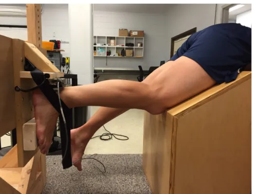
Hamstring stiffness was assessed as described by Blackburn et al.18 Maximum
the hip and thigh supported in 30° of flexion just off of the edge of a plinth (Figure 1). The foot was fixed to a loading device such that the knee will be maintained in 30° of flexion with the calcaneus in contact with a load cell (model 41; Honeywell Sensotec, Columbus, OH). Participants had the arms placed by their sides without grasping the edge of the table, and performed a submaximal warm-up contraction followed by a series of three, 5-second maximal knee-flexion efforts during which load cell data was sampled at 1,000 Hz. Load cell data was lowpass filtered at 10 Hz, and MVIC calculated using a 100-millisecond moving average from which we used the largest hamstrings force value to represent MVIC. This value was then used to determine the loading parameters for the stiffness assessment (i.e. 45% MVIC).
Figure 1- MVIC Assessment Position
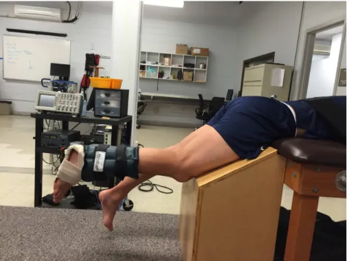
hamstrings on oscillatory knee flexion and extension. Participants were placed in the same position on the plinth as during MVIC testing but with the foot and shank free to move. A splint was secured to the plantar aspect of the foot and the posterior distal portion of the shank to maintain neutral talocrural position and to keep gastrocnemius length consistent, and an accelerometer (model 356A32; PCB Piezotronics, Inc, Depew, NY) was attached to the splint to measure tangential acceleration of the shank segment. Weights representing 45% of MVIC were secured near the ankle. The investigator then positioned the shank parallel to the floor, which positioned the knee in 30° of flexion, and the participants contracted the hamstrings to support the limb in the testing position (Figure 2). Within 5 seconds after this contraction, the investigator applied a downward manual perturbation to the calcaneus that forced the knee into extension and initiated oscillatory sagittal plane knee motion. The participant was instructed to allow the shank to oscillate, but to try to maintain the initial testing position and to not intervene with the perturbation.
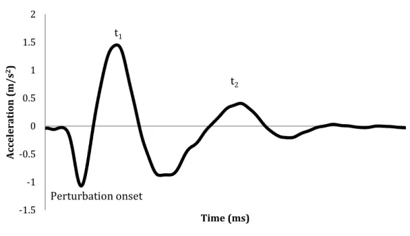
The oscillatory motion was recorded via the tangential acceleration of the shank segment sampled from the accelerometer at 1,000 Hz. Acceleration data was lowpass filtered at 10 Hz, and the damped frequency of oscillation was calculated as the inverse of the time interval between the first 2 oscillatory peaks (t1 and t2) in the acceleration signal.
Stiffness was calculated using the equation k= 4π2mf2 where k is stiffness, m is the total
mass of the system (shank and foot segment + 45% MVIC) and f is the damped frequency of oscillation. Shank and foot segment mass were calculated as 6.1% of total body mass 59.
Participants performed 5 trials separated by 30-second rest periods to reduce likelihood of fatigue.
Figure 3- Hamstring Stiffness Data
Landing Biomechanics Assessment
Landing biomechanics were assessed during five double-leg and single leg jump landings from a 30-cm height located 50% of the participant’s height away from 2 force plates (Bertec 4060- Bertec Corporation, Columbus, OH). For the double-leg jump landing, participants were instructed to land with each foot centered on a single force plate, to
minimize upward displacement upon leaving the box, and to immediately perform a maximal vertical jump following the initial landing. For the single-leg jump landing, participants were instructed to land with their right foot on the right force plate and to minimize upwards displacement upon leaving the box. Pilot testing revealed difficulty with participants completing a counter movement following SL landing, therefore no vertical jump was required following landing for SL condition. Trials that did not meet these criteria were eliminated and repeated until five recorded trials were available.
Biomechanical measurements were obtained from an electromagnetic motion-capture system (Ascension trakSTAR, Ascension Technology Corp., Shelburne, VT) interfaced with the forceplaces. Electromagnetic motion-capture sensors were placed on the pelvis over the sacrum, lateral midthigh, proximal anteromedial shank, and dorsum of the foot to measure lower extremity kinematics. Sensor position selected to minimize soft tissue artifact during jump landing tasks. The left and right anterior superior iliac spines, left and right femoral epicondyles and the medial and lateral malleoli were digitized to create a segment linkage model of the lower extremity. Locations of the knee and ankle joint centers were defined as the midpoints between the femoral epicondyles and malleoli, respectively. The hip joint center were estimated as a function of the 3-dimensional
distance between the digitized left and right anterior superior iliac spines as described by Bell et al. 60. Three-dimensional coordinate data and ground reaction forces were sampled
Data was sampled and processed via The Motion Monitor motion capture software (Innovative Sports Training, Inc., Chicago, IL). Kinematic and kinetic data was lowpass filtered at 10 Hz and 75 Hz, respectively, and combined via a standard inverse dynamics solution to yield net internal joint moments and forces.61 Knee joint angles were calculated
as motion of the shank relative to the thigh using Grood & Suntay angles. Kinematic and kinetic variables were identified during the loading phase of the landing, defined as the interval from initial ground contact (point at which vertical ground reaction force >10 N) to the peak knee-flexion angle. Peak knee flexion angle, peak anterior tibial shear force, and peak internal knee varus moments (i.e. the internal response to external valgus loading) were identified for each trial. Forces were normalized to body weight (xBW), and joint moments will be normalized to the product of body weight and height (xBW*Ht). Hamstring Strength Assessment
Participants were seated in a dynamometer (HUMAC Norm, CSMi, Stoughton, MA) with the hip in 55° rather than 90° of flexion to eliminate flexibility restraints and to allow for full knee extension during trials. The distal shank of the right limb was strapped into the leg attachment of the device with the foam pad placed approximately 1 inch proximal to the medial malleolus and the knee joint sagittal axis of rotation aligned with the axis of rotation of the dynamometer. Eccentric hamstring strength was be tested at 60°/s as this isokinetic speed has the greatest reliability between dynamometers62-64 and has been used
minutes of rest given between tests. The peak torque was derived as the largest value out of each three total trials of 9 eccentric knee flexion actions. These three values were then averaged across the three trials and will be referred to as peak torque. Peak eccentric torque was also evaluated between 10-30° of knee flexion as the ACL undergoes more strain as the knee approaches full extension.8 Eccentric peak torque was normalized to
body mass. Data Analyses
Chapter IV: Results
All variables were confirmed as being normally distributed. Three subjects were excluded from SL landing analyses due to errors during data reduction (consistent gimbal lock). Normalized eccentric torque values were consistent with previous research.63
DL Landing Biomechanics and Strength
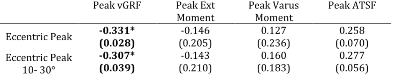
Associations between eccentric strength and double leg landing kinetics are displayed in Table 1. Eccentric peak torque was not associated with peak internal knee extension moment (r = -0.146, P = 0.205), peak internal varus moment (r = 0.127, P = 0.236), or ATSF (r = 0.258, P = 0.070). However, greater eccentric peak torque was associated with lesser peak vGRF (r = 0.331, P = 0.028). Similarly, eccentric torque at 10- 30° of knee flexion was not associated with peak internal knee extension moment (r = -0.143, P = 0.210), peak internal knee varus moment (r = 0.160, P = 0.183), or ATSF (r = 0.277, P = 0.056), but eccentric torque was associated with lesser peak vGRF (r = 0.307, P = 0.039).
Table 1 Correlations betweeneccentric hamstring strength and double leg jump landing kinetics.
Peak vGRF Peak Ext Moment
Peak Varus Moment
Peak ATSF
Eccentric Peak -0.331* (0.028) -0.146 (0.205) 0.127 (0.236) 0.258 (0.070) Eccentric Peak 10- 30° -0.307* (0.039) -0.143 (0.210) 0.160 (0.183) 0.277 (0.056)
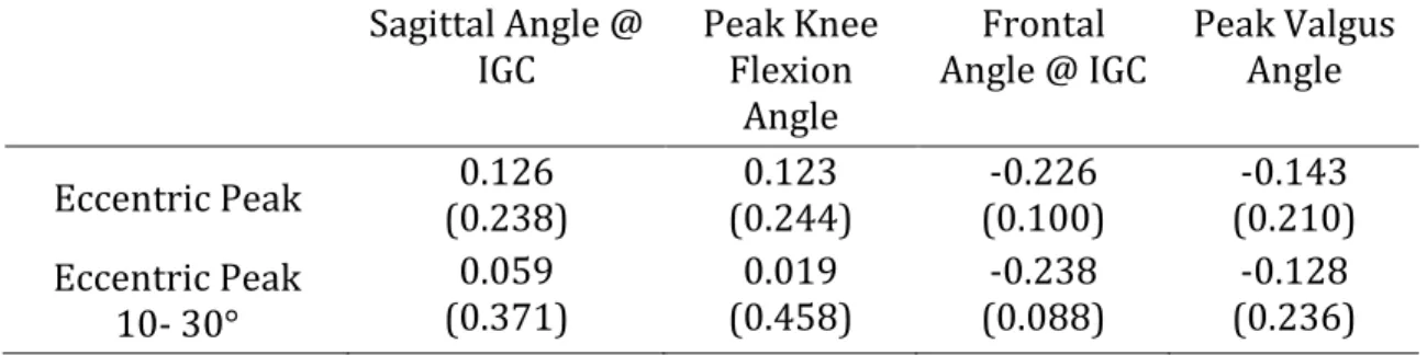
Associations between eccentric strength and double leg landing kinematics are displayed in Table 2. Eccentric peak torque was not associated with the sagittal knee angle at IGC (r = 0.126, P = 0.238), peak knee flexion angle (r = 0.123, P = 0.244), frontal knee angle at IGC (r = -0.226, P = -0.100), or peak knee valgus angle (r = -0.143, P = 0.210). Similarly, eccentric peak torque at 10- 30° of knee flexion was not associated with sagittal knee angle at IGC (r = 0.059, P = 0.371), peak knee flexion angle (r = 0.019, P = 0.458), frontal knee angle at IGC (r = -0.238, P = -0.088), or peak knee valgus angle (r = -0.128, P = 0.236).
Table 2 Correlations between eccentric hamstring strength and double leg jump landing kinematics.
Sagittal Angle @ IGC
Peak Knee Flexion
Angle
Frontal Angle @ IGC
Peak Valgus Angle
Eccentric Peak (0.238) 0.126 (0.244) 0.123 (0.100) -0.226 (0.210) -0.143 Eccentric Peak 10- 30° 0.059 (0.371) 0.019 (0.458) -0.238 (0.088) -0.128 (0.236)
SL Landing Biomechanics and Strength
Associations between eccentric strength and single leg landing kinetics are
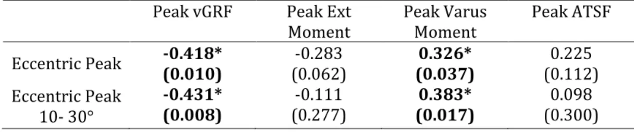
displayed in Table 3. Greater eccentric peak torque was associated with lesser peak vGRF (r = 0.418, P = 0.010) and peak internal knee varus moment (r = 0.326, P = 0.037).
However, eccentric peak torque at 10- 30° of knee flexion was not associated with peak internal knee extension moment (r = -0.111, P = 0.277) or peak ATSF (r = 0.098, P = 0.300). Table 3 Correlations between eccentric hamstring strength and single leg jump landing kinetics.
Peak vGRF Peak Ext Moment
Peak Varus Moment
Peak ATSF
Eccentric Peak -0.418* (0.010) -0.283 (0.062) 0.326* (0.037) 0.225 (0.112) Eccentric Peak 10- 30° -0.431* (0.008) -0.111 (0.277) 0.383* (0.017) 0.098 (0.300)
Bold text indicates significant correlations.
Associations between eccentric strength and single leg landing kinematics are displayed in Table 4. Eccentric peak torque was not associated with sagittal knee angle at IGC (r = -0.050, P = 0.394), peak knee flexion angle (r = 0.077, P = 0.341), frontal knee angle at IGC (r = -0.075, P = 0.343), or peak knee valgus angle (r = -0.023, P = 0.450). Similarly, eccentric peak torque at 10- 30° of knee flexion was not associated with sagittal knee angle at IGC (r = -0.159, P = 0.197), peak knee flexion angle (r = -0.028, P = 0.440), frontal knee angle at IGC (r = 0.034, P = 0.429), or peak knee valgus angle (r = 0.072, P = 0.351).
Table 4 Correlations between eccentric hamstring strength and single leg jump landing kinematics.
Sagittal Angle @ IGC
Peak Knee Flexion
Angle
Frontal Angle @ IGC
Peak Valgus Angle
Stiffness and Strength
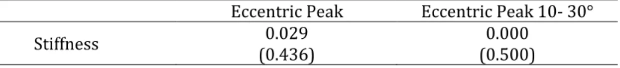
Association between eccentric strength and hamstring stiffness are displayed in Table 5. Eccentric peak torque (r = 0.029, P = 0.436) and eccentric peak torque at 10- 30° of knee flexion (r = 0.000, P = 0.500) were not associated with hamstring stiffness.
Table 5 Correlations between eccentric hamstring strength and hamstring stiffness.
Eccentric Peak Eccentric Peak 10- 30°
Stiffness 0.029
(0.436)
Chapter V: Discussion
The purpose of this study was to investigate relationships between eccentric hamstring strength, jump landing biomechanics linked to ACL injury risk, and hamstring stiffness. The results indicated that individuals with greater eccentric hamstring strength displayed lesser vGRF during both DL and SL landing conditions as well as lesser peak internal varus moment in the SL landing condition. There was no significant association between eccentric hamstring strength and hamstring stiffness.
Hewett et al.30 reported that greater peak external knee valgus moment was
predictive of greater ACL injury risk. Our data indicate that greater eccentric hamstring strength is associated with lesser peak internal varus moment (i.e. the internal response to external valgus moment) during SL landing. Therefore, greater eccentric hamstring
strength may assist in mitigating ACL injury risk. It is unclear, however, why this association was observed during SL landing but not during DL landing. Potentially, the hamstrings may be more active during SL landing to accommodate the greater relative lower extremity loading and joint stability demands. Leporace et al.65 found an increase in
the instantaneous median frequency of the biceps femoris EMG signal during SL landing compared to DL landing. Struminger et al.66 also reported greater preparatory phase
activation of the medial hamstrings during SL sagittal plane landing compared to a series of DL landing tasks. Similarly, McCurdy et al.67 described significantly greater hamstring EMG
activity during a SL squat compared to a DL squat. These findings suggest that the
the knee joint. Participants with greater eccentric hamstring strength also had lesser vGRF in both landing conditions. Cerulli et al.68 reported that the peak ACL strain and peak vGRF
occurred at roughly the same time. Hewett et al.30 reported that vGRF was 20% greater in
individuals who went on to injury their ACLs compared to those who did not. Thus, a smaller vGRF may be advantageous in ACL injury prevention.
Our finding that hamstring strength was generally not associated with landing biomechanics (other than peak vGRF and internal varus moment) agrees with previous literature. Shultz et al.69 found no association between isometric hamstring strength and
landing biomechanical factors such as anterior shear force and knee extensor moment. Homan et al.70 reported that isometric hip abduction strength was not associated with
landing biomechanics. Similarly, Norcross et al.71 reported that eccentric strength of the hip
abductors and external rotators was not associated with frontal plane hip motion during a lateral step-down. These findings are consistent with our study, and may indicate that strength (i.e. maximal force production capacity) is not an adequate indicator of muscle function during dynamic tasks. This disparity may be due to the fact that while strength assessments involve maximal activation of the muscle being tested, most dynamic tasks do not require maximal effort. Hamstring activity is submaximal during landing, ranging from 12% MVIC during a counter movement jump to 76% MVIC during the braking phase of landing72. Peng et al.73 found that hamstring activity was greater during the takeoff phase
than during the landing phase of landing. As our study was evaluating a counter movement jump and only collected data from the landing phase, hamstring activity may have been minimal. Padulo et al.72 Bennett et al.74 reported that quadriceps and hamstring strength
weaker individuals employed greater neural drive to the gluteal muscles (i.e. greater EMG amplitudes) during landing compared to stronger individuals to achieve the same
kinematic profile. Shultz et al.69 also reported a similar effect of muscle activation from the
quadriceps and the hamstrings, and also found that quadriceps and hamstring muscle activation during landing explained 16% of the variance in ATSF. Our study did not evaluate quadriceps strength or activation. The evaluation of both quadriceps and
hamstrings may have provided a more holistic representation of what influences landing profiles. It may be necessary to evaluate the quadriceps as well due to their influence on landing biomechanics.27, 69, 73, 75-77 Potentially, EMG activity may have been greater in
individuals with lesser hamstring strength to produce the same kinematic profile, similar to the findings of Homan et al.70 and Shultz et al.69 This may partially explain why eccentric
hamstring strength was not associated with landing biomechanics.
The biarticular nature of the hamstrings muscle group may also explain why eccentric hamstring strength was not correlated with landing biomechanics. During the impact phase of landing, both the hip and knee display flexion. This causes proximal lengthening of the hamstrings due to hip flexion, but distal shortening due to knee flexion. An isolated isokinetic measurement of strength may not adequately represent the function of the muscle during a dynamic task. Furthermore, the method by which we assessed hamstring strength specifically evaluated the knee flexion component of hamstrings
function. However, Cleather et al.78 reported that the hamstrings function predominately as
difference in proximal versus distal hamstrings activity during stiff-legged deadlift and lying leg curl. Nonuniform muscle activation was noted in the medial hamstrings, suggesting that the proximal and distal portions of the muscle can be preferentially recruited.79 This may also partially explain why there was no correlation between
hamstring strength and stiffness, as only the distal component of hamstrings function was assessed.
Hamstring stiffness has been linked with ACL loading. Blackburn et al. 17, 18 found
that greater hamstring stiffness was associated with lesser ATT during controlled joint perturbations. Furthermore, individuals with greater hamstring stiffness demonstrated smaller peak knee valgus and extension moments and greater knee flexion at the instant of peak anterior tibial shear force18, suggesting lesser ACL loading. Given the
laboratory-intensive nature of hamstring stiffness assessments, we sought to determine if eccentric hamstring strength could serve as a proxy for hamstring stiffness due to the fact that it can be obtained relatively easily in the clinical setting. However, eccentric hamstring strength was not significantly correlated with hamstring stiffness. This is consistent with the findings of Blackburn et al.17 and Hannah and Folland45 that isometric hamstring strength
There are limitations to our study that should be considered when interpreting the results. First, ACL loading in vivo is multifactorial and occurs in multiple planes. During our study, ACL loading factors were assessed independently and only in the sagittal and frontal planes. Similarly, the stiffness assessment modeled the knee as a single degree of freedom mass spring system for which motion was restricted to the sagittal plane. During the stiffness assessment, the perturbation is intended to produce isolated flexion/extension, but is likely to produce frontal and transverse plane motion as well due to the open chain nature of the assessment.
Conclusions
Eccentric hamstring strength is not an acceptable clinical estimate of hamstring stiffness. The hamstrings appear to play a pivotal role in restricting ACL loading. Although eccentric hamstring strength was generally not associated with landing biomechanics (with the exception of vGRF and internal knee varus in SL landing) other indices of hamstring function should be evaluated to determine their roles in knee joint stability. As stiffness is correlated with lesser ATSF17 and a more favorable landing biomechanics profile in terms
of ACL loading17, 18, further research is warranted to determine its potential role in ACL
injury prevention. Stiffness is also modifiable through training15, 19-21 and can be maximized
REFERENCES
1. Mall, N.A., et al., Incidence and trends of anterior cruciate ligament reconstruction in the United States. Am J Sports Med, 2014. 42(10): p. 2363-70.
2. Lohmander, L.S., et al., The long-term consequence of anterior cruciate ligament and meniscus injuries: osteoarthritis. Am J Sports Med, 2007. 35(10): p. 1756-69.
3. Shelbourne, K.D., et al., Loss of normal knee motion after anterior cruciate ligament reconstruction is associated with radiographic arthritic changes after surgery. Am J Sports Med, 2012. 40(1): p. 108-13.
4. Mather, R.C., 3rd, et al., Societal and economic impact of anterior cruciate ligament tears. J Bone Joint Surg Am, 2013. 95(19): p. 1751-9.
5. Ardern, C.L., et al., The impact of psychological readiness to return to sport and recreational activities after anterior cruciate ligament reconstruction. Br J Sports Med, 2014. 48(22): p. 1613-9.
6. Lentz, T.A., et al., Comparison of Physical Impairment, Functional, and Psychosocial Measures Based on Fear of Reinjury/Lack of Confidence and Return-to-Sport Status After ACL Reconstruction. Am J Sports Med, 2014.
7. Li, G., et al., The effects of ACL deficiency on mediolateral translation and varus-valgus rotation. Acta Orthopaedica, 2007. 78(3): p. 355-360.
8. Markolf, K.L., et al., Combined knee loading states that generate high anterior cruciate ligament forces. J Orthop Res, 1995. 13(6): p. 930-5.
9. Li, G., et al., The importance of quadriceps and hamstring muscle loading on knee kinematics and in-situ forces in the ACL. J Biomech, 1999. 32(4): p. 395-400. 10. Boden, B.P., et al., Mechanisms of anterior cruciate ligament injury. Orthopedics,
2000. 23(6): p. 573-8.
11. Blackburn, J.T. and D.A. Padua, Influence of trunk flexion on hip and knee joint kinematics during a controlled drop landing. Clin Biomech (Bristol, Avon), 2008. 23(3): p. 313-9.
12. DeMorat, G., et al., Aggressive quadriceps loading can induce noncontact anterior cruciate ligament injury. Am J Sports Med, 2004. 32(2): p. 477-83.
13. Withrow, T.J., et al., Effect of varying hamstring tension on anterior cruciate ligament strain during in vitro impulsive knee flexion and compression loading. J Bone Joint Surg Am, 2008. 90(4): p. 815-23.
15. Blackburn, J.T., et al., Sex comparison of hamstring structural and material properties. Clin Biomech (Bristol, Avon), 2009. 24(1): p. 65-70.
16. More, R.C., et al., Hamstrings--an anterior cruciate ligament protagonist. An in vitro study. Am J Sports Med, 1993. 21(2): p. 231-7.
17. Blackburn, J.T., M.F. Norcross, and D.A. Padua, Influences of hamstring stiffness and strength on anterior knee joint stability. Clin Biomech (Bristol, Avon), 2011. 26(3): p. 278-83.
18. Blackburn, J.T., et al., Hamstrings stiffness and landing biomechanics linked to anterior cruciate ligament loading. J Athl Train, 2013. 48(6): p. 764-72.
19. Pousson, M., J. Van Hoecke, and F. Goubel, Changes in elastic characteristics of human muscle induced by eccentric exercise. Journal of Biomechanics, 1990. 23(4): p. 343-348.
20. Foure, A., A. Nordez, and C. Cornu, Effects of plyometric training on passive stiffness of gastrocnemii muscles and Achilles tendon. Eur J Appl Physiol, 2012. 112(8): p. 2849-57.
21. Blackburn, J.T. and M.F. Norcross, The effects of isometric and isotonic training on hamstring stiffness and anterior cruciate ligament loading mechanisms. J
Electromyogr Kinesiol, 2014. 24(1): p. 98-103.
22. Enoka, R.M., Eccentric contractions require unique activation strategies by the nervous system. J Appl Physiol (1985), 1996. 81(6): p. 2339-46.
23. Hootman, J.M., R. Dick, and J. Agel, Epidemiology of collegiate injuries for 15 sports: summary and recommendations for injury prevention initiatives. J Athl Train, 2007. 42(2): p. 311-9.
24. Griffin, L.Y., et al., Noncontact anterior cruciate ligament injuries: risk factors and prevention strategies. J Am Acad Orthop Surg, 2000. 8(3): p. 141-50.
25. Arendt, E. and R. Dick, Knee injury patterns among men and women in collegiate basketball and soccer. NCAA data and review of literature. Am J Sports Med, 1995. 23(6): p. 694-701.
26. Harmon, K.G. and R. Dick, The relationship of skill level to anterior cruciate ligament injury. Clin J Sport Med, 1998. 8(4): p. 260-5.
28. Georgoulis, A.D., et al., Three-dimensional tibiofemoral kinematics of the anterior cruciate ligament-deficient and reconstructed knee during walking. Am J Sports Med, 2003. 31(1): p. 75-9.
29. Yoo, J.D., The Effect of Anterior Cruciate Ligament Reconstruction on Knee Joint Kinematics Under Simulated Muscle Loads. American Journal of Sports Medicine, 2005. 33(2): p. 240-246.
30. Hewett, T.E., et al., Biomechanical measures of neuromuscular control and valgus loading of the knee predict anterior cruciate ligament injury risk in female athletes: a prospective study. Am J Sports Med, 2005. 33(4): p. 492-501.
31. Louie, J.K. and C.D. Mote Jr, Contribution of the musculature to rotatory laxity and torsional stiffness at the knee. Journal of Biomechanics, 1987. 20(3): p. 281-300. 32. Renstrom, P., et al., Non-contact ACL injuries in female athletes: an International
Olympic Committee current concepts statement. Br J Sports Med, 2008. 42(6): p. 394-412.
33. Dienst, M., et al., Correlation of intercondylar notch cross sections to the ACL size: a high resolution MR tomographic in vivo analysis. Arch Orthop Trauma Surg, 2007. 127(4): p. 253-60.
34. Smith, H.C., et al., Risk factors for anterior cruciate ligament injury: a review of the literature - part 1: neuromuscular and anatomic risk. Sports Health, 2012. 4(1): p. 69-78.
35. Orchard, J.W., et al., Rye grass is associated with fewer non-contact anterior cruciate ligament injuries than bermuda grass. Br J Sports Med, 2005. 39(10): p. 704-9. 36. Olsen, O.E., et al., Relationship between floor type and risk of ACL injury in team
handball. Scand J Med Sci Sports, 2003. 13(5): p. 299-304.
37. Lambson, R.B., B.S. Barnhill, and R.W. Higgins, Football cleat design and its effect on anterior cruciate ligament injuries. A three-year prospective study. Am J Sports Med, 1996. 24(2): p. 155-9.
38. Agel, J., E.A. Arendt, and B. Bershadsky, Anterior cruciate ligament injury in national collegiate athletic association basketball and soccer: a 13-year review. Am J Sports Med, 2005. 33(4): p. 524-30.
39. Hewett, T.E. and G.D. Myer, The mechanistic connection between the trunk, hip, knee, and anterior cruciate ligament injury. Exerc Sport Sci Rev, 2011. 39(4): p. 161-6. 40. Besier, T.F., D.G. Lloyd, and T.R. Ackland, Muscle activation strategies at the knee
41. Claiborne, T.L., et al., Relationship between hip and knee strength and knee valgus during a single leg squat. J Appl Biomech, 2006. 22(1): p. 41-50.
42. Zhang, L.Q. and G. Wang, Dynamic and static control of the human knee joint in abduction-adduction. J Biomech, 2001. 34(9): p. 1107-15.
43. Dhaher, Y.Y., A.D. Tsoumanis, and W.Z. Rymer, Reflex muscle contractions can be elicited by valgus positional perturbations of the human knee. J Biomech, 2003. 36(2): p. 199-209.
44. Blackburn, J.T. and D.N. Pamukoff, Geometric and architectural contributions to hamstring musculotendinous stiffness. Clin Biomech (Bristol, Avon), 2014. 29(1): p. 105-10.
45. Hannah, R. and J.P. Folland, Muscle-tendon unit stiffness does not independently affect voluntary explosive force production or muscle intrinsic contractile properties. Appl Physiol Nutr Metab, 2015. 40(1): p. 87-95.
46. McNair, P.J., G.A. Wood, and R.N. Marshall, Stiffness of the hamstring muscles and its relationship to function in anterior cruciate ligament deficient individuals. Clin Biomech (Bristol, Avon), 1992. 7(3): p. 131-7.
47. Burgess, K.E., et al., Plyometric vs. isometric training influences on tendon properties and muscle output. J Strength Cond Res, 2007. 21(3): p. 986-9.
48. Kubo, K., et al., Effects of static and dynamic training on the stiffness and blood volume of tendon in vivo. J Appl Physiol (1985), 2009. 106(2): p. 412-7.
49. Kovanen, V., H. Suominen, and E. Heikkinen, Mechanical properties of fast and slow skeletal muscle with special reference to collagen and endurance training. J Biomech, 1984. 17(10): p. 725-35.
50. Goubel, F. and J.F. Marini, Fibre type transition and stiffness modification of soleus muscle of trained rats. Pflugers Arch, 1987. 410(3): p. 321-5.
51. Kelly, S.B., et al., Comparison of Concentric and Eccentric Bench Press Repetitions to Failure. J Strength Cond Res, 2014.
52. Leger, A.B. and T.E. Milner, The effect of eccentric exercise on intrinsic and reflex stiffness in the human hand. Clin Biomech (Bristol, Avon), 2000. 15(8): p. 574-82. 53. Kinikli, G.I., et al., The effect of progressive eccentric and concentric training on
54. Dragicevic-Cvjetkovic, D., et al., The effects of rehabilitation protocol on functional recovery after anterior cruciate ligament reconstruction. Med Arch, 2014. 68(5): p. 350-2.
55. Yu, B., C.F. Lin, and W.E. Garrett, Lower extremity biomechanics during the landing of a stop-jump task. Clin Biomech (Bristol, Avon), 2006. 21(3): p. 297-305.
56. Pollard, C.D., S.M. Sigward, and C.M. Powers, Limited hip and knee flexion during landing is associated with increased frontal plane knee motion and moments. Clin Biomech (Bristol, Avon), 2010. 25(2): p. 142-6.
57. Wild, C.Y., J.R. Steele, and B.J. Munro, Insufficient hamstring strength compromises landing technique in adolescent girls. Med Sci Sports Exerc, 2013. 45(3): p. 497-505. 58. Jennings, A.G. and B.B. Seedhom, The measurement of muscle stiffness in anterior
cruciate injuries -- an experiment revisited. Clin Biomech (Bristol, Avon), 1998. 13(2): p. 138-140.
59. Dempster, W.T., W.C. Gabel, and W.J. Felts, The anthropometry of the manual work space for the seated subject. Am J Phys Anthropol, 1959. 17: p. 289-317.
60. Bell, A.L., D.R. Pedersen, and R.A. Brand, A comparison of the accuracy of several hip center location prediction methods. J Biomech, 1990. 23(6): p. 617-21.
61. Gagnon, D. and M. Gagnon, The influence of dynamic factors on triaxial net muscular moments at the L5/S1 joint during asymmetrical lifting and lowering. J Biomech, 1992. 25(8): p. 891-901.
62. de Araujo Ribeiro Alvares, J.B., et al., Inter-machine reliability of the Biodex and Cybex isokinetic dynamometers for knee flexor/extensor isometric, concentric and eccentric tests. Phys Ther Sport, 2015. 16(1): p. 59-65.
63. Sugiura, Y., et al., Strength deficits identified with concentric action of the hip extensors and eccentric action of the hamstrings predispose to hamstring injury in elite sprinters. J Orthop Sports Phys Ther, 2008. 38(8): p. 457-64.
64. Koller, A., et al., Decrease in eccentric hamstring strength in runners in the Tirol Speed Marathon. Br J Sports Med, 2006. 40(10): p. 850-2; discussion 852.
65. Leporace, G., et al., Differences in time-frequency representation of lower limbs myoelectric activity during single and double leg landing in male athletes. J Electromyogr Kinesiol, 2011. 21(3): p. 506-11.
67. McCurdy, K., et al., Comparison of lower extremity EMG between the 2-leg squat and modified single-leg squat in female athletes. J Sport Rehabil, 2010. 19(1): p. 57-70. 68. Cerulli, G., et al., In vivo anterior cruciate ligament strain behaviour during a rapid
deceleration movement: case report. Knee Surg Sports Traumatol Arthrosc, 2003. 11(5): p. 307-11.
69. Shultz, S.J., et al., Thigh strength and activation as predictors of knee biomechanics during a drop jump task. Med Sci Sports Exerc, 2009. 41(4): p. 857-66.
70. Homan, K.J., et al., The influence of hip strength on gluteal activity and lower extremity kinematics. J Electromyogr Kinesiol, 2013. 23(2): p. 411-5.
71. Norcross M, H.S., Hawkey T, Blackburn J, Padua D., Evaluation of the Lateral Step-Down Test as a Clinical Assessment of Hip Musculature Strength. Athletic Training and Sports Health Care., 2009. 1: p. 272-278.
72. Padulo, J., et al., EMG amplitude of the biceps femoris during jumping compared to landing movements. Springerplus, 2013. 2: p. 520.
73. Peng, H.T., T.W. Kernozek, and C.Y. Song, Quadricep and hamstring activation during drop jumps with changes in drop height. Phys Ther Sport, 2011. 12(3): p. 127-32. 74. Bennett, D.R., et al., The relationship between anterior tibial shear force during a jump
landing task and quadriceps and hamstring strength. Clin Biomech (Bristol, Avon), 2008. 23(9): p. 1165-71.
75. Elias, A.R., C.D. Hammill, and R.L. Mizner, Changes in quadriceps and hamstring cocontraction following landing instruction in patients with anterior cruciate ligament reconstruction. J Orthop Sports Phys Ther, 2015. 45(4): p. 273-80. 76. Blackburn, J.T. and D.A. Padua, Sagittal-plane trunk position, landing forces, and
quadriceps electromyographic activity. J Athl Train, 2009. 44(2): p. 174-9.
77. Withrow, T.J., et al., The relationship between quadriceps muscle force, knee flexion, and anterior cruciate ligament strain in an in vitro simulated jump landing. Am J Sports Med, 2006. 34(2): p. 269-74.
78. Cleather, D.J., D.F. Southgate, and A.M. Bull, The role of the biarticular hamstrings and gastrocnemius muscles in closed chain lower limb extension. J Theor Biol, 2015. 365: p. 217-25.