Description of
Immundisolibacter cernigliae
gen. nov., sp. nov., a
high-molecular-weight polycyclic aromatic
hydrocarbon-degrading bacterium within the class
Gammaproteobacteria
, and
proposal of
Immundisolibacterales
ord. nov. and
Immundisolibacteraceae
fam. nov.
Elizabeth M. Corteselli, Michael D. Aitken and David R. Singleton*
Abstract
The bacterial strain TR3.2Twas isolated from aerobic bioreactor-treated soil from a polycyclic aromatic hydrocarbon (PAH)-contaminated site in Salisbury, NC, USA. Strain TR3.2Twas identified as a member of‘Pyrene Group 2’or‘PG2’, a previously uncultivated cluster of organisms associated with the degradation of high-molecular-weight PAHs by stable-isotope probing. Based on its 16S rRNA gene sequence, the strain was classified as a member of the class Gammaproteobacteria but possessed only 90.5 % gene identity to its closest described relative, Methylococcus capsulatus strain Bath. Strain TR3.2T
grew on the PAHs pyrene, phenanthrene, anthracene, benz[a]anthracene and fluorene, as well as the azaarene carbazole, and could additionally metabolize a limited number of organic acids. Optimal growth occurred aerobically under mesophilic temperature, neutral pH and low salinity conditions. Strain TR3.2Twas catalase and oxidase positive. Predominant fatty acids
were C17 : 0cyclo and C16 : 0. Genomic G+C content of the single chromosome was 67.79 mol% as determined by complete
genome sequencing. Due to the high sequence divergence from any cultivated species and its unique physiological properties compared to its closest relatives, strain TR3.2Tis proposed as a representative of a novel order, family, genus and species within the class Gammaproteobacteria, for which the name Immundisolibacter cernigliae gen. nov., sp. nov. is proposed. The associated order and family are therefore proposed as Immundisolibacterales ord. nov. and
Immundisolibacteraceaefam. nov. The type strain of the species is TR3.2T(=ATCC TSD-58T=DSM 103040T).
A prior stable-isotope probing (SIP) experiment of polycy-clic aromatic hydrocarbon (PAH)-contaminated, aerobic bioreactor-treated soil using the four-ring PAH pyrene identified several clusters of 16S rRNA genes derived from uncultivated Proteobacteria [1]. One group, designated
‘Pyrene Group 2’ or‘PG2’, had no cultivated relatives and could only be classified to the class Gammaproteobacteria. Subsequent SIP experiments using pyrene, benz[a ]anthra-cene or fluoranthene [2, 3] further implicated bacteria belonging to PG2 in the degradation of high-molecular-weight PAHs as well as the low-molecular-high-molecular-weight PAH phenanthrene [4] and potentially the low-molecular-weight PAH anthracene [5]. The 16S rRNA gene sequences com-prising PG2 from these SIP projects possessed a relatively high (96 %) intragroup 16S rRNA gene identity. Other globally distributed environmental 16S rRNA gene
sequences with high similarity to those designated as PG2 have been detected in hydrocarbon-impacted environments contaminated with oils [6–8], creosote [9], pyridine [10] and aliphatic compounds [11]. Other 16S rRNA gene sequences from PG2 have been recovered from natural asphalts [12] as well as pristine sediment [13] and water samples [14–16]. The most closely related isolate to organ-isms within PG2 wasMethylococcus capsulatusstrain Bath (~90 % 16S rRNA gene similarity) [17], a methanotroph iso-lated from soil [18].
A bacterial strain whose 16S rRNA gene sequence placed it phylogenetically within the cluster of SIP-derived PG2 sequences, designated TR3.2T, was successfully isolated from an aerobic bioreactor-treated, PAH-contaminated soil from the site of a former manufactured gas plant in Salisbury, NC,
Author affiliation:Department of Environmental Sciences and Engineering, Gillings School of Global Public Health, University of North Carolina, Chapel Hill, NC 27599-7431, USA.
*Correspondence:David R. Singleton, [email protected]
Keywords:Gammaproteobacteria; polycyclic aromatic hydrocarbons; Pyrene Group 2.
Abbreviations:PAH, polycyclic aromatic hydrocarbon; SIP, stable-isotope probing.
The GenBank/EMBL/DDBJ accession number for the 16S rRNA gene sequence of strain TR3.2Tis CP014671. Two supplementary figures are available with the online Supplementary Material.
USA [19]. Strain TR3.2Twas isolated through serial dilutions of bioreactor-treated soil onto a modified version of the aero-bic cultivation medium forSulfuritalea hydrogenivorans[20] using a layer of oversprayed pyrene as a carbon source; this medium was also used to obtain the PAH-degrading bacte-rium‘Rugosibacter aromaticivorans’Ca6 [21]. Prior isolation attempts may have been hindered by the lack of required trace minerals or vitamins in the medium in addition to the slow growth of the organism. Subsequent tests of strain TR3.2Tgrown in the liquid isolation medium excluding vari-ous components indicated that the following medium com-ponents were required for maximum growth:‘reactor buffer’
(5 mM Na K phosphate buffer, 5 mM NH4NO3, pH 7.0),
1 mM MgSO4, 1 mM CaCl2, 1 ml l 1 of a trace element solution [containing (per litre) 12.5 ml HCl (25 %), 2.1 g FeSO4.7H2O, 30 mg H3BO3, 100 mg MnCl2.4H2O, 190 mg
CoCl2.6H2O, 24 mg NiCl2.6H2O, 2 mg CuCl2.2H2O, 144 mg
ZnSO4.7H2O, 36 mg Na2MoO4.2H2O] and vitamin B12
solution (1 ml l 1; containing 5 mg cyanocobalamin in 100 ml distilled water, filter sterilized and added after autoclaving), to a final pH of 7.0. Carbon was added before autoclaving as either 0.02 % (w/v) pyrene dissolved in ace-tone to the flask, allowing the solvent to evaporate prior to adding other components, or as 0.2 % (w/v) sodium pyru-vate. No growth at all occurred in the absence of MgSO4,
CaCl2 or trace element solutions, and reduced growth rates
and cell density compared to the complete growth medium were observed when vitamin B12 was omitted from the medium. All subsequent tests used these five components (referred to as ‘sRB2 medium’) with sodium pyruvate as a carbon source unless otherwise noted. For solid medium (‘sRB2-agar’), 1.5 % agar (Acros Organics) was added to liq-uid medium prior to autoclaving.
The optimal growth conditions of strain TR3.2T in sRB2 medium with pyruvate as a carbon source were determined for a range of temperatures, pH and salinities. Optimal growth was defined as the maximum growth rate during the exponential growth phase as measured by turbidity at OD600 with a HACH DR3000 spectrophotometer
(Love-land). Optimal temperature for growth was determined in triplicate 5 ml cultures in liquid sRB2 medium at pH 7.0 and 225 r.p.m. for temperatures of 26, 28, 30, 32, 34, 35, 36 and 37
C. Additional temperatures were evaluated on solid sRB2 plates at 20 and 4
C. The effects of pH were tested in triplicate 5 ml tubes of sRB2 medium buffered to pH values of 5.0, 5.5, 6.0, 6.5 (buffered with MES, 50 mM), 7.0, 7.5, 8.0 (HEPES, 50 mM) and 8.5, 9.0 (Tris/HCl, 50 mM), which were incubated at 225 r.p.m. and 30
C. Salinity effects were determined using triplicate 5 ml cultures of sRB2, pH 7.0, incubated at 30
C and 225 r.p.m., amended with either 0, 0.25, 0.5, 1, 2, 3, 4 or 5 % (w/v) NaCl. The optimal tempera-ture for growth was from 28 to 30
C, with obvious growth observed between 20 and 36
C. Strain TR3.2Tgrew between pH 6.5 and 8.0 with an optimum at 7.0. No growth was observed at salt concentrations >1 %, and strain TR3.2Thad the highest growth rate with no amended NaCl in the medium. Under optimal conditions, inoculated tubes of
strain TR3.2Tgrew to turbidity in liquid culture with con-stant shaking at 225 r.p.m. in approximately 6 days, with a doubling time of 25 h. Colonies on sRB2 plates with pyru-vate as a carbon source developed more slowly over a period of 2 weeks when incubated at 30
C.
The ability of TR3.2T to grow under anaerobic conditions was assessed using the GasPak 100 system containing a Gas-Pak EZ Anaerobe Container System sachet (BD Bioscien-ces). Plates containing sRB2-agar and pyruvate were inoculated and sealed in the system according to manufac-turer’s directions. The system achieves an atmosphere of <1 % oxygen and 13 % carbon dioxide within 2.5 h. Anaerobic conditions after 48 h were confirmed by visual observation of an indicator strip soaked in 1 mM resazurin solution. Plates were incubated at 30
C with concurrent aerobic controls. TR3.2Tdid not display growth in anaero-bic conditions after 4 weeks. Unless otherwise noted, all subsequent tests of TR3.2T occurred at 30
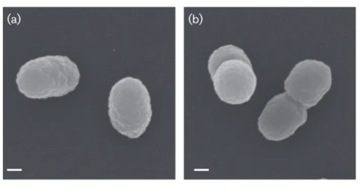
C, pH 7.0 and 0 % salinity under aerobic conditions. Liquid cultures were additionally incubated with constant shaking at 225 r.p.m. The cellular morphology of strain TR3.2T was investigated using scanning electron microscopy. For the preparation of samples, cells were grown in liquid sRB2 with pyruvate as a carbon source. Specimens were observed and images taken using a Zeiss Supra 25 FESEM operating at 5 kV, 5 mm working distance and 10 µm aperture (Carl Zeiss SMT). Average sizes were determined from digital micrographs of single, well-distinguished cells. Strain TR3.2Tcells were coc-cobacilli with an average size of 0.56±0.08 µm by 0.35 ±0.02 µm (n=100 cells). Cells appeared slightly wrinkled, generally as singles or doubles, with no obvious extracellular features (Fig. 1). Gram staining performed using the stan-dard reaction and light microscopy indicated strain TR3.2T was Gram-type negative. Cellular motility was tested using sRB2-agar stab-tubes with agar added at 0.3 % (w/v) and pyruvate as the carbon source. No motility was observed in the stab-tubes, and no flagella were observed in micro-graphs. Log-phase cells grown in sRB2 stained with the Remel Flagella Stain (Thermo Scientific) and examined under oil-immersion light microscopy did not appear to
(a) (b)
Fig. 1.Scanning electron micrographs of strain TR3.2Tgrown on
have flagella. However, analysis of the genome of strain TR3.2T indicated a complete set of genes required for assembly of a flagellum [22]. Expression of flagella by strain TR3.2T therefore may be possible under some conditions, but was not observed under those tested here. Strain TR3.2T colonies on sRB2 plates with pyruvate were small (0.50–
0.75 mm in diameter), circular, with a convex elevation and were yellow-orange in colour.
Metabolism of a variety of carbon substrates was tested using the Biolog GN2 Microplate. For each plate, cells were grown in liquid sRB2 medium amended with pyruvate and washed three times in PBS (pH 7.5) before being resus-pended in Biolog GN/GP Inoculating Fluid as per the man-ufacturer’s instructions. The microplates were incubated overnight at 30
C and scored through visual examination in comparison to the no-carbon control. In triplicate tests, the only substrates that strain TR3.2Tactively metabolized were
a-ketobutyric acid, methyl pyruvate,
mono-methyl-succinate and a-ketovaleric acid. The other 91 substrates,
including a variety of sugars, other organic acids, nucleo-sides and amino acids, were not metabolized.
Utilization of nitrogen sources was tested by substituting either 5 mM KNO3 or NH4Cl for NH4NO3 in sRB2 liquid
medium. Strain TR3.2T displayed growth on pyruvate as a carbon source with both nitrogen sources tested. Nitrate reduction was evaluated using the culture grown in sRB2 medium substituted with 5 mM KNO3. No nitrate reduction
to nitrite beyond what was required for assimilation and growth was observed. Starch hydrolysis and cellulase activ-ity were evaluated by supplementing sRB2 plates with either starch (10 g l 1) or cellulose (1 g l 1) (adapted from Kasana
et al.[23]). Protease activity was tested by aseptically adding a filter-sterilized skimmed milk solution (10 % skimmed milk powder dissolved in distilled water) to sRB2-agar after autoclaving. Plates were incubated at 30
C for 4 weeks and monitored for zones of clearing; for starch and cellulose plates, Gram’s iodine was also added at the end of the incu-bation to help detect zones of clearing. Gelatin hydrolysis was assessed using a nutrient gelatin stab method wherein powdered gelatin (120 g l 1) was added to sRB2, gently heated to dissolve and aliquoted into 5 ml tubes. Gelatin tubes containing TR3.2Twere incubated at 30
C and moni-tored for liquefaction of the medium. Strain TR3.2T was negative for starch hydrolysis, cellulase, skimmed milk pro-tease and gelatinase activities.
Strain TR3.2T was tested for catalase activity by adding 3 % (v/v) hydrogen peroxide solution to cells freshly scraped from the surface of a sRB2 plate. Oxidase activity was deter-mined by adding a few drops of freshly prepared 1 %N¢,N¢,N¢,
N¢-tetramethyl-p-phenylenediamine dihydrochloride (Acros Organics) to cells scraped from the same plate onto filter paper. Strain TR3.2Twas catalase and oxidase positive. Lipase activity was assessed by adding tributyrin (98 %; Acros Organics) to sRB2-agar (1 %, v/v). Urease activity was determined using a medium prepared with the sRB2
phosphate buffer with urea as a nitrogen source (20 g l 1) and phenol red (10 mg l 1) in addition to the standard concentrations of MgSO4, CaCl2, trace element
solution and vitamin B12 solutions with pyruvate as a growth substrate. Strain TR3.2Twas positive for lipase activ-ity and negative for urease activactiv-ity.
Indole production was tested by supplementing sRB2 medium with tryptone (10 g l 1). After growth to turbidity, a few drops of Kovac’s reagent were added to each tube. The lack of a colour change indicated no production of indole by strain TR3.2T. Production of indigo from exogenous indole was assessed by adding a crystal of indole to the lids of inverted Petri plates containing sRB2-pyruvate medium 24 h after inoculation. Indigo production from indole is indicative of activity for some ring-hydroxylating dioxygenases involved in the initial step in the aerobic degradation of PAHs [24, 25]. A purple colour developed by colonies on the plate after exposure to indole confirmed the likely presence of an active ring-hydroxylating dioxygenase.
Growth of strain TR3.2Ton select PAHs as sole sources of carbon and energy was tested in sRB2 medium amended with individual PAHs (final concentration 0.2 g l 1). PAHs were added to tubes in either acetone or dichloromethane and the solvent allowed to evaporate prior to adding the remaining medium components. After autoclaving, the tubes were briefly placed in an ultrasonic water bath to dis-perse PAH crystals before washed TR3.2T cells grown in sRB2-pyruvate were inoculated in triplicate tubes for each PAH and incubated at 30
over the course of the incubation; the HPLC assay for quan-tifying PAHs was not suitable for this compound.
Mineralization of the partially14C-labelled PAHs phenan-threne, fluoranthene, chrysene, benz[a]anthracene and benzo[a]pyrene was performed as previously described [27]. Strain TR3.2Tcells grown in sRB2-pyrene and washed three times with reactor buffer were used to inoculate triplicate flasks. The extent of mineralization was measured after 24 h. Mineralization was assessed as percentage of added PAH mineralized and compared among biotic and acidified repli-cates. Statistically significant mineralization (P0.01) occurred for phenanthrene (56 % mineralization), benz[a] anthracene (29 %), benzo[a]pyrene (41 %) and chrysene (52 %), but not fluoranthene.
Growth on BTEX compounds was investigated by creating an atmosphere in separate, tightly sealed metal containers of either benzene (99 %), toluene (99.8 %), ethylbenzene (99.8 %) or xylenes [o-xylene (98.5 %),m-xylene (99 %),p -xylene (99 %), in equal volumes] by adding 0.5 ml of the chemical(s) to a piece of filter paper in a glass beaker. Inocu-lated sRB2-agar plates without amended carbon were incu-bated in sealed containers at room temperature for 1 month and compared to concurrent plates incubated outside the closed systems with and without additional carbon sources. Strain TR3.2Tdid not display growth on any of the tested BTEX compounds under these conditions.
Cellular fatty acid profiling was performed using the Sherlock Microbial Identification System by Microbial ID on cells freshly grown on sRB2 plates with pyruvate [28]. The dominant fatty acids in TR3.2T were C17 : 0 cyclo
(32.1 %), C16 : 0(29.6 %) and summed feature 3 (C16 : 1!7cor
C16 : 1!6c) (12.0 %). TR3.2Talso contained C19 : 0 cyclo !8c
(8.3 %), C12 : 03-OH (6.4 %), C12 : 0(2.0 %), summed feature
8 (C18 : 1!7cor C18 : 1!6c) (1.9 %), summed feature 5 (C18 : 0
anteiso or C18 : 2!6,9c) (1.6 %), C14 : 0 (1.4 %) and C10 : 0
(1.4 %). Trace amounts (<1 %) of C10 : 03-OH, C14 : 03-OH,
C17 : 1!7c, C16 : 0 3-OH, C18 : 0, C18 : 0!7c 11-methyl, C19 : 0,
C18 : 12-OH and C20 : 2!6,9cwere present. Analysis of
respi-ratory quinones and polar lipids was carried out by the Iden-tification Service, DSMZ, Braunschweig, Germany [29–31]. Major polar lipids in TR3.2T were diphosphatidylglycerol, phosphatidylmethylethanolamine, phosphatidylethanol-amine, phosphatidylglycerol and phospholipids (Fig. S1, available in the online Supplementary Material). Also pres-ent in minor amounts were lipid, phosphoglycolipid and unidentified pigments. Respiratory quinones present were Q7 (9 %), Q8 (90 %) and Q9 (1 %).
The genome of strain TR3.2Twas acquired and annotated as described previously [22]. The genome was a singular, circular chromosome of 3 243 537 bp with a G+C content of 67.79 mol%. No extrachromosomal elements were detected. Annotation predicted 3114 total genes with 3054 protein coding genes, a single rRNA gene operon and 45 tRNA-encoding genes [22]. The complete 16S rRNA gene of strain TR3.2Twas compared to publicly available sequences using
BLASTNof the non-redundant GenBank database in separate
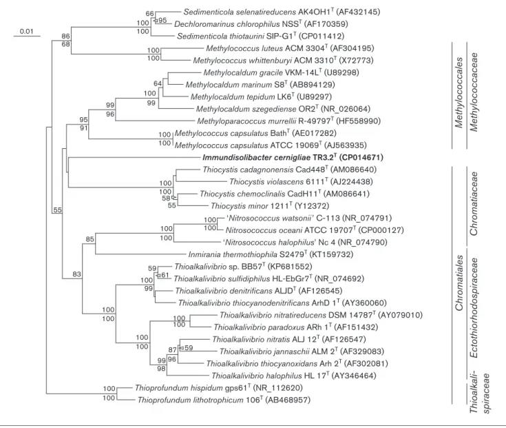
searches including and excluding environmental sequences. Some of the closest described relatives to TR3.2Tbased on 16S rRNA gene similarity (Fig. 2) wereM. capsulatusstrain Bath (90.5 % similarity) [17], M. capsulatus strain ATCC 19069T (strain Texas; 90.2 %), ‘Thioalkalivibrio sulfidophi-lus’ strain HL-EbGr7 (90.1 %) [32] and Inmirania thermothiophilastrain S2479T(90.3 %) [33]. An estimate of DNA–DNA hybridization using the complete genome of strain TR3.2Tcompared to those ofM. capsulatusBath and
‘Thioalkalivibrio sulfidophilus’HL-EbGr7 with the genome-to-genome distance calculator [34] predicted a DNA–DNA hybridization value of 18.5 % to both organisms using the recommended formula. Based on the complete 16S rRNA gene, the naive Bayesian classifier of the Ribosomal Data-base Project [35] indicated that the highest taxonomic level that TR3.2T could reliably be grouped in with confidence (confidence threshold >80 %; training set 16, released June 2016) was the class Gammaproteobacteria. Within the
Gammaproteobacteria, strain TR3.2Twas equally dissimilar to described bacteria within the ordersMethylococcalesand
Chromatiales. Bootstrapping with multiple algorithms did not reliably cluster strain TR3.2T with members of either order, so its phylogenetic position within the Gammaproteo-bacteriais uncertain.
Despite the lack of phylogenetic similarity to any character-ized bacteria, large numbers of 16S rRNA gene sequences with high similarity (>97 %) to strain TR3.2T have been recovered from a variety of environmental sources (Fig. S2). Many of those studies, including those involving SIP of PAH-contaminated soils within our research group, investi-gated the diversity of communities in which the sample medium was impacted by petroleum or hydrocarbon con-tamination [7–9, 11, 12, 36]. The prevalence of environmen-tal sequences closely related to strain TR3.2T in similarly contaminated but geographically separated samples suggests a potential widespread association of PG2-type organisms with the degradation of those compounds.
In addition to phylogenetic dissimilarity to the most closely related and characterized bacteria, strain TR3.2T was also physiologically differentiated from those organisms. Members of the orderMethylococcales, which includes the genera Meth-ylococcus,MethylocaldumandMethyloparacoccus, are obligate users of C1compounds for carbon and energy [37, 38]. This is
in stark contrast to the complex aromatics and organic acids that strain TR3.2Tcan use as growth substrates. Additionally, no obvious homologues of particulate methane monooxyge-nase genes, common among methanotrophic members of the
facultatively anaerobic autotroph that grows optimally at high temperatures and salinity [33]. Members of the genusNitrosococcusare marine, ammonia-oxidizing chemoli-thotrophs [41, 42]. While some Sedimenticola strains are capable of growth on aromatic compounds, they are also strict anaerobes that use selenium or nitrogen oxyanions as electron acceptors [43].Thioprofundumspecies are thermophilic, pie-zophilic chemolithoautotrophs isolated from deep-sea hydro-thermal vents [44, 45].
On the basis of distinct physiological, phylogenetic and environmental habitat differences to cultivated and charac-terized bacterial relatives, the novel genus and species
Immundisolibacter cernigliaegen. nov., sp. nov. is proposed
for this organism, with TR3.2Tas the type strain. Due to the physiological and phylogenetic dissimilarity to organisms within described orders of the classGammaproteobacteria, we also propose a novel order and family of which strain TR3.2T is currently the sole member, and would thus be designatedImmundisolibacteralesord. nov. and Immundiso-libacteraceaefam. nov., respectively.
DESCRIPTION OF
IMMUNDISOLIBACTER
GEN. NOV.
Immundisolibacter (Im.mun.di.so.li.bac¢ter. L. adj. immun-dusunclean, impure; L. n.solumsoil; N.L. n.bacterrod; N. L. masc. n.Immundisolibactera rod from unclean soil).
Thioalkalivibrio sp. BB57T (KP681552)
Thioprofundum hispidum gps61T (NR_112620)
Methylococcales
Chromatiale
s
Ectothiorhodospiraceae
Chromatiaceae
Thioalkali- spiraceae
Methylococcaceae
Sedimenticola selenatireducens AK4OH1T (AF432145)
66 95 100 100
100 100
99 99
96 86
0.01
68
95 91
100
100 100
100 100
100 100 100
100
100 100
100 100
99 98
96 87 59
100 100
100 100 55
85
83 59 61
100 99
55 58 64
Dechloromarinus chlorophilus NSST (AF170359)
Sedimenticola thiotaurini SIP-G1T (CP011412)
Methylococcus luteus ACM 3304T (AF304195)
Methylococcus whittenburyi ACM 3310T (X72773)
Methylocaldum gracile VKM-14LT (U89298)
Methylocaldum marinum S8T (AB894129)
Methylocaldum tepidum LK6T (U89297)
Methylocaldum szegediense OR2T (NR_026064)
Methyloparacoccus murrellii R-49797T (HF558990)
Methylococcus capsulatus BathT (AE017282)
Methylococcus capsulatus ATCC 19069T (AJ563935)
Immundisolibacter cernigliaeTR3.2T (CP014671)
Thiocystis cadagnonensis Cad448T (AM086640)
Thiocystis violascens 6111T (AJ224438)
Thiocystis chemoclinalis CadH11T (AM086641)
Thiocystis minor 1211T (Y12372)
‘Nitrosococcus watsonii ’ C-113 (NR_074791)
Nitrosococcus oceani ATCC 19707T (CP000127)
‘Nitrosococcus halophilus’ Nc 4 (NR_074790)
Inmirania thermothiophila S2479T (KT159732)
Thioalkalivibrio sulfidiphilus HL-EbGr7T (NR_074692)
Thioalkalivibrio denitrificans ALJDT (AF126545)
Thioalkalivibrio thiocyanodenitrificans ArhD 1T (AY360060)
Thioalkalivibrio nitratireducens DSM 14787T (AY079010)
Thioalkalivibrio paradoxus ARh 1T (AF151432)
Thioalkalivibrio nitratis ALJ 12T (AF126547)
Thioalkalivibrio jannaschii ALM 2T (AF329083)
Thioalkalivibrio thiocyanoxidans Arh 2T (AF302081)
Thioalkalivibrio halophilus HL 17T (AY346464)
Thioprofundum lithotrophicum 106T (AB468957)
Fig. 2.16S rRNA gene neighbour-joining phylogenetic tree of strain TR3.2Twith 16S rRNA gene sequences of the most similar
culti-vated and described strains. Sequences were aligned and the neighbour-joining tree created usingCLUSTALX2.1 [46] and without
con-sidering positions with gaps. The maximum-parsimony tree was determined using thePAUP[47] implementation within Geneious 9.1.3
Cells are Gram-negative, non-motile and grow aerobically. Catalase and oxidase positive. Heterotrophic growth occurs on a limited number of organic acids. Predominant fatty acids are C17 : 0 cyclo, C16 : 0 and summed feature 3 (C16 : 1
!7cor C16 : 1!6c). The major polar lipids are
didylglycerol, phosphatidylmethylethanolamine, phosphati-dylethanolamine, phosphatidylglycerol and phospholipids. The major respiratory quinone is Q8. Phylogenetically, the genus is a member of the classGammaproteobacteria. The type species isImmundisolibacter cernigliae.
DESCRIPTION OF
IMMUNDISOLIBACTER
CERNIGLIAE
SP. NOV.
Immundisolibacter cernigliae(cer.nig¢li.ae. N.L. gen. n.
cerni-gliae named after the prominent researcher of
PAH-degrading bacteria Carl Cerniglia).
Cells are coccobacilli approximately 0.560.35 µm. Colonies on solid medium are small, convex and circular with a yel-low-orange colour. Growth occurs aerobically between 20 and 36
C (optimum between 28 and 30
C), pH 6.5–8.0 (optimum 7.0) and at salinity1 % (optimum 0 %). Growth was not observed under anaerobic conditions. Cells can metabolize a-ketobutyric acid, methyl pyruvate,
mono-methyl-succinate and a-ketovaleric acid and can grow on
polycyclic aromatic compounds as sole sources of carbon, and both nitrate and ammonia as nitrogen sources. Cells are negative for gelatin hydrolysis, cellulase, skimmed milk pro-tease, starch hydrolysis, dissimilatory nitrate reduction and urease, and positive for lipase activity. In addition to those listed in the genus description, fatty acids present in lesser amounts are C19 : 0cyclo!8cand C12 : 03-OH. In addition to
the major respiratory quinone, Q7 and Q9 are also present. Polar lipids are consistent with the genus.
The type strain, TR3.2T(=ATCC TSD-58T=DSM 103040T), was isolated from PAH-contaminated soil from Salisbury, NC, USA. Genomic G+C content is 67.79 mol%.
DESCRIPTION OF
IMMUNDISOLIBACTERACEAE
FAM. NOV.
Immundisolibacteraceae (Im.mun.di.so.li.bac.ter.a.ce¢ae. N. L. masc. n.Immundisolibactertype genus of the family; N.L. suff. -aceae ending denoting a family; N.L. fem. pl. n.
Immundisolibacteraceaethe family ofImmundisolibacter). Description is the same as for the genusImmundisolibacter. The type genus isImmundisolibacter.
DESCRIPTION OF
IMMUNDISOLIBACTERALES
ORD. NOV.
Immundisolibacterales (Im.mun.di.so.li.bac.ter.a¢les. N.L. masc. n.Immundisolibacterthe type genus of the order; N.L. suff. -alesending denoting an order; N.L. fem. pl. n. Immun-disolibacteralesthe order ofImmundisolibacter).
Order of the classGammaproteobacteria. Segregation of this genus into a new order is justified by its phylogenetic
distance and physiological differences from organisms in the most closely related extant orders. Organisms within the order are Gram-type negative with aerobic growth under mesophilic temperature, neutral pH and low salinity condi-tions. The type genus isImmundisolibacter.
Funding information
This work was supported by the U.S. National Institute of Environmen-tal Health Sciences (NIEHS) as part of the Superfund Research Pro-gram (5 P42ES005948).
Conflicts of interest
The authors declare that there are no conflicts of interest.
References
1. Singleton DR, Sangaiah R, Gold A, Ball LM, Aitken MD. Identifica-tion and quantificaIdentifica-tion of uncultivated Proteobacteria associated with pyrene degradation in a bioreactor treating PAH-contaminated soil.Environ Microbiol2006;8:1736–1745.
2. Jones MD, Crandell DW, Singleton DR, Aitken MD.Stable-isotope probing of the polycyclic aromatic hydrocarbon-degrading bacte-rial guild in a contaminated soil.Environ Microbiol2011;13:2623–
2632.
3. Jones MD, Singleton DR, Carstensen DP, Powell SN, Swanson JS
et al.Effect of incubation conditions on the enrichment of pyrene-degrading bacteria identified by stable-isotope probing in an aged, PAH-contaminated soil.Microb Ecol2008;56:341–349.
4. Singleton DR, Hunt M, Powell SN, Frontera-Suau R, Aitken MD.
Stable-isotope probing with multiple growth substrates to deter-mine substrate specificity of uncultivated bacteria. J Microbiol Methods2007;69:180–187.
5. Dunlevy SR, Singleton DR, Aitken MD. Biostimulation reveals functional redundancy of anthracene-degrading bacteria in poly-cyclic aromatic hydrocarbon-contaminated soil. Environ Eng Sci
2013;30:697–705.
6. Li D, Midgley DJ, Ross JP, Oytam Y, Abell GCet al.Microbial biodi-versity in a Malaysian oil field and a systematic comparison with oil reservoirs worldwide.Arch Microbiol2012;194:513–523. 7. Liu R, Zhang Y, Ding R, Li D, Gao Yet al.Comparison of archaeal
and bacterial community structures in heavily oil-contaminated and pristine soils.J Biosci Bioeng2009;108:400–407.
8. Païsse S, Goñi-Urriza M, Coulon F, Duran R.How a bacterial com-munity originating from a contaminated coastal sediment responds to an oil input.Microb Ecol2010;60:394–405.
9. Tauler M, Vila J, Nieto JM, Grifoll M.Key high molecular weight PAH-degrading bacteria in a soil consortium enriched using a sand-in-liquid microcosm system.Appl Microbiol Biotechnol2016; 100:3321–3336.
10. Bai Y, Sun Q, Sun R, Wen D, Tang X.Bioaugmentation and adsorp-tion treatment of coking wastewater containing pyridine and quin-oline using zeolite-biological aerated filters.Environ Sci Technol
2011;45:1940–1948.
11. Militon C, Boucher D, Vachelard C, Perchet G, Barra Vet al. Bacte-rial community changes during bioremediation of aliphatic hydro-carbon-contaminated soil.FEMS Microbiol Ecol2010;74:669–681. 12. Kim JS, Crowley DE.Microbial diversity in natural asphalts of the
Rancho La Brea tar pits. Appl Environ Microbiol 2007;73:4579–
4591.
13. Handley KM, Verberkmoes NC, Steefel CI, Williams KH, Sharon I
et al.Biostimulation induces syntrophic interactions that impact C, S and N cycling in a sediment microbial community.ISME J2013; 7:800–816.
15. Liu R, Yu Z, Zhang H, Yang M, Shi Bet al.Diversity of bacteria and mycobacteria in biofilms of two urban drinking water distribution systems.Can J Microbiol2012;58:261–270.
16. Xia N, Xia X, Zhu B, Zheng S, Zhuang J.Bacterial diversity and community structure in the sediment of the middle and lower reaches of the Yellow River, the largest turbid river in the world.
Aquat Microb Ecol2013;71:43–55.
17. Ward N, Larsen Ø, Sakwa J, Bruseth L, Khouri Het al.Genomic insights into methanotrophy: the complete genome sequence of
Methylococcus capsulatus(Bath).PLoS Biol2004;2:e303.
18. Whittenbury R, Phillips KC, Wilkinson JF. Enrichment, isolation and some properties of methane-utilizing bacteria.J Gen Microbiol
1970;61:205–218.
19. Singleton DR, Richardson SD, Aitken MD.Pyrosequence analysis of bacterial communities in aerobic bioreactors treating polycyclic aromatic hydrocarbon-contaminated soil.Biodegradation2011;22: 1061–1073.
20. Kojima H, Fukui M.Sulfuritalea hydrogenivoransgen. nov., sp. nov., a facultative autotroph isolated from a freshwater lake.Int J Syst Evol Microbiol2011;61:1651–1655.
21. Corteselli EM, Aitken MD, Singleton DR.Rugosibacter aromaticivor-ansgen. nov., sp. nov., a bacterium within the family Rhodocycla-ceae, isolated from contaminated soil, capable of degrading aromatic compounds.Int J Syst Evol Microbiol2017;67:311–318. 22. Singleton DR, Dickey AN, Scholl EH, Wright FA, Aitken MD.
Com-plete genome sequence of a bacterium representing a deep uncultivated lineage within the Gammaproteobacteria associated with the degradation of polycyclic aromatic hydrocarbons.
Genome Announc2016;4:e01086-16.
23. Kasana RC, Salwan R, Dhar H, Dutt S, Gulati A.A rapid and easy method for the detection of microbial cellulases on agar plates using Gram’s iodine.Curr Microbiol2008;57:503–507.
24. Kanaly RA, Harayama S.Advances in the field of high-molecular-weight polycyclic aromatic hydrocarbon biodegradation by bacter-ia.Microb Biotechnol2010;3:136–164.
25. Schell MA.Cloning and expression inEscherichia coliof the naph-thalene degradation genes from plasmid NAH7.J Bacteriol1983; 153:822–829.
26. Richardson SD, Lebron BL, Miller CT, Aitken MD. Recovery of phenanthrene-degrading bacteria after simulatedin situ persul-fate oxidation in contaminated soil.Environ Sci Technol2011;45: 719–725.
27. Singleton DR, Ramirez LG, Aitken MD.Characterization of a poly-cyclic aromatic hydrocarbon degradation gene cluster in a phen-anthrene-degradingAcidovoraxstrain.Appl Environ Microbiol2009; 75:2613–2620.
28. Sasser M. Identification of Bacteria by Gas Chromatography of Cellular Fatty Acids, MIDI Technical Note 101. 2001. www.micro-bialid.com/PDF/TechNote_101.pdf
29. Tindall BJ.A comparative study of the lipid composition of Halo-bacterium saccharovorum from various sources. Syst Appl Microbiol1990;13:128–130.
30. Tindall BJ. Lipid composition of Halobacterium lacusprofundi.
FEMS Microbiol Lett1990;66:199–202.
31. Tindall BJ, Sikorski J, Smibert RM, Kreig NR.Phenotypic charac-terization and the principles of comparative systematics. In: Reddy CA, Beveridge TJ, Breznak JA, Marzluf G, Schmidt TMet al. (edi-tors).Methods for General and Molecular Microbiology. Washington, DC: ASM Press; 2007. pp. 330–393.
32. Muyzer G, Sorokin DY, Mavromatis K, Lapidus A, Clum Aet al.
Complete genome sequence of "Thioalkalivibrio sulfidophilus" HL-EbGr7.Stand Genomic Sci2011;4:23–35.
33. Slobodkina GB, Baslerov RV, Novikov AA, Viryasov MB, Bonch-Osmolovskaya EAet al. Inmirania thermothiophila gen. nov., sp. nov., a thermophilic, facultatively autotrophic, sulfur-oxidizing
gammaproteobacterium isolated from a shallow-sea hydrother-mal vent.Int J Syst Evol Microbiol2016;66:701–706.
34. Meier-Kolthoff JP, Auch AF, Klenk HP, Göker M. Genome sequence-based species delimitation with confidence intervals and improved distance functions.BMC Bioinformatics2013;14:60. 35. Wang Q, Garrity GM, Tiedje JM, Cole JR.Naive Bayesian classifier
for rapid assignment of rRNA sequences into the new bacterial taxonomy.Appl Environ Microbiol2007;73:5261–5267.
36. Martin F, Torelli S, Le Paslier D, Barbance A, Martin-Laurent F
et al. Betaproteobacteria dominance and diversity shifts in the bacterial community of a PAH-contaminated soil exposed to phen-anthrene.Environ Pollut2012;162:345–353.
37. Bowman JP.Order VII. Methylococcalesord. nov. In: Brenner DJ and Staley JT (editors).Bergey’s Manual of Systematic Bacteriology. Volume 2: The Proteobacteria Part B: The Gammaproteobacteria. New York: Springer; 2005.
38. Hoefman S, Van der Ha D, Iguchi H, Yurimoto H, Sakai Yet al. Methyloparacoccus murrellii gen. nov., sp. nov., a methanotroph isolated from pond water.Int J Syst Evol Microbiol2014;64:2100–
2107.
39. Sorokin DY, Lysenko AM, Mityushina LL, Tourova TP, Jones BE
et al.Thioalkalimicrobium aerophilumgen. nov., sp. nov. and Thioal-kalimicrobium sibericumsp. nov., andThioalkalivibrio versutusgen. nov., sp. nov.,Thioalkalivibrio nitratissp. nov., andThioalkalivibrio denitrificancssp. nov., novel obligately alkaliphilic and obligately chemolithoautotrophic sulfur-oxidizing bacteria from soda lakes.
Int J Syst Evol Microbiol2001;51:565–580.
40. Imhoff JF.Order I.Chromatialesord. nov. In: Brenner DJ and Sta-ley JT (editors). Bergey’s Manual of Systematic Bacteriology. Volume 2: The Proteobacteria Part B: The Gammaproteobacteria. New York: Springer; 2005.
41. Campbell MA, Chain PS, Dang H, El Sheikh AF, Norton JMet al. Nitrosococcus watsoniisp. nov., a new species of marine obligate ammonia-oxidizing bacteria that is not omnipresent in the world’s oceans: calls to validate the names’Nitrosococcus halophilus’and
’Nitrosomonas mobilis’.FEMS Microbiol Ecol2011;76:39–48. 42. Klotz MG, Arp DJ, Chain PS, El-Sheikh AF, Hauser LJet al.
Com-plete genome sequence of the marine, chemolithoautotrophic, ammonia-oxidizing bacteriumNitrosococcus oceani ATCC 19707.
Appl Environ Microbiol2006;72:6299–6315.
43. Narasingarao P, H€aggblom MM. Sedimenticola selenatireducens, gen. nov., sp. nov., an anaerobic selenate-respiring bacterium iso-lated from estuarine sediment.Syst Appl Microbiol2006;29:382–
388.
44. Mori K, Suzuki K, Urabe T, Sugihara M, Tanaka Ket al. Thioprofun-dum hispiThioprofun-dumsp. nov., an obligately chemolithoautotrophic sulfur-oxidizing gammaproteobacterium isolated from the hydrothermal field on Suiyo Seamount, and proposal ofThioalkalispiraceaefam. nov. in the orderChromatiales.Int J Syst Evol Microbiol2011;61: 2412–2418.
45. Takai K, Miyazaki M, Hirayama H, Nakagawa S, Querellou Jet al.
Isolation and physiological characterization of two novel, piezo-philic, thermophilic chemolithoautotrophs from a deep-sea hydro-thermal vent chimney.Environ Microbiol2009;11:1983–1997. 46. Thompson JD, Gibson TJ, Plewniak F, Jeanmougin F, Higgins DG.
The CLUSTAL_X windows interface: flexible strategies for multiple sequence alignment aided by quality analysis tools.Nucleic Acids Res1997;25:4876–4882.
47. Cummings MP, Hancock JM, Zvelebil MJ. PAUP* (phylogenetic analysis using parsimony (and other methods)).
Dictionary of Bioinformatics and Computational Biology. Hoboken, NJ: John Wiley & Sons, Ltd; 2004.
48. Kearse M, Moir R, Wilson A, Stones-Havas S, Cheung Met al.
Geneious basic: an integrated and extendable desktop software platform for the organization and analysis of sequence data.