UNRAVELING THE COMPLEXITIES OF IDIOPATHIC PAIN DISORDERS: FROM MOUSE TO MAN
Brittney Pauline Ciszek
A dissertation submitted to the faculty at the University of North Carolina at Chapel Hill in partial fulfillment of the requirements for the degree of Doctor of Philosophy in Oral Biology
in the School of Dentistry.
Chapel Hill 2016
© 2016
ABSTRACT
Brittney Pauline Ciszek: Unraveling the Complexities of Idiopathic Pain Disorders: From Mouse to Man
(Under the direction of Andrea G. Nackley)
Idiopathic pain disorders (IPDs) are common in the population, yet poorly understood
and managed. It is generally accepted that IPDs result from variability in genetic and
environmental factors that impact the expression of pain-relevant genes, such as
catechol-O-methyltransferase (COMT; an enzyme that metabolizes catecholamines). Human genetic
variants coding for reduced levels of COMT are associated with increased experimental pain
and IPD risk. Preclinical work in our lab has further shown that COMT-dependent pain is
mediated via β2- and β3-adrenergic receptors (ARs) and downstream signaling mediators.
Emerging evidence also implicates a role for microRNAs, regulators of gene expression, in
IPDs and adrenergic signaling. Yet, more work is required to elucidate the contribution of
distinct populations of βARs and microRNAs to persistent pain in preclinical and clinical
settings. Thus, the present studies aim to 1) determine which βAR-enriched tissues drive
dependent pain and 2) identify specific microRNAs associated with
COMT-dependent and idiopathic pain. To identify a site-of-action for COMT-COMT-dependent pain, we
measured the ability of peripheral, spinal, and supraspinal β2- and β3-AR antagonists to
block the development of pain initiated by a COMT inhibitor and to reverse pain established
in COMT-/- mice. Peripheral antagonist administration blocked pain produced by COMT
pharmacologic inhibition, but not gene knockdown. This suggests that peripherally-located
β2- and β3-ARs initiate, but do not maintain COMT-dependent pain. Next, to elucidate the role of microRNAs in persistent pain, we examined whole blood microRNA expression
demonstrated that COMT-dependent pain and IPDs are each associated with significant
microRNA dysregulation. Clinical results further demonstrated that microRNA expression
profiles differentiate IPD patients into two subtypes with distinct pain phenotypes,
psychological characteristics, and biological correlates. Collectively, the results presented in
these studies suggest that βARs and microRNAs represent novel targets for the treatment of
idiopathic pain. Peripherally-acting βAR antagonists may be beneficial in acute clinical settings to prevent the development of idiopathic pain among susceptible individuals. MicroRNAs and/or microRNA inhibitors may also represent novel treatments for established
pain as well as to diagnose and differentiate IPD subtypes.
To Nancy SanCartier Morgan, for showing me that no dream is ever out of reach. Thanks, mum.
“One of the problems of opening yourself to gratitude is that the big things have no words. There is only being there in the wonder of it, not knowing if you’ll ever speak again…”
ACKNOWLEDGEMENTS
I am indebted to my advisor, Dr. Andrea Nackley, for her unwavering support,
relentless pursuit of excellence, and unmatched integrity as both a scientist and a mentor.
Andi - your perseverance and passion towards this field are unmatched, and yet you remain
compassionate and thoughtful always. This is truly a unique combination of traits, making
you an incredible role model and leader. Thank you for valuing my goals as if they were your
own. I also extend special thanks to Dr. Asma Khan, Dr. Denniz Zolnoun, and Dr. Bill
Maixner, for their clinical and translational research mentorship. You have each set a strong
example for me by dedicating your careers towards improving clinical care through basic
science research. Greatest thanks to my dissertation committee members, Dr. Praveen
Sethupathy and Dr. Gary Slade, for contributing their time and technical expertise in support
of my goals and education. The methods and projects described in this dissertation were
strengthened by your assistance. Finally, thank you to Dr. Pat Flood, Dr. Ceib Phillips, Cindy
Blake, Nathan Kotecki, and Meagan Solloway for their behind-the-scenes work in directing
and managing a successful PhD program and creating an environment in which students
can thrive.
I am incredibly grateful for the lab family that I have gained and extend the warmest
appreciation for my fellow graduate students in the Oral Biology program, as well as in the
Nackley Lab. Bomi Oladosu and Jane Hartung, you are my sisters in science. I am
incredibly grateful for your camaraderie. It has been a pleasure to mature as a scientist
alongside the two of you. Additionally, I am grateful for past and current Nackley Lab staff
technical advice, informative discussions, and experimental assistance you never hesitated to provide. Together, you all have made this experience a fun and memorable journey.
TABLE OF CONTENTS
LIST OF TABLES ... xiii
LIST OF FIGURES ... xiv
LIST OF ABBREVIATIONS AND SYMBOLS ... xvi
CHAPTER 1: INTRODUCTION ... 1
1.1 Persistent Pain Conditions ... 1
1.1.1 Definition and Impact of Persistent Pain ... 1
1.1.2 Characteristics of Idiopathic Pain Disorders ... 3
i. Pain Amplification ... 3
ii. Psychological Mood ... 3
iii. Immune Response ... 4
1.1.3 Vestibulodynia Represents a Common, yet Understudied Idiopathic Pain Disorder ... 6
1.1.4 Current Treatment Strategies for Vestibulodynia and Related Idiopathic Pain Disorders ... 7
1.2 Known Causes and Correlates ... 8
1.2.1 Environmental Factors ... 8
1.2.2 Genetic Factors ... 9
1.2.3 Epigenetic Factors ... 10
i. MicroRNAs ... 11
ii. MicroRNAs Associated with Pain and Inflammation ... 12
1.3 The Contribution of Catechol-O-Methyltransferase ... 14
1.3.1 Catechol-O-Methyltransferase and Persistent Pain ... 14
and Environmental Factors ... 15
1.3.3 An Animal Model of Catechol-O-methyltransferase-Dependent Pain ... 16
i. β-Adrenergic Receptors and Catechol-O- methyltransferase-Dependent Pain ... 17
ii. MicroRNAs and Catechol-O- methyltransferase-Dependent Pain ... 18
1.4 Summary and Specific Aims ... 19
CHAPTER 2: PERSISTENT CATECHOL-O-METHYLTRANSFERASE- DEPENDENT PAIN IS INITIATED BY PERIPHERAL BETA- ADRENERGIC RECEPTORS ... 24
2.1 Introduction ... 24
2.2 Materials and Methods ... 26
2.2.1 Animals ... 26
2.2.2 General Experimental Conditions ... 26
2.2.3 Drug Preparation ... 27
2.2.4 Surgical Procedures ... 28
2.2.5 Assessment of Behavioral Responses to Mechanical and Thermal Stimuli ... 29
2.2.6 Statistical Analyses ... 30
2.3 Results ... 31
2.3.1 Sustained catechol-O-methyltransferase inhibition produces persistent pain ... 31
2.3.2 Adrenalectomized rats fail to develop persistent catechol-O-methyltransferase-dependent pain ... 31
2.3.3 Peripheral βAR antagonist administration prevents the development of persistent catechol-O- methyltransferase-dependent pain ... 32
2.3.4 Intrathecal βAR antagonist administration does not alter persistent catechol-O-methyltransferase- dependent pain ... 33
dependent pain ... 34
2.4 Discussion ... 35
2.5 Acknowledgements ... 40
2.6 Footnotes ... 41
CHAPTER 3: MECHANICAL AND THERMAL PAIN IN CATECHOL-O-METHYLTRANSFERASE KNOCKOUT MICE IS NOT MAINTAINED BY PERIPHERAL BETA-ADRENERGIC RECEPTORS ... 52
3.1 Introduction ... 52
3.2 Materials and Methods ... 54
3.2.1 Animals ... 54
3.2.2 General Experimental Conditions ... 54
3.2.3 Drug Preparation ... 55
3.2.4 Surgical Procedures ... 56
3.2.5 Assessment of Behavioral Responses to Mechanical and Thermal Stimuli ... 57
3.2.6 Assessment of Non-Evoked Behavioral Responses ... 58
3.2.7 Statistical Analyses ... 58
3.3 Results ... 59
3.3.1 Genetic catechol-O-methyltransferase deficiencies produce mechanical and thermal pain ... 59
3.3.2 Peripheral βAR antagonist administration does not alter persistent catechol-O-methyltransferase- dependent pain in catechol-O-methyltransferase knockout mice ... 60
3.4 Discussion ... 61
3.5 Acknowledgements ... 65
3.6 Footnotes ... 65
4.2 Materials and Methods ... 73
4.2.1 Subject Consent and Enrollment ... 73
4.2.2 Inclusion and Exclusion Criteria ... 73
4.2.3 Mucosal Pressure Pain Measurement ... 74
4.2.4 Muscle Pressure Pain Detection Measurement ... 75
4.2.5 Remote Bodily Pressure Pain Threshold Measurement ... 76
4.2.6 Remote Bodily Thermal Windup Measurement ... 76
4.2.7 Assessment of Psychological and Self-Reported Health Phenotypes ... 76
4.2.8 Assessment of Circulating Cytokine Protein Levels ... 77
4.2.9 MicroRNA Profiling ... 78
4.2.10 MicroRNA Pathway Analysis ... 79
4.2.11 Statistical Analysis ... 79
4.2.12 MicroRNA Data Analysis ... 80
4.3 Results ... 81
4.3.1 Presence of comorbid conditions ... 81
4.3.2 Pelvic pressure pain associated with case status ... 81
4.3.3 Remote bodily pain associated with case status ... 82
4.3.4 Self-reported clinical pain and psychological characteristics associated with case status ... 82
4.3.5 Cytokines associated with case status ... 83
4.3.6 MicroRNAs associated with case status ... 83
4.3.7 MicroRNA pathways associated with case status ... 84
4.4 Discussion ... 86
4.5 Acknowledgements ... 91
4.6 Footnotes ... 91
APPENDIX 1: ONGOING RODENT STUDIES EXPLORING
THE RELATIONSHIP BETWEEN MICRORNA EXPRESSION AND
LIST OF TABLES
Table 4.1 Demographic data ... 93
Table 4.2 Self-reported pain, function, and psychological characteristics associated with case status ... 94
Table 4.3 MicroRNA expression is correlated with intermediate phenotypes ... 95
Table 4.4 MicroRNA pathway dysregulation in VBD ... 96
Table 4.5 MicroRNA pathway dysregulation in VBD+IBS ... 100
Table A1.1 RNA concentrations and RIN values ... 121
Table A1.2 MicroRNA dysregulation in a rodent model of OR486- induced COMT-dependent Pain ... 122
Table A1.3 Predicted microRNA pathway dysregulation in rats receiving OR486/Veh ... 123
LIST OF FIGURES
Figure 1.1 Idiopathic pain disorders are influenced by environmental
and genetic factors ... 21
Figure 1.2 Decreased COMT activity is associated with idiopathic
pain disorders ... 22
Figure 1.3 MicroRNA dysregulation can affect immune cell function and is associated with pain-relevant animal models and
clinical conditions ... 23
Figure 2.1 Sustained administration of the COMT inhibitor OR486
leads to mechanical and thermal pain ... 42
Figure 2.2 Sustained administration of the COMT inhibitor OR486 leads to increased mechanical and thermal pain in male
and female rats ... 43
Figure 2.3 Adrenalectomized rats fail to develop OR486-induced pain ... 44
Figure 2.4 Male and female adrenalectomized rats fail to develop
OR486-induced pain ... 45
Figure 2.5 Peripheral administration of βAR antagonists blocks
OR486-induced pain ... 46
Figure 2.6 Intrathecal administration of βAR antagonists does not
alter OR486-induced pain ... 47
Figure 2.7 Intrathecal administration of high dose propranolol does
not alter OR486-induced pain ... 48
Figure 2.8 Intracerebroventricular administration of βAR antagonists
does not alter OR486-induced pain ... 49
Figure 2.9 Intracerebroventricular administration of high dose
propranolol does not alter OR486-induced pain ... 50
Figure 2.10 Sustained peripheral, intrathecal, or intracerebroventricular administration of βAR antagonists, in the absence of
systemic administration of OR486, does not alter pain ... 51
Figure 3.1 COMT-/-, but not COMT +/-, mice have enhanced mechanical
and thermal pain ... 66
Figure 3.2 Enhanced mechanical and thermal pain sensitivity occurs in
both male and female COMT -/- mice ... 67
Figure 3.4 Sustained peripheral administration of β2/3AR antagonists
does not alter COMT-dependent pain in COMT-/- mice ... 69
Figure 3.5 Acute administration of β2/3AR antagonists does not alter
COMT-dependent pain in COMT-/- mice ... 70
Figure 4.1 Pelvic muscle and mucosa pressure pain is enhanced in women with VBD and VBD+IBS, while remote bodily muscle pain
and thermal windup is enhanced only in women with VBD+IBS ... 103
Figure 4.2 Pelvic muscle pressure pain is enhanced in patients with
VBD and VBD+IBS ... 104
Figure 4.3 Pelvic mucosa pressure pain is enhanced in patients with
VBD and VBD+IBS ... 105
Figure 4.4 Cytokine expression is altered in women with VBD and
VBD+IBS ... 106
Figure 4.5 MicroRNA expression signatures are altered in women with
VBD and VBD+IBS ... 107
Figure 4.6 MicroRNA expression in VBD and VBD+IBS is validated by
real-time PCR ... 108
Figure 4.7 MicroRNA dysregulation affects multiple genes and pathways
in women with VBD and VBD+IBS ... 109
LIST OF ABBREVIATIONS AND SYMBOLS
α alpha
ADCY9 adenylate cyclase type 9
AdoMet S-adenosyl-L-methionine
Adx adrenalectomized
AKT1 protein kinase B
ANCOVA analysis of covariance
ANOVA analysis of variance
AR adrenergic receptor
β beta
βAR β-adrenergic receptor
BDNF brain-derived neurotrophic factor
BL baseline
BPS bladder pain syndrome
cAMP-PKA cyclic adenosine monophosphate kinase
CCL2 chemokine C-C motif ligand 2
CFA Complete Freund’s Adjuvant
CIP congenital insensitivity to pain
CNS central nervous system
COL1A1 alpha-1 type I collagen
COMT catechol-O-methyltransferase
COX cyclooxygenase
CPSQ Comprehensive Pain and Symptoms Questionnaire
CREB1 cyclic AMP responsive element-binding protein 1
CT cycle threshold
δ delta
DAG1 dystroglycan 1 DRG dorsal root ganglion
DRPLA dentatorubral-pallidoluysian atrophy DUSP deubiquitinating enzymes
ECM extracellular matrix
EGFR epidermal growth factor receptor ERBB4 receptor tyrosine-protein kinase erbB-4 ERK extracellular signal-regulation kinase
ETS1 protein C-ets-1 FDR false detection rate
FM fibromyalgia
FNDC1 fibronectin type III domain containing 1
GCH1 GTP cyclohydrolase 1
GnRH gonadotropin-releasing hormone GPCR G protein-coupled receptors G Protein guanine nucleotide-binding protein HC healthy control
HLA human lymphocyte antigen HTR2A serotonin receptor 2A
IASP International Association for the Study of Pain IBS irritable bowel syndrome
ICHD-2 International Classification of Headache Disorders ICI ICI-118,511
IKK I kappa B kinase
IL interleukin
INS insulin
INSR insulin receptor
i.p. intraperitoneal
IPD idiopathic pain disorder
i.pl. intraplantar
i.t. intrathecal
ITGA5 integrin alpha 5
ITGB1 integrin beta 1
JAK-STAT janus kinase and signal transducer and activator of transcription
Kegg Kyoto Encyclopedia of Genes and Genomes
KRAS GTPase KRas
LPAR1 lysophosphatidic acid receptor 1
LPS lipopolysaccharide
MB-COMT membrane bound catechol-O-methytransferase
MAP3K14 mitogen-activated protein 3-kinase 14
MAPK mitogen-activated protein kinase
MAX myc-associated factor X
MOA monoamine oxidase
mRNA messenger RNA
miRNA microRNA
MPQ McGill Pain Questionnaire
NF-ĸB nuclear factor kappa-light-chain-enhancer of activated B cells
NK1R neurokinin-1 receptor
NMDA N-methyl-D-aspartate
NO nitric oxide
NRS numerical rating scale
NSAID non-steroidal anti-inflammatory drug
OA osteoarthritis
OPRM1 mu-opioid receptor
PAMP pathogen-associated molecular patterns
PIK3CA phosphatidylinositol-4,5-bisphosphate 3-kinase, catalytic subunit alpha
PILL Pennebaker Index of Limbic Languidness
PLCB4 phospholipase C beta-4
pri-miRNAs primary microRNAs
PRKCA protein kinase C
PRKACB cAMP-dependent protein kinase catalytic subunit beta
prop propranolol
RAF1 RAF proto-oncogene serine/threonine-protein kinase
RIN RNA integrity number
RISC RNA-induced silencing complex
s.c. subcutaneous
SCL-90R Symptom Checklist 90-Revised
S-COMT soluble catechol-O-methytransferase
SF12v2 Short Form 12 version 2
Shm Sham
SLC6A4 solute carrier family 6 member 4
SNL spinal nerve ligation
SR SR59230A
SSRI selective serotonin reuptake inhibitors
TGF-β transforming growth factor beta
TMD temporomandibular disorder
TMJ temporomandibular joint
TNF tumor necrosis factor
TRPV transient receptor potential vanilloid cation channel V
VBD vestibulodynia
CHAPTER 1
INTRODUCTION
1.1 The Persistent Pain Epidemic
1.1.1 Definition and Impact of Persistent Pain
The International Association for the Study of Pain (IASP) describes pain as “an
unpleasant sensory and emotional experience associated with actual or potential tissue
damage, or described in terms of such damage.”1 Pain exists prior to tissue damage as a
warning sign, motivating an individual to withdraw from a stimulus that could cause further
damage and encouraging the individual to avoid similar stimuli in the future. In the presence
of tissue damage pain promotes protection of the injured region, allowing healing to occur.1
Acute pain is therefore important and necessary for protection of the body.1 In fact,
Congenital Insensitivity to Pain (CIP) is a rare condition in which a person is unable to
perceive pain and is therefore extremely susceptible to wounds and injuries, resulting in
reduced life expectancy and increased susceptibility to disease.2 In contrast, some
individuals experience exaggerated pain in response to an acute stimulus or spontaneous
pain in the absence of a stimulus. This type of pain, which is neither protective nor adaptive,
is a hallmark of chronic pain conditions.
Chronic pain, commonly referred to as persistent pain, is defined by the IASP as pain
that exists for an extended duration.1 Persistent pain conditions represent one of the nation’s
most significant healthcare problems, affecting over 100 million Americans and costing the
United States economy over $600 billion every year. Patients with persistent pain conditions
experience uncontrollable and debilitating symptoms nearly six days of the week, often
disease, cancer, stroke and diabetes combined, accounting for 20% of medical visits and
10% of prescription drug sales.3-5
Persistent pain conditions may be categorized as neuropathic, inflammatory, or
idiopathic. Neuropathic pain results from injury or disease that affects the peripheral, spinal,
and/or supraspinal nervous systems. It is often accompanied by maladaptive changes to the
somatosensory nervous system1 such as alterations in action potential firing, synaptic
transmission, synaptic connectivity and circuiting, and neuroimmune interactions. These
alterations lead to amplification of pain. Examples of neuropathic pain include conditions
such as chemotherapy-induced peripheral neuropathy, diabetic neuropathy, and spinal cord
injury.6 Animal studies utilize peripheral or spinal nerve ligations (SNLs) to mimic
neuropathic pain conditions in humans.7
Inflammatory pain is characterized by an ongoing inflammatory response and
nociceptive activation that may or may not exist as a result of tissue damage or injury. For
example osteoarthritis is a persistent inflammatory condition that can develop as a result of
trauma or infection to the joint, whereas rheumatoid arthritis is an autoimmune condition.
Other examples of inflammatory pain conditions include complex regional pain syndrome
(CRPS) and endometriosis. Animal studies utilize injections of inflammatory agents such as
carrageenan, capsaicin, or complete Freund’s adjuvant (CFA) to mimic inflammatory pain
conditions.7
In contrast to neuropathic and inflammatory pain, idiopathic pain exists without
underlying neural or tissue damage. Idiopathic pain is heterogeneous in nature and can be
described as burning, itching, stinging, irritating, stabbing, and/or sharp. Pain severity and
type can vary with factors such as age, gender, culture, and economic status.8-10 Idiopathic
pain disorders (IPDs) such as vestibulodynia (VBD), irritable bowel syndrome (IBS),
the aforementioned neuropathic and inflammatory animal pain models (i.e., SNLs,
carrageenan injections) to study IPDs. These do not adequately represent the absence of
neural and/or tissue damage that defines IPDs. This topic is discussed in greater detail in
Section 1.3.3.
1.1.2 Characteristics of Idiopathic Pain Disorders
While clinical manifestations of idiopathic pain are heterogeneous, IPDs are
characterized by a state of pain amplification, psychological distress, and enhanced
inflammation:
i. Pain Amplification
As previously mentioned, IPDs are characterized by pain amplification that occurs in
the absence of an apparent cause.11 The pain may be continuous or intermittent and it may
be provoked, only present with an accompanying stimulus (i.e., sexual intercourse or
tampon insertion in VBD) or spontaneous, occurring in the absence of a stimulus.12 Pain
may be localized to one area of the body in IPDs such as TMD, VBD, IBS, low back pain,
and migraines; or it can be distributed across several regions of the body in IPDs such as
FM. Persistent idiopathic pain at one site often co-occurs with pain in one or more distinct
regions of the body.13 Additionally, IPD patients often report enhanced widespread bodily
pain as well as decreased thresholds in response to stimuli at various sites. For example,
TMD patients are more likely to report a history of headaches or back pain, and often
possess a greater number of irritable bowel syndrome symptoms.14 Patients with migraines
report skin hypersensitivity as well as decreased pain thresholds at various sites of the
body.15
ii. Psychological Mood
Patients with IPDs also frequently suffer from severe psychological and
socioeconomic consequences in addition to their physiological symptoms.16,17 Depression is
has been linked to back pain,18 migraines,19 FM,20 VBD21 and IBS.22 Uncontrolled pain at
severe levels, particularly when in coexistence with depression, increases the risk of
suicide.23 Anxiety and anhedonia are also among psychological conditions that commonly
coexist with IPDs.24,25 Physical and emotional pain are thought to share neural pathways
and connectivity, which could explain why psychological distress frequently co-occurs with
IPDs. For example, exposing a subject to noxious heat and unpleasant pictures causes
overlapping activation of the brain.26 In line with this, psychological conditions such as
depression are known to intensify pain sensitivity,27 while positive emotions can help to
decrease pain perception.28 The relationship between a psychological condition such as
depression and physical pain is bidirectional. Each can intensify the other, leading to a
vicious “mutual maintenance” cycle that promotes distress and disability in IPD patients.25,29
This mutually reinforcing relationship makes treatment an even greater challenge for
clinicians.
iii. Immune Response
IPDs are also associated with abnormalities in immune signaling. In a healthy
individual an interactive network of organs, factors, and cytokines, which comprise the
immune system, work to defend the body from pathogens and disease. Cells that serve the
immune system include mononuclear cells (macrophages, monocytes, T cells, natural killer
(NK) cells, leukocytes) and granulocytes (neutrophils, eosinophils, basophils). Immune cells
secrete cytokines, which are small messenger proteins that can alter the behavior of
surrounding cells by influencing processes such as cell activation, division, or movement.30
Following injury, immune cells are activated within minutes.
In a healthy individual, pro-inflammatory cytokines such as monocyte chemotactic
protein (MCP-1), interleukin-1β (IL-1β), IL-6, and tumor necrosis factorα (TNFα) and
damage. In patients with IPDs, cytokine expression is altered.31 For example, TMD is
associated with elevated levels of MCP-1 and IL1-ra
,
32 FM with elevated levels of IL-6 andIL-8,33 and VBD with elevated levels of TNF
α. A reduction in anti-inflammatory cytokine
levels has similarly been associated with IPDs, particularly when the pain is widespread or
exists at multiple sites.32,33 Collectively, these studies suggest that immune cell
dysregulation, and/or an imbalance between pro- and anti- inflammatory cytokines, may
facilitate the development of IPDs.34 Cytokine accumulation following injury may lead to
IPDs via peripheral and/or central sensitization.35
Peripheral sensitization begins when inflammatory mediators such as cytokines
interact with primary afferent neurons in the tissue. Primary afferent Aβ fibers are
responsible for transmitting non-noxious stimuli, whereas Aδ and C fibers transmit noxious
signals. Aδ fibers are lightly myelinated with a small diameter, responsible for sharp pain
immediately following injury. C fibers are unmyelinated with a very small diameter,
responsible for slow, burning pain. Aδ and C fibers synapse with second-order afferent
neurons in the dorsal horn of the spinal cord, relaying sensory information to supraspinal
structures including the thalamus and the brainstem.36 Following exposure to inflammatory
mediators and/or damaged tissue, nociceptor sensitivity is enhanced. Activation thresholds
are reduced, resulting in amplified nociceptor responsiveness.36,37 Many pathways, such as
the mitogen-activated protein kinase (MAPK)38 and cyclic adenosine monophosphate kinase
(cAMP-PKA)39 pathways are expected to play a role in the sensitization downstream from
enhanced pro-inflammatory cytokine production. In an healthy individual, peripheral
sensitization is usually coupled to the presence of an acute peripheral noxious stimuli.40
Peripheral sensitization that continues to exist in the absence of a stimulus, however, is
pathological and is associated with IPD onset and idiopathic pain localized to the area of
Sustained or repetitive activation of primary afferents can also alter central pathways,
leading to central sensitization. In central sensitization, neurotrophic factors such as
substance P and brain-derived neurotrophic factor (BDNF) induce long-lasting spinal
excitability. Further, wide diameter Aβ fibers, which normally transmit non-noxious signals,
begin to act like C fibers. As a result, normally innocuous stimuli such as light touch or
pressure becomes painful.36,37,40 Central sensitization is a normal response that can be
induced in healthy individuals via mechanical, thermal, or chemical activation of nociceptors.
This sensation generally lasts for many hours beyond the inducing stimulus. Unsurprisingly,
central sensitization can also become pathological, if it exists for an extended or persistent
duration.42 Central sensitization may explain why patients with one IPD are susceptible to
developing IPDs at separate, distinct areas. Further, it may explain why patients with IPDs
such as TMD,43 FM,44 neck pain,45 and headache46 demonstrate widespread mechanical
and/or thermal pain sensitivity. To summarize, IPDs are associated with immunological
abnormalities and pathological peripheral and central sensitization.
1.1.3 Vestibulodynia Represents a Common, yet Understudied Idiopathic Pain Disorder
VBD is an understudied IPD characterized by debilitating pain or discomfort to the
vulvar region and is a main focus of the studies outlined in Chapter 4 of this dissertation.
Like other pain conditions, VBD exists in the absence of a visible cause or inciting
neurological disorder.47 The vulvar pain experienced by patients with VBD is often described
as burning, stinging, irritating, or raw48 and is associated with substantial disability.49 The
pain may be provoked by sexual intercourse or activities such as walking, or it may be
completely unprovoked and continuous in the absence of a stimulus.50 Like other IPDs, VBD
can have a significant effect on a woman’s psychological health. In addition to the common
from problems with body image, sexual function, and social relationships, leading to a
significant decrease in their quality of life.49-51
Despite the physical and emotional characteristics of VBD, patients frequently fail to
seek treatment for fear of neglect or skepticism.52 Many professionals in the medical
community believe that VBD is solely a psychological problem, and others misdiagnose
and/or mistreat patients as a result of not understanding of the condition.49 Though studies
by the National Institutes of Health (NIH) report that over 15% of American women seek
treatment for VBD, population-based studies predict that close to 40% of women actually
suffer from the condition.52 In this sense research and care for VBD is far behind that for
conditions such as TMD and FM. A greater awareness and understanding of VBD is
necessary not only to improve treatment for this condition, but also to encourage individuals
suffering from vulvar pain to seek care for their symptoms.53
1.1.4 Current Treatment Strategies for VBD and Related Idiopathic Pain Disorders
Current pharmacological treatments for VBD and other IPDs are rarely effective and
often associated with adverse side effects that can be severe and at times life threatening.
Opioids, one of the most commonly prescribed drugs for idiopathic pain, reduce pain by
activating opioid receptors, thus mimicking the actions of endorphins.54 Unfortunately opioids
are associated with several adverse side effects including dependence, addiction,
gastrointestinal problems, altered mental state, muscle rigidity, nausea and respiratory
depression.55-58 Furthermore, long-term use of opioids is associated with increased pain
sensitivity and may actually enhance pain further through a phenomenon known as
opioid-induced hyperalgesia.59,60
Non-steroidal anti-inflammatory drugs (NSAIDs), also commonly prescribed for
patients with IPDs such as VBD, reduce pain by inhibiting cyclooxygenases (COX-1 and
COX-2) and subsequently preventing the synthesis of pro-inflammatory prostaglandins.
cardiovascular risk, impaired hemostasis and asthma.61 Many of the other drugs used for the
treatment of idiopathic pain, such as tricyclic antidepressants, selective serotonin reuptake
inhibitors (SSRIs), antiepileptic drugs, and cannabinoids, are mitigated by similar side
effects and marginal efficacy.61 Topical applications of substances or drugs such as
estrogen, lidocaine jelly, nitroglycerine, amitriptyline, baclofen, or capsaicin or injections of
triamcinolone or botulinum toxin A may help to reduce pain in patients, but the relief is often
short-lived.50 Current research is focused on the development of drugs as well as
non-pharmacological treatment strategies to overcome these limitations.
Non-pharmacological interventions for IPDs can be behavioral, cognitive, integrative
or physical in nature.62 Unlike drugs, which target the biological causes of pain, these
interventions work to address the psychological and social consequences of idiopathic
pain.63 Examples include behavioral therapy, cognitive-behavioral therapy, hypnosis, guided
imagery, diaphragmatic breathing, muscle relaxation, mindfulness-based stress reduction,
acupuncture, stretching, exercising, and physical therapy.62 VBD-specific examples include
physical therapy that targets the pelvic floor, pelvic floor surface electromyography and
biofeedback, a low-oxalate diet with calcium citrate supplementation, surgery (e.g., localized
excision, vestibulectomy, perineoplasty), and sexual counseling.50 Though
non-pharmacological interventions for IPDs usually are not sufficiently effective alone, they can
be used in combination with pharmacologic treatment strategies to enhance drug efficacy.62
Still, future research is necessary to develop better therapeutics as well as to understand
individual variability that may impact the efficacy of treatment for VBD and other IPDs.
1.2 Known Causes and Correlates
1.2.1 Environmental Factors
develop as a result of a previous trauma, injury, or event. For example, 30-50% of patients
suffer from idiopathic pain between one and one half and two years after a surgery such as
hernia repair, amputation, mastectomy, or thoracotomy.64-67 The presence of severe acute
pain in post-operative patients 24 hours following surgery, both at rest and following
movement, can be predictive of IPDs later in life.65 Infections have also been associated with
IPDs.68,69 Many VBD patients have a history of frequent yeast infections.50,70 Other
environmental stressors that commonly lead to idiopathic pain or IPDs include car
accidents,71,72 bone fractures,73,74 physical abuse,75 sexual abuse,76,77 and cancer.78-80
Scientists hypothesize that traumatic events or environmental factors such as these may
result in inflammation or nerve damage that never resolves.81 Chapter 1.1.2 provides a
detailed description of possible mechanisms that may facilitate idiopathic pain following
traumatic events or injuries. Collectively, these studies provide strong evidence for a
relationship between environmental stressors and idiopathic pain. Despite this evidence, the
development of an IPD cannot be attributed to environmental factors alone.
1.2.2 Genetic Factors
Not all individuals develop one or more debilitating IPDs following an event such as
surgery or a car accident. This may be explained by genetics which, in conjunction with the
environmental factors listed above, can also play a vital role in the development of idiopathic
pain. Hundreds of pain- and analgesia-relevant genes have been identified from gene
microarray studies. Acute, inflammatory, cancer, musculoskeletal, neuropathic, idiopathic,
visceral, widespread, and experimental pain phenotypes have been associated with various
genes. Mouse studies predict that the heritability for nociceptive and analgesic sensitivity is
somewhere between 28-76%.82,83
The most popular candidate genes for pain include those that code for
catechol-O-methyltransferase (COMT), GTP cyclohydrolase 1 (GCH1), human lymphocyte antigen
solute carrier family 6 member 4 (SLC6A4), transient receptor potential cation channel V1
(TRPV1), and TNF. Unsurprisingly, many of these genes are involved in the activation or
expression of receptors (e.g., OPRM1, TRPV1), molecules (e.g., ILs, TNF) or pathways
involved in pain processing.81COMT is one of the most widely studied genes in the context
of pain. COMT is of particular interest to the studies outlined in this dissertation and is
discussed further in Chapter 1.3. Of note, genetic associations have also been established
for several psychological traits and conditions that can influence pain perception and the
susceptibility of developing IPDs (See Chapter 1.1.2 for more information on the
psychological traits and conditions associated with IPDs). For example the heritability of
depression is estimated to be within 50-70%. Genes that contribute to pain perception
and/or a pained psychological state may influence an individual’s susceptibiltiy of developing
persistent idiopathic pain and IPDs.
1.2.3 Epigenetic Factors
Environmental stressors or factors may be involved in the activation of idiopathic
pain or onset of IPDs in individuals who have genetic predispositions. Perhaps the best
evidence of this comes from studies that demonstrate significant differences in inflammatory
and persistent pain phenotypes in monozygotic twins.84,85 Though twins may have identical
or similar epigenomes at birth, they are later subjected to environmental differences and,
consequentially, epigenetic differences that may or may not activate their genetic
predisposition to develop idiopathic pain.86
The field of epigenetics explores the interactions between genes and the
environment. Emerging evidence suggests that epigenetic mechanisms can silence
expression of genes that are pro- or anti-nociceptive, thus altering pain pathways.86,87
Factors such as those listed in Chapter 1.2.1 as well as environmental toxins, medications,
diet, psychological stressors, age, nutrition, and social context can alter epigenetic
Epigenetic modifications like these likely play a pivotal role in the development of IPDs, as
they can impact pro- and anti-inflammatory cytokine expression, steroid responsiveness,
and opioid sensitivity.86 To summarize, IPDs are a product of genetic and environmental
factors, an interaction that is important to consider in future studies (Figure 1.1).91
i. MicroRNAs
MicroRNAs (miRNAs) are a type of epigenetic regulation that may affect
pain-relevant processes.67 MiRNAs are small, non-coding pieces of RNA that are usually
nineteen to twenty-five nucleotides in length and can regulate gene expression by binding to
downstream mRNA targets.92 Primary miRNAs (pri-miRNAs) are initially transcribed in the
nucleus, then processed into precursor pre-miRNAs by the “Drosha” endonuclease.
Intracellular transport protein exportin 5 facilitates the export of pre-miRNAs into the
cytoplasm, where they are converted into mature miRNAs by the endonuclease “Dicer”.
Double-stranded, mature miRNAs become single-stranded following attachment to the
RNA-induced silencing complex (RISC). At this point miRNAs can regulate gene expression,
either by completely binding to their downstream mRNA targets and thereby cleaving the
transcript or by incompletely binding and thereby inducing translational repression.92,93
MiRNAs represent a complex regulatory network, as each individual miRNA binds to several
downstream mRNAs and, similarly, each mRNA transcript complements several miRNAs.92
Dysregulation of miRNAs can cause subsequent dysregulation of downstream
pathways and processes, often resulting in disease. MiRNA dysregulation has been
observed in various conditions including multiple sclerosis,94 peripheral artery disease,95
major depressive disorder,96 cardiovascular disease,97 liver injury,98 and cancer.99 MiRNAs
may represent a therapeutic target for many of these conditions. In fact, phase two clinical
trials are currently assessing the efficacy of a miRNA-based therapeutic for the treatment of
cancer. The aim of this therapeutic, known as “miRNA replacement therapy,” is to
be used in the future to diagnose disease. Circulating miRNAs are stable in serum and
plasma, making them ideal biomarkers.101
ii. MicroRNAs Associated with Pain and Inflammation
Dysregulation of miRNAs that are involved in pain- and inflammation-relevant
processes may facilitate the development of persistent idiopathic pain.93 Several miRNAs
are known to play a role in the development and normal function of the immune system.
Abnormal immune system function can lead to overproduction of inflammatory mediators
(i.e., cytokines), contributing to IPDs.102,103 Many studies have confirmed the importance of
miRNAs in regulating immune cell development and fine-tuning the immune cell response.
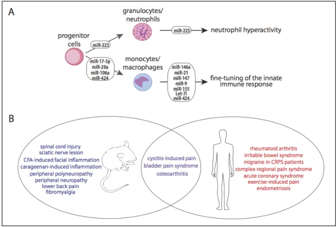
Granulocytes (neutrophils, eosinophils, basophils) and mononuclear cells
(monocytes, lymphocytes, macrophages) each play important roles in defending the body
(see Chapter 1.2.2). MiR-223 plays an important role in granulocyte differentiating, as well
as in the development and function of neutrophils, which act as the first line of defense
against pathogens. Other miRNAs, including miR-17-5p, miR-20a, miR-106a, and miR-424
are involved in the regulation and differentiation of monocytopoiesis, or the production of
mononuclear cells. Several different miRNAs are either upregulated or downregulated in
response to pathogen-associated molecular patterns (PAMPs) such as lipopolysaccharide
(LPS) stimulation in mononuclear cells. Activation of toll-like receptors (TLR2, TLR4, TLR5)
or inflammatory cytokines (IL-1 β) affects the expression of miR-146a and miR-147 in
mononuclear cells. MiR-21 and miR-9 are each responsible for the negative regulation of
LPS-activated inflammatory signaling. MiR-155 and miR-17-92 play distinct roles in
lymphocyte development and function.104 Let-7i and miR-125b are decreased during innate
immune responses. A summary of miRNA regulation of immune cells can be found in Figure
1.3A.
MiRNAs have also been directly linked to the expression of inflammatory cytokines.
the regulation of inflammatory cytokine secretion. Additionally, miRNAs are regulators of
nuclear factor kappa-light-chain-enhancer of B cells (NF-κB), which controls inflammatory
genes and cytokine secretion and is chronically active in many inflammatory diseases.109,110
Collectively this demonstrates that miRNAs play an important role in the immune system
and can alter the immune response.
Aberrant miRNA profiles have been observed in various rodent models of pain and
inflammation. This includes inflammatory pain models such as CFA,111,112 carrageenan,113
and capsaicin112 injections as well as neuropathic pain models such as peripheral nerve
ligations and SNLs.112,114,115 Rodent studies have also linked miRNA expression to
nociceptor excitability and pain thresholds.116 Furthermore, intrathecal injections of miR-103
and miR-124, which were previously identified as regulators of pain signaling, have been
shown to prevent and alleviate pain in rodent models.117,118 These are the first studies to
support the use of miRNAs as treatment for pain.119
MiRNA dysregulation has similarly been associated with pain-related diseases in
humans. For example, miR-146a is significantly upregulated in peripheral knee joint tissues
of osteoarthritis (OA) patients. It regulates pain-related factors, modulates inflammatory
molecules, and may be used in the future as a treatment target for OA.120 Likewise, miRNA
dysregulation has been linked to rheumatoid arthritis104 and complex regional pain syndrome
(CRPS). In CRPS, miRNA profiles can be used to stratify patients into sub-groups that may
respond to treatment plans differently.121 More recently, miRNA dysregulation has been
linked to IPDs such as fibromyalgia,122 bladder pain syndrome (BPS),123 and migraines.124 A
list of additional pain-relevant animal models and clinical conditions that have been linked to
miRNA dysregulation is shown in Figure 1.3B.
To summarize, miRNA dysregulation plays an important role in many pain-relevant
processes and conditions. The dysregulation of pain-regulating miRNAs may facilitate the
relationship between miRNAs and pain and how that relationship might be exploited to
develop more effective therapeutic strategies for IPDs. The studies presented in this
dissertation aim to elucidate the role of miRNAs in idiopathic pain, utilizing experiments in an
animal model as well as in clinical IPDs.
1.3 The Contribution of Catechol-O-methyltransferase
1.3.1 Catechol-O-methyltransferase and Persistent Pain
As discussed in Chapter 1.2.1, there is strong evidence of a role for COMT in
persistent and idiopathic pain. COMT is an enzyme that metabolizes catecholamines such
as dopamine, epinephrine, and norepinephrine. Its active site consists of an
S-adenosyl-L-methionine (AdoMet) binding site and a catalytic site. In the presence of Mg2+ COMT
catalyzes the transfer of the AdoMet methyl group to one of the two hydroxyl groups of the
catechol. The COMT gene codes for two forms: membrane bound (MB-COMT) and soluble
(S-COMT). S-COMT is dominantly expressed in the majority of tissues; however, in the
brain 70% of total COMT is MB-COMT.125 COMT expression varies across tissue types as a
result of tissue-specific transcription factor-mediated gene regulation.126
Decreased activity of COMT has been directly linked to pain-relevant conditions such
as facial pain.127,128 Diminished COMT activity leads to abnormalities in catecholamine levels and physiology,125 which have also been linked to pain and IPDs. In fact, previous studies
have utilized catecholamine excretion as a measure of pain-relevant conditions such as
rheumatoid arthritis.129 Patients with FM demonstrate increased levels of norepinephrine,
which increase further in response to enhanced pro-inflammatory cytokine expression.130,131
Patients with myofacial pain132,133 and chronic bladder pain134 similarly demonstrate
sustained elevation of catecholamines and augmented sympathetic signaling.
Catecholamine abnormalities have also been linked to psychological characteristics
1.3.2 Catechol-O-methyltransferase is Influenced by Genetic and Environmental Factors
Historically, COMT research has focused on a common single nucleotide
polymorphism (SNP) in the gene locus (rs4680), which causes a valine (Val) to methionine
(Met) substitution at codon 158 and leads to a four-fold reduction of the enzyme. Individuals
with the Met/Met genotype have reported higher sensory and affective pain ratings and have
a higher regional density of µ-opioid receptors. Additionally, this polymorphism can influence
the efficacy of morphine treatment for pain.139 This polymorphism alone, however, has not
been able to fully account for individual variation in pain sensitivity, as results across studies
have been largely inconsistent.140
Our laboratory has also demonstrated the importance of haplotypes consisting of
rs4680 along with three other common SNPs (rs6269, rs4633, rs4818, and rs4680) to
persistent pain. We identified three haplotypes in the COMT gene locus, which account for
variation in pain sensitivity among individuals: the G_C_G_G haplotype, associated with a
low pain sensitivity (LPS) phenotype; the A_T_C_A haplotype, associated with an average
pain sensitivity (APS) phenotype; and the A_C_C_G haplotype, associated with a high pain
sensitivity (HPS) phenotype. The LPS haplotype provides 4.8 times higher levels of COMT
activity compared with the APS haplotype. The HPS haplotype provides 11.4 times lower
levels of COMT activity compared with the LPS haplotype. In addition to increased
experimental pain sensitivity, the HPS phenotype is associated with TMD onset. During a
three-year observational period, individuals with only HPS and/or APS haplotypes were
more than twice as likely to develop TMD than those with at least one LPS haplotype.141
Individuals with HPS COMT haplotypes are also more likely to develop pain or IPDs
following traumatic events such as surgeries,142,143 car accidents,144 and orthodontic
treatment.90 Additionally,COMT haplotypes are associated with variance in pain severity for
Variability in the COMT gene also determines efficacy in the treatment of pain
disorders by propranolol, a non-selective beta-adrenergic receptor (βAR) antagonist that
effectively blocks COMT-dependent mechanical and thermal pain in rodents147 and is
commonly used to treat chronic daily headaches in the clinic.148 Propranolol substantially
improves experimental and clinical pain in TMD subjects not carrying a LPS haplotype. The
pain is however only moderately improved in LPS heterozygotes, and not affected at all in
LPS homozygotes.140
To summarize, decreased COMT activity levels lead to increased catecholamine
levels, resulting in heightened pain sensitivity (Figure 1.2). Genetic variability in the COMT
gene signficantly alters an individual’s response to experimental pain, susceptibility to IPDs
and responsiveness to treatment. This is an important consideration that must be
considered by clinicians who treat individuals with idiopathic pain. Future research should
focus on elucidating the mechanisms underlying COMT-dependent pain.
1.3.3 An Animal Model of COMT-Dependent Pain
Though animal models have been instrumental in developing a better understanding
of the mechanisms underlying pain, many existing models do not accurately reflect human
IPDs. Instead, they often utilize inflammatory agents to provoke pain. Many of these models
require persistent injections or applications of the chosen drug or chemical. Other animal
models induce pain by inflicting nerve damage. These models, though useful for the study of
inflammatory or neuropathic pain, do not accurately represent IPDs (Chapter 1.1.1). As a
result, many potential analgesics have failed in the clinic despite strong efficacy in a
pre-clinical animal model.7 Our laboratory has developed a rat model of persistent idiopathic
pain in which a COMT inhibitor is administered to rats to mimic the endogenously low levels
of COMT activity observed in patients (Chapters 1.3.1 and 1.3.2 explore the role of COMT in
animal models that rely on repeated administration of noxious stimuli. We have utilized this
model to gain a better understanding of the mechanisms underlying COMT-dependent pain.
Consistent with clinical IPDs, administration of a COMT-inhibitor to rats results in
mechanical and thermal pain and alters cognitive-affective behaviors linked to pain (e.g.,
avoidance of painful heat and bright light).147,149,150 Because catecholamines stimulate the
sympathetic nervous system via activation of adrenergic receptors (ARs), pharmacologic
studies were utilized to assess the efficacy of alpha (α1 and α2) and beta (β1, β2, and β3)
adrenergic as well as dopaminergic receptor antagonists in blocking OR486-induced pain.
Results demonstrated that OR486-induced pain is blocked by the non-selective β
-adrenergic receptor (βAR) antagonist propranolol or by the combined administration of
selective β2- and β3AR antagonists. In contrast OR486-induced pain is not blocked by β1
-adrenergic, α-adrenergic, or dopaminergic receptor antagonists.147 These results are in line
with those from clinical studies, showing that propranolol alleviates pain among FM and TMD patients.140,151 Collectively these studies suggest that increased catecholamine levels,
resulting from reduced COMT activity, drive pain viaβ2- and β3ARs.
i. β-Adrenergic Receptors and COMT-Dependent Pain
ARs are G protein-coupled receptors (GPCRs), responsible for transmitting signals
from extracellular ligands to the intracellular environment to control various cellular events
and physiological processes. Though several ARs exist—alpha-adrenergic (α1 and α2),
beta-adrenergic (β1, β2, and β3) and dopaminergic—only β2- and β3ARs are known to contribute
to COMT-dependent pain (see Chapter 1.3.3). βARs were first discovered in 1948 and have
been heavily studied in the context of heart disease. Though βAR antagonists, otherwise
known as β-blockers, were developed for the treatment of heart failure in 1975,152 it was not
long before their efficacy was discovered for pain reduction in conditions such as arthritis
and joint pain.153-155 Today they represent a promising possibility for the treatment of IPDs
Like all GPCRs,βARs have seven transmembrane-spanning α-helices, which fold to
create three extracellular and three intracellular loops. Upon activationβ2- and β3ARs couple
to the Gs guanine nucleotide-binding protein (G protein), activating adenylyl cyclase and
thereby leading to the formation of cAMP and activation of PKA. This represents a specific
and rapid signaling cascade.156,157 G
s interaction sites for both β2- and β3ARs are located in the membrane proximal regions of the second and third intracellular loops as well as at the
carboxy-terminal domains.157 Although many of the actions of both β2- and β3ARs are
mediated by Gs proteins, studies have demonstrated that both receptor types can also
couple to Gi proteins, leading to the activation of extracellular signal-regulation kinase (ERK)
and MAPK pathways.156,158
β2- and β3ARs are expressed in peripheral and central regions where they could
drive pain. β2ARs are located on peripheral terminals159-163 and cell bodies164-166 of primary
afferent nociceptors, keratinocytes,167-169 immune cells,170-173 and adipocytes174 in the
periphery and neurons175,176 and glial cells177 in the central nervous system. β
3ARs are
located on primary afferent nociceptors,178 adipocytes,174 and immune cells171,172 in the
periphery and noradrenergic neurons in the brain.179 A site of action for the β
2- and β3ARs
that mediate COMT-dependent pain has yet to be determined. The studies presented in this
dissertation aim to identify a site of action for the βARs receptors that mediate
COMT-dependent pain using clinically-relevant rodent models of COMT-COMT-dependent pain that
improve upon those used in prior studies.
ii. MicroRNAs and COMT-Dependent Pain
It is important to note that miRNAs, which are discussed in detail in Chapter 1.2.3,
may also play a role in COMT-dependent pain. It is likely that COMT-dependent pain is
associated with the dysregulation not only of miRNAs involved in pain and immune
regulating β2AR expression.180 Furthermore, βAR stimulation induces expression of
miR-21181 as well as miR-199a-5p.182 It is thought that the cardioprotective effects of βAR
antagonist propranolol are in part a result of miR-1 downregulation.183 Several miRNAs have
been associated with reduced catecholamine sensitivity in the heart, a characteristic of
chronic heart failure.184 Though many ongoing studies are examining the relationship
between miRNAs and βAR-mediated cardiac disease; the relationship between miRNAs,
βAR signaling, and COMT-dependent pain has yet to be studied.
The studies presented in this dissertation aim to elucidate the role of miRNAs not only in
persistent idiopathic pain, but also specifically in COMT-dependent pain.
1.4 Summary and Specific Aims
Though IPDs are heterogeneous in nature, they are generally characterized by a
physical state of pain amplification, psychological distress, and enhanced inflammation
(Chapter 1.1.2). Persistent idiopathic pain manifests without an apparent cause, but it is
facilitated by both environmental (Chapter 1.2.1) and genetic (Chapter 1.2.2) factors. One
gene of particular interest codes for COMT and has been linked to increased pain sensitivity
and susceptibility to developing IPDs (Chapter 1.3). Though studies have demonstrated that
COMT-dependent pain is mediated viaβ2- and β3ARs, the site of action whereby these
receptors mediate COMT-dependent pain remains unknown (Chapter 1.3.3). Chapters 2
and 3 therefore aimed to determine the site of action whereby β-adrenergic systems drive
persistent COMT-dependent pain. We hypothesized that peripheral, spinal, and supraspinal
βARs would contribute to COMT-dependent pain. We found that peripheral but not spinal or
supraspinal β2- and β3ARs contribute to the initiation, but not the maintenance, of
COMT-dependent pain.
Epigenetic modifications such as miRNA regulation can be initiated by environmental
susceptible individuals (Chapters 1.2.3 and 1.3.3). Though emerging evidence suggests a
role for miRNAs in idiopathic pain, the relationship between miRNAs and COMT-dependent
pain has not been widely studied. Chapter 4 aimed to identify miRNAs associated with IPDs.
We hypothesized that miRNA dysregulation would be associated with idiopathic pain in a
clinical setting. As hypothesized, we observed miRNA dysregulation in patients with IPDs.
We also found that miRNAs may be useful for distinguishing between separate subtypes of
IPDs. Collectively, the present studies may help to inform future care for patients by
elucidating the etiologies and identifying targets for treatment of idiopathic pain.
Figure 1.1 Idiopathic pain disorders are influenced by environmental and genetic factors. This model depicts environmental and genetic factors that determine an individual’s psychological profile and pain amplification status. These two domains can influence the risk of onset and maintenance of IPDs in patients. Adapted from: “Idiopathic pain disorders: Pathways of vulnerability” by L.L. Diatchenko, 2006, PAIN.91 Abbreviations: brain-derived
Figure 1.2 Decreased COMT activity is associated with idiopathic pain disorders. Genetic variability in the COMT gene can result in decreased levels of COMT activity and subsequently increased levels of circulating catecholamines (e.g., epinephrine,
Figure 1.3 MicroRNA dysregulation can affect immune cell function and is associated with pain-relevant animal models and clinical conditions. (A) MiRNAs play an important role in the development and function of immune cells, subsequently fine-tuning the innate immune response and protecting the host from injury. Dysregulation of the miRNAs that are involved with immune system regulation may result in chronic inflammation and pain. Adapted from: “MicroRNA, a new paradigm for understanding immunoregulation,
inflammation, and autoimmune diseases” by R. Dai and S.A. Ahmed, 2011, Translational Research.104(B) MiRNA dysregulation has been associated with various animal models of
CHAPTER 2
PERSISTENT CATECHOL-O-METHYLTRANSFERASE-DEPENDENT PAIN IS INITIATED BY PERIPHERAL BETA-ADRENERGIC RECEPTORS 1,2
2.1 Introduction
Idiopathic pain disorders (IPDs) including fibromyalgia (FM), headache,
temporomandibular disorder (TMD), and vestibulodynia (VBD) constitute a significant
healthcare problem, affecting over 100 million Americans.185-191 These disorders occur more
frequently in females than males192 and are chronic in nature, characterized by pain that
occurs daily and spans years. While the mechanisms underlying IPDs are poorly
understood, emerging evidence indicates a role for adrenergic pathways. Patients exhibit
increased levels of catecholamines131-133 alongside diminished activity of
catechol-O-methyltransferase (COMT),127,128 a ubiquitously expressed enzyme that metabolizes
catecholamines to their inactive derivatives.125 An increase in catecholaminesis is similarly
observed in patients with inflammatory conditons such as arthritis and complex regional pain
syndrome (CRPS).193-195 Furthermore, functional variants in the COMT gene that reduce
COMT activity128,196,197 are associated with increased susceptibility to FM,145,146,198-200
TMD,141 and experimental pain141,201 as well as impaired response to treatment.139,202 It is
estimated, based on the frequency of allele variation, that nearly two-thirds of patients with
persistent pain disorders possess the low-activity COMT variants.203,204
Consistent with clinical disorders, our lab found that administration of the COMT
inhibitor OR486 in rodents produces increased hypersensitivity at multiple body sites and
alters cognitive-affective behaviors linked to pain (e.g., avoidance of painful heat and bright
by administration of the non-selective βAR antagonist propranolol or by combined
administration of selective β2- and β3AR antagonists.147,149,150 These results are in line with
those from clinical studies, showing that propranolol alleviates pain among FM and TMD
patients.140,151 Collectively, these studies suggest that increased catecholamine levels,
resulting from reduced COMT activity, drive pain viaβ2- and β3ARs.
β2- and β3ARs are G protein-coupled receptors (GPCRs) expressed in peripheral
and central regions where they could mediate pain perception and/or processing. β2ARs are
located on peripheral terminals159-163 and cell bodies164-166 of primary afferent nociceptors,
keratinocytes,167-169 immune cells,170-173 and adipocytes174 in the periphery; and neurons175,176
and glial cells177 in the central nervous system. β3ARs are located on primary afferent
nociceptors,178 adipocytes,174 and immune cells171,172 in the periphery and noradrenergic
neurons in the brain.179 Thus, the aim of this study was to determine a site-of-action for
COMT-dependent pain. We hypothesized that peripheral, spinal, and/or supraspinalβ2- and
β3ARs contribute topersistent COMT-dependent pain.
To test this hypothesis, we employed a clinically-relevant model of persistent
COMT-dependent pain and evaluated responses to mechanical and thermal stimuli in
adrenalectomized rats, lacking peripheral epinephrine, and in intact rats receiving
continuous delivery of βAR antagonists via intraplantar (i.pl.), intrathecal (i.t.), or
intracerebroventricular (i.cv.) routes. Potential sexual dimorphism in the contribution of
adrenergic systems to persistent COMT-dependent pain was also assessed.
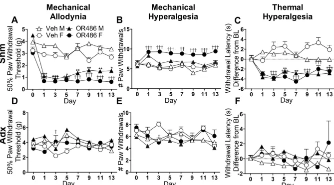
Results demonstrated that male and female rats receiving sustained OR486
exhibited COMT-dependent mechanical and thermal pain, persisting for two-weeks. In
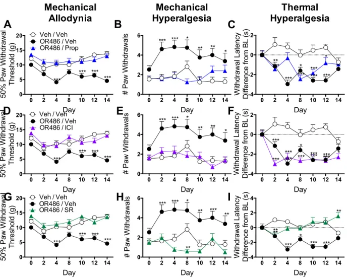
contrast, adrenalectomized rats failed to develop OR486-induced pain. Furthermore, i.pl.,
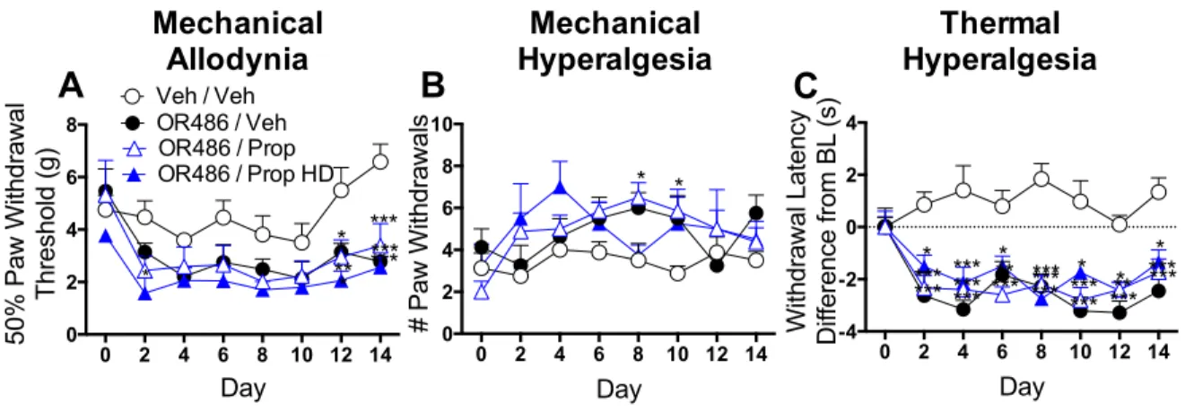
but not i.t. or i.c.v., administration of the non-selective βAR antagonist propranolol, β2AR
antagonist ICI118,551, or β3AR antagonist SR59230A blocked OR486-induced pain. These
pain, and suggest that peripherally-acting βAR antagonists may provide an effective
treatment option for patients with persistent pain disorders.
2.2 Materials and Methods
2.2.1 Animals
Adult male and female Sprague-Dawley rats (N=24 intact, N=24 adrenalectomized
and N=23 sham) were purchased (Charles River Laboratories, Raleigh, NC) for the first set
of experiments. For subsequent βAR antagonist experiments, adult male Sprague-Dawley
rats (N=111) were bred in-house. Rats weighed between 200 and 400g for all experimental
studies. Rats had ad libitum access to standard laboratory chow and water.
Adrenalectomized rats were provided with saline water (0.9%) to compensate for the loss of
sodium in urine due to the absence of aldosterone. All animal procedures were approved by
the Institutional Animal Care and Use Committee (IACUC) at the University of North
Carolina at Chapel Hill (UNC). Though rodent models of pain only partially correlate with
human conditions, rats were chosen for these experiments because an extensive body of
literature exists regarding nociceptive pathways and behavior in this species, and because
rat pain behavior assays are readily available and well characterized.7,205,206
2.2.2 General Experimental Conditions
First, the effects of sustained COMT inhibition on pain were evaluated in intact rats
receiving the COMT inhibitor OR486 or vehicle systemically for a two-week period via a
2002 Alzet osmotic pump (Durect Corporation, Cupertino, CA). Next, the contribution of
peripheral adrenergic systems to persistent OR486-induced pain was evaluated in
adrenalectomized rats, lacking peripheral epinephrine, or sham rats receiving OR486 or
vehicle systemically for two-week period via an osmotic pump. Finally, the contribution of
peripheral, spinal and supraspinal βARs to persistent OR486-induced pain was evaluated in
delivery of OR486 or vehicle for a two-week period via an osmotic pump. The βAR
antagonists were delivered via a catheter attached to a separate 2002 Alzet osmotic pump.
Animals were handled and habituated to the experimenter and environment for four
days prior to testing. Responses to punctuate mechanical and thermal stimuli were
assessed in intact and adrenalectomized animals 1 day prior to and on days 1, 3, 5, 7, 9, 11,
and 13 following pump implantation. For βAR antagonist experiments, pain behaviors were
assessed one day prior to and on days 2, 4, 6, 8, 10, 12, and 14 following pump
implantation. The rest day between surgery and testing allowed animals to fully recover from
catheter implantation. On baseline and testing days, rats were habituated to the mechanical
and thermal testing environments for ten to fifteen minutes. Though we were unable to
eliminate all environmental factors (e.g., season, humidity, noise) from this study, we
minimized others (e.g., experimenter consistency, testing time of day, cage density) that
were in our control.207,208 Animals were randomly assigned to groups, were tested by a
single, blinded experimenter at a consistent time of day (morning), and were housed with
one to two other rats. The primary outcome reported in this study is behavioral changes, in
the form of mechanical allodynia, mechanical hyperalgesia, and thermal hyperalgesia, which
are described in detail below under their respective subtitles.
2.2.3 Drug Preparation
OR486 (Tocris, Ellisville, MO) was dissolved in a 5:3:2 ratio of dimethylsulfoxide
(DMSO), 0.9% saline, and ethanol.147 For peripheral experiments βAR antagonists
propranolol hydrochloride (Tocris, Ellisville, MO), ICI-118,511 (Tocris, Ellisville, MO), and
SR59230A (Tocris, Ellisville, MO) were each dissolved in 5:3:2 ratios of DMSO, 0.9% saline,
and ethanol. For i.t. and i.c.v. experiments, βAR antagonists were dissolved in 0.9% saline.
Drug solutions were injected into pumps, which were placed in 15mL conical tubes
containing sterile 0.9% saline and primed overnight in a dry heat bath (Lab Armor,