S
URGICAL ASPECTS OF
S
URGICAL ASPECTS OF
PEDIATRIC URINARY INCONTINENCE
(met een samenvatting in het Nederlands)Proefschrift
ter verkrijging van de graad van doctor aan de Universiteit Utrecht
op gezag van de Rector Magnificus, Prof. dr H.O. Voorma, ingevolge het besluit van het College voor Promoties
in het openbaar te verdedigen op 12 januari 2001 te 16.15 uur
door
Thomas Pius Vianney Maria de Jong Geboren op 15 januari 1952 te Heerlen
Promotor : Prof. dr T.A. Boon Co-promotor : Prof. dr J.D. van Gool
CIP-data Koninklijke Bibliotheek, den Haag Jong, Thomas Pius Vianney Maria de
Surgical aspects of pediatric Urinary Incontinence Tom P.V.M. de Jong
Utrecht, Universiteit Utrecht, Faculteit Geneeskunde Thesis Universiteit Utrecht
ISBN 90-9014211-8
Dit proefschrift is opgedragen aan de ouders van de geopereerde kinderen die steeds opnieuw het vertrouwen hebben moeten schenken om hun kind operaties te laten ondergaan waarvan het resultaat vooraf vaak onzeker is geweest. Zij zijn de helden in dit proefschrift.
Daarnaast vanzelfsprekend aan Anke, Jet, Jelle en Simone die dit alles met minimaal mopperen hebben laten gebeuren.
Contents
Introduction ix Chapter 1: Structural incontinence 1 Chapter 2: Functional incontinence 11 Chapter 3: Ectopic ureteroceles 27 Chapter 4: Post-Otis incontinence 45 Chapter 5:Surgery for female ambiguous genitalia 59
Chapter 6:
Bladder extrophy and epispadias 69
Chapter 7:
Female epispadias repair 89
Discussion 100 Summary 102 Samenvatting 107 Dankwoord 112 Curriculum vitae 114 Publications 116
Introduction
Incontinence at pediatric age is a problem that can harm the psychological and physical development of children. Starting in 1986 we have searched for better solutions to treat incontinent children, by medication as well as by training and surgery. Important questions have been what surgical or non-surgical solutions do exist and what are the possibilities to improve the current techniques? This thesis is a synopsis of our publications on this theme.
The seven Chapters in this thesis deal with structural incontinence in chil-dren caused by congenital or acquired malformations of the urinary tract. Chapters 1, 2, 3 and 4 describe the sequelae of anomalous development
Figure 1. A. Wolffian duct zones are shown in relation to nephrogenic area. Note direction of migration of kidney cranially and Wolffian duct into developing bladder. B. Migration of Wolffian duct zones into the bladder is completed. C and D show the development of a normal functioning double system with orthotopic ureteral orifices when double ureteral buds arising from normal Wolffian zone strike nephrogenic area and migration occurs in direction of arrows. E. Orthotopic bud arises from normal zone and strikes normal nephrogenic area. Ectopic bud strikes diminished tissue and migration occurs. F. After migration, the lower pole ureter is too lateral resulting in reflux and/or lower pole dysplasia. G. Ectopic bud arises too far laterally on Wolffian duct and strikes abnormal nephrogenic area. H. The ectopic upper pole ureter ends in the caudal zone and the upper pole of the kidney will be dysplastic. (with permission by the Williams and Wilkins Co.)
of Wolffian ducts. Chapter 5 delineates the concept of structural inconti-nence in children born with ambiguous genitalia and a patent urogenital sinus. Chapters 6 and 7 present new insights in the treatment of children that have been born with bladder extrophy and epispadias.
Anomalous development of Wolffian and Müllerian ducts
During the first weeks of gestation, the interactions between the Wolffian and Müllerian ducts, the urogenital sinus and the metanephros define the functional and anatomical outcome of the urinary tract, from the top of the kidney to the tip of the urethra in females, and to the end of the pro-static urethra in males.
The Wolffian duct delivers material that develops into ureter, trigone, bladder neck and urethra. For a normally functioning urinary tract the Wolffian duct must deliver one or two ureteral buds at the right place to the core of the metanephros, in order to develop into a functioning kid-ney and collecting system. When the ureteral bud develops from a site on the Wolffian duct that prohibits contact with the optimal part of the metanephros, a dysplastic kidney or a dysplastic moiety of a double sys-tem will result. Moreover, ectopic delivery of the ureteral bud from the Wolffian duct results in an ectopic site for the ureteral orifice. This ectopic site can be located either cranially or caudally of the orthotopic position. A cranial ectopic position of the ureteric orifice results in vesicoureteral reflux, in severe cases combined with renal dysplasia. A caudal ectopic position of the ureteric orifice, below the level of the bladder neck, may result in dribbling incontinence and dysplasia of the corresponding renal moiety. An ectopic ureter may be accompanied by a congenital gap in the bladder neck, leading to bladder outlet insufficiency.
The clinical result of this combination of congenital malformations can be, simultaneously, dribbling incontinence, true congenital stress inconti-nence, and non-neuropathic bladder/sphincter dysfunction due to the constant presence of urine in the proximal urethra1. In conclusion, a small
derailment in the embryological development of the ureteral bud and the Wolffian duct is able to cause dysplastic changes of the kidney as well as anatomical and functional anomalies of the trigone, bladder neck and ure-thra. This mechanism has been described superbly by Mackie and Douglas Stephens2(Figure 1).
chapter 1
In Chapter 1, the types of structural incontinence based on anomalous development of the ureteral bud and the Wolffian duct are described, jux-taposed with the standard-textbook-causes of structural incontinence in children. A complete list of structural causes is mandatory in the manage-ment of incontinent children, and true congenital stress incontinence should not be left out.
chapter 2
Patients with lower urinary tract symptoms based on anomalous develop-ment of the ureteral bud and the Wolffian duct will generally present with symptoms that fit into the category of non-neuropathic bladder/sphinc-ter dysfunction, the clinical signs of which are symptoms of urge syn-drome or dysfunctional voiding. In most patients, suspicion of structural anomalies arises only when treatment with a well-structured bladder train-ing program has been proven a failure. Even in the case where a structur-al anomstructur-aly of bladder neck or urethrstructur-al sphincter is the principstructur-al suspect, the first step in the treatment will often be a try-out with a training pro-gram, to prove that the condition is resistant to conservative treatment. This implies that the availability of a structured training program for the treatment of non-neuropathic bladder/sphincter dysfunction is a conditio sine qua non for departments dealing with structural pediatric inconti-nence. Without specific training experience the patients will be subjected to undue long conservative treatment plans that conceal the underlying surgical condition. In an editorial comment in the Journal of Urology the statement is made that a urotherapist needs to have the anatomical and physiological insight of a pediatric urologist, the patience of a pediatri-cian, and the tact of a psychologist3. Chapter 2 reviews non-neuropathic
bladder/sphincter dysfunction of which urge syndrome and dysfunctional voiding are the main elements. The review is helpful to clarify the distinc-tion between funcdistinc-tional and structural incontinence. The review also delineates the therapeutical approaches that need to be followed in all patients, before deciding on surgery.
chapter 3
Patients with ectopic ureteroceles, not detected by prenatal ultrasonogra-phy, are presented with recurrent urinary tract infections, dysfunctional voiding and incontinence. Much discussion exists on two major items. Firstly, the question whether one should opt for primary complete recon-struction, or try and treat these patients with minimal surgery, implying a multi-stage approach with endoscopic incision of the ureterocele, with or without removal of the upper moiety of the affected kidney, possibly fol-lowed by lower urinary tract surgery. Secondly, the question whether major bladder surgery in neonates can be justified, in view of the long-standing (non-evidenced) belief that such surgery in the first year of life can seriously impair future bladder function. Backed up by urodynamic expertise and because of disappointing results with patients treated with a staged approach, we have opted for a one-stage complete reconstruction in all of our patients, regardless of age.
chapter 4
Historically, patients with vesicoureteral reflux and recurrent urinary tract infections have been treated by blind internal Otis-urethrotomy or ure-thral dilatation. This treatment was based on the assumption that the so-called spinning-top urethra, as visualised with the voiding cysto-urethrography used to detect reflux, represents a urethral obstruction. Urodynamics in children with urge syndrome or with dysfunctional void-ing has taught us that the spinnvoid-ing-top urethra represents a functional anomaly, caused by simultaneous contraction of the detrusor muscle and the pelvic floor muscles during voiding or during filling of the bladder. As a result of a wide-spread urethrotomy policy we have been confronted with a series of girls with severe iatrogenic stress incontinence, combined with symptoms of the primary disorder: urge syndrome or dysfunctional voiding. In Chapter 4 we describe the difficult quest for a safe and defini-tive solution of the incontinence in these girls.
chapter 5
Both chromosomal anomalies and endrocrinological disorders can pro-duce ambiguous genitalia with a patent urogenital sinus. Whenever female gender is assigned to these children, both a genitoplasty and a vaginal
pull-through operation needs to be done. Historically, staged procedures and delayed reconstructions have been performed, in many cases with lower urinary tract dysfunction as a result. These lower urinary tract symptoms are based on urinary retention in the vagina and on urethral insufficiency due to the shortness of urethra and/or a bladder neck defect. Clinically, the lower urinary tract symptoms will fit into the category of non-neuro-pathic bladder/sphincter dysfunction.
Next, the psychological impact of major genital surgery at a later age is an important factor in gender identity disturbances, as well as a possible con-founder in the treatment of the bladder/sphincter dysfunction. In search for a way to avoid the lower urinary tract complications and to minimise psychological disturbances for the children and parents, we ended up with a one-stage procedure at neonatal age as the best possible option.
chapters 6 & 7
Patients with bladder extrophy/epispadias are generally presented as a neonatal emergency. Girls with epispadias are sometimes misdiagnosed as intersex patients. Major congenital anomalies such as the bladder extro-phy/epispadias complex force us to constantly pursue an optimal result in terms of the number of operations, the time of hospitalisation, the uri-nary tract morbidity, the preservation of renal function, the rate of conti-nence and the quality of life for the patient. The complex problem of female epispadias (girls born without a urethra and without a sphincteric mechanism) has also raised the question how to diminish morbidity and reduce the number of operative procedures. This question eventually prompted us to design a one-stage operative procedure that shows prom-ise when compared with existing approaches. In Chapter 6, an overview of the treatment of bladder extrophy/epispadias is presented, and in Chapter 7 the results of our technique for female epispadias repair are described.
References
1. van Gool JD, Tanagho EA. External sphincter activity and recurrent urinary infection in girls. Urology 1977; 10:348-353
2. Mackie GG, Douglas Stephens F. Duplex kidneys: a correlation of renal dysplasia with position of the ureteral orifice. J.Urol 1975;114:274-280.
Structural Incontinence
Structural incontinence in childhood
CHAPTER 1
Introduction
Many structural anatomic causes for incontinence exist at pediatric age. Several, such as the exstrophy-epispadias complex, are well known to everybody; many others are unknown to the majority of physicians deal-ing with children. The embryology of the urinary tract teaches us that the ureter, trigone, bladder neck and proximal urethra in the male, and the whole urethra in the female, are derivates of the distal part of the Wolffian duct. This means that derailment of a small part of the distal Wolffian duct can lead to a cascade of anomalies in the urinary tract. In fact, embryology can provide a completely logical explanation for severe reflux, ipsilateral renal dysplasia and bladder neck insufficiency in one and the same patient. The same holds true for the combination of dribbling incontinence due to an ectopic ureter and stress incontinence based on bladder neck insufficiency. This is a subject with very sparse backing from published data.
Anatomic causes for incontinence in children
Primary stress-incontinenceIn children, stress incontinence occurs as a stand-alone disease or as a symptom in children with hyperlaxity of the joints. Clinically, it can pres-ent as an urge syndrome, due to forced hold manoeuvres with the pelvic floor, resulting in squatting and urge incontinence, predominantly pro-voked by stress. Anatomically, the lower urinary tract is usually normal, except for a relatively flat vesico-urethral angle. It occurs more frequently in conjunction with a hypospadiac urethral orifice, inside the hymenal ring, in patients with a relatively short urethra. There is only sparse litera-ture on this subject.1-9
Stress and/or urge incontinence due to bladder neck insufficiency
When dealing with children suffering from incontinence and recurrent UTI due to urge syndrome, an open bladder neck on cystography or ultra-sound is a common finding. In the anatomically normal child the bladder
neck will present as a ring with a dilated urethra down to a mid-urethral ring at the level of the pelvic floor, with the bladder filled to capacity under anesthesia. However, a group of patients exists with a clear gap in the bladder neck, a gap that appears to be a congenital bladder neck defect. When such a gap leads to urinary leakage into the urethra, the child will try to cope with this leak by using the pelvic floor as emergency brake. Clinically, the child may present as a case of urge syndrome. A con-genital bladder neck defect can be suspected when biofeedback training fails. Such a defect has been described in combination with an ectopic ureter, it is apparent with double ectopic ureters, and it can occur as a sin-gle anomaly leading to incontinence.
Bladder neck insufficiency also occurs in girls with ectopic ureteroceles-ureteroceles running through the bladder neck into the urethra. However, a large group of these children appears not to suffer from incontinence. The literature rarely includes urinary continence as a factor in the follow-up of the treatment of ectopic ureteroceles.
Urogenital sinus with bilateral ectopic ureters is another candidate for bladder neck insufficiency. Also at risk for urethral insufficiency are patients with adrenogenital syndrome with a high ending vagina. We do not know how to classify the female hypospadias, with an urethral meatus placed inside the hymenal ring and a relatively short urethra, but these children can present clinically with either stress incontinence or urge syn-drome.10-15
Urethral obstruction in boys
In boys, urethral obstruction leading to bladder overactivity and urge-incontinence is common. Urethral obstruction in boys can occur along the whole course of the urethra. Bladder neck obstruction can be primary, or secondary to a more distal obstruction. Utricle cysts or a cystic cap covering the colliculus can lead to obstruction.
Posterior urethral valves are the commonest cause of urethral obstruction in boys. Several types are recognised. By far the most common valves arise from the colliculus and present as a membrane that partially closes the urethra.16-19A few millimeters past the sphincteric mechanism the
pas-sage of the urethra through the pelvic floor can cause obstruction, a con-stricting ring known as Cobb’s web or Moorman’s ring.20-24Just distal of
the pelvic floor, cystic anomalies of Cowper’s gland duct can present as syringocele.25 In the same area, anterior diverticula, that can be a direct
result of a destroyed syringocele, can arise. More distally in the pars bul-bosa or pars pendulans of the urethra, diverticula arise very rarely.
A rare and very nasty anomaly is congenital urethral stenosis, that occurs mostly starting from the penoscrotal angle.
In the penile urethra rare obstructions can be found based on obstruction of the duct of a urethral gland, a cyst of the duct of Littré’s gland. More commonly, obstruction is caused by the fossa naviculare, a stenosis of the last centimeter of the urethra.26-32
Common obstructions occur in boys at the urethral meatus, especially after circumcision, by friction of the meatus to the clothes. Rarely is an obstruction caused by debris in a fossa naviculare sinus, a sinus running parallel to the distal urethra. Relatively rare is true outlet obstruction based on phimosis in the uncircumcised boy.
It is important to state that voiding cystourethrography (VCUG) is an unreliable investigation in diagnosing urethral obstruction in boys. In our experience, comparing urodynamic results with VCUG, false-negative results of VCUG are found in 60%, while only 6% false negative results occurr with urodynamics.
Urethral obstruction in girls
In girls, urethral obstruction is rare. In the past, it has been diagnosed and treated very frequently, based on the finding of a spinning-top urethra at VCUG.33, 34 We now know that this aspect is caused by a functional
obstruction in the majority of cases.35-47Urodynamically, in our
experi-ence, approximately 2% of the spinning top urethras reflect a true ure-thral obstruction. In the cases with a proximal dilation of the urethra at VCUG, the obstruction can be found 5-10 mm proximal of the meatus. Meatal stenosis does occur in girls. When inspecting the meatus, an anteri-or position is often present. The histanteri-ory often reveals a urinary stream that runs under the toilet seat on the floor in front of the toilet. Very rarely primary bladder neck obstruction can occur in girls. Urethral diver-ticula in girls are rare and can give obstruction. Also rarely, urethral obstruction and incontinence can be provoked by anomalies of the
hymen.48-51A hymen annulare can completely cover the meatus and lead
to micturition into the vagina with vulvovaginitis and post-void dribbling as a result. Finally, severe forms of synechia of the labia minora can lead to obstruction, infections and post-void dribbling.
Epispadias and exstrophy
Nobody doubts the incontinence problems in exstrophy patients. However, we tend to forget that epispadias in boys come with congenital sphincter insufficiency, with or without a gap between the pubic bones. Rare variants of bladder exstrophy such as covered exstrophy or abdomi-nal fissura can come with sphincteric incompetence. The very rare female epispadias present with complete absence of the urethra and the pelvic floor, with the bladder neck at the level of the hymenal ring.
Rare congenital anomalies of the lower urinary tract in females can cause all kinds of urinary leakage. Impressive collections of cavities and sinuses can cause incontinence in combination with variations of cloacal anom-alies and urogenital sinus.
Dribbling incontinence
In girls, true dribbling incontinence, continuously manifest, is mostly caused by ureteral ectopy. In general, an upper pole ureter from a duplex system is involved, rarely a single ectopic ureter can be seen. The ectopic ureter can end in bladder neck, urethra, vulva, vagina, uterus and even in the rectum. The ectopic ureter always will end in a remnant of the Wolffian system. In boys, all Wolffian remnants are proximal of the exter-nal sphincter, thus not resulting in incontinence except for urge com-plaints based on irritation by a ureter ending into the prostate, a seminal vesicle or a vas deferens.
Iatrogenic incontinence
Internal urethrotomy (Otis) has been a standard procedure for recurrent urinary tract infections and reflux until the late 80’s. Fortunately, nature has been mild in most cases. However, incontinence based on urethral scarring does occur and is difficult to treat, usually by complete recon-struction of the urethra after excision of the complete scar.52-58
vesico-vaginal fistulas in less experienced hands. Ectopic ureteroceles can be accompanied by incontinence: unfortunately, incontinence is seldom included as a criterium in assessing the results of ureterocele therapy, and because of that we do not know the prevalence. Incontinence can occur after intersex surgery, especially when a high ending vagina has been pulled through.
Literature
1. Ball, T.L. and Woodruff, J.J.: Stress incontinence-revision of an unsuccessful Marshall-Marchetti operation in a 5-year-old child. J. Urol., 100:492-497, 1968.
2. Murray, K., Nurse, D., Borzyskowski, M. and Mundy, A.R.: The “congenital” wide bladder neck anomaly-a common cause of incontinence in children. Brit. J. Urol., 59:533-535, 1987. 3. Hjälmås, K.: Urinary incontinence in children-suggestions for definitions and terminology
[see discussion]. Scand. J. Urol. Nephrol., Suppl 141, 26:1-6, 1992.
4. Lopez, A.E., Padron, O.F., Patsias, G. and Politano, V.A.: Transurethral polytetrafluoroethyl-ene injection in female patients with urinary continence. J. Urol., 150:856-858, 1993. 5. Stanton, S.L.: Stress urinary incontinence [see discussion]. Ciba Found. Symp., 151:182-189,
1990.
6. Norton, P.A., Baker, J.E., Sharp, H.C. and Warenski, J.C.: Genitourinary prolapse and joint hypermobility in women. Obstet. Gynecol., 85:225-228, 1995.
7. Couillard, D.R. and Webster, G.D.: Detrusor instability. Urol. Clin. North Am., 22:593-612, 1995.
8. McIntosh, L.J., Stanitski, D.F., Mallett, V.T., Frahm, J.D., Richardson, D.A. and Evans, M.I.: Ehlers-Danlos syndrome-relationship between joint hypermobility, urinary incontinence, and pelvic floor prolapse. Gynecol. Obstet. Invest., 41:135-139, 1996.
9. Hendren, W.H.: Construction of a female urethra using the vaginal wall and a buttock flap: experience with 40 cases. J. Pediatr. Surg., 33:180-187, 1998.
10. Mogg, R.A.: The single ectopic ureter. Brit. J. Urol., 46:3-10, 1974.
11. Ogawa, A., Kakizawa, K. and Akaza, H.: Ectopic ureter passing through the external ure-thral sphincter-report of a case. J. Urol., 116:109-110, 1976.
12. Saltutti, C., Stefanucci, S., Di Cello, V., Bartoletti, R. and Bracci, G.: [Ectopic ureter. Description of a clinical case with appearance of stress urinary incontinence]. Minerva Urol. Nefrol., 41:17-22, 1989.
13. Passerini Glazel, G., Milani, C., Bassi, P., Chiozza, L., Rizzoni, G. and Pagano, F.: Bilateral single ectopic ureter. Eur. Urol., 14:454-457, 1988.
14. Ritchey, M.L., Kramer, S.A., Benson, R.C., Jr. and Kelalis, P.P.: Bilateral single ureteral ectopia. Eur. Urol., 14:41-55, 1988.
15. Barrows, B., Bavendam, T. and Leach, G.E.: Ectopically draining dysplastic kidney associat-ed with genuine stress urinary incontinence-unusual combinassociat-ed cause of incontinence. Urology, 40:283-285, 1992.
16. Atwell, J.D.: Posterior urethral valves in the British Isles-a multicenter B.A.P.S. review. J. Pediatr. Surg., 18:70-74, 1983.
17. De Gennaro, M., Mosiello, G., Capitanucci, M.L., Silveri, M., Capozza, N. and Caione, P.: Early detection of bladder dysfunction following posterior urethral valves ablation. Eur. J. Pediatr. Surg., 6:163-165, 1996.
18. Kim, Y.H., Horowitz, M., Combs, A.J., Nitti, V.W., Borer, J. and Glassberg, K.I.: Management of posterior urethral valves on the basis of urodynamic findings. J. Urol., 3 Pt 2, 158:1011-1016, 1997.
19. Holmdahl, G.: Bladder dysfunction in boys with posterior urethral valves. Scand. J. Urol. Nephrol., Suppl 188, 31:1-36, 1997.
20. Mori, Y., Matsui, T., Ogino, T., Hosokawa, S., Tsujimoto, S., Ihara, H., Terakawa, T., Shima, H., Shimada, K., et al.: [Treatment of congenital urethral stenosis (urethral ring) in children. Optic internal urethrotomy in the congenital bulbar urethral stenosis in boys]. Nippon Hinyokika Gakkai Zasshi, 80:704-10, 1989.
21. Cranston, D., Davies, A.H. and Smith, J.C.: Cobb’s collar-a forgotten entity [see comments]. Brit. J. Urol., 66:294-296, 1990.
22. Corriere, J.N.: Re: Cobb’s collar-a forgotten entity [letter; comment]. Brit. J. Urol., 69:220, 1992.
23. Dewan, P.A., Keenan, R.J., Morris, L.L. and Le Quesne, G.W.: Congenital urethral obstruc-tion-Cobb’s collar or prolapsed congenital obstructive posterior urethral membrane (COPUM). Brit. J. Urol., 73:91-95, 1994.
24. Dewan, P.A.: A study of the relationship between syringoceles and Cobb’s collar. Eur. Urol., 30:119-124, 1996.
25. Campobasso, P., Schieven, E. and Sica, F.: [Cowper’s syringocele in children: report on ten cases]. Minerva Pediatr., 47:297-302, 1995.
26. Sommer, J.T. and Stephens, F.D.: Dorsal urethral diverticulum of the fossa navicularis-symptoms, diagnosis and treatment. J. Urol., 124:94-947, 1980.
27. Duszlak, E.J., Jr., Bellinger, M.F., Boal, D.K. and Stanford, A.: Dorsal diverticulum of the distal male urethra. Am. J. Roentgenol., 138:931-3, 1982.
28. Bellinger, M.F., Purohit, G.S., Duckett, J.W. and Cromie, W.J.: Lacuna magna-a hidden cause of dysuria and bloody spotting in boys. J. Pediatr. Surg., 18:163-6, 1983.
29. Hentati, M., Sayed, S., Ben Attia, M., Gharbi, H.A. and Saied, H.: [“Lacuna magna”: a little known cause of dysuria and urethrorrhagia in children]. Tunis Med., 63:353-356, 1985. 30. Schäfer, J., Pinter, A. and Jainsch, M.: [Lacuna magna: diverticulum of the anterior urethra
in childhood]. Urologe [A], 27:214-216, 1988.
31. Friedman, R.M. and King, L.R.: Valve of Guerin as a cause of dysuria and hematuria in young boys-presentation and difficulties in diagnosis. J. Urol., 150:159-161, 1993.
32. Seskin, F.E. and Glassberg, K.I.: Lacuna magna in 6 boys with post-void bleeding and dysuria-alternative approach to treatment. J. Urol., 152:980-982, 1994.
33. Kondo, A., Kapoor, R., Ohmura, M. and Saito, M.: Functional obstruction of the female urethra: relevance to refractory bed wetting and recurrent urinary tract infection. Neurourol. Urodyn., 13:541-546, 1994.
34. Batista, J.E., Caffaratti, J., Arano, P., Regalado, R. and Garat, J.M.: The reliability of cysto-urethrographic signs in the diagnosis of detrusor instability in children [In Process Citation]. Brit. J. Urol., 81:900-904, 1998.
35. Vincent, S.A.: Postural control of urinary incontinence. The curtsy sign. Lancet, ii:631-632, 1966.
36. van Gool, J. and Tanagho, E.A.: External sphincter activity and recurrent urinary tract infec-tion in girls. Urology, 10:348-353, 1977.
37. Schmidt, R.A., Witherow, R., van Gool, J.D. and Tanagho, E.A.: Urethral pressure profilom-etry with membrane catheter compared with perfusion catheter systems. Urol. Int., 33:345-354, 1978.
38. van Gool, J.D., Kuijten, R.H., Donckerwolcke, R.A., Messer, A.P. and Vijverberg, M.: Bladder-sphincter dysfunction, urinary infection and vesico-ureteral reflux-with special ref-erence to cognitive bladder training. Contrib. Nephrol., 39:190-210, 1984.
39. Hellström, A.L., Hjälmås, K. and Jodal, U.: Rehabilitation of the dysfunctional bladder in children-method and 3- year followup. J, Urol,, 4, 138:847-849, 1987.
40. Hjälmås, K.: Urodynamics in normal infants and children. Scand. J. Urol. Nephrol., Suppl 114, 22:20-27, 1988.
41. Williams, M.A., Noe, H.N. and Smith, R.A.: The importance of urinary tract infection in the evaluation of the incontinent child. J. Urol., 151:188-190, 1994.
42. Chandra, M.: Reflux nephropathy, urinary tract infection, and voiding disorders. Curr. Opin. Pediatr., 7:164-170, 1995.
43. van Gool, J.D.: Dysfunctional voiding-a complex of bladder/sphincter dysfunction, urinary tract infections and vesicoureteral reflux. Acta Urol. Belg., 63:27-33, 1995.
44. Ewalt, D.H. and Bauer, S.B.: Pediatric neurourology. Urol Clin. North Am., 23:501-509, 1996.
45. de Jong, T.P.: Treatment of the neonatal and infant megaureter in reflux, obstruction and complex congenital anomalies. Acta Urol. Belg., 65:45-47, 1997.
46. Vijverberg, M.A., Elzinga-Plomp, A., Messer, A.P., van Gool, J.D. and de Jong, T.P.: Bladder rehabilitation-the effect of a cognitive training programme on urge incontinence. Eur. Urol., 31:68-72, 1997.
47. Jayanthi, V.R., Khoury, A.E., McLorie, G.A. and Agarwal, S.K.: The nonneurogenic neuro-genic bladder of early infancy. J. Urol., 3 Pt 2, 158:1281-1285, 1997.
48. Dell’Agnola, C.A., Fedele, L., Zamberletti, D., Fagnani, A. and Marchini, M.: The manage-ment of hydrocolpos. Acta Eur. Fertil., 15:51-7, 1984.
49. Reynolds, M.: Neonatal disorders of the external genitalia and vagina. Semin. Pediatr. Surg., 7:2-7, 1998.
50. Sersiron, D.: [Examination of the vulva of small girls. A too often neglected examination]. Sem. Hôp., 54:669-673, 1978.
51. Tolete-Velcek, F., Hansbrough, F., Kugaczewski, J., Coren, C.V., Klotz, D.H., Price, A.F., Laungani, G. and Kottmeier, P.K.: Utero-vaginal malformations-a trap for the unsuspecting surgeon. J. Pediatr. Surg., 24:736-740, 1989.
52. Smith, G.I.: Internal urethrotomy for recurrent or chronic urethritis-clinical experience in 40 women. Urology, 2:144-147, 1973.
53. Farrar, D.J., Green, N.A. and Ashken, M.H.: An evaluation of Otis urethrotomy in female patients with recurrent urinary tract infections. Brit. J. Urol., 45:610-615, 1973.
54. Mahony, D.T. and Laferte, R.O.: Studies of enuresis. VII. Results of distal internal urethro-tomy in girls with juvenile urinary incontinence. Urology, 4:162-172, 1974.
55. Moolgaoker, A.S., Rizvi, J.H., Payne, P.R. and Parker, J.C.: The effect of internal urethroto-my and urethral dilatation on the postoperative course of patients undergoing surgery for stress incontinence. Brit. J. Obstet. Gynaecol., 83:484-488, 1976.
56. Tanagho, E.A.: Bladder neck reconstruction for total urinary incontinence-10 years experi-ence. J. Urol., 125:321-326, 1981.
57. Kessler, R. and Constantinou, C.E.: Internal urethrotomy in girls and its impact on the ure-thral intrinsic and extrinsic continence mechanisms. J. Urol., 136:1248-1253, 1986. 58. Leadbetter, G.W.: Re: Internal urethrotomy in girls and its impact on the urethral intrinsic
Functional Incontinence
Enuresis versus incontinence, diagnosis and treatment of the dysfunctional voiding sequence in children
CHAPTER 2
Tom P.V.M. de Jong, Jan D. van Gool, Aart J. Klijn and Marianne A.W. Vijverberg
Abstract
Incontinence in childhood, especially in girls, is predominantly based on non-neurogenic bladder/sphincter dysfunction, the so-called dysfunction-al voiding sequence. Within this sequence three voiding patterns that fol-low each other closely can be recognised. The sequence starts with the urge syndrome and urge incontinence. The patients are frequent voiders, empty their bladder with a near normal flow on detrusor contraction and are wet on unstable detrusor contractions. The patients remain in this pat-tern or develop gradually the classical dysfunctional voiding patpat-tern. These patients have a normal micturition frequency, empty their bladder straining with staccato or disrupted flow pattern and sustained detrusor action and are wet with unstable detrusor contractions or with overflow. Finally, patients can develop a true lazy bladder syndrome. These patients have a very low micturition frequency, empty their bladders straining with interrupted flow, without detrusor action, and are wet on overflow. The question whether one can be born with dysfunctional voiding or that it always follows some mishap such as a cystitis or urethritis due to infection or irritation is debatable. Both pathways appear to exist. The fact that the dysfunctional voiding sequence is accompanied by urinary tract infections in most cases and is complicated by vesico-ureteral reflux in 40% of cases, makes it into a serious disease that has to be recognised separately from urine loss due to behavioral and developmental problems that generally comes with a normal, uncompromised urinary tract.
Treating a child with dysfunctional voiding with behavioral therapy only is frustrating for the child and dangerous for the upper urinary tract. The authors have therefore adopted the strict rule to discriminate between enuresis, a complete emptying of the bladder at the wrong time by a child with a normal urinary tract, and incontinence that encapsules all other forms of involuntary urine loss. The English literature generally names all wetting children enuretics. Evidence from our clinical practice produces many arguments to discard this nomenclature and start to discriminate strictly between the diagnoses of incontinence and enuresis at pediatric age.
Introduction
In the treatment of children with involuntary urine loss we discriminate strictly between incontinence and enuresis. Enuresis is classified as a com-plete bladder emptying with normal urinary tract function due to behav-ioral immaturity. It occurs in 20% of our, selected, patient population and predominantly in boys. All other forms of urine loss in small quantities are called incontinence.
Functional incontinence due to the ‘dysfunctional voiding sequence’ in girls is the most common cause of involuntary daytime urine loss in the pediatric age group.1,2,3,4,5,6,7Rarely, dysfunctional voiding occurs in boys
as well. Clinically dysfunctional voiding (DV) is characterised by com-plaints of urge and urge incontinence. The micturition pattern can devel-op from simple urge and urge incontinence into the dysfunctional voiding pattern, non-neurogenic bladder/sphincter dyssynergia, and eventually into the lazy bladder syndrome. The most important complications of DV are, in the majority of female cases, recurrent urinary tract infections (UTI), and in approximately 40% of all cases, vesicoureteral reflux (VUR). Thirty percent of the children have reflux nephropathy at refer-ral.8,9,10,11,12,13Discussion exists whether DV can cause VUR or that DV
can be the result of VUR. This question will probably never be resolved properly notwithstanding the fact that conservative treatment of reflux in a patient with DV will be unsuccessful as long as the DV persists.14,15,16,17
Furthermore it is important to realise that several rare congenital and iatrogenic anomalies of the bladder neck and urethra can produce a pat-tern of incontinence that is clinically indistinguishable from classical DV.
Pathophysiology of dysfunctional voiding
The pathophysiology of DV has been clarified thanks to the pioneering work of Vincent, Allen, Koff, Hjalmas, van Gool, Vijverberg and few oth-ers.9,13,18,19,20,21The complex falls into three distinct micturition patterns
that can progress from one onto the other. Firstly an urge syndrome and urge incontinence with normal micturition; secondly a bladder/sphincter
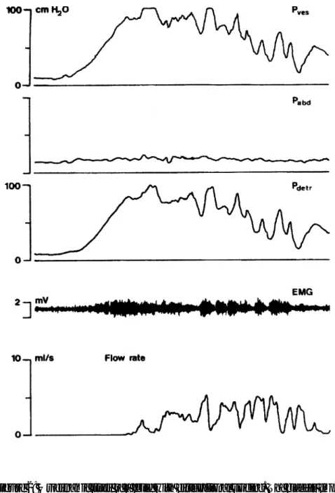
dyscoordination which begins with a staccato, and later a fractionated mic-turition. Finally, a lazy bladder syndrome with huge amounts of residual urine in an atonic bladder occurs. The urge syndrome and urge-incontinence aris-es when a child starts to postpone micturition, mostly during an episode of painful voiding due to UTI, vulvovaginitis, or urethritis. Simple prob-lems like oxyuriasis or irritating detergents can be the primary reason for postponement of micturition in girls. The children hold back their mic-turition with active pelvic floor contractions, thus producing a functional urethral obstruction resulting in an overactive bladder with small capacity and high micturition frequency. Incontinence is based on high pressure detrusor instability. The patients have lost central inhibition of their blad-der and use the pelvic floor as an emergency brake in the event of a detrusor contraction. In the typical urge syndrome urodynamic studies usually demonstrate normal pelvic floor relaxation while voiding. Small amounts of residual urine are the result of active pelvic floor contractions before the bladder is emptied. Some children maintain this voiding pattern for many years, others develop the dysfunctional voiding pattern. They learn over time to contract their pelvic floor effectively with more power, so that the bladder gives up most of its overactivity, and they have lost the ability to relax their pelvic floor properly when voiding. These children empty the bladder with near- normal frequency assisted by abdominal pressure with a typical staccato or fractionated flow. Incontinence is most-ly based on overflow. Urodynamic studies in these children show a nearmost-ly normal or hypo-active detrusor with unstable contractions that can be inhibited easily, and a micturition phase with unsustained contractions. Non-neurogenic bladder/sphincter dyssynergia is remarkable. They have important amounts of residual urine after micturition. Eventually the dys-functional micturition pattern can develop into the lazy bladder syndrome in which the detrusor muscle gives up completely. A decompensated, atonic bladder with very low micturition frequency is the result. The only driving force during micturition is the abdominal pressure. Incontinence is based on overflow with imperative urge at maximal capacity. Due to the pelvic floor overactivity these children develop enormous amounts of residual urine. Figures 1,2 and 3 give details on the urodynamic characteristics of these children.
Figure 1: Typical urodynamic study of a child with urge syndrome and urge inconti-nence. Unstable detrusor contractions are countered with straining of the pelvic floor . When, during an unstable contraction the child gives up and relaxates the pelvic floor, the bladder is emptied with normal flow and normal relaxation of the pelvic floor. Note the drop in detrusor pressure that comes with opening the urethra.
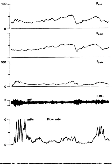
Figure 2: Urodynamic study of a child with dysfunctional voiding. The bladder emp-ties on a sustained detrusor contraction that is countered by the pelvic floor. With inter-mittent relaxation and contraction of the pelvic floor a staccato urinary stream is produced. Voiding will stop before the bladder is empty thus resulting in large residual volumes.
Figure 3: Urodynamic study of a child with a lazy bladder syndrome. Note the absence of detrusor contraction. The only driving force to empty is the abdominal pres-sure. The pelvic floor does not relax. By straining the abdominal muscles the bladder is emptied in a fractionated flow with large residuals of urine.
In addition to voiding dysfunction, nearly all children with micturition complaints due to the dysfunctional voiding complex have some form of fecal constipation. Clinically, in many cases, the fecal dysfunction only comes forward as skipping daily stools on a regular basis. The pelvic floor dysfunction hampers the rectum in the same manner as the bladder. Abdominal complaints connected to defecation are common in these girls. In addition, this problem often remains unrecognised by the parents in many cases. Only careful interview with specific questions will reveal the constipation
The patient’s history
The characteristic patient with DV is a girl between age 3 and 12 years with frequency, urge and urge incontinence. In most cases this is com-bined with simple or complicated UTIs. Pathognomonic is some form of Vincent’s curtsy sign: unstable detrusor contractions are felt as urge and suppressed by maximal contraction of the pelvic floor, when needed combined with compression of the perineum. This is accomplished by crossing their legs or by squatting or by sitting on a heel. When the unsta-ble contraction has subsided and the interrupted daily action is continued they may have lost a small amount of urine in their pants.
Specific micturition history reveals a tendency to postpone voiding, to interrupt the stream (staccato stream or fractionated stream) and finally to strain when voiding. These patients are never enuretics: they never or nearly never empty their bladder completely in their pants. In nearly all cases they wet their pants more during the afternoon then in the morning, because after several hours of concentration on the bladder they tend to give up. Parents are often annoyed by the behaviour of their child: when squatting the children say they cannot go to the toilet, when the detrusor contraction has subsided children deny any urgency to go to the toilet. The child is right in both cases: standing up during the suppression of the unstable contraction would result in profuse wetting. When the contrac-tion has subsided there is no further urgency to empty the bladder, because there is no relation between the amount of urine in the bladder
and unstable contractions. Clarifying this mechanism to the parents often works as an eye-opener. Finally, when a child has been successful in cop-ing with unstable bladder contractions durcop-ing a long period of time it can end with a decompensated bladder with low micturition frequency and large quantities of residual urine after voiding.
The majority of patients with dysfunctional voiding also wet during night. This does not always result in classical nocturnal enuresis. Often, it con-sists of wet spots in the underwear or bed during the night. Thus this should be classified as nocturnal incontinence instead of enuresis.
Apart from the micturition problems the majority of the patients are more or less constipated with or without fecal soiling.
The diagnosis
The preceding discussion automatically leads to the conclusion that the patient’s history is the most important factor in coming to the proper diagnosis. Most of the efforts of the consulted urologist or pediatrician should therefore be spent on the micturition anamnesis. Extensive urora-diological and nuclear investigations are indicated when complicated UTI’s have occurred. In most cases ultrasound of kidneys and bladder, uroflowmetry and determination of residual urine are sufficient at first. Urodynamic studies with good EMG recording are most typical in these patients (figure), but a thorough patient’s history will allow the experi-enced investigator to draw the urodynamic study without actually doing it. The authors reserve urodynamic study for the patients that don’t improve on a standard therapeutic scheme after 3 to 6 months. However, a major role for urodynamic studies in these patients remains. In rare cases only, an unsuspected urethral obstruction or a neurogenic component is detect-ed by urodynamic study.
Psychology of incontinence
Children tend to cope with disability by ignoring the disabled part of their bodies. This natural defense mechanism is responsible for the fact that wet children are generally called enuretics, suggesting a behavioural ground for the wetting, thus resulting in incontinent children in educa-tional circuits. The incontinent child will ignore the urgency of the blad-der and wet pants, the enuretic child will deny these. The typical enuretic patient will answer no to a parent’s question, shouldn’t you empty your bladder, and then do a complete voiding within seconds or minutes after-wards. The incontinent patient will ignore the fact that pants gradually wet and smell and continue daily affairs as if nothing is wrong. The treating urologist or pediatrician must discriminate between these subtle differ-ences. In general, one can state that incontinence as a sign of mental dis-comfort is very rare. A child in distress has many easier ways to express this discomfort besides wetting his/her pants. This makes the discrimina-tion between true enuresis and incontinence a major priority in the treat-ment of wet children.
True daytime enuresis, complete emptying of the bladder in the pants in one run, occurs predominantly in boys. In girls there may be a behavioural factor in DV, but it remains indistinguishable in most cases. Thus treat-ment of DV should be straightforward along the following lines with an open eye for psychologic complications.
Treatment
Treatment of children with DV requires some form of cognitive bladder training after thorough education of the child and the parents on the underlying mechanism of the dysfunction. A comparison to a mouth full of water after tooth-brushing often elucidates the mechanism to the child. The water can be sprayed out with force by squeezing the cheeks and the lips both at the same time, this results in residual water in the mouth. The water in the mouth can also simply be evacuated by opening the lips so that it drops out. In the last case the evacuation will be complete.
In the out-patient clinic the child and parents are informed on measures to avoid urethral irritation: no soap or washing-glove for cleaning, shower bi-daily to rinse the genitalia, no irritating or perfumed bath-salts and no tight stretch-clothing. Avoid extra mechanical irritation by a narrow bicy-cle-saddle or improper gymnastic exercises. The child should be taught to maintain a voiding frequency of 6-7 times a day. Instruct the parents to monitor this, because many children detest voiding so much that they will stay in the bathroom for a few minutes, flush the toilet and return with the happy statement that they have emptied their bladder while in fact they have not even lowered their pants. Children with a large bladder capacity for age should learn that, lifelong, they have to empty their blad-der when the time to do so has arrived instead of the urgency. The child should be taught to sit relaxed on the toilet with the thighs in a horizontal position, and when needed, the feet on a small stool or block. Make the child read one page of a comic-book when voiding and take care that the child does not strain during micturition. The easiest way to achieve this is by having them whistle a tune while voiding: it is very difficult to whistle and strain at the same time! Require a minimal daily fluid intake because many children with day-time incontinence stop drinking adequate vol-umes of fluid, thus producing urethral irritation by voiding concentrated urine. Dietary advice and adjustment to prevent constipation is essential. Finally have the child keep a calendar on micturition, incontinence and stools.
The above rules have to be supported by medication in the training peri-od. Routinely prescribe chemoprophylaxis for several months to prevent infections. Once a day 2 mg/kg nitrofurantoin or trimethoprim will usual-ly do the job. When frequency, urge and urge-incontinence are the most important symptoms and residual urine is minimal, spasmolytic support is needed in the form of oxybutinin 0,4 mg/kg up to three times a day. Oxybutinin works effectively for 6 hours. When major complaints occur only in the afternoon one dose of oxybutinin at noon will do. At the least suspicion of constipation, especially when spasmolytic therapy is needed, spasmolytics are postponed and laxatives are added to the treatment until daily stools are achieved. New spasmolytics have been developed, effect in these children has to be proven yet.
When supported and controlled properly this therapy will cure 30-50% of the patients in the out-patient clinic. The remaining hard-core group of patients will need a proper biofeedback training program to get rid of their symptoms.
Bio-feedback training
Biofeedback training can be done on an out-patient basis or during a peri-od of hospitalisation.21,22Psychological determination of the maturity of
the child and the motivation is important. In general, training before the age of 6 years, in girls, and 7 years, in boys, is disappointing because of lack of cognitive insight resulting in lack of motivation. The older the child the better the results. In our hospital we provide a training package of 10 days for the hard-core patients. Apart from psychological support they learn how, when and how often to void. They learn how to void by doing every micturition on a flowmeter with direct visualisation of the flow on a screen, thus eliminating interruptions of the stream with the pelvic floor. Bladder emptying is controlled by ultrasound. They learn when to void and to recognise bladder signals by wearing urine detector pants with an alarm, that rings when wet, during daytime. They must try to go to the toilet before the alarm rings. Finally they learn how often to void by making a chart during training. In general 80% of the hard-core patients obtain complete cure or significant improvement of incontinence and UTI’s by this training schedule. This result remains permanent over 10 years follow-up in our patient group. After the training they have regained central inhibition of the bladder signals instead of using the pelvic floor as an ‘urgency-brake’. After hospitalised training the children are followed up by the incontinence-therapists by weekly telephone con-tact for several months. When residual urine after micturition does not subside after training some children with a full-blown lazy bladder syn-drome have to be treated by clean intermittent self-catheterisation.
The failed therapy
It is important to realize that dysfunctional voiding in a small group of patients can be a symptom of congenital bladder neck insufficiency as a result of embryological derangement of the Wolffian duct. Also, a prior generous internal urethrotomy at young age can produce a bladder neck insufficiency. In severe primary bilateral reflux, in single or bilateral ectopic ureters, in ectopic ureteroceles, in intersex cases with adrenogenital syn-drome and a short urethra after reconstruction and in several other rare anomalies an insufficient bladder neck can force the child to try to with-hold urine that leaks into the urethra with the pelvic floor. This can eventu-ally result in a typical DV pattern that is resistant to training. Operative therapy is needed to cure these children. This operative therapy can consist of a simple colposuspension in uncomplicated cases. When severe reflux is coexisting with DV colposuspension must be considered during ureteral reimplantation. Needle suspension in the pediatric age group provides only temporary success. The authors sometimes use this procedure in difficult cases to judge the chances of a proper Burch type colposuspension. When a scarred bladder neck and urethra due to a previous internal urethrotomy appears to be the underlying cause of the incontinence simple colposus-pension has been disappointing in our hands. Nowadays we excise the scar completely and reconstruct the urethra. Although very succesfull in a few cases it is too early to tell wether this is the best way to cure this problem.
Developments
An international prospectively controlled multicenter trial with financial support of the EEC has been conducted in Europe to determine which way of training for children with DV provides the best results without the exorbitant costs of hospitalisation. Surprisingly, the amount of attention given to the child appears to be the most important factor for success. We have started a new study to the treatment of DV with a controlled amount of attention and the addition of a new biofeedback device, a portable flowmeter and toilet that is used by the child at home every day.
Ultrasosonography of the pelvic floor and the rectum gives new insights in the pathophysiology and the treatment of DV. Recently we have proven that a direct correlation exists between the diameter of the rectum on ultrasound and the amount of fecal constipation. This has proven to be of help to convince both the parents and the children of the existence of constipation and it provides an easy, non-invasive control of therapy. Dynamic ultrasonography of the pelvic floor muscles has learned us that approximately 40% of the children with DV has a paradoxal movement of the musculature. They contract the puborectal muscles when ordered to relax and they relax the musculature when ordered to squeeze. Physical therapy to correct this paradoxal movement appears to have a 100% suc-cess rate. The effect of this correction on the results of DV therapy remains to be proven yet.
The authors do hope that after reading this paper on dysfunctional void-ing therapists will recognize that the misused overactive pelvic floor is the primary problem in these patients and thus realise that the worst advice to give these patients is to do the ‘stop-test’ during micturition. This manoeuvre will only make the bladder derailment worse! Finally one must realise that these children are incontinent, they are not enuretics. Calling them enuretics delays cognitive treatment by inappropriate behavioural therapies and produces unneccesary trauma for the child.
Literature
1. Hellstrom A-L, Hanson E, Hansson S, Hjalmas K, Jodal U. Micturition habits and inconti-nence in 7-year old Swedish school entrants. Eur J Pediatrics 1990; 149:434-437.
2. Hellstrom A-L, Hanson E, Hansson S, Hjalmas K, Jodal U. Micturition symptoms at age 7 related to previous urinary infection. Brit J Urol 1989; 63:358-362.
3. Gool JD van, Hjalmas K, Tamminen-Mobius T, Olbing H (on behalf of the International Reflux Study in Children). Historical clues to the complex of dysfunctional voiding, urinary tract infection and vesicoureteral reflux. J Urol part 2 1992; 148:1699-1702.
4. Gool JD van, Jonge GA de. Urge syndrome and urge incontinence. Arch Dis Child 1989; 64:1629-1634.
5. Hansson S, Hjalmas K, Jodal U, Sixt R. Lower urinary tract dysfunction in girls with untreated asymptomatic or covert bacteriuria. J Urol 1990; 143:333-335.
6. Koff SA, Murtagh DS. The uninhibited bladder in children - effect of treatment on recur-rence of urinary infection and on vesicoureteral reflux resolution. J Urol 1983; 130: 1138-1141.
7. Bloom DA, Faerber G, Bomalaski MD. Urinary incontinence in girls. Evaluation, treatment, and its place in the standard model of voiding dysfunctions in children. Urologic Clinics of North America 1995; 22 No.3:521-538.
8. Allen TD. Vesicoureteral reflux and the unstable bladder. J Urol 1985; 134: 1180-1183. 9. Gool JD van, Kuijten RH, Donckerwolcke RA, Messer AP, Vijverber M. Bladder-sphincter
dysfunction, urinary tract infection and vesicoureteral reflux - with special reference to cog-nitive bladder training. Contr Nephrol 1984; 39:190-210.
10. Taylor CM, Corkery JJ, White RHR. Micturition symptoms and unstable bladder activity in girls with primary vesicoureteric reflux. Brit J Urol 1982; 54:494-498.
11. Taylor CM. Unstable bladder activity and the rate of resolution of vesicoureteric reflux. Contr Nephrol 1984; 39:238-246.
12. Gool JD van, Vijverberg MAW, Jong TPVM de. Functional daytime incontinence - clinical and urodynamic assessment. Scand J Urol Nephrol 1992; S141:58-69.
13. Griffiths DJ, Scholtmeijer RJ. Vesicoureteral reflux and lower urinary tract dysfunction -evidence for 2 different reflux/dysfunction complexes. J Urol 1987; 137:240-244.
14. Koff SA, Lapides J, Piazza DH. Association of urinary tract infection and reflux with unin-hibitied bladder contractions and voluntary sphincteric obstruction. J Urol 1978; 122:373-376.
15. Nielsen JB, Djurhuus JC, JNrgensen TM. Lower urinary tract dysfunction in vesicoureteral reflux. Urol Int 1984; 39:29-31.
16. Nielsen JB, Norgaard JP, Schwartz-Sorensen S, JNrgensen TM, Djurhuus JC. Continuous overnight monitoring of bladder activity in vesicoureteral reflux patients. Neurourol and Urodyn 1984; 3:1-6.
17. Vincent SA. Treatment of enuresis with a perineal pressure apparatus-the irritable bladder syndrome. Dev Med Child Neurol 1964; 6:23-9
18. Koff SA, Lapides J, Piazza DH. Association of urinary tract infection and reflux with unin-hibited bladder contractions and voluntary sphincter obstruction. J Urol 1979;122:373-6 19. Allen TD. Vesicoureteral reflux as a manifestation of dysfunctional voiding. In: Hodson CJ,
Kincaid-Smith P (eds). Reflux nephropathy. Masson, New York, 1979.
20. Gool JD van, Tanagho EA. External sphincter activity and recurrent urinary tract infections in girls. Urology 1977;10:348-353
21. Vijverberg MAW, Elzinga-Plomp A, Messer AP, de Jong TPVM, van Gool JD. Bladder rehabilitation: the effect of a cognitive training programme on urge incontinence. Eur urol 1997;31(1):68-72
22. Hoebeke P. et.l. Outpatient pelvic-floor therapy in girls with daytime incontinence and dys-functional voiding. Urology 1996;48(6):923-7
Ectopic Ureteroceles
Ectopic ureteroceles, results of open surgical therapy in 40 patients
CHAPTER 3
Tom P.V.M. de Jong, Pieter Dik, Aart J. Klijn, Cuno S.P.M. Uiterwaal and Jan D. van Gool
Abstract
Purpose: The treatment policy of ectopic ureteroceles is controversial.
Besides debate on the optimal treatment of ectopic ureteroceles, discus-sion exists whether there is an extra risk for deterioration of bladder func-tion after extensive bladder surgery during the first year of life thus giving reason to postpone surgery. We have treated 40 patients with an ectopic ureterocele in a prospective, non-randomised trial, with complete surgical reconstruction of the lower urinary tract combined with upper pole resec-tion of malfuncresec-tioning upper pole moieties at the time of referral, regard-less of age. Three patients with only 1 affected renal moiety initially were excluded from the study.
Materials and methods: Forty patients, 31 female and 9 male, were treated for
ectopic ureteroceles that extended into the bladder neck and urethra. Age at operation was between 0.0 and 8.8 years with a mean age of 2.17 years and 19 patients were under the age of 1 year at the time of operation. Of the patients, 37 underwent primary excision of the ureterocele with recon-struction of the urethra, bladder neck and trigone and with ureteral reim-plants. In 3 patients, because of small size of the ureterocele, the ureterocele was left in situ, leading to secondary removal of the uretero-cele in two, one because of obstructive voiding and one to treat urinary incontinence. In 5 neonates, a staged procedure was done with primary reconstruction of the lower urinary tract combined with upper pole cuta-neous ureterostomies, followed by upper pole resection or ureteral reim-plantation a few months later. In 16 patients, after bladder neck reconstruction, a colposuspension was performed as well to create a nor-mal vesico-urethral angle. All patients had a clinical and urodynamic work-up at least 1.25 year after operation with a mean follow-work-up of 5.59 years. Patients who by Jan. 1999 were too young to have their continence clini-cally assessed were excluded from the study.
Results: All our patients are continent. Thirteen patients needed a
second-ary endoscopic procedure (cystoscopy only in 2, scar incision near ureteral orifice in 3, endoscopic reflux treatment in 4, resection of remnants of ureterocele in 2, bladder neck incision to treat obstructive voiding in 2), one patient needed a secondary open reconstruction of the bladder because of a diverticulum. Eleven patients had postoperative only one or
two uncomplicated urinary tract infection (UTI). Recurrent UTI’s were seen in 4 patients, 1 complicated patient had severe infections leading to renal scarring. This patient had a pre-existing loss of renal function. A normal voiding pattern is seen in 29 patients, 11 have a dysfunctional voiding type of micturition with recurrent UTI’s in 5 cases. Urodynamic follow-up confirms these clinical findings. Bladder capacity in these patients is relatively high and averages 124% of expected capacity for age. There was no statistically significant difference in the follow-up parame-ters between children operated under the age of 1 year and those operated at a later age. The additional colposuspension in 16 patients did not pro-duce any significant change in outcome compared to the patients that did not have this procedure.
Conclusion: Compared to the literature, complete primary reconstruction of
the lower urinary tract in patients with ectopic ureteroceles appears to have better results then a staged approach with initial endoscopic treat-ment. Moreover, an important conclusion of this study is that no proof exists that extensive reconstructive surgery of the bladder in neonates and infants leads to deterioration of the bladder function at later age.
Introduction
In the treatment of ureteroceles, strict discrimination between intravesical and ectopic ureteroceles is mandatory. In contrast to ectopic ureteroceles, intravesical ureteroceles rarely have consequences for the urodynamic behaviour of the bladder and can be treated as seems best for each indi-vidual patient by endoscopic incision or by open removal with reimplanta-tion of the affected ureters. The choice of therapy in intravesical ureteroceles is determined by the size of the ureterocele, by the function and degree of obstruction of the affected renal unit and by the presence or absence of vesicoureteral reflux (VUR).1-6
Ectopic ureteroceles, in general, can have consequences for the urody-namic properties of the bladder7and have a high morbidity for urinary
tract infections (UTI), loss of renal function and incontinence. In contrast to intravesical ureteroceles, ectopic ureteroceles, in general, drain the
upper renal moiety of a double system, which shows a severely compro-mised function.8Literature review of ectopic ureteroceles reveals a
ten-dency to start with upper pole removal or in the newborn with endoscopic incision of the affected unit , although in the last years more aggressive therapy for ectopic ureteroceles with reflux in 1 or more moi-eties is advocated.5In many reviews the authors were forced to do a
com-plete removal of the ureterocele as a secondary procedure. VUR and recurrent UTI’s formed the most important reasons to do so. As early as 1979 Hendren, for ectopic ureteroceles, proposed to do a complete recon-struction of the lower urinary tract with upper pole removal. He conclud-ed that this type of surgery is not suitable for neonates and infants .9In
addition, a non-evidenced concern exists that extensive lower tract gery in infancy produces higher urodynamic morbidity compared to sur-gery at a later age. With advances in pediatric anesthesiology, nowadays duration of surgery hardly presents an extra risk or contraindication in deciding the time of reconstruction in neonatal cases. Because of our pre-vious experiences with staged procedures that had a high morbidity for UTI’s and incontinence, we began to search for an approach that is defini-tive and safe. Therefore, we have treated our patients with ectopic uretero-celes by primary, complete surgical reconstruction from 1989 on as a prospective, non-randomised study. All the patients were followed with respect to clinical outcome, urodynamic behaviour of the bladder, num-ber of infections and renal function. In addition, we have compared the urodynamical and clinical outcome of patients operated before and after the age of one year.
Materials and methods
Between 1988 and 1998, 40 patients with 41, previously untreated ectopic ureteroceles presented to our hospital. Distribution of sexes was 31 female and 9 male patients.
In the 40 patients, 24 ureteroceles were on the left side, 15 on the right, 1 bilateral. Age at operation ranged from 0 to 8.84 years with a mean age of 2.17 years. Nineteen patients were less then 1 year old at the time of
oper-ation with a mean age of 0,22 years. Eleven cases were detected by prena-tal ultrasound, 2 in evaluating incontinence and 27 by urosepsis or recur-rent UTI’s. Of these patients 10 were straining while voiding.
Nine patients presenting with life-threatening septicaemia were primarily treated by drainage of the affected renal units by endoscopic incision of the ureterocele and/or a nephrostomy drain in the distended upper pole. One infant with life-threatening urinary ascites and pleuritis in bilateral ectopic ureteroceles was first treated by a vesicostomy with intubation of the 4 ureters combined with evacuation of the urine from the abdominal and thoracic cavities. His lower urinary tract was reconstructed in a sec-ondary procedure and included bladder augmentation with both upper pole ureters.
Excision of the ectopic ureteroceles with reconstruction of the lower uri-nary tract was performed as a primary procedure in 28 patients, as a sec-ondary procedure, within a few weeks after presentation, in the aforementioned 10 patients. In five neonates reconstruction of the lower urinary tract was combined with the installation of upper pole ureteros-tomies followed after 2 months by upper pole resection in 3 cases, upper pole ureter reimplantation in 2, after establishing the function of the affected upper pole moieties. Reason for this delayed upper tract repair was the fact that no information on the function of the upper pole moi-eties existed at the time of the first procedure.
Reimplantation of all the refluxing contralateral ureters and of all the remaining lower pole ureters was performed after ureterocele resection. In cases of megaureters, tapering by plication was performed when the flat width of the ureter exceeded 10 mm during the first 6 years of the study, 6 mm from 1994 on. When, after reconstruction of the urethra, bladder neck and bladder base a vesico-urethral angle was considered non-exist-ing, a Burch-type colposuspension was performed by the end of the pro-cedure.
Preoperative diagnostics consisted of ultrasonography (US) and voiding cystourethrography (VCUG) in all the patients, DMSA-scanning in 23 patients and urodynamic studies in 10 patients. In all cases, the operative procedure started with cystoscopy and colposcopy. The differential diag-nosis intravesical/ectopic ureterocele could not always be determined by US and VCUG. In approximately 25% of the cases only cystoscopy could
exactly determine the caudal extension of the ureterocele and discriminate between ectopic and intravesical ureterocele. A DMSA scan was not per-formed in neonatal cases and in cases that, on ultrasound, showed a very small and scarred upper pole moiety of the kidney in the presence of a normal looking lower pole.
Operative approach
Heminephroureterectomy was performed by lumbotomy with selective ligation of upper pole vessels. Nephropexia was done only when the whole of the lower pole needed to be mobilised. The reconstruction of the lower urinary tract was carried out by a low transverse Pfannenstiel incision with transverse opening of the fascia and splitting of the rectus muscles in the midline. The adventitial layer of the bladder is opened selectively, the bladder is opened in the midline, and the bladder dome is filled to capacity with 1 or more gauzes and stretched with the third blade of the wound retractor. The ureterocele is identified and grabbed in a clamp, the bladder mucosa around the ureterocele is incised with a diathermic needle and the ureterocele is peeled out, partly by blunt dissec-tion, partly by cautery. The part of the ureterocele that runs through the bladder neck into the urethra is freed from mucosal lining as far as is nec-essary and possible. Surprisingly, even in a neonatal female urethra, one can reach down into the urethra for approximately 1.5 centimetres in cases of large cecoureteroceles. In cases of cecoureteroceles the whole urethral part of the ureterocele is incised endoscopically at the start of the procedure. Manipulation of the urethra is made easier when a small addi-tional suction tube is placed retrograde into the distal urethra and the ele-vation of the anterior wall of the urethra can be optimised by putting an 8 Fr. silicon drain through the urethra that is subsequently tied around the symphysis. The upper pole ureter is excised, and, when necessary, the dis-tal portion of the lower pole ureter. The lower pole ureter passes into the bladder cranially in the large bladder defect that is the result of this proce-dure. Starting as deeply as possible in the urethra, the dorsal urethral wall is closed with polyglycolic 5.0 interrupted sutures. Care is taken to grab
the urethral wall in such a way that the suture is positioned submucosally with the knot outside. In general, 3-5 sutures can be put into the urethra and the bladder neck. The defect of the bladder base is closed similarly with 4.0 sutures, leaving room in the cranial part of the defect for the pas-sage of the lower pole ureter. The lower pole ureter ending is fixed in the physiological position by 2 5.0 polyglycolic sutures and covered with blad-der mucosa.
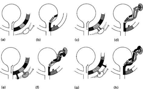
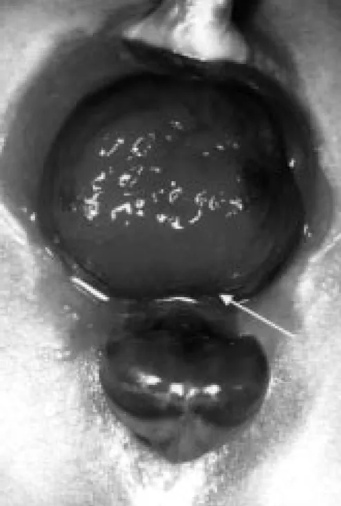
When in cases of very large or bilateral ureteroceles this procedure appears to lead to a narrow, funnel-shaped trigonal part of the bladder, it may be necessary to bring the cranial rim of the defect down to the level of the bladder neck, resulting in a remodeling of the bladder. When after this procedure, macroscopically no vesico-urethral angle can be seen, a colposuspension is thought to be appropriate to restore a normal vesico-urethral angle. This is performed by lifting the anterior vaginal wall left and right of the bladder neck with a metal sound through the introitus and grabbing it into a clamp. Depending on the size and age of the child the anterior vaginal wall is lifted to Cooper’s ligament with a 2.0 or 3.0 polyglycolic acid suture. The reimplanted ureters are splinted with 4-6 Fr. feeding tubes, a 12 Fr. silicon Malecot-type catheter is left as a suprapubic catheter. A 10 Fr. silicon drain is left transurethrally and fixed to the blad-der wall by a 5.0 rapid soluble polyglycolic acid suture. The bladblad-der is closed with 4.0 polyglycolic acid sutures, the adventitional layer is closed separately (figure)
The ureteral splints are taken out after 4 days; after 7 days in ureters that have been tapered. Suprapubic and transurethral drainage is maintained for 6-9 postoperative days. All patients were on chemoprophylaxis for at least 6 months after the operation.
A VCUG and a urodynamic study were done in all patients postoperative-ly. All patients repeatedly underwent US of the upper tracts and the blad-der combined with uroflowmetry and US determination of post-void residual urine
Figure: Schematic drawing of a left double system with an ectopic ureterocele opening in the proximal urethra. The lower pole ureteric orifice is on the proximal part of the ureterocele. After upper pole nephrectomy and excision of the cele, a large defect runs from the bladder into the urethra. Closure of the defect results in a reconstruction of the urethra, the bladder neck and the trigone. After closure of the defect the lower pole ureter is fixed in the physiological position running over a part of the closed defect and covered by mucosa.