doi: 10.20506/rst.35.3.2580
Evaluation of the bacteriological quality of raw
cow’s milk at various stages of the milk production
chain on farms in Algeria
M. Hamiroune
(1, 2)*, A. Berber
(3)& S. Boubekeur
(3)(1) Department of Agronomic and Veterinary Sciences, Faculty of Natural and Life Sciences, Ziane Achour University, B.P. 3117, Moudjbara road, Djelfa, Algeria
(2) High National Veterinary School, Issad Abbes road, Oued Smar, Algiers, Algeria (3) Institute of Veterinary Sciences, Blida 1 University, B.P. 270, Soumâa road, Blida, Algeria * Corresponding author: mouradhamiroune@gmail.com
Summary
This study evaluates hygiene practices on 53 dairy farms in the Jijel and Blida regions of Algeria. A survey questionnaire was drawn up covering milking conditions and cleaning of the equipment. In parallel, bacteriological analyses were carried out to estimate the rate, source and development of bacterial contamination in raw milk produced on the farm. In addition, screening was performed to detect the presence of inhibitor residues.
The results of the survey revealed poor livestock conditions and milking practices that could explain the presence of bacteria in cow’s milk.
The bacteriological results showed that 76.1% of milk samples taken from cow udders complied with legal standards, compared with only 35.8% of milk samples taken from storage tanks. Moreover, bacterial inhibitors were detected in 28.8% of milk samples. These results showed that the hands of milkers, udders, teat cups, utensils, the water used during milking and the milking environment were all potential sources of milk contamination by the bacteria under investigation.
These results suggest that, to improve the bacteriological quality of milk, there is a need to introduce a quality policy which places a premium on milk of high bacteriological quality and aims to generalise good hygiene practices throughout the dairy production chain.
Keywords
Algeria – Bacterial contamination − Bacteriological quality – Cows − Hygiene practices – Milking – Raw milk.
Introduction
Milk figures prominently in the diet of Algerians, which explains why the dairy sector has seen annual growth of 8% (1). Algeria is the leading consumer of raw milk in the Maghreb region, with almost 3 billion litres a year. The hygiene quality of raw milk is therefore vital (2).
To maintain hygiene conditions on farms and up to the arrival of milk in dairies, the bacteriological quality of the milk must be monitored (3).
There are several risk factors for milk contamination at different stages of production on the farm, prompting the authors to conduct this study with the principal aim of
evaluating the bacteriological quality of raw milk in the Jijel and Blida regions and identifying raw milk contamination risk factors on the farm.
Materials and methods
Farm selection
This study was conducted on 53 dairy farms covering 360 milking cows in the Jijel and Blida regions in Algeria, during the period from March 2013 to July 2014.
A non-random convenience sampling plan was defined to include herds that differed in terms of size, milking method
and equipment or in preparation of the udders for milking (washing and disinfection).
Epidemiological survey
On each dairy farm, a survey was carried out and the milking process was monitored on the sampling day. The survey questionnaire form indicates the cows sampled, milk production systems and milking hygiene.
Sampling
In order to assess the bacteriological quality and sources of contamination of milk produced on the farm, raw milk samples were taken, as well as samples from the environment and the milking equipment.
Before milking, a 100 ml sample of the water used for milking was taken, as well as 100 ml of the water used for rinsing the milking utensils. Swab samples were taken from the milkers’ hands, teat cups and skin of the udders of each of the cows.
During milking, a flask containing sterile water was exposed for 30 minutes to assess environmental contamination (environmental sample).
The samples were taken aseptically and placed in labelled sterile vials. The authors used the individual milk sampling technique described by Mialot (4).
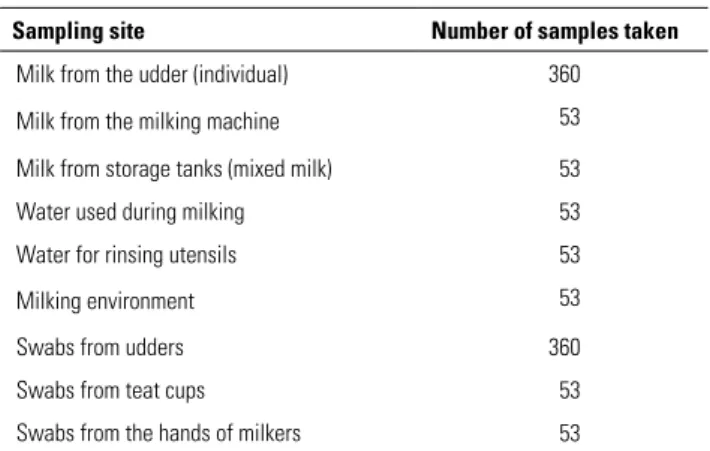
The samples were then placed in a cooler and transported directly to the testing laboratories (the laboratory of the Algerian centre for quality control and packaging [CACQE] and the laboratory for veterinary testing, quality control, compliance and applied research [AVCQ-LAB] in Baraki), where they were refrigerated at +4°C. The time between sampling and the first analyses barely exceeded 24 hours. Table I shows the number of samples taken by sampling site.
Detection and enumeration of contaminating
microorganisms
Different dilutions with a tryptone salt broth (TSB) were used depending on the nature of the sample; they varied between 10–1 and 10–8.
In each sample, a search was made for five groups of bacteria: total aerobic mesophilic flora, faecal streptococci, faecal coliforms, Staphylococcus aureus and Clostridium
sulphite-reducers (CSR) (5).
The detection and enumeration of total aerobic mesophilic flora (TAMF) was carried out on glucose agar with yeast
extract (plate count agar [PCA]) after incubation at 30°C for 72 hours (6).
Faecal coliforms (FC) were detected and enumerated on violet red bile lactose agar (VRBL), incubated for 24 hours at 44°C. All red colonies (lactose+) that appeared with a minimum diameter of 0.5 mm were considered to be faecal coliforms (7).
Staphylococcus aureus (SA) was detected and enumerated on Baird Parker agar with egg yolk and tellurite incubated at 37°C for 24–48 hours. Black, shiny convex colonies appeared surrounded by a clear halo of 2–5 mm in diameter. This was confirmed by Gram stain test (+), catalase test (+) and coagulase test (+) (8).
Faecal streptococci (FS) were enumerated on Rothe broth (Pasteur Institute, Algeria). A millilitre of each sample to be analysed was added to 9 ml of TSB. In this way, the authors obtained a mother dilution of 10–1from which the decimal
dilutions were made. A millilitre of each dilution was then placed in three tubes of Rothe broth. Following incubation for 48 hours at 37°C, the contents of the positive tubes (those that were cloudy) were then seeded on bile agar and bile esculin azide (BEA) for confirmation and allowed to incubate at 37°C for 24 and 48 hours (9).
For CSR at 46°C, an aliquot of milk placed in a sterile test tube was preheated for 10 minutes at 80°C to destroy vegetative forms and to activate the spores. Using a sterile pipette, 1 ml of the test sample (milk heated for 10 minutes at 80°C) was then injected deep into the tryptose-sulfite-cycloserine agar (TSC) (Pasteur Institute, Algeria) and the inoculum was mixed gently into the culture medium, without forming bubbles to avoid oxygenation of the medium. The tubes were then plunged into cold water to solidify the mixture. Following incubation at 46°C for 20 ± 2 hours, only the characteristic colonies, i.e. those surrounded by a black halo, were counted (10).
Table I
Distribution of samples from different sampling sites on the 53 Algerian dairy cattle farms that participated in the study
Sampling site Number of samples taken Milk from the udder (individual) 360
Milk from the milking machine 53
Milk from storage tanks (mixed milk) 53
Water used during milking 53
Water for rinsing utensils 53
Milking environment 53
Swabs from udders 360
Swabs from teat cups 53
The enumeration results obtained for the different flora were interpreted according to the standards laid down in interministerial decree No. 35-1998 of January 1998 on the microbiological specifications of certain foodstuffs (5) (Table II).
Detection of bacterial inhibitors in milk
The DelvoTest® SP-NT (DSM Food, the Netherlands) was
used to detect bacterial inhibitors in raw milk. It is based on inhibiting the growth of Bacillus stearothermophilus var.
calidolactis, a bacterium which is very sensitive to a wide range of antibiotics and sulphonamides. It takes the form of ampoules containing an agar medium seeded with spores of B. stearothermophilus and enriched with growth nutrients. In each previously identified ampoule, 100 µl of a milk sample were introduced using a micropipette fitted with a disposable tip. The ampoules were placed in a water bath at 64 ± 1°C for three hours. On removal, the colour of the medium was examined by the naked eye. If a sample had clearly changed from violet to yellow, it indicated that the sample contained no bacterial inhibitors. In the presence of bacterial inhibitors the sample remained a violet colour.
Statistical analyses
Geometric mean calculations were performed for the enumeration of bacteria isolated at different points of the raw milk production chain on the farm.
The chi-square (χ2) test was used to test the relationships
between the bacterial composition of milk and milk production points on the farm, as well as between the presence of bacterial inhibitors and milk production points on the farm.
Results
Description of milking practices
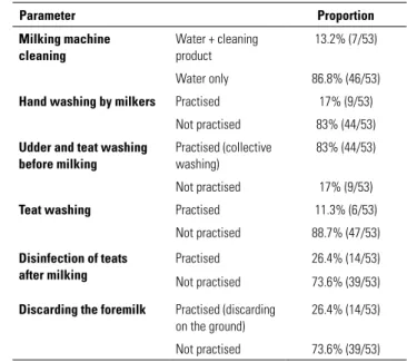
The results of the survey on milk production systems are presented in Table III. It transpires that:
– on almost all farms (86.8%), the milking machine was cleaned using only water
– very few milkers washed their hands (17%)
– on 83% of farms, the cows’ udders and teats were washed before milking using the same washing cloth for all the cows
– 88.7% of farmers neglected to wipe down the teats – only 26.4% of farmers soaked the teats in a disinfectant solution, the remaining 73.6% failed to do this
– 73.6% of farmers neglected to discard the foremilk on the ground, compared with only 26.4% who did use this practice.
Overall bacteriological quality of milk
With regard to the criteria required by interministerial decree No. 35-1998 of 24 January 1998 on the microbiological specifications of raw milk (5), the results obtained from the 466 samples can be summarised as follows:
– 8.6% (31/360) of the individual milk samples, 15.1% (8/53) of milk samples from the milking machine and 47.2% (25/53) of milk samples from storage tanks did not comply with the legal criteria for all the enumerated bacteria
Table II
Microbiological specifications of raw milk (acceptability thresholds) in force in Algeria at the time of the study (5)
Microbiological parameter Acceptability threshold in raw milk Total aerobic mesophilic flora at 30°C 105 cfu/ml
Faecal streptococci Absence/0.1 ml
Faecal coliforms 103 cfu/ml
Staphylococcus aureus Absence
Clostridium sulphite-reducers at 46°C 50 cfu/ml
Bacterial inhibitors Absence
Table III
Characteristics of milking practices on the 53 Algerian dairy cattle farms that participated in the study
Parameter Proportion Milking machine cleaning Water + cleaning product 13.2% (7/53) Water only 86.8% (46/53) Hand washing by milkers Practised 17% (9/53)
Not practised 83% (44/53) Udder and teat washing
before milking
Practised (collective washing)
83% (44/53) Not practised 17% (9/53) Teat washing Practised 11.3% (6/53)
Not practised 88.7% (47/53) Disinfection of teats
after milking
Practised 26.4% (14/53) Not practised 73.6% (39/53) Discarding the foremilk Practised (discarding
on the ground)
26.4% (14/53) Not practised 73.6% (39/53)
– 15.3% (55/360) of the individual milk samples, 24.5% (13/53) of milk samples from the milking machine and 17% (9/53) of milk samples from storage tanks complied with the legal criteria for only some enumerated bacteria – 76.1% (274/360) of the individual milk samples, 60.4% (32/53) of milk samples from the milking machine and 35.8% (19/53) of milk samples from storage tanks complied with the legal criteria for all the enumerated bacteria. A deterioration in milk quality was observed between the udder and the storage tank. The proportion of good quality milk fell from 76.1% to 35.8%, while poor quality milk rose from 8.6% to 47.2% (Fig. 1).
Frequency of bacterial inhibitors in milk on the
farm
Bacterial inhibitors were detected in 28.8% (134/466) of all the raw milk samples. The 134 positive samples were distributed as follows:
– 30.6% (110/360) were individual milk samples
– 26.4% (14/53) were milk samples from the milking machine
– 18.9% (10/53) were milk samples from the storage tanks. The rate of bacterial inhibitors in raw milk was particularly high in the individual milk samples (30.6%) (Table IV). The frequencies varied significantly depending on the sampling site (p < 0.05).
Sources of milk contamination
The proportion of samples contaminated by the bacteria studied (TAMF, FS, FC, SAand CSR) varied from 0% for samples from the hands of milkers (FS, FC and CSR), the milking environment (SA and CSR) and the milking water (FS and FC) to 98.1% for samples of water used during milking (TAMF) (Table V).
TAMF was found in all sample types at levels varying from 79.2% for samples taken from the hands of milkers to 98.1% for those from milking water.
While FS and FC were not detected in the samples taken from the hands of milkers or in the samples of water used during milking, they were often found in the samples taken from utensils (60.4% and 66% respectively), from udders (51.9% and 57.8% respectively) and from teat cups (41.5% and 45.3% respectively).
While CSRwere detected in the samples taken from udders (10.8%), from utensils (9.4%), from teat cups (5.7%) and from the water used at different stages of milking (18.9%),
they were not found in the samples taken from the hands of milkers or in the environmental samples.
Staphylococcus aureus was isolated mainly from the water used at the different stages of milking (50.9%), from samples taken from the hands of milkers (39.6%) and from udders (28.9%). The lowest levels were found on utensils (5.7%) and teat cups (7.5%).
Critical points and the presence of
contaminating bacteria
A comparison of the bacterial counts in milk at different sampling points on the farm (from the cow’s udder to the storage tank) showed a significant difference (p < 0.05) for each group of bacteria identified (Table VI).
In individual milk samples, 78.9% contained TAMF, 23.6% contained FS, 32.8% contained FC, 16.1% contained SA and 3.3% contained CSR.
Table IV
Proportion of milk samples containing bacterial inhibitors taken from the 53 Algerian dairy cattle farms that participated in the study
Source of the sample Proportion
Milk from the udder 30.6% (110/360)
Milk from the milking machine 26.4% (14/53)
Milk from storage tanks 18.9% (10/53)
A: Good quality B: Average quality C: Poor quality
Fig. 1
Overall quality of raw milk as measured by the bacterial concentration detected in samples taken at different stages of the milk production chain on the 53 Algerian dairy cattle farms that participated in the study
Hygiene assessment of milking practices
The survey conducted on these dairy farms revealed that, in general, neither milking conditions, nor equipment cleaning, nor milk storage were optimal. On all of the farms covered by the study, milking was carried out under poor hygiene conditions and cleaning products were rarely used for udder preparation or for equipment cleaning.
Cleaning milking machines
The majority of milkers (86.8%) rinsed the milking machine with water only, compared with 13.2% who used a mixture of water and a cleaning product. These results are similar to those of the study conducted in Monastir (11), where 10% of farmers alternated the use of acid and alkaline detergents during cleaning operations.
Hand washing by milkers
The level of hygiene among the majority of milkers was unacceptable: only 17% of them washed their hands before each milking, while most (83%) did not.
According to Thomelin (12), it is vital to ensure the best possible hygiene conditions in order to reduce In the milk in storage tanks, the proportions were
respectively 96.2% (TAMF), 64.2% (FS), 75.5% (FC), 58.5% (SA) and 5.7% (CSR).
The bacterial load in raw milk samples rose progressively along the farm production chain (Fig. 2).
Discussion
Samples from raw cow’s milk and the environment, as well as from milking equipment, were taken on several dairy farms in the Jijel and Blida regions of Algeria. Convenience sampling was employed in order to include farms of different sizes using different methods and different milking equipment. Milk quality was assessed according to the Algerian standards in force for the microbiological specifications of raw milk.
Poor hygiene conditions during milking and milk storage, as well as lack of hygiene among milkers (dirty hands, poor-quality milking water, etc.) and in the equipment used for milk production, were the causes of the poor hygiene quality of the milk produced. In fact, bacterial contamination of milk worsened as it progressed along the production chain.
Table V
Proportion of environmental samples containing bacterial contaminants taken from the 53 Algerian dairy cattle farms that participated in the study
Source of the sample Bacterial contaminant
TAMF FS FC SA CSR
Water used during milking 98.1% (52/53) 0% (0/53) 0% (0/53) 50.9% (27/53) 18.9% (10/53)
Utensils 94.3% (50/53) 60.4% (32/53) 66% (35/53) 5.7% (3/53) 9.4% (5/53)
Milking environment 81.1% (43/53) 13.2% (7/53) 18.9% (10/53) 0% (0/53) 0% (0/53)
Udders 83.9% (302/360) 51.9% (187/360) 57.8% (208/360) 28.9% (104/360) 10.8% (39/360)
Teat cups 96.2% (51/53) 41.5% (22/53) 45.3% (24/53) 7.5% (4/53) 5.7% (3/53)
Hands of milkers 79.2% (42/53) 0% (0/53) 0% (0/53) 39.6% (21/53) 0% (0/53)
TAMF: Total aerobic mesophilic flora at 30°C FS: Faecal streptococci
FC: Faecal coliforms SA: Staphylococcus aureus
CSR: Clostridium sulphite-reducers at 46°C
TAMF: Total aerobic mesophilic flora at 30°C FS: Faecal streptococci
FC: Faecal coliforms SA: Staphylococcus aureus
CSR: Clostridium sulphite-reducers at 46°C
Table VI
Proportion of bacteria isolated at various stages of the milk production chain on the 53 Algerian dairy cattle farms that participated in the study
Source of the sample Bacterial contaminant
TAMF FS FC SA CSR
Milk from the udder (individual) 78.9% (284/360) 23.6% (85/360) 32.8% (118/360) 16.1% (58/360) 3.3% (12/360) Milk from the milking machine 83% (44/53) 26.4% (14/53) 41.5% (22/53) 22.6% (12/53) 3.8% (2/53) Milk from storage tanks 96.2% (51/53) 64.2% (34/53) 75.5% (40/53) 58.5% (31/53) 5.7% (3/53)
contamination of udders and milk by bacteria that can enter when a cow’s sphincters are open.
Udder and teat washing before milking
Most milkers (83%) performed collective washing of teats and udders before milking. These results are similar to those of M’Sadak etal. (13), who found that the majority of farmers (93%) prepared the udder by pre-washing with water using the same cloth for all the cows.
According to Noireterre (14), this udder preparation method increases the risk of transmitting pathogens from an infected quarter to a healthy quarter of the udder with the subsequent onset of mastitis.
Teat washing
This stage can minimise the risk of mastitis, improve milk quality and prevent slippage of teat cups and the entry of air (vacuum fluctuation) into milking units (15).
This study found a teat-washing frequency (11.3%) well below that reported by M’Sadak etal. (67%) in a study of the effect of milking conditions on the udder health of dairy cows in the Mahdia region of Tunisia (16).
Disinfection of teats
This study found that very few milkers (26.4%) disinfected the teats after milking. These results are significantly lower
than those reported by M’Sadak etal. (11) in a recent study in the Monastir region, where 59% of farmers applied this practice.
According to Bareille and Lemarchand (17), teat disinfection after milking can reduce the rate of new intra-mammary staphylococcus infections by 50–95%. According to Hanzen (18), teat disinfection after milking can reduce the number of microorganisms transferred via the teat canal during milking, which may have developed at the tips between milking sessions. It can also be used to treat any injuries to the teats.
Discarding the foremilk
The authors found that only 26.4% of milkers discarded the foremilk, even though doing so has advantages over early detection of clinical mastitis and the elimination of micro-organisms in the teat canal (18). This corroborates the observations of M’Sadak etal. (11) in the Monastir region, where 28% of farmers used this practice.
Bacterial inhibitors in milk
According to the interministerial decree on the microbiological specifications of certain foodstuffs (5), good quality milk should not contain bacterial inhibitors. However, 28.8% of the 466 raw milk samples analysed in this study contained bacterial inhibitors and the majority of farmers treated mastitis with penicillin and/or tetracycline (according to the results of the authors’ survey questionnaire). These figures show the scale of bacterial inhibitor use on the dairy farms studied.
These results are in line with those of Srairi etal.(19) and Kouame etal. (20), who reported levels of around 25% and 24.7% respectively.
Heavy contamination of milk samples with tetracycline and/ or penicillin was also confirmed by a study by Ben Mahdi and Ouslimani (21), who reported a contamination rate of around 97.3%.
In addition, these results revealed differences in the proportion of bacterial inhibitors, depending on the site. They were found in 30.6% of individual samples of milk (on leaving the udder), in 26.4% of milk samples taken from milking machines and in 18.9% of samples from storage tanks. These figures indicate the scale of antibiotic use on dairy farms where the milk is collected and hence the extent of the resulting risk to consumer health.
High levels of bacterial inhibitors in individual raw milk samples are probably caused by the widespread, uncontrolled use of intra-mammary pharmaceutical
Fig. 2
Results of the enumeration of five bacterial indicators in raw milk at different stages of the milk production chain on the 53 Algerian dairy cattle farms that participated in the study (Geometric mean in log10 cfu/ml)
A: Individual milk samples (milk from the udder)
B: Milk from the milking machine C: Milk from storage tanks
TAMF: Total aerobic mesophilic flora at 30°C
FS: Faecal streptococci FC: Faecal coliforms SA: Staphylococcus aureus
preparations for the curative and preventive treatment of bovine mastitis, coupled with failure to respect waiting periods. Moreover, the practice of deliberately adding bacterial growth inhibitors, such as antibiotics, to stabilise raw milk should not be underestimated (22).
According to Ameur et al. (23) who carried out a survey on the use of intra-mammary antibiotics in the wilaya
(province) of Tizi Ouzou (Fréha, Azazga and Yakouren districts), intra-mammary antibiotic syringes are used routinely to treat acute mastitis. The most widely used (or prescribed) products are based on tetracycline, penicillin and, more rarely, macrolides. The choice of these molecules is based mainly on effectiveness and price.
Bacterial inhibitors in milk can partially or totally inhibit the growth of the lactic starters used to make dairy products such as cheese and yoghurt, which often causes problems during the manufacture of fermented dairy products. The commonest problems are milk failing to coagulate, insufficient draining and the risk of uncontrolled spread of gas-forming bacteria that are immune to antibiotics, such as coliforms, Bacillus, Clostridium or Proteus. Such widespread incidents result in heavy economic losses for the dairy industry each year (24).
Overall bacterial quality
of milk and sources of contamination
These results show that milk becomes increasingly contaminated as it progresses through the different stages of milking. Between the udder and the milk storage tank, the proportion of good quality milk samples fell from 76.1% to 35.8%.
This rapid decline in the bacteriological quality of milk as it passes along the farm production chain is the result of successive instances of contamination from utensils, the udders, the teat cups, the milking environment and the hands of milkers. Milk becomes contaminated during milking operations and the more it is handled, the greater the risk of bacterial contamination.
Bacteria detection revealed that the udder skin, utensils and teat cups carried all the bacteria under investigation (TAMF, FS, FC, SA and CSR). The udders of some cows were more contaminated than others and the mixing of raw milk from several cows contributed to the drop in milk quality in storage tanks.
In addition, S. aureus was found in the water used at the different milking stages (50.9%), on the hands of milkers (39.6%) and on the udders (28.9%). Faecal streptococci and faecal coliforms were found on utensils
(60.4% and 66% respectively), on the udders (51.9% and 57.8% respectively), on teat cups (41.5% and 45.3% respectively) and in the milking environment (13.2% and 18.9% respectively). Clostridium
sulphite-reducers were found at low levels in the water used at the different milking stages (18.9%), on the udders (10.8%), on utensils (9.4%) and on teat cups (5.7%). All these elements are therefore sources of contamination of raw milk.
The presence of TAMF in raw milk is an indicator of the overall level of hygiene on farms. TAMF includes microorganisms that cause spoilage or contamination, acidifying lactic flora and sometimes pathogenic bacteria. Enumeration of these flora is the method most commonly used by dairy processing plants to assess the bacterial quality of milk and it is therefore an important indicator of hygiene conditions during milking (25). The high levels of these flora found in samples from milk cooling tanks is probably the result of intensive bacterial growth arising from failure to control hygiene conditions during milking and milk storage.
The presence of faecal coliforms and faecal streptococci in raw milk indicates an environmental source of contamination. Their proliferation in raw milk reflects a failure to observe the required hygiene measures during milking, and probably contamination during milk storage. Faecal coliforms and streptococci in raw milk are strongly associated with faeces-soiled udder skin and poorly designed and improperly cleaned milking equipment (25). Bonfoh et al. (26) report that poor cleaning of recipients in contact with milk on the farm left residual levels of contamination of around 4.1 log10 colony-forming units (cfu)/ml.
Clostridium sulphite-reducers were found in animal feed (that had been in contact with the ground), which contaminates the milk either directly or via faeces. These are pathogenic bacteria and their presence indicates recent or older faecal contamination of the ground (27).
Staphylococcus aureus is a contagious agent living on cow udders that can be transmitted from one cow to another (28). This bacterium can enter milk either directly, by excretion from udders infected with clinical or subclinical staphylococcal mastitis, or by environmental contamination during the handling and processing of raw milk (29, 30). When the udder is infected, S. aureus is excreted in the milk in highly variable quantities from 0 to 108 cfu/ml (31).
These results, which support those of Kouame et al. (20), show that this bacterium came mainly from the water used at the different stages of milking (50.9%), from the hands of milkers (39.6%) and from udders (28.9%).
References
1. Salon international du lait (SILAIT) (2008). – Actes du Premier Salon international du lait et de ses dérivés, 27–29 mai 2008, Alger. Available at: www.agroligne.com/ contenu/silait-2008-1er-salon-international-lait (accessed on 15 August 2014).
2. Kirat S. (2007). – Les conditions d’émergence d’un système d’élevage spécialisé en engraissement et ses conséquences sur la redynamisation de l’exploitation agricole et la filière des viandes rouges bovines. Cas de la wilaya de Jijel, en Algérie. Thèse de master en Sciences. Centre international de hautes études agronomiques méditerranéennes, Institut agronomique méditerranéen, Montpellier, 139 pp. Available at: www.iamm. ciheam.org/ress_doc/opac_css/doc_num.php?explnum_ id=4370 (accessed on 10 August 2014).
3. Faye B. & Loiseau G. (2002). – Sources de contamination dans les filières laitières et exemples de démarches qualité.
In Gestion de la sécurité des aliments dans les pays en
développement, Actes de l’atelier international CIRAD-FAO, 11–13 décembre 2000, Montpellier. Available at: http:// agritrop.cirad.fr/514114/ (accessed on 5 August 2014).
4. Mialot J.P. (1983). – Technique de prélèvement de
lait pour examen bactériologique. Rec. Méd. Vét., 159 (11), 1057–1058.
5. Ministère du Commerce (Algérie) (1998). – Arrêté
interministériel du 25 Ramadhan 1418 correspondant au 24 janvier 1998 modifiant et complétant l’arrêté du 14 Safar 1415 correspondant au 23 juillet 1994 relatif aux spécifications microbiologiques de certaines denrées. J. Off. Rép. Algér. Dém.
Pop., 35, 7–24. Available at: www.qualilab.dz/.../V-Arrete_
24-janvier-1998_specifications_microbiologiques.pdf (accessed on 8 August 2014).
6. International Organization for Standardization (ISO) (2003). – Standard ISO 4833 (F). Microbiologie des aliments. Méthode horizontale pour le dénombrement des micro-organismes. Technique par comptage des colonies à 30 °C [Microbiology of food and animal feeding stuffs – Horizontal method for the enumeration of microorganisms – Colony-count technique at 30 degrees C]. ISO, Geneva, 9 pp.
7. Agence française de normalisation (AFNOR) (1996). – Norme F V08-060. Microbiologie alimentaire. Dénombrement des coliformes thermotolérants par comptage des colonies à 44 °C. Méthode de routine. AFNOR, Paris, 10 pp.
8. Agence française de normalisation (AFNOR) (2004). – Norme F V08-057-01. Microbiologie des aliments. Méthode de routine pour le dénombrement des staphylocoques à coagulase positive par comptage des colonies à 37 °C. Partie 1 : Technique avec confirmation des colonies. AFNOR, Paris, 11 pp.
Conclusion
This study shows that the increasing bacterial load in milk as it passes along the farm production chain is the result of successive instances of contamination associated with poor hygiene practices during milking.
The search for sources of contamination along the entire raw milk chain showed that udders, milkers’ hands, teat cups, utensils, the milking environment and the water used during milking were all sources of milk contamination by the bacteria under investigation. In addition, bacterial inhibitors were detected in the milk samples analysed. To improve the quality of raw milk, farmers need to implement a range of hygiene measures in cowsheds and
during milking, all the more rigorously and systematically because the animals’ environment is highly contaminated. This environmental contamination could be reduced by introducing manure storage and spreading practices to prevent the recycling and spread of bacteria. This will be difficult to achieve without the effective participation of farmers following information campaigns targeted at them.
Acknowledgements
The authors would like to thank all the staff of the laboratory of the Algerian centre for quality control and packaging (CACQE) and the laboratory for veterinary testing, quality control, compliance and applied research (AVCQ-LAB).
9. Maury M. (1987). – Milieux et réactifs de laboratoire. Microbiologie et immunologie. Diagnostics Pasteur, Paris, 728 pp.
10. Agence française de normalisation (AFNOR) (2009).
– Norme NF V08-061. Microbiologie des aliments. Dénombrement en anaérobiose des bactéries sulfito-réductrices par comptage des colonies à 46 °C. AFNOR, Paris, 11 pp.
11. M’Sadak Y., Mighri L., Ben Omrane H. & Kraiem K. (2012). – Évaluation des chantiers et des équipements de traite chez des élevages bovins laitiers hors sol dans la région de Monastir. Rev.
Nature & Technol.,7, 96–101.Available at: www.webreview.
dz/IMG/pdf/art_07_12.pdf (accessed on 8 August 2014). 12. Thomelin R. (2009). – Mammites, cellules : Tous les conseils
pour lutter efficacement. GIE Élevage des Pays de la Loire, Angers, 57 pp. Available at: www.charte-elevage.fr/sites/ default/files/files/Mammites_Cellules_-_Tous_les_conseils_ pour_lutter_efficacement.pdf (accessed on 20 August 2014). 13. M’Sadak Y., Mighri L. & Kraiem K. (2011). – Étude de la
situation sanitaire mammaire et estimation des pertes laitières engendrées chez des ateliers bovins hors sol en Tunisie. Rev.
Nature & Technol., 4, 8–14. Available at: www.webreview.
dz/?Etude-de-la-situation-sanitaire-mammaire-a-partir (accessed on 20 August 2014).
14. Noireterre P. (2006). – Suivi de comptages cellulaires et d’examens bactériologiques lors de mammites cliniques chez la vache laitière. Thèse de doctorat en médecine vétérinaire, École nationale vétérinaire de Lyon, 98 pp. Available at: www2. vetagro-sup.fr/bib/fondoc/th_sout/dl.php?file=2006lyon099.
pdf(accessed on 25 August 2014).
15. Wattiaux M. (2004). – Procédure de traite. Lactation et récolte du lait. Babcock Institute for International Dairy Research and Development, University of Wisconsin, 5 pp. Available at: https://federated.kb.wisc.edu/images/group226/52750/19-25/
de_25.fr.pdf(accessed on 15 August 2014).
16. M’Sadak Y., Mighri L. & Kraiem K. (2010). – Effet des conditions de traite sur la santé mammaire des vaches laitières et estimation des pertes en lait consécutives dans la région de Mahdia en Tunisie. Rev. Elev. Méd. Vét. Pays Trop., 63 (1–2), 35–39. Available at: http://remvt.cirad.fr/cd/derniers_num/ 2010/REMVT10_035_039.pdf (accessed on 30 August 2014). 17. Bareille N. & Lemarchand F. (2004). – La désinfection des
trayons avant et après la traite ; comment choisir les méthodes et les produits ? In Dossier : hygiène de la mamelle et traitement des mammites. Bull. GTV, 24, 21–27.
18. Hanzen C. (2010). – Pathologie infectieuse de la glande mammaire, étiopathogénie et traitements, approche individuelle et de troupeau. Faculté de médecine vétérinaire, Université de Liège. Available at: www.therioruminant. ulg.ac.be/notes/200910/R22_Mammites_etiopathogenie_ traitement_2010.pdf (accessed on 15 August 2014).
19. Srairi M.T., Hasni Alaoui I., Hamama A. & Faye B. (2004). – Qualité physicochimique et contamination par les antibiotiques
du lait de mélange en étables intensives au Maroc. Renc.
Rech. Rumin., 11, 115. Available at: www.journees3r.fr/IMG/
pdf/2004_qualite_17_Srairi.pdf (accessed on 30 August 2014). 20. Kouame Sina S., Bassa A., Dadie A., Makita K., Grace D.,
Dje M. & Bonfoh B. (2010). – Analyse des risques microbiens du lait cru local à Abidjan (Côte d’Ivoire). Rev. Afr. Santé Prod.
Anim., 8 (S), 35–42. Available at: www.researchgate.net/
publication/281571059_Analyse_des_risques_microbiens_ du_lait_cru_local_a_Abidjan_Cote_d’Ivoire (accessed on 31 August 2014).
21. Ben Mahdi M. & Ouslimani S. (2009). – Mise en évidence des résidus d’antibiotiques dans le lait de vache produit dans l’Algérois. Eur. J. Sci. Res.,36 (3), 357–362. Available at: www. researchgate.net/publication/237343688_Mise_en_Evidence_ De_Residus_d’Antibiotiques_Dans_le_Lait_de_Vache_ Produit_Dans_l’Algerois (accessed on 31 August 2014). 22. Zinedine A., Faid M. & Benlemlih M. (2007). – Détection des
résidus d’antibiotiques dans le lait et les produits laitiers par la méthode microbiologique. Rev. Microbiol. Ind. San. Environn.,
1 (1), 1–9. Available at: www.remise.ma/images/V2007/
zinedine2007.pdf (accessed on 29 August 2014).
23. Ameur A., Rahal K., Guedioura A., Bouyoucef A. & Kaidi R. (2008). – Utilisation des antibiotiques intra-mammaires dans la région de Tizi Ouzou. Premiers résultats. Communication aux Sixièmes Journées des sciences vétérinaires, École nationale des Services vétérinaires (ENSV), 19–20 avril 2008. 24. Berger J.L. & Lenoir J. (1997). – Les accidents de fromagerie
et les défauts des fromages. In Le fromage, de la science à
l’assurance qualité, 3e éd. Tec & Doc, Lavoisier, Paris.
25. Mhone T.A., Matope G. & Saidi P.T. (2011). – Aerobic bacterial, coliform, Escherichia coli and Staphylococcus aureus counts of raw and processed milk from selected smallholder dairy farms of Zimbabwe. Int. J. Food Microbiol., 151, 223– 228. doi:10.1016/j.ijfoodmicro.2011.08.028.
26. Bonfoh B., Roth C., Traore A.N., Fane A., Simbe C.F., Alfaroukh I.O., Nicolet J., Farah Z. & Zinsstag J. (2006). – Effect of washing and disinfecting containers on the microbiological
quality of fresh milk sold in Bamako. Food Control,
17 (2), 153–161. doi:10.1016/j.foodcont.2004.09.015.
27. Joffin C. & Joffin J.N. (1999). – Microbiologie alimentaire.
Collection ‘Biologie technique’, 5e éd. Centre régional de
documentation pédagogique d’Aquitaine, 212 pp.
28. Makovec J.A. & Ruegg P.L. (2003). – Results of milk samples submitted for microbiological examination in Wisconsin from 1994 to 2001. J. Dairy Sci., 86, 3466–3472. doi:10.3168/jds. S0022-0302(03)73951-4.
29. Donkor E.S., Aning K. & Quaye J. (2007). – Bacterial contaminations of informally marketed raw milk in Ghana.
Ghana Med. J., 41 (2), 58–61. Available at: www.ncbi.nlm.
nih.gov/pmc/articles/PMC1976296/ (accessed on 31 August 2014).
30. Normanno G., Firinu A., Virgilio S., Mula G., Dambrosio A., Poggiu A., Decastelli L., Mioni R., Scuota S., Bolzoni G., Di Giannatale E., Salinetti A.P., La Salandra G., Bartoli M., Zuccon F., Pirino T., Sias S., Parisi A., Quaglia N.C. & Celano G.V. (2005). – Coagulase-positive staphylococci
and Staphylococcus aureus in food products marketed in
Italy. Int. J. Food Microbiol., 98 (1), 73–79. doi:10.1016/j. ijfoodmicro.2004.05.008.
31. Asperger H. & Zangerl P. (2003). – Staphylococcus aureus. In Encyclopedia of dairy sciences (H. Roginski, J.W. Fuquay & P.F. Fox, eds). Vol. 4. Academic Press & Elsevier Science, Amsterdam, 2563–2569.