R E S E A R C H A R T I C L E
Open Access
Root of
Polygonum cuspidatum
extract reduces
progression of diabetes-induced mesangial cell
dysfunction via inhibition of platelet-derived
growth factor-BB (PDGF-BB) and interaction
with its receptor in streptozotocin-induced
diabetic rats
Eunjin Sohn, Junghyun Kim, Chan-Sik Kim, Kyuhyung Jo, Yun Mi Lee and Jin Sook Kim
*Abstract
Background:Platelet-derived growth factor–BB (PDGF-BB) is highly expressed in the renal tissues of patients with diabetic nephropathy, and it plays an important role in the initiation and progression of diabetic nephropathy. The aim of this study was to evaluate the protective effects of root ofPolygonumcuspidatum extract (PCE) on early renal glomerular proliferation in streptozotocin (STZ)-induced diabetic rats.
Methods:PCE (100, 350 mg/kg/day) was administered to diabetic rats for 16 weeks. Blood glucose and albuminuria were measured. Renal histology,α-smooth muscle actin (α-SMA), and proliferating cell nuclear antigen (PCNA) expression levels were also examined.
Results:After 16 weeks of treatment with PCE, severe hyperglycemia and albuminuria were observed in the diabetic rats. The expressions levels ofα-SMA and PCNA proteins were significantly increased in the glomeruli of the diabetic rats. The expression levels of PDGF-BB and its receptor expressions were greatly increased in the glomeruli of the diabetic rats. However, PCE markedly reduced albuminuria in the diabetic rats. PCE inhibited
α-SMA and PCNA up-regulation and ameliorated PDGF-BB and PEGFR-ß protein expression in the diabetic rats. In addition, the binding of PDGF-BB/PDGFR-ß was inhibited by PCE as shown by anin vitroassay.
Conclusions:These results suggest that PCE has an inhibitory effect on mesangial proliferation in diabetic renal tissues via the inhibition of the interaction of PDGF-BB with its receptor. PCE may have beneficial effects in preventing the progression of diabetic nephropathy.
Keywords:Diabetic nephropathy, Glomerular proliferation,Polygonum cuspidatum
Background
Diabetic nephropathy causes the majority of case of end-stage renal disease, and it is an important cause of increased mortality in patients with diabetes mellitus (DM) worldwide. Platelet-derived growth factor (PDGF) has been suggested to play a role in the pathophysiology
of various fibroproliferative diseases, including pulmon-ary fibrosis, systemic sclerosis, liver cirrhosis, cardiovas-cular disease and kidney fibrosis [1,2]. The up-regulation of the PDGF pathway has been shown in the kidneys of diabetic animals and patients with DM [2-4]. PDGF po-tently increases cell proliferation and matrix synthesis. PDGF-BB, which is an isoform of PDGF that has been the most closely linked with the development of various renal diseases, is produced by glomerular mesangial cells [5]. Moreover, PDGF-BB is highly expressed in diabetic
* Correspondence:[email protected]
Korean Medicine Based Herbal Drug Development Group, Herbal Medicine Research Division, Korea Institute of Oriental Medicine (KIOM), 1672 Yusengdaero, Yuseong-gu, Daejeon 305-811, South Korea
renal tissues and plays important role in the initiation and progression of diabetic nephropathy [6]. Hyperglycemia has been is well characterized and albuminuria is consid-ered to be one of the most sensitive markers of renal injury, and albuminuria significantly increased within 6–8 weeks in the streptozotocin (STZ)-induced diabetic animal model [7-10]. Moreover, the expression of the PDGF-B chain or PDGFR-ß in mesangial cells is actually increased in the glomeruli in this animal model [2].
Medicinal plants have been used as traditional remedies for hundreds of years.Polygonum cuspidatum (P. cupida-tum) is the dried root of Polygonum cuspidatumSieb. et Zucc. (Polygonaceae). It has been widely used as a medi-cinal herb for a variety of purposes in Asian countries, such as the treatment of inflammatory diseases, hepatitis, tumors, and diarrhea [11]. This herb has also been used to control oral disease in Korea [12]. In traditional Chinese medicine, P. cuspidatum has been used for its anti-diabetic, antibacterial properties [13-15]. Recently, it has been reported that an herbal formulation containing ofP. cuspidatum prevented hepatic fibrosis by decreasing the expression of transforming growth factor-beta (TGF-ß) and α-smooth muscle actin (α-SMA) [16]. The general understanding of the pathogenic factors leading to renal fibrosis in diabetic nephropathy patients has considerably expanded, and the mechanisms involving TGF-ß and α-SMA have been found to be important to the develop-ment of diabetic renal fibrosis [17-19]. Therefore, in this study, we investigated the protective effect of an ethanol extract of P. cuspidatum (PCE) against renal injury and fibrosis in STZ-induced diabetic rats.
Methods
Preparation ofPolygonum cuspidatiumextract (PCE)
Root of Polygonum cuspidatum was purchased from a commercial supplier in Jung-dong, Daejeon, Korea, in November 2008 and identified by Prof. Ju Han Kim at the Department of Life Sciences, Gachon University. A voucher specimen was deposited at the Herbarium of the Diabetic Complication Research Team, Korea Institute of Oriental Medicine (KIOM). The dried and ground plant material (6.8 kg) was extracted with ethanol (3 × 36 L) by maceration at room temperature for 3 days, and the ex-tracts were combined and concentrated in vacuoat 40°C to create lyophilized PCE (580 g).
Animals and induction of diabetes
The experiments were performed in accordance with the National Institutes of Health (NIH) Guide for the Care and Use of Laboratory Animals and approved by the Korea Institute of Oriental Medicine Institutional Animal Care and Use Committee. In our study, six-week-old male SD rats purchased from the Charles River Laboratory (Waltham, MA, USA) were acclimated for 1 week prior to
the study. Our study was initiated using 8-week-old male SD rats (weighting, ~200 g), which were monitored for 16 weeks. Diabetes was induced in the rats by a single injection of streptozotocin (STZ, 60 mg/kg, i.p.). Age-matched control rats received an equal volume of vehicle (0.01 M citrate buffer, pH 4.5). Two days after the injec-tion, a blood sample was collected from the ratil vein to measure the blood glucose level. Rats with a blood glucose level over 300 mg/dl were considered diabetic rats. At 8 weeks of age, the rats were randomly assigned to one of four groups (n = 8). PCE was dissolved in vehicle (0.5% w/ v carboxyl methylcellulose solution) to a concentration of 50 mg/ml. Two groups of STZ-induced diabetic rats received daily gastric gavage of PCE (100 and 350 mg/kg), and the other groups were administered the same amount of vehicle gavage for 16 weeks. Blood glucose levels and body weight were monitored consecutively.
Metabolic and morphological analyses
After 16 weeks of treatment, blood glucose was mea-sured using an automated analyzer (Hitachi, Tokyo, Japan). Blood samples were collected from the tail vein after a 16-h fast. Individual rats were placed in metabolic cages to obtain 24-h urine collections, and urinary albu-min excretion levels were measured using a sandwich enzyme-linked immunosorbent ELISA assay kit accord-ing to the manufacturer’s manual (Life Diagnostics, Inc., PA, USA). Rat albumin present in the urine sample was captured by an anti-rat albumin antibody that had been pre-adsorbed on the surfaces of microtiter wells. After sample binding, unbound proteins and molecules were washed off with washing buffer, and a biotinylated detec-tion antibody was added to the wells to bind to the captured albumin. Strepavidin-conjugated horseradish peroxidase (SA-HRP) was then added to catalyze a col-orimetric reaction with the chromogenic substrate 3,3′,5,5′-tetramethylbenzidine(TMB). This colorimetric reaction produces a blue product, which turns yellow when the reaction is terminated by the addition of stop solution (0.1 M H2SO4). The resulting, yellow reaction
products were read at 450 nm using a microtiter plate reader (Bio-Tek, Winooski, VT, USA). Renal cortexes were fixed in 10% formaldehyde and embedded in paraf-fin, and 4-μm-thick sections were prepared. The sections were stained with periodic acid-Schiff (PAS) reagent and hematoxylin as a counterstain.
Western blot analysis
anti-PDGF-BB and anti-PDGFR-ß (R&D Systems, MN, USA). The immunoblots were developed using an en-hanced chemiluminescence detection system (Amersham Bioscience, NJ, USA) with HRP-conjugated secondary antibodies. Protein expression levels were determined by analyzing the signals captured on the PVDF membranes using an image analyzer (Las-3000, Fuji Photo, Tokyo, Japan).
Immunohistochemical and immunofluorescent staining
Renal cortexes were fixed in 10% formaldehyde and em-bedded in paraffin, and 4-μm-thick sections were prepared. Staining was performed as previously described [15]. The antibodies used included rabbit anti-proliferating cell nuclear antigen (PCNA, Santa Cruz, CA, USA), mouse PDGF-BB (R&D Systems, MN, USA), mouse anti-PDGFR-ß (R&D Systems), rabbit anti-α-SMA (Santa Cruz, CA, USA) and mouse anti-thy 1.1 (a mesangial cell marker; Millipore, MA, USA). For the detection of PDGF-BB and PDGFR-ß, the sections were incubated using an Envision kit (DAKO, CA, USA) and visualized by 3,3′ -diaminoben-zidine tetrahydrochloride (DAB). To detect PCNA and α-SMA, the sections were incubated with Texas red-conjugated goat anti-rabbit IgG (Santa Cruz, CA, USA) and fluorescein isothiocyanate-conjugated goat anti-rabbit IgG (Santa Cruz, CA, USA), respectively, and detected by fluorescence microscopy (Olympus, Tokyo, Japan). To characterize the proliferation of the mesangial cells, we performed double staining for PCNA and Thy 1.1. using the same section. To prevent cross-reactions between the two labeling procedures, the slides were incubated with normal mouse serum (Dako, CA, USA). To detect PCNA and Thy 1.1, the sections were incubated with fluorescein isothiocyanate-conjugated goat anti-rabbit IgG (Santa Cruz, CA, USA) and Texas red-conjugated goat anti-mouse IgG (Santa Cruz, CA, USA), respectively. Negative controls for immunohistochemistry were prepared by incu-bating the sections with nonimmune serum instead of primary antibody. For morphometric analysis, the number of positive cells or positive signal areas per one glomerulus in a total of 50 glomeruli was determined using ImageJ software (NIH, Bethesda, MD, USA).
In vitroassay for ligand receptor binding inhibition
To investigate the inhibitory effect of PCE on PDGF-BB/ PDGFR-ß ligand receptor binding, ligand binding was examined by a sandwich ELISA assay in vitro. A total of 50μl of a 0.05μg dose of recombinant human PDGF-BB (R&D Systems, MN, USA) was pre-coated and incubated on a microplate for 24-h at 4°C and was then washed with PBS (pH 7.4). Next, the sample was blocked with 100 μl of CAS block solution (Life Technologies, CA, USA) for 30 min at 37°C and then washed with PBS. The sample was then added to 50 μl recombinant
human (rh) PDGFR-ß /FC chimera labeled with peroxid-ase (Dojindo, Kumanoto, Japan) and 50 μl of a serial dilution of the PCE mixture in a microplate pre-coated with rh PDGF-BB. The samples were incubated for 1 h at 37°C and then washed with PBS. A TMB chromogen solution was used as a substrate for the horseradish peroxidase. After the reaction was terminated with stop buffer (0.1 M H2SO4), and a yellow reaction product
formed, its absorbance was measured at 450 nm using a microtiter plate reader (Bio-Tek, Winooski, VT, USA). The inhibition of PDGF-BB ligand receptor binding was expressed as the percentage decrease in optical density (OD 450 nm). We calculated the IC50 concentration
(ug/ml) as the 50% inhibition of PDGF-BB/ PDGFR-ß ligand binding.
Statistical analysis
The data are expressed as the mean ± SEM and were analyzed by one-way analysis of variance (ANOVA), followed by Tukey’s multiple comparison test or by an unpaired Student’s t-test using GraphPad Prism 5.0
software (GraphPad, San Diego, CA, USA). Differences of P < 0.01 were considered statistically significant.
Results
Body weights and metabolic parameters
At 16 weeks of treatment, the body weights were signifi-cantly decreased in the vehicle-treated diabetic rats com-pared with the normal control rats. However, there was no difference in body weight between the vehicle-treated diabetic rats and PCE-treated diabetic rats. The blood glucose levels increased by approximately 3-fold in the vehicle-treated diabetic rats compared with normal con-trol rats (P < 0.01). There was no statistically significant difference between the vehicle-treated diabetic rats and PCE-treated diabetic rats with respect to blood glucose levels (Table 1). These resulting data demonstrated that PCE maybe has independent effect on glucose-lowering and body weight.
Morphology and urinary albumin levels
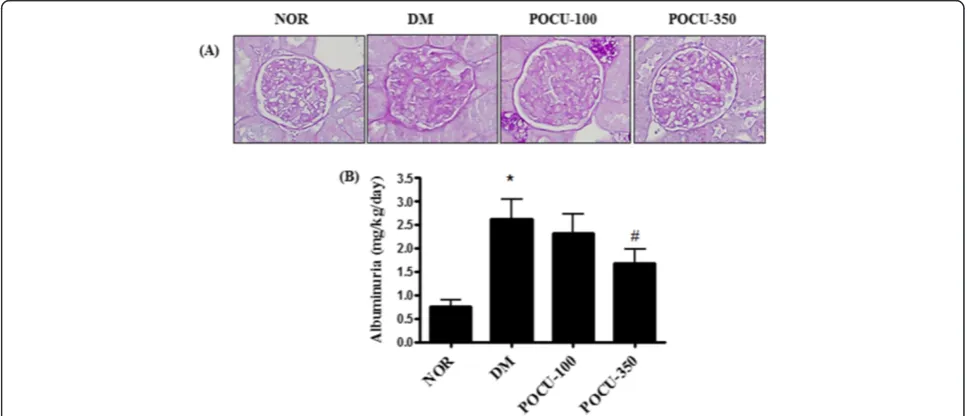
Mesangial matrix expansion is considered a hallmark of diabetic nephropathy [21]. At 24 weeks of age, vehicle-treated diabetic rats showed focal mesangial matrix expan-sion, and albuminuria was significantly increased in the vehicle-treated diabetic rats compared to the normal con-trol rats (Figure 1A). However, PCE treatment ameliorated the mesangial expansion and albuminuria compared with the vehicle-treated diabetic rats (Figure 1A and B).
PDGF expression in renal glomeruli
Immunohistochemical staining of PDGF-BB and PDGFR-ß in the glomeruli were mainly localized to the glomerular mesangial cells revealing a significant increase in the vehicle-treated diabetic rats compared with the normal control rats. This increase of PDGF-BB and PDGFR-ß expression in the glomeruli in the vehicle-treated diabetic rat was attenuated by PCE treatment (Figure 2A and B). Western blot analysis showed that, PDGF-BB and its receptor in the renal cortex were also markedly increased in the vehicle-treated diabetic rats compared with the nor-mal control rats. PCE reduced these diabetes-induced increases in PDGF-BB and PDGFR-ß expressions in a dose-dependent manner (Figure 2C and D). This result demonstrated that PCE prevents renal mesangial prolifera-tion in diabetic nephropathy.
Inhibition of PDGF-BB ligand receptor binding assay
To investigate whether PCE could inhibit the binding of the PDGF ligand to its receptor, a sandwich ELISA assay was performed. PCE showed inhibition effect on binding of the PDGF ligand to its receptor in a concentration-dependent manner. PCE exhibited an inhibitory effect on the binding of the PDGF-BB ligand to its receptor (IC50= 0.185 ± 0.14μg/ml) (Figure 3).
Expression ofα-SMA and PCNA in renal glomeruli
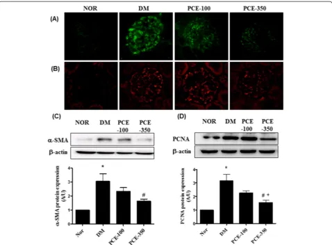
Mesangial proliferation is considered a prominent fea-ture of diabetic nephropathy. The expression level of proliferation markers, such as α-SMA and PCNA, were measured using immunohistochemistry and western blot analysis. Immunohistochemical staining of α-SMA and PCNA was markedly more visible in the diabetic renal glomeruli compared with normal glomeruli. PCE
treat-ment showed decreased expression of α-SMA and
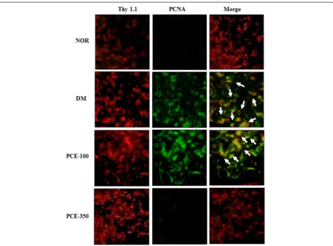
[image:4.595.54.544.101.141.2]PCNA in the diabetic glomeruli (Figure 4A and B). The western blot assay indicated that, the expressions levels ofα-SMA and PCNA were significantly increased in the vehicle-treated diabetic rats compared with the normal control rats. The high-dose PCE treatment markedly reduced these diabetes-induced increases inα-SMA and PCNA expression (Figure 4C and D). In addition, to investigate whether the enhanced protein expression was colocalized to the mesangial cells of the glomeruli, PCNA and Thy 1.1, which is a mesangial cell marker [22], were visualized by double immunofluorescence staining. Thy 1.1 was positively stained in the glomerular mesangial cells for all of the experimental rats. PCNA expression was significantly increased in the vehicle-treated diabetic rats compared with the normal control
Table 1 Metabolic and physical parameters
NOR DM PCE-100 PCE-350
Blood glucose (mg/dl) 162.1 ± 11.0 587.3 ± 43.5* 530.3 ± 58.1 474.2 ± 88.4
Body weight (g) 475.9 ± 10.7 208.5 ± 12.1* 205.5 ± 23.1 206.0 ± 16.4
NOR, normal rat; DM, STZ-induced diabetic rat; PCE-100, DM treated with PCE (100 mg/kg); PCE-350, DM treated with PCE (350 mg/kg). All data are expressed as the mean ± SEM (n=8). *P <0.01 vs. NOR group.
[image:4.595.56.540.487.695.2]rats. In the dual-labeled section, PCNA expression was localized to a region of Thy 1.1-positive mesangial cells in the diabetic renal tissue (Figure 5). However, the high-dose PCE treatment significantly decreased PCNA expression in the diabetic rats (Figure 5).
Discussion
Our current study demonstrated that PCE treatment sig-nificantly improved albuminuria, which is a biochemical marker of renal function in diabetic rats. In addition, PCE prevented mesangial expansion and proliferation in these diabetic rats. Of particular interest, our study showed that PCE exhibits inhibitory activity against the binding of the PDGF-BB ligand to its receptor, PDGFR-ß. Moreover, PCE reduced the expression ofα-SMA and PCNA in the diabetic rats.
[image:5.595.59.538.89.436.2]Hyperglycemia and albuminuria are present in STZ-induced diabetic rats, and diabetic nephropathy progresses rapidly in this animal model [23]. Hyperglycemia leads to the destruction of the glomerular filtration barrier, leading to glomerular damage and resulting in urinary protein or albumin leakage, exacerbating the progression of diabetic nephropathy [24,25]. Albuminuria is the main pathologic feature of many primary glomerular diseases including diabetic nephropathy [7]. Consistent with this interpret-ation, our study demonstrated that albuminuria and focal Figure 2Effects of PCE on PDGF-BB and PDGFR-ß in the renal glomeruli. (A)Immunohistochemical staining of PDGF-BB and(B)PDGFR-ß. 400× magnification. Morphometric analysis of western blots showing(C)PDGF-BB and(D)PDGFR-ß expression in the renal cortex for each group. All data are expressed as the mean ± SEM (n=8). *P <0.01 vs. NOR group; #P <0.01 vs. DM group, and +P <0.01 vs. PCE-100 group.
[image:5.595.57.292.566.693.2]glomerular matrix expansion were markedly increased in the vehicle-treated diabetic rats. However, our study showed that PCE treatment ameliorated the enhanced diabetes-induced renal dysfunction, such as albuminuria glomerular matrix expansion. AlthoughP. cuspidatumhas anti-diabetic property [13], our data shows that PCE inactived on blood glucose and body weight in diabetic induced rat. However, various anti-diabetic drugs, such as metformin, dipeptidyl peptidase 4 (DPP4) inhibitors, al-though it has been successfully work to blood glucose low-ering in type 2 diabetes, have been repurposed from other clinical indications to treat renal injury. Actually, Kanasaki et al. and Liu et al. investigated the anti-fibrotic effect of linagliptin, DPP4 inhibitor, in type 1 model of diabetic nephropathy [26,27]. These studies demonstrate that an insulin-deficient model of diabetes enable the evaluation of the effects of the DPP4 inhibitor independent of gly-cemic control and body weight. Also, these studies provide information regarding renal benefit of DPP4 inhibitor inde-pendent of glycemic control and body weight. Moreover,
metformin, a well-known anti-diabetic drug for type 2 dia-betes mellitus (T2DM), had no effect on body weight and blood glucose in type 1 diabetes mellitus (T1DM) [28,29].
However, our study demonstrated that PCE has effect on the reno-protective action. Therefore, PCE possibly has reno-protective effect independent body weight and hypoglycemic effect.
[image:6.595.59.538.88.440.2]in the initiation and progression of diabetic nephropathy [32]. PDGF-BB has received considerable attention as the major mediator of mesangial cell proliferation, promoting the up-regulation of PDGF and its receptor in mesangial cells [5,33]. In the early stages of diabetic nephropathy, mesangial cells express high levels of PDGF-BB, which binds to PDGFR-ß, resulting in an increase glomerular volume, inducing the proliferation and overgrowth of mesangial cells [5]. The up-regulation of PDGF-BB and its receptor in the mesangial cells of diabetic rats may be re-sponsible for the proliferation of these cells and the accu-mulation of ECM proteins in the mesangium as observed in the early stages of diabetes [2,34]. In addition, ECM proteins, such as α-SMA and PCNA, have been regarded as appropriate indicators of mesangial proliferative glom-erulonephritis, including diabetic nephropathy [35,36].
P. cuspidatumhas been broadly utilized as a medicinal herb in Asian countries, including Korea. In our study, PCE exhibited inhibitory activity on the binding of the PDGF-BB ligand to its receptor, PDGFR-ß, showing an
IC50 value of 0.185±0.14 μg/ml. We also performed a
dot blot assay to determine the affinity of PCE for PDGF-BB or PDGFR-ß (see Additional file 1). This assay showed that PCE did not directly bind to PDGF-BB or PDGFR-ß, indicating its lack of selective affinity. Resver-atrol and emodin, which are major components of the activated form of PCE, are phenolic compounds that are concentrated in P. cuspidatum and have been studied for their anti-diabetic effectsin vitro[37,38]. An ethanolic extract ofP. cuspidatumhas been demonstrated to exhibit comprehensive and suppressive effects on inflammatory and oxidative stresses [39] and significantly enhance the rate of wound healing in rats and humans [40,41]. In our previous study, an extract ofP. cuspidatumradix has been shown to exhibit strong anti-lipase activity [42]. Resvera-trol is a natural anti-oxidant polyphenol that is present in
[image:7.595.61.537.87.438.2]emodin has an inhibitory effect on high-glucose induced TGF- ß1 and fibronectin expression in cultured mesangial cells [44]. In our study, PCE showed potential for prevent-ing mesangial proliferation and ECM overgrowth in the diabetic kidney due to its inhibitory effects on PDGF-BB/ PDGFR-ß binding. Although the major chemical com-pounds of PCE include resveratrol and emodin, we did not assess the combined effect of these compounds on diabetic nephropathy. However, a previous study has demonstrated that resveratrol or/and emodin ameliorates renal function in diabetic rodents and suppresses high glucose-induced glomerular mesangial cell proliferation by inhibiting the NF-kB pathway [44-47]. Based on previous reports and our
in vivo results, the ability of PCE to protect against renal damage may be due to the effect of these compounds.
Conclusions
In conclusion, renal mesangial expansion and albumin-uria in diabetic rats were ameliorated by treatment with PCE. PCE exerts anti-proliferative effects via inhibition of the PDGF-BB/PDGFR-ß signaling pathway in diabetic renal glomeruli. Therefore, our study indicates that ther-apies targeting mesangial proliferation may be signifi-cantly beneficial to patients with diabetic nephropathy
Additional file
Additional file 1:Supporting Data.
Competing interests
The authors declare that they have no competing interests.
Authors’contributions
This work was carried out in collaboration between all authors. ES and JSK designed the study and wrote the paper; ES, JK, CSK, KJ and YML performed research and analyzed data. All authors read and approved the final manuscript.
Acknowledgements
This research was supported by grants from the Korea Institute of Oriental Medicine (KIOM) [K13040].
Received: 26 March 2014 Accepted: 3 December 2014 Published: 11 December 2014
References
1. Gilbert RE, Kelly DJ, McKay T, Chadban S, Hill PA, Cooper ME, Atkins RC, Nikolic-Paterson DJ:PDGF signal transduction inhibition ameliorates experimental mesangial proliferative glomerulonephritis.Kidney Int2001,
59(4):1324–1332.
2. Nakagawa H, Sasahara M, Haneda M, Koya D, Hazama F, Kikkawa R:
Immunohistochemical characterization of glomerular PDGF B-chain and PDGF beta-receptor expression in diabetic rats.Diabetes Res Clin Pract
2000,48(2):87–98.
3. Kelly DJ, Gilbert RE, Cox AJ, Soulis T, Jerums G, Cooper ME:Aminoguanidine ameliorates overexpression of prosclerotic growth factors and collagen deposition in experimental diabetic nephropathy.J Am Soc Nephrol2001,
12(10):2098–2107.
4. Langham RG, Kelly DJ, Maguire J, Dowling JP, Gilbert RE, Thomson NM:
Over-expression of platelet-derived growth factor in human diabetic nephropathy.Nephrol Dial Transplant2003,18(7):1392–1396.
5. Wang QY, Guan QH, Chen FQ:The changes of platelet-derived growth factor-BB (PDGF-BB) in T2DM and its clinical significance for early diagnosis of diabetic nephropathy.Diabetes Res Clin Pract2009,85(2):166–170. 6. Gilbert RE, Zhang Y, Williams SJ, Zammit SC, Stapleton DI, Cox AJ, Krum H,
Langham R, Kelly DJ:A purpose-synthesised anti-fibrotic agent attenuates experimental kidney diseases in the rat.PLoS One2012,7(10):e47160. 7. Yamashita H, Nagai Y, Takamura T, Nohara E, Kobayashi K:Thiazolidinedione
derivatives ameliorate albuminuria in streptozotocin-induced diabetic spontaneous hypertensive rat.Metabolism2002,51(4):403–408. 8. Pandhare R, Sangameswaran B:Extract of Adenanthera pavonina L. seed
reduces development of diabetic nephropathy in streptozotocin-induced diabetic rats.Avicenna J Phytomed2012,2(4):233–242.
9. Tesch GH, Allen TJ:Rodent models of streptozotocin-induced diabetic nephropathy.Nephrology (Carlton)2007,12(3):261–266.
10. Flynn ER, Marbury DC, Sawyer RT, Lee J, Teutsch C, Kauser K, Maric-Bilkan C:
Amlodipine reduces inflammation despite promoting albuminuria in the streptozotocin-induced diabetic Rat.Nephron extra2012,2(1):205–218. 11. Bralley EE, Greenspan P, Hargrove JL, Wicker L, Hartle DK:Topical
anti-inflammatory activity of Polygonum cuspidatum extract in the TPA model of mouse ear inflammation.J Inflamm (Lond)2008,5:1. 12. Ban SH, Kwon YR, Pandit S, Lee YS, Yi HK, Jeon JG:Effects of a bio-assay
guided fraction from Polygonum cuspidatum root on the viability, acid production and glucosyltranferase of mutans streptococci.Fitoterapia
2010,81(1):30–34.
13. Zhang H, Li C, Kwok ST, Zhang QW, Chan SW:A review of the pharmacological effects of the dried root of (Hu Zhang) and its constituents.Evid Based Complement Alternat Med2013,2013:208349. 14. Lu Y, Jeong YT, Li X, Kim MJ, Park PH, Hwang SL, Son JK, Chang HW:
Emodin isolated from polygoni cuspidati radix inhibits TNF-alpha and IL-6 release by blockading NF-kappaB and MAP kinase pathways in mast cells stimulated with PMA plus A23187.Biomol Ther2013,21(6):435–441. 15. Fan HT, Ding SL, Lin HS:[Pharmacological of Polygoni cuspidati rhizoma].
Zhongguo Zhong Yao Za Zhi2013,38(15):2545–2548.
16. Zhou YX, Chen J, Li JP, Wang YL, Jin XD:Chinese medicinal herbs in treating model rats with hepatic fibrosis.Afr J Tradit Complement Altern Med
2010,7(2):104–108.
17. Brosius FC 3rd:New insights into the mechanisms of fibrosis and sclerosis in diabetic nephropathy.Rev Endocr Metab Disord2008,9(4):245–254. 18. Leask A, Abraham DJ:TGF-beta signaling and the fibrotic response.FASEB J
2004,18(7):816–827.
19. Wynn TA, Ramalingam TR:Mechanisms of fibrosis: therapeutic translation for fibrotic disease.Nat Med2012,18(7):1028–1040.
20. Kim CS, Sohn EJ, Kim YS, Jung DH, Jang DS, Lee YM, Kim DH, Kim JS:
Effects of KIOM-79 on hyperglycemia and diabetic nephropathy in type 2 diabetic Goto-Kakizaki rats.J Ethnopharmacol2007,111(2):240–247. 21. Sohn E, Kim J, Kim CS, Kim YS, Jang DS, Kim JS:Extract of the aerial parts
of Aster koraiensis reduced development of diabetic nephropathy via anti-apoptosis of podocytes in streptozotocin-induced diabetic rats. Biochem Biophys Res Commun2010,391(1):733–738.
22. Ricono JM, Xu YC, Arar M, Jin DC, Barnes JL, Abboud HE:Morphological insights into the origin of glomerular endothelial and mesangial cells and their precursors.J Histochem Cytochem2003,51(2):141–150. 23. Siu B, Saha J, Smoyer WE, Sullivan KA, Brosius FC 3rd:Reduction in
podocyte density as a pathologic feature in early diabetic nephropathy in rodents: prevention by lipoic acid treatment.BMC Nephrol2006,7:6. 24. Jarad G, Miner JH:Update on the glomerular filtration barrier.Curr Opin
Nephrol Hypertens2009,18(3):226–232.
25. Ma ST, Liu DL, Deng JJ, Niu R, Liu RB:Effect of arctiin on glomerular filtration barrier damage in STZ-induced diabetic nephropathy rats. Phyto Res2013,27(10):1474–1480.
26. Kanasaki K, Shi S, Kanasaki M, He J, Nagai T, Nakamura Y, Ishigaki Y, Kitada M, Srivastava SP, Koya D:Linagliptin-mediated DPP-4 inhibition ameliorates kidney fibrosis in streptozotocin-induced diabetic mice by inhibiting endothelial-to-mesenchymal transition in a therapeutic regimen. Diabetes2014,63(6):2120–2131.
27. Liu WJ, Xie SH, Liu YN, Kim W, Jin HY, Park SK, Shao YM, Park TS:Dipeptidyl peptidase IV inhibitor attenuates kidney injury in streptozotocin-induced diabetic rats.J Pharmacol Exp Ther2012,340(2):248–255.
29. Meyer L, Bohme P, Delbachian I, Lehert P, Cugnardey N, Drouin P, Guerci B:
The benefits of metformin therapy during continuous subcutaneous insulin infusion treatment of type 1 diabetic patients.Diabetes Care2002,
25(12):2153–2158.
30. Chow FY, Nikolic-Paterson DJ, Atkins RC, Tesch GH:Macrophages in streptozotocin-induced diabetic nephropathy: potential role in renal fibrosis.Nephrol Dial Transplant2004,19(12):2987–2996.
31. Ostendorf T, Kunter U, Grone HJ, Bahlmann F, Kawachi H, Shimizu F, Koch KM, Janjic N, Floege J:Specific antagonism of PDGF prevents renal scarring in experimental glomerulonephritis.J Am Soc Nephrol2001,12(5):909–918. 32. Bessa SS, Hussein TA, Morad MA, Amer AM:Urinary platelet-derived
growth factor-BB as an early marker of nephropathy in patients with type 2 diabetes: an Egyptian study.Ren Fail2012,34(6):670–675. 33. Pietras K, Sjoblom T, Rubin K, Heldin CH, Ostman A:PDGF receptors as
cancer drug targets.Cancer Cell2003,3(5):439–443.
34. Mauer SM:Structural-functional correlations of diabetic nephropathy. Kidney Int1994,45(2):612–622.
35. Geleilete TJ, Costa RS, Dantas M, Coimbra TM:Alpha-smooth muscle actin and proliferating cell nuclear antigen expression in focal segmental glomerulosclerosis: functional and structural parameters of renal disease progression.Braz J Med Biol Res2001,34(8):985–991.
36. Kuno Y, Iyoda M, Shibata T, Hirai Y, Akizawa T:Sildenafil, a
phosphodiesterase type 5 inhibitor, attenuates diabetic nephropathy in non-insulin-dependent Otsuka Long-Evans Tokushima Fatty rats. Br J Pharmacol2011,162(6):1389–1400.
37. Park CE, Kim MJ, Lee JH, Min BI, Bae H, Choe W, Kim SS, Ha J:Resveratrol stimulates glucose transport in C2C12 myotubes by activating AMP-activated protein kinase.Exp Mol Med2007,39(2):222–229.
38. Xue J, Ding W, Liu Y:Anti-diabetic effects of emodin involved in the activation of PPARgamma on high-fat diet-fed and low dose of streptozotocin-induced diabetic mice.Fitoterapia2010,81(3):173–177. 39. Ghanim H, Sia CL, Abuaysheh S, Korzeniewski K, Patnaik P, Marumganti A,
Chaudhuri A, Dandona P:An antiinflammatory and reactive oxygen species suppressive effects of an extract of Polygonum cuspidatum containing resveratrol.J Clin Endocrinol Metab2010,95(9):E1–E8. 40. Wu XB, Luo XQ, Gu SY, Xu JH:The effects of Polygonum cuspidatum
extract on wound healing in rats.J Ethnopharmacol2012,141(3):934–937. 41. Zahedi HS, Jazayeri S, Ghiasvand R, Djalali M, Eshraghian MR:Effects of
polygonum cuspidatum containing resveratrol on inflammation in male professional basketball players.Int J Prev Med2013,4(Suppl 1):S1–S4. 42. Kim YS, Lee YM, Kim JH, Kim JS:Polygonum cuspidatum inhibits
pancreatic lipase activity and adipogenesis via attenuation of lipid accumulation.BMC Complement Altern Med2013,13:282.
43. Ikizler M, Ovali C, Dernek S, Erkasap N, Sevin B, Kaygisiz Z, Kural T:Protective effects of resveratrol in ischemia-reperfusion injury of skeletal muscle: A clinically relevant animal model for lower extremity ischemia.Chin J Physiol
2006,49(4):204–209.
44. Yang J, Zeng Z, Wu T, Yang Z, Liu B, Lan T:Emodin attenuates high glucose-induced TGF-beta1 and fibronectin expression in mesangial cells through inhibition of NF-kappaB pathway.Exp Cell Res2013,319(20):3182–3189. 45. Gao J, Wang F, Wang W, Su Z, Guo C, Cao S:Emodin suppresses
hyperglycemia-induced proliferation and fibronectin expression in mesangial cells via inhibiting cFLIP.PLoS One2014,9(4):e93588. 46. Xu F, Wang Y, Cui W, Yuan H, Sun J, Wu M, Guo Q, Kong L, Wu H, Miao L:
Resveratrol prevention of diabetic nephropathy is associated with the suppression of renal inflammation and mesangial cell proliferation: possible roles of Akt/NF-kappaB pathway.Int J Endocrinol2014,2014:289327. 47. Zhang L, Pang S, Deng B, Qian L, Chen J, Zou J, Zheng J, Yang L, Zhang C,
Chen X, Liu Z, Le Y:High glucose induces renal mesangial cell proliferation and fibronectin expression through JNK/NF-kappaB/NADPH oxidase/ROS pathway, which is inhibited by resveratrol.Int J Biochem Cell Biol
2012,44(4):629–638.
doi:10.1186/1472-6882-14-477
Cite this article as:Sohnet al.:Root ofPolygonum cuspidatumextract reduces progression of diabetes-induced mesangial cell dysfunction via inhibition of platelet-derived growth factor-BB (PDGF-BB) and interaction with its receptor in streptozotocin-induced diabetic rats.BMC Complementary and Alternative Medicine201414:477.
Submit your next manuscript to BioMed Central and take full advantage of:
• Convenient online submission
• Thorough peer review
• No space constraints or color figure charges
• Immediate publication on acceptance
• Inclusion in PubMed, CAS, Scopus and Google Scholar
• Research which is freely available for redistribution