
R E S E A R C H A R T I C L E
Open Access
Khz-cp (crude polysaccharide extract obtained
from the fusion of
Ganoderma lucidum
and
Polyporus umbellatus
mycelia) induces apoptosis
by increasing intracellular calcium levels and
activating P38 and NADPH oxidase-dependent
generation of reactive oxygen species in SNU-1
cells
Tae Hwan Kim
1, Ju Sung Kim
2, Zoo Haye Kim
3, Ren Bin Huang
4, Young Lye Chae
5and Ren Sheng Wang
1*Abstract
Background:Khz-cp is a crude polysaccharide extract that is obtained after nuclear fusion inGanoderma lucidum andPolyporus umbellatusmycelia (Khz). It inhibits the growth of cancer cells.
Methods:Khz-cp was extracted by solvent extraction. The anti-proliferative activity of Khz-cp was confirmed by using Annexin-V/PI-flow cytometry analysis. Intracellular calcium increase and measurement of intracellular reactive oxygen species (ROS) were performed by using flow cytometry and inverted microscope. SNU-1 cells were treated with p38, Bcl-2 and Nox family siRNA. siRNA transfected cells was employed to investigate the expression of apoptotic, growth and survival genes in SNU-1 cells. Western blot analysis was performed to confirm the expression of the genes.
Results:In the present study, Khz-cp induced apoptosis preferentially in transformed cells and had only minimal effects on non-transformed cells. Furthermore, Khz-cp was found to induce apoptosis by increasing the intracellular Ca2+ concentration ([Ca2+]i) and activating P38 to generate reactive oxygen species (ROS)viaNADPH oxidase and the
mitochondria. Khz-cp-induced apoptosis was caspase dependent and occurredviaa mitochondrial pathway. ROS generation by NADPH oxidase was critical for Khz-cp-induced apoptosis, and although mitochondrial ROS production was also required, it appeared to occur secondary to ROS generation by NADPH oxidase. Activation of NADPH oxidase was shown by the translocation of the regulatory subunits p47phoxand p67phoxto the cell membrane and was necessary for ROS generation by Khz-cp. Khz-cp triggered a rapid and sustained increase in [Ca2+]ithat activated P38.
P38 was considered to play a key role in the activation of NADPH oxidase because inhibition of its expression or activity abrogated membrane translocation of the p47phoxand p67phoxsubunits and ROS generation.
Conclusions:In summary, these data indicate that Khz-cp preferentially induces apoptosis in cancer cells and that the signaling mechanisms involve an increase in [Ca2+]i, P38 activation, and ROS generationviaNADPH oxidase and
mitochondria.
* Correspondence:[email protected]
1Department of Radiotherapy, The First Affiliated Hospital, Guangxi Medical
University, Nanning, China
Full list of author information is available at the end of the article
Background
Cancer develops because of abnormal cellular prolifera-tion or defective apoptosis that leads to uncontrolled growth [1]. Therefore, new treatments that target the proliferation and apoptosis of cancer cells are necessary. Under normal conditions, programmed cell death occurs after exposure to pathological factors. Apoptosis involves cell shrinkage, condensation of nuclei and chromatin, and DNA fragmentation, all of which result in unmistak-able cellular morphology. Apoptosis is initiated by exter-nal sigexter-nals through a series of cysteine acid proteases, including important regulatory factors such as caspases. Cytochrome c–mediated Casp3 activation may be utilized by a specific and restricted set of external apoptosis stim-uli. Defective signaling during the regulation of cell death can result in the abnormal proliferation of cells and can cause cancer. Therefore, repairing defective cell death mechanisms or developing drugs or food components that induce cell differentiation may be a promising approach for the generation of anticancer agents [2,3]. In particular, many studies are being performed to identify natural products that can be used as anticancer drugs and that do not have the toxicity and adverse effects associated with chemotherapeutic drugs. Several biologically active ingre-dients that show effective anticancer activity have been de-rived from edible or medicinal mushrooms [4-6], and the anticancer effects of Ganoderma lucidum have been de-scribed in various studies [7-10]. Additionally, Polyporus umbellatusinduces G2/M cell cycle arrest and apoptosis in HepG2 cells, thereby causing growth suppression [11].
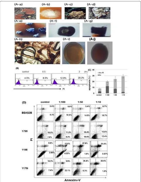
Khz-cp is an extract mixture from the mycelia of a G. lucidum and P. umbellatus nuclear fusion (Figure 1A). The anticancer effect of the fusion of G. lucidum and P. umbellatus has been previously demonstrated [12,13]. In this study, we investigated the mechanism underlying Khz-induced cell death in gastric cells.
Oxidative stress is widely implicated in apoptotic and non-apoptotic cell death [14-16]. The major sources of intracellular reactive oxygen species (ROS) include NADPH oxidase and the mitochondrial electron transport chain (ETC.). When ROS production, either by the mito-chondria or by NADPH oxidase, becomes excessive, the natural cellular antioxidant defense system is over-whelmed, which results in oxidative stress. Cancer cells are more susceptible to oxidative stress than healthy cells, and some anticancer agents such as cisplatin, arsenic tri-oxide (As2O3), and 2-methoxyestradiol exert their effects
by inducing ROS production [17-19]. Much of the avail-able data indicate that the ROS that accumulate during cell death are generated by the mitochondria in response to impairment of the mitochondrial respiratory chain [20-23]. Although mitochondrial ROS production is regarded as an integral component of the apoptotic pro-gram, the role of NADPH oxidase as a primary source of
ROS during the induction of apoptosis has also been re-ported [24-26]. Sustained elevation of the intracellular Ca2+ concentration ([Ca2+]i) is associated with the in-duction of apoptosis [27]. When the cytoplasmic [Ca2+]i increases, the mitochondria take up Ca2+ and function as a Ca2+ buffer; however, excessive accumulation of mitochondrial Ca2+triggers apoptosis, at least in part by inducing ROS generation via the mitochondrial ETC. An increase in cytoplasmic [Ca2+]i can also activate NADPH oxidase, which has been well documented in neutrophils [28]. In some cell types, the activation of protein kinase C via intracellular Ca2+ leads to the phosphorylation of the p47phox subunit and subsequent enzyme assembly [29].
In the present study, we investigated the role of Khz-cp in cellular apoptosis and found that Khz-Khz-cp induced a sustained increase in [Ca2+]ithat resulted in ROS gener-ation by NADPH oxidase via P38 and, finally, cellular apoptosis.
Methods
Cell lines and Khz-cp treatment
The BEAS-2B (normal immortalized), 1799 (non-trans-formed), 1198 (transformed but non-tumorigenic), and 1170-I (tumorigenic) cell lines that compose the in vivo lung carcinogenesis model used in this study have been previously described [30,31]. The human gastric cancer cell line SNU-1 was maintained in RPMI 1640 media supplemented with 10% fetal bovine serum, 100 U/ml penicillin G sodium, 100 μg/ml streptomycin sulfate, and 0.25 μg/ml amphotericin B. Unless otherwise indi-cated, all the cells were treated with Khz-cp diluted 1:100 in the media.
Extraction of Khz-cp (crude polysaccharide extract obtained from the fusion ofG. lucidumandP. umbellatus
mycelia)
First, 1 kg of powder was added to 8.5 L of clean water, heated to 115°C, and extraction was performed for 60 min under pressure. This was followed by a 60-min maturation period. Next, the remaining water from the first extraction was added to 7.5 L of clean water and heated to 115°C; extraction was performed under pres-sure for 60 min, followed by maturation for a further 60 min. The first and second extracts were then mixed, boiled, and placed in bottles after 5 min.
denatured protein content was minimized. Preliminary ex-periments showed that a fourfold excess of 95% ethanol with respect to the sample volume was appropriate for pre-cipitation. Therefore, after the removal of the denatured protein, 95% ethanol (4 times the sample volume) was added slowly until additional precipitation did not occur. The mixture was centrifuged and the supernatant was re-moved. The sediment collected was a brown precipitate and represented the total bacterial crude polysaccharide fraction. The polysaccharides were then dissolved and fil-tered by membrane ultrafiltration (molecular weight cutoff: 100,000 Da). Khz-cp was obtained from BrainGroup (Seoul, South Korea).
Reagents and antibodies
Mitochondrion-targeted ubiquinone (MitoQ) is an ubiqui-nol antioxidant attached to a lipophilic triphenylphospho-nium (TPP) cation [32]. MitoQ and TPP were kind gifts from Dr. Michael P. Murphy (Medical Research Council Dunn Human Nutrition Unit, UK). SB203580, apocynin, and cyclosporin A (CsA) were purchased from Calbio-chem (San Diego, CA, USA), andN-acetyl cysteine (NAC) and ethylene glycol tetraacetic acid (EGTA) were pur-chased from Sigma (St. Louis, MO, USA). z-VAD-fmk was obtained from R&D Systems (Minneapolis, MN, USA), diphenylene iodonium (DPI) was from Cayman Chemical (Ann Arbor, MI, USA), and BAPTA-AM was from Invi-trogen (Eugene, OR, USA).
Antibodies against p38 (sc-7972), p47phox (sc-14015), p67phox(sc-15342), caspase 3 (sc-7148), PARP (sc-7150), and cytochromec(sc-13561) were purchased from Santa Cruz Biotechnology (Santa Cruz, CA, USA). Anti-p-P38 (9255) antibodies were obtained from Cell Signaling (Danvers, MA, USA), and a COX IV antibody (A21347) was obtained from Invitrogen.
Western blot analysis
Cells were lysed in an extraction buffer (31.25 mM Tris–HCl [pH 6.8], 1% sodium dodecyl sulfate [SDS], 10% glycerol, and 2.5% mercaptoethanol), and the whole cell lysate was subjected to 10% SDS-polyacrylamide gel electrophoresis. Size-fractionated proteins on the gel were transferred onto a nitrocellulose membrane. The membrane was blocked in 5% skim milk in Tris-buffered
saline containing 0.05% Tween 20 and was incubated with a primary antibody. After washing, the membrane was in-cubated with the peroxidase-conjugated secondary anti-body. The protein band of interest was detected using enhanced chemiluminescence reagents (Amersham).
Apoptosis assay
Cells treated with Khz-cp were washed twice in cold phosphate-buffered saline (PBS) and stained with annexin-V-FITC (A13199; Invitrogen) and propidium iod-ide (PI) according to the manufacturer’s instructions. Briefly, annexin-V-FITC (5 μL) was added to the cells, which were resuspended in 100 μL of binding buffer (10 mM HEPES, 140 mM NaCl, and 2 mM CaCl2;
pH 7.4). The cells were then incubated at room temperature for 15 min, and PI was added before flow cy-tometry or fluorescence microscopy analysis. Apoptosis were determined using a FACS calibur (Becton and Dick-son) and analysed using Cell Quest pro software. Images were analyzed using NIS Elements software (Nikon).
Assessment of cytoplasmic and mitochondrial ROS levels
The levels of cytoplasmic ROS were estimated using the oxidation-sensitive fluorescent dye H2DCF-DA (2′,7′
-dichlorodihydrofluorescein diacetate; Invitrogen) or the Amplex Red hydrogen peroxide assay kit (Invitrogen). For DCF staining, the cells were loaded with H2DCF-DA
(100 nM) for 1 h at 37°C and washed once with PBS. After treatment with Khz-cp, ROS levels were analyzed using a flow cytometer (FACSCalibur; Becton Dickinson, San Jose, CA, USA) or a fluorescence microscope (Eclipse 80i; Nikon, Tokyo, Japan). The Amplex Red hydrogen peroxide assay was performed according to the manufacturer’s protocol. In brief, the cells were lysed in 50 μM Amplex Red solution supplemented with 0.1 U/mL horseradish peroxidase and incubated in the dark for 30 min. Fluorescence was measured using a plate reader (Victor 2; Perkin-Elmer Life Sciences, Boston, MA, USA) with an excitation wavelength of 540 nm and an emission wavelength of 590 nm.
Mitochondrial superoxide anion levels were analyzed by staining with MitoSOX™ Red (Invitrogen). Cells were loaded with MitoSOX Red (5μM) for 30 min at 37°C and
(See figure on previous page.)
Figure 1Khz-cp induces apoptosis in transformed cells. (A)(A-a) The shape and type of fused fruiting bodies. (A-b) Hyphae isolated from a
Ganoderma lucidummushroom on a petri dish. (A-c) Shape ofG. lucidum. (A-d) Shape and type of fused fruiting bodies and hyphae from
then treated with Khz-cp. The fluorescence was then ana-lyzed by flow cytometry or fluorescence microscopy.
Preparation of subcellular fractions
To prepare the mitochondrial and cytosolic fractions, the cells (1 × 10⁷) were washed once in PBS and dis-rupted by passing them through a glass homogenizer 80 times in ice-cold isolation buffer (250 mM sucrose, 20 mM HEPES, 10 mM KCl, 1.5 mM MgCl2, 1 mM
EGTA, 1 mM EDTA, 1 mM DTT, and 0.1 mM PMSF). Nuclei and non-disrupted cells were removed by cen-trifugation at 750 ×g for 20 min at 4°C. The super-natant was further centrifuged at 10,000 ×gfor 15 min at 4°C to obtain a mitochondrion-enriched pellet and a cytoplasm-enriched supernatant.
Membrane and cytosolic fractions were prepared using the Compartmental Protein Extraction kit (Millipore, Temecula, CA, USA) according to the manufacturer’s instructions.
Ca2+imaging
Digital imaging of the intracellular free Ca2+ was per-formed using the 2 AM dye (Invitrogen). When fura-2 binds to Ca2+, its maximal absorption wavelength shifts from 363 to 335 nm. SNU-1 cells (1 × 10⁴) were cultured in 35-mm glass-bottomed dishes and loaded with fura-2 AM (fura-2μM) for 30 min at 37°C. Fluorescence images of fura-2 were digitally captured at excitation wavelengths of 340 and 380 nm and an emission wavelength of 510 nm with an IX70 fluorescence microscope (Olympus, Tokyo, Japan) equipped with a digital cooled charge-coupled de-vice camera. Paired 340/380 ratiometric images were ana-lyzed using the Metafluor software (Molecular Devices, Sunnyvale, CA, USA). Confocal images of intracellular free Ca2+were obtained using the fluo-4 AM Ca2+ -sensi-tive fluorescent dye (Invitrogen). The cells were loaded with fluo-4 AM (1μM) for 30 min at 37°C, and Ca2+ im-aging was performed using a confocal laser scanning microscope (LSM510; Carl Zeiss, Jena, Germany).
Transfection of siRNA and plasmids
SNU-1 cells were transfected with siRNA using Lipofec-tamine 2000 (Invitrogen), as described previously. The coding strand sequences of the siRNA were as follows: 5′-CUG GUA UGA UCC UUC UGA AdTdT-3′(P381), 5′-GAG GUA UAC ACA UAC UGA dTdT-3′ (Nox2), 5′-CUG UUG UGG ACC CAA UUC AdTdT-3′(Nox4), and 5′-GUU CAG CGU GUC CGG CGA GdTdT-3′ (GFP). Bcl-2 cDNA was transfected into cells using the Lipofectamine-PLUS reagent (Invitrogen) according to the manufacturer’s instructions. Stably transfected cells were selected using G418 (3 mg/mL).
Results
Khz-cp induces apoptosis in transformed cells
Khz-cp is shown in Figure 1A. The aim of the present study was to examine whether Khz-cp causes apoptosis in human cancer cells and, if so, to identify the signaling mechanisms involved. As shown in Figure 1b and c, staining with annexin-V-FITC and PI showed that Khz-cp triggered apoptosis in SNU-1 cells (annexin-V single-positive or annexin-V/PI double-single-positive cells) as early as 60 min after treatment. As apoptosis progressed, the population of cells stained with annexin-V alone de-clined, whereas that of cells stained with both annexin-V and PI increased (Figure 1c). The population of cells positive for PI alone may represent necrotic cells. The induction of preferential apoptosis by Khz-cp in trans-formed cells was then evaluated in a series of cell lines that comprise an in vivo lung epithelial carcinogenesis model. BEAS-2B is an immortalized normal human bronchial epithelial cell line, and 1198 and 1170-I are transformed cell lines derived from BEAS-2B cells ex-posed in vivo to beeswax pellets containing cigarette smoke condensate (CSC). The 1799 cell line is a non-transformed line derived from BEAS-2B cells exposed to beeswax alone. Khz-cp induced apoptosis in the trans-formed 1198 and 1170-I cells but not in non-transformed BEAS-2B and 1799 cells (Figure 1d). These data indicate that Khz-cp induces apoptosis preferen-tially in cancer cells, which suggests that it has potential as a therapeutic agent against cancer.
Khz-cp-induced apoptosis is caspase dependent and occurs through a mitochondrial pathway
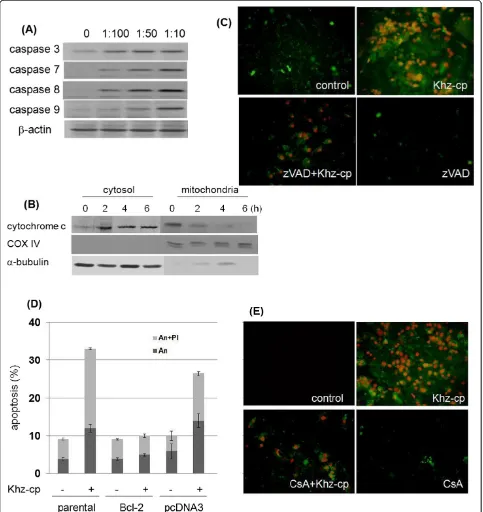
To determine whether the Khz-cp-induced apoptosis was caspase dependent, caspase activation was analyzed after Khz-cp treatment. Cleavage of caspase 3 and PARP (indicating their activation) increased in SNU-1 cells after Khz-cp treatment (Figure 2a). Moreover, pretreat-ment of these cells with the pan-caspase inhibitor z-VAD-fmk completely blocked Khz-cp-induced apoptosis (Figure 2b), which indicates that Khz-cp induces caspase-dependent apoptosis.
to cyclophilin D) abrogated Khz-cp-induced apoptosis, which suggests that mitochondrial permeability transi-tion is required for apoptosis (Figure 2e).
Oxidative stress mediates Khz-cp-induced apoptosis
[image:6.595.57.540.90.602.2](See figure on previous page.)
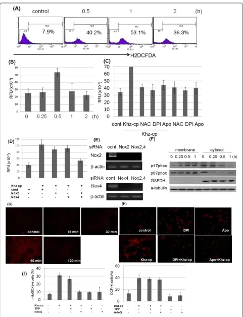
Figure 3Khz-cp triggers cytoplasmic and mitochondrial ROS generation. (A)SNU-1 cells were loaded with H2DCF-DA and treated with
Khz-cp (diluted 1:100), and the cytoplasmic ROS levels were assessed by flow cytometry.(B)Intracellular ROS levels in SNU-1 cells treated with Khz-cp (diluted 1:100) were analyzed using an Amplex Red hydrogen peroxide assay.(C)The cells were pretreated with NAC (5 mM), DPI (10μM), or apocynin (Apo; 300μM) for 1 h. Intracellular ROS levels in SNU-1 cells were analyzed 30 min after Khz-cp treatment by using the Amplex Red hydrogen peroxide assay.(D)SNU-1 cells were transfected with siRNA targeting Nox2 and Nox4. After 48 h, the cells were treated with Khz-cp (1:100, 0.5 h), and ROS generation was measured after 60 min by using the Amplex Red hydrogen peroxide assay.(E)Silencing of Nox2 and Nox4 by siRNA transfection was assessed by RT-PCR.(F)SNU-1 cells were treated with Khz-cp, and the membrane and cytosol fractions were separated using the Compartmental Protein Extraction kit. The expression of the p47phoxand p67phoxproteins in the membrane and cytosol fractions was analyzed by immunoblotting.(G)SNU-1 cells were loaded with MitoSOX Red for 30 min and treated with Khz-cp. Mitochondrial ROS generation was then assessed by fluorescent microscopy at the indicated time points.(H)SNU-1 cells were pretreated with DPI or apocynin for 1 h, and mitochondrial ROS generation was analyzed 60 min after Khz-cp treatment as in (G).(I)SNU-1 cells were pretreated with 0.5μM MitoQ or TPP for 30 min. Right panel: Cytoplasmic ROS generation was measured 30 min after Khz-cp treatment by DCF staining and flow cytometry. Left panel: Mitochondrial ROS generation was assessed 60 min after Khz-cp treatment by MitoSOX Red staining and flow cytometry.
Figure 3a and b show that cytoplasmic ROS levels in-creased 30 min after Khz-cp treatment. Therefore, NADPH oxidase was investigated as a potential source of ROS generation after Khz-cp treatment. Figure 3c shows that DPI (flavoprotein inhibitor) and apocynin (p47phox in-hibitor) prevented ROS production. Moreover, silencing of Nox2 and Nox4 by using specific siRNAs almost com-pletely abrogated ROS production (Figure 3d, e). Trans-fection with Nox2 or Nox4 siRNA alone partially blocked ROS production, which suggests that Nox2 and Nox4 contribute to Khz-cp-induced ROS generation in combin-ation. Activation of NADPH oxidase by Khz-cp was also shown by the translocation of the cytosolic subunits of NADPH oxidase p47phox and p67phox to the cell mem-brane 15 min after Khz-cp treatment (Figure 3f). Taken together, these data indicate that NADPH oxidase pro-duces ROS upon Khz-cp treatment.
Because the mitochondrial respiratory chain is another major source of cellular ROS, mitochondrial ROS pro-duction was evaluated using MitoSOX Red staining. Mitochondrial ROS levels in SNU-1 cells increased 60 min after Khz-cp treatment (Figure 3g); therefore, it appears that ROS production by mitochondria occurs later than that by NADPH oxidase. Mitochondrial ROS generation by Khz-cp was prevented by pretreatment with DPI or apocynin, which suggests that NADPH oxi-dase plays a critical role in mitochondrial ROS production
(Figure 3h). Furthermore, the mitochondria-targeting anti-oxidant MitoQ did not significantly block cytoplasmic ROS generation at a concentration that completely blocked mitochondrial ROS production (Figure 3i). TPP, which is the lipophilic moiety of MitoQ, was used as a control. Taken together, these data show that mitochon-drial ROS generation induced by Khz-cp was mediated in-directly by NADPH oxidase-derived ROS.
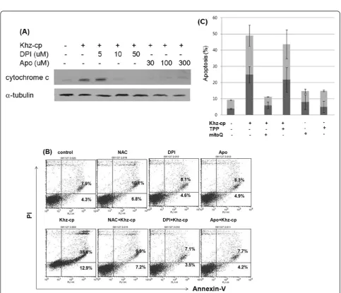
We further examined whether ROS generation through NADPH oxidase and/or mitochondria was necessary for Khz-cp-induced apoptosis. Pretreatment of cells with DPI or apocynin suppressed mitochondrial cytochrome c re-lease and apoptosis induced by Khz-cp treatment, which indicates that ROS generation through NADPH oxidase was required for the induction of apoptosis (Figure 4a, b). Furthermore, pretreatment with MitoQ, but not TPP, pre-vented Khz-cp-induced apoptosis, which suggests that mitochondrial ROS generation was also necessary for the induction of apoptosis (Figure 4c). Therefore, NADPH oxidase-derived ROS appear to trigger apoptosisvia mito-chondrial ROS generation.
Khz-cp induces a rapid and sustained increase in [Ca2+]i
[image:9.595.53.540.87.332.2]Because increased [Ca2+]ihas been widely implicated in mitochondria-mediated apoptosis, we evaluated whether Khz-cp affected [Ca2+]i and, if so, whether it played a role in the activation of NADPH oxidase and ROS Figure 5An increase in [Ca2+]
iis necessary for Khz-cp-induced ROS generation and apoptosis. (A)SNU-1 cells were loaded with fura-2
AM for 30 min, and changes in [Ca2+]
iafter Khz-cp treatment (50μg/ml) were analyzed by digital imaging microscopy.(B)SNU-1 cells were
pretreated with EGTA (10μM) or BAPTA-AM (10μM) for 30 min, and membrane fractions were prepared for immunoblot analysis 12 h after Khz-cp treatment. (C-E) Cells were pretreated with EGTA or BAPTA-AM for 30 min and treated with Khz-cp (1:100 dilution). After 12 h, ROS generation was measured in SNU-1 cells by an Amplex Red hydrogen peroxide assay. The data are presented as the mean ± SD values
generation. Figure 5a shows that Khz-cp induced an im-mediate and sustained increase in [Ca2+]iin SNU-1 cells. The effects of the increase in [Ca2+]ion the activation of NADPH oxidase were then examined. EGTA and BAPTA-AM, which are extracellular and intracellular Ca2+ chelators, respectively, prevented NADPH oxidase activation as measured by the membrane translocation of the p47phoxand p67phoxsubunits (Figure 5b). Further-more, both of these chelators inhibited ROS generation (Figure 5c) and blocked Khz-cp-induced mitochondrial cytochromecrelease and apoptosis (Figure 5d, e). These results indicate that Khz-cp triggers a rapid and sus-tained increase in [Ca2+]ithat activates NADPH oxidase to induce ROS generation and, finally, apoptosis.
P38 mediates Ca2+-dependent activation of NADPH oxidase
[image:10.595.59.538.267.635.2]The mitogen-activated protein kinase P38 is activated in response to oxidative stress [32,33]. Because Khz-cp in-duced ROS generation, the activation of P38 and its pos-sible role in Khz-cp-induced apoptosis were examined. As shown in Figure 6a, P38 was activated as early as 15 min after Khz-cp treatment. Considering that ROS generation was observed 30 min after Khz-cp treatment (Figure 4b), it is unlikely that the activation of P38 was caused by oxidative stress. Nonetheless, P38 activity was required for the induction of apoptosis because a chemical inhibitor of P38 (SB203580) blocked mitochondrial cytochromecrelease and apoptosis induced by Khz-cp treatment, but not an inactive analog of SB202474
Figure 6P38 mediates the increase in [Ca2+]
ito induce NADPH oxidase-derived ROS generation. (A)Activation of P38 was analyzed in
Khz-cp-treated SNU-1 cells by immunoblot analysis of phosphorylated P38.(B)Cells were pretreated with 20μM SB203580 (SB) and SB202474 (SBA) for 1 h. Apoptosis was analyzed 1 h after Khz-cp treatment as in Figure 2(C).(C)SNU-1 cells were pretreated with P38 inhibitors as in (B), and cytochrome c levels in the cytosol were analyzed by immunoblotting 2 h after Khz-cp treatment.(D, E)SNU-1 cells were transfected with control or P38-1 siRNA. After 48 h, ROS generation was analyzed 30 min after Khz-cp treatment using the Amplex Red hydrogen peroxide assay. The data represent the mean ± SD values(D). P38 protein expression was analyzed by immunoblotting 48 h after transfection(E). (F, G)SNU-1 cells were pretreated with 20μM SB203580 (SB) and SB202474 (SBA) for 1 h, and ROS generation induced by Khz-cp treatment was measured using the Amplex Red hydrogen peroxide assay. The data represent the mean ± SD values(F). The levels of p47phoxand p67phoxproteins in the
(Figure 6b, c). Inhibition of P38 by siRNA transfection or pretreatment with chemical inhibitors suppressed Khz-cp-induced ROS production, which indicates that the activa-tion of P38 caused ROS generaactiva-tion rather than the opposite (Figure 6d–f). In agreement with these findings, pretreat-ment of SNU-1 cells with SB203580 also suppressed the ac-tivation of NADPH oxidase, as assessed by the membrane translocation of p47phoxand p67phox(Figure 6g). Therefore, we examined whether P38 activation was dependent on [Ca2+]i. Collectively, these results strongly indicate that P38 is activated by Khz-cp via an increase in [Ca2+]i, thereby
triggering ROS generation by NADPH oxidase and the induction of apoptosis.
Discussion
In the present study, Khz-cp triggered caspase-dependent and mitochondria-mediated apoptotic death in human cancer cells. Khz-cp-induced apoptosis was selective for transformed cells, and Khz-cp only minimally affected non-transformed cells, which suggests that it is a potential anticancer therapeutic agent. Intrinsic oxidative stress in transformed cells may render them more susceptible to apoptosis induced by Khz-cp. Alternatively, cancer cells may have defective restoration of glutathione (GSH) re-serves. However, the GSH levels in tumor tissues are higher than those in normal tissues [34], and depleting GSH reserves often sensitizes cancer cells to ROS-induced cell death, which suggests that the GSH present in cancer cells protects them from oxidative stress.
Khz-cp-induced apoptosis was observed to be caspase dependent, as indicated by the activation of caspases and
inhibition of apoptosis by pretreatment with the pan-caspase inhibitor z-VAD-fmk (Figure 2a, b). The involve-ment of a mitochondria-mediated pathway was confirmed by the release of mitochondrial cytochromec, and inhib-ition of apoptosis was confirmed by the overexpression of Bcl-2 (Figure 2c, d). Oxidative stress is associated with apoptotic and non-apoptotic cell death, although pro-oxidative conditions are not a prerequisite for apoptosis [35]. The present study shows that the induction of apop-tosis by Khz-cp required ROS generation by both NADPH oxidase and mitochondria. Several studies have implicated ROS generated from mitochondria in the induction of apoptosis; however, the results of the present study indi-cate that NADPH oxidase-derived ROS were critical for Khz-cp-induced apoptosis (Figure 4a, b). Although mito-chondrial ROS generation was also necessary for Khz-cp-induced apoptosis (Figure 4c), its effect was secondary to that of the initial ROS production by NADPH oxidase be-cause the generation of mitochondrial ROS occurred after that of cytoplasmic ROS and was prevented by pretreat-ment with NADPH oxidase inhibitors (Figure 3g, h). Of the 7 members of the human NADPH oxidase family, Nox2 and Nox4 were found to be responsible for the ROS generation induced by Khz-cp treatment in SNU-1 cells (Figure 3d, e).
It is widely accepted that calcium signaling plays an im-portant role in apoptosis [29,36]. Cross-talk between ROS and calcium signaling pathways may lead to synergistic ef-fects on mitochondrial permeabilization and cell death. The present study showed that an increase in the [Ca2+]i induced by Khz-cp treatment resulted in ROS generation by NADPH oxidase. The [Ca2+]iincreased before the gen-eration of ROS, and Ca2+ chelators such as EGTA and BAPTA-AM abrogated the activation of NADPH oxidase and ROS generation (Figure 5b, c). However, the inhibitors of NADPH oxidase did not affect the increase in [Ca2+]i
induced by Khz-cp treatment (data not shown). The acti-vation of NADPH oxidase by elevated [Ca2+]i in neutro-phils is well known and has also been reported for other cell types [37,38].
P38 was identified as the mediator of Ca2+-dependent NADPH oxidase activation. P38 was activated within 15 min of Khz-cp treatment in a Ca2+-dependent manner (Figure 6a) Furthermore, treatment with P38 siRNA or chemical inhibitors prevented Khz-cp-induced ROS gener-ation (Figure 6d–g). Most studies place ROS upstream of P38 activation [39,40] however, in this study, ROS gener-ation by NADPH oxidase was found to be mediated by the activation of P38.
Conclusions
[image:11.595.58.290.90.321.2]In this study, we investigated that Khz induces apoptosis by increasing intracellular calcium levels and activating JNK and NADPH oxidase-dependent generation of ROS Figure 7Summary of the signaling events leading to
[12]. We showed that Khz-cp induces mitochondrion-mediated apoptosis preferentially in transformed cells, and the signaling pathway of Khz-cp-induced apoptosis involves an increase in [Ca2+]i, activation of P38, and ROS generation through NADPH oxidase and mito-chondria (Figure 7).
Competing interests
The authors declare that they have no competing interests. Authors’contributions
KTH, JSK, ZHK, RBH,YLC carried out the study and prepared the manuscript. KTH and RSW critically revised manuscript. All authors have read and approved the manuscript for publication.
Acknowledgements
This research was supported by Brain Group Co., Ltd, Young Lye Chae (chief executive officer).
Author details
1
Department of Radiotherapy, The First Affiliated Hospital, Guangxi Medical University, Nanning, China.2Clinical Medicine, Harbin Medical University, Harbin, China.3Graduate School of Information Science, Nagoya University, Nagoya, Japan.4Department of Pharmacology, Guangxi Medical University, Nanning, China.5Korea Institute of Science and Management Career College, Seoul, South Korea.
Received: 11 September 2013 Accepted: 1 July 2014 Published: 10 July 2014
References
1. Debatin KM:Apoptosis pathways in cancer and cancer therapy.Cancer Immunol Immunother2004,53:153–159.
2. Arends MJ, Wyllie AH:Apoptosis: mechanisms and roles in pathology.Int Rev Exp Pathol1991,32:223–254.
3. Mesner PW Jr, Budihardjo II, Kaufmann SH:Chemotherapy-induced apoptosis.Adv Pharmacol1997,41:461–499.
4. Palombo JD, Ganguly A, Bistrian BR, Menard MP:The antiproliferative effects of biologically active isomers of conjugated linoleic acid on human colorectal and prostatic cancer cells.Cancer Lett2002,177:163–172. 5. Wasser SP, Weis AL:Therapeutic effects of substances occurring in higher
Basidiomycetes mushrooms: a modern perspective.Crit Rev Immunol
1999,19:65–96.
6. Jiang J, Sliva D:Novel medicinal mushroom blend suppresses growth and invasiveness of human breast cancer cells.Int J Oncol2010,
37(6):1529–1536.
7. Lin ZB, Zhang HN:Anti-tumor and immunoregulatory activities of Ganoderma lucidum and its possible mechanisms.Acta Pharmacol Sin
2004,25:1387–1395.
8. Sanodiya BS, Thakur GS, Baghel RK, Prasad GB, Bisen PS:Ganoderma lucidum: a potent pharmacological macrofungus.Curr Pharm Biotechnol
2009,10:717–742.
9. Gao Y, Gao H, Chan E, Tang W, Xu A, Yang H, Huang M, Lan J, Li X, Xu C, Zhou S, Duan W:Antitumor activity and underlying mechanisms of ganopoly, the refined polysaccarides extracted from Ganoderma lucidum, in mice.Immunol Investig2005,34:171–198.
10. Yue GG, Fung KP, Tse GM, Leung PC, Lau CB:Comparative studies of various ganoderma species and their different parts with regard to antitumor and immunomodulating activities in vitro.J Altern Complement Med2006,12:777–789.
11. Zhao YY, Chao X, Zhang Y, Lin RC, Sun WJ:Cytotoxic steroids from Polyporus umbellatus.Planta Med2010,76(15):1755–1758.
12. Kim TH, Kim JS, Kim ZH, Huang RB, Wang RS:Khz (fusion of ganoderma lucidum and polyporus umbellatus mycelia) induces apoptosis by increasing intracellular calcium levels and activating JNK and NADPH oxidase-dependent generation of reactive oxygen species.PLoS One
2012,7(10):e46208.
13. Kim TH, Kim J, Kim Z, Huang RB, Wang RS:Khz (Fusion of Ganoderma lucidum and Polyporus umbellatus Mycelia) Induces Apoptosis in A549 Human Lung Cancer Cells by Generating Reactive Oxygen Species and
Decreasing the Mitochondrial Membrane Potential.Food Sci. Biotechnol.
2014,23(3):859–864.
14. Ryter SW, Kim HP, Hoetzel A, Park JW, Nakahira K, Wang X, Choi AM:Mechanisms of cell death in oxidative stress.Antioxid Redox Signal2007,9:49–89.
15. Buttke TM, Sandstrom PA:Oxidative stress as a mediator of apoptosis.
Immunol Today1994,15:7–10.
16. Jacobson MD:Reactive oxygen species and programmed cell death.
Trends Biochem Sci1996,21:83–86.
17. Miyajima A, Nakashima J, Yoshioka K:Role of reactive oxygen species in cis-dichlorodiammineplatium-induced cytotoxicity on bladder cancer cells.Br J Cancer1997,76:206–210.
18. Zhou Y, Hileman EO, Plunkett W, Keating MJ, Huang P:Free radical stress in chronic lymphocytic leukemia cells and its role in cellular sensitivity to ROS-generating anticancer agents.Blood2003,101:4098–4104. 19. Schulze-Osthoff K, Bakker AC, Vanhaesebroeck B, Beyaert R, Jacob WA, Fiers
W:Inhibition of mitochondrial respiration: a novel strategy to enhance drug-induced apoptosis in human leukemia cells by a reactive oxygen species-mediated mechanism.J Biol Chem2003,278:37832–37839. 20. Quillet-Mary A, Jaffrézou JP, Mansat V, Bordier C, Naval J, Laurent G:
Cytotoxic activity of tumor necrosis factor is mediated by early damage of mitochondrial functions. Evidence for the involvement of
mitochondrial radical generation.J Biol Chem1992,267:5317–5323. 21. Fleury C, Mignotte B, Vayssiere JL:Implication of mitochondrial hydrogen
peroxide generation in ceramide-induced apoptosis.J Biol Chem1997,
272:21388–21395.
22. Fleury C, Mignotte B, Vayssière JL:Mitochondrial reactive oxygen species in cell death signaling.Biochimie2002,84:131–141.
23. Ott M, Gogvadze V, Orrenius S, Zhivotovsky B:Mitochondria, oxidative stress and cell death.Apoptosis2007,12:913–922.
24. Hiraoka W, Vazquez N, Nieves-Neira W, Chanock SJ, Pommier Y:Role of oxygen radicals generated by NADPH oxidase in apoptosis induced in human leukemia cells.J Clin Invest1998,102:1961–1968.
25. Qin F, Patel R, Yan C, Liu W:NADPH oxidase is involved in angiotensin II-induced apoptosis in H9C2 cardiac muscle cells: effects of apocynin.Free Radic Biol Med2006,40:236–246.
26. Brennan AM, Suh SW, Won SJ, Narasimhan P, Kauppinen TM, Lee H, Edling Y, Chan PH, Swanson RA:NADPH oxidase is the primary source of superoxide induced by NMDA receptor activation.Nat Neurosci2009,
12:857–863.
27. Nicotera P, Orrenius S:The role of calcium in apoptosis.Cell Calcium1998,
23:173–180.
28. Granfeldt D, Samuelsson M, Karlsson A:Capacitative Ca2+ influx and activation of the neutrophil respiratory burst. Different regulation of plasma membrane- and granule-localized NADPH-oxidase.J Leukoc Biol
2002,71:611–617.
29. Wang G, Anrather J, Glass MJ, Tarsitano MJ, Zhou P, Frys KA, Pickel VM, Iadecola C:Nox2, Ca2+, and protein kinase C play a role in angiotensin II-induced free radical production in nucleus tractus solitaries.
Hypertension2006,48:482–489.
30. Kim JE, Koo KH, Kim YH, Sohn J, Park YG:Identification of potential lung cancer biomarkers using an in vitro carcinogenesis model.Exp Mol Med
2008,40:709–720.
31. Klein-Szanto AJ, Iizasa T, Momiki S, Garcia-Palazzo I, Caamano J, Metcalf R, Welsh J, Harris CC:A tobacco-specific N-nitrosamine or cigarette smoke condensate causes neoplastic transformation of xenotransplanted human bronchial epithelial cells.Proc Natl Acad Sci U S A1992,89:6693–6697. 32. Kelso GF, Porteous CM, Coulter CV, Hughes G, Porteous WK, Ledgerwood EC,
Smith RA, Murphy MP:Selective targeting of a redox-active ubiquinone to mitochondria within cells: antioxidant and antiapoptotic properties.J Biol Chem2001,276:4588–4596.
33. Matsuzawa A, Ichijo H:Redox control of cell fate by MAP kinase: physiological roles of ASK1-MAP kinase pathway in stress signaling.
Biochim Biophys Acta2008,1780:1325–1336.
34. Kuppusamy P, Li H, Ilangovan G, Cardounel AJ, Zweier JL, Yamada K, Krishna MC, Mitchell JB:Noninvasive imaging of tumor redox status and its modification by tissue glutathione levels.Cancer Res2002,62:307–312. 35. Jacobson MD, Raff MC:Programmed cell death and Bcl-2 protection in
very low oxygen.Nature1995,374:814–816.
37. Yu JH, Lim JW, Kim KH, Morio T, Kim H:NADPH oxidase and apoptosis in cerulein-stimulated pancreatic acinar AR42J cells.Free Radic Biol Med
2005,39:590–602.
38. Gandhi S, Wood-Kaczmar A, Yao Z, Plun-Favreau H, Deas E, Klupsch K, Downward J, Latchman DS, Tabrizi SJ, Wood NW, Duchen MR, Abramov AY:
PINK1-associated Parkinson’s disease is caused by neuronal vulnerability to calcium-induced cell death.Mol Cell2009,33:627–638.
39. Benhar M, Dalyot I, Engelberg D, Levitzki A:Enhanced ROS production in oncogenically transformed cells potentiates c-Jun N-terminal kinase and p38 mitogen-activated protein kinase activation and sensitization to genotoxic stress.Mol Cell Biol2001,21:6913–6926.
40. Saeki K, Kobayashi N, Inazawa Y, Zhang H, Nishitoh H, Ichijo H, Saeki K, Isemura M, Yuo A:Oxidation-triggered c-Jun N-terminal kinase (JNK) and p38 mitogen-activated protein (MAP) kinase pathways for apoptosis in human leukaemic cells stimulated by epigallocatechin-3-gallate (EGCG): a distinct pathway from those of chemically induced and receptor-mediated apoptosis.Biochem J2002,368:705–720.
doi:10.1186/1472-6882-14-236
Cite this article as:Kimet al.:Khz-cp (crude polysaccharide extract
obtained from the fusion ofGanoderma lucidumandPolyporus umbellatus
mycelia) induces apoptosis by increasing intracellular calcium levels and activating P38 and NADPH oxidase-dependent generation of reactive oxygen species in SNU-1 cells.BMC Complementary and Alternative
Medicine201414:236.
Submit your next manuscript to BioMed Central and take full advantage of:
• Convenient online submission
• Thorough peer review
• No space constraints or color figure charges
• Immediate publication on acceptance
• Inclusion in PubMed, CAS, Scopus and Google Scholar
• Research which is freely available for redistribution
![Figure 5 An increase in [Ca2+]i is necessary for Khz-cp-induced ROS generation and apoptosis](https://thumb-us.123doks.com/thumbv2/123dok_us/5808.1000270/9.595.53.540.87.332/figure-increase-necessary-khz-induced-ros-generation-apoptosis.webp)
![Figure 6 P38 mediates the increase in [Ca2+]i to induce NADPH oxidase-derived ROS generation](https://thumb-us.123doks.com/thumbv2/123dok_us/5808.1000270/10.595.59.538.267.635/figure-mediates-increase-induce-nadph-oxidase-derived-generation.webp)
