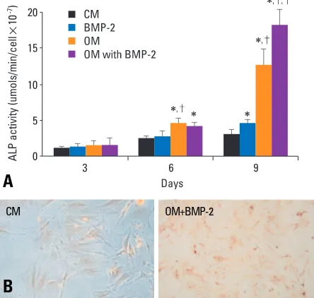
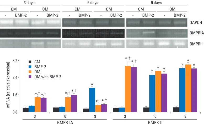
Bone Morphogenetic Protein Receptor in the Osteogenic Differentiation of Rat Bone Marrow Stromal Cells
6
0
0
Full text
Figure
Related documents