http://dx.doi.org/10.4236/jbise.2016.94014
How to cite this paper: Gündüz, D. and Aslam, M. (2016) MicroRNAs as Modulators of Endothelial Differentiation of Stem Cells. J. Biomedical Science and Engineering, 9, 177-190. http://dx.doi.org/10.4236/jbise.2016.94014
MicroRNAs as Modulators of Endothelial
Differentiation of Stem Cells
Dursun Gündüz, Muhammad Aslam
*Department of Cardiology/Angiology, University Hospital Giessen (UKGM), Giessen, Germany
Received 21 September 2015; accepted 26 March 2016; published 29 March 2016
Copyright © 2016 by authors and Scientific Research Publishing Inc.
This work is licensed under the Creative Commons Attribution International License (CC BY). http://creativecommons.org/licenses/by/4.0/
Abstract
MicroRNAs (miRs) are a class of small (~22 nucleotides), widely distributed, and highly conserved non-coding RNA molecules and play an important post-transcriptional regulatory role by targeting mRNA. Embryonic and induced pluripotent stem cells (ESCs and iPSC, respectively) hold great promise for vascular regenerative therapies. However, several limitations currently prohibit their therapeutic use. The importance of miRs in controlling the gene expression profile of a particular cell type is emerging and a multitude of miRs have been identified that play key roles in vascular development and regeneration. A combination of pluripotency transcription factors and different miRs not only enhances the pluripotency of stem cells but also has been reported to enhance their endothelial differentiation. This review will summarize the findings that focus different miR clus-ters in the induction, maintenance, and directed endothelial differentiation of ESCs and iPSCs.
Keywords
EPCs, ESCs, iPSC, miRs, Vascular Regeneration
1. Introduction
Endothelial cells (ECs) form the innermost lining of the blood vessels and play an active role in the maintenance of vascular integrity and homeostasis through the synthesis and release of numerous vasoactive molecules [1]. In vertebrates, the cardiovascular system is the first functional organ formed during embryonic development. Dur-ing embryogenesis, first the ECs form a rudimentary vascular meshwork that undergoes a series of developmen-tal stages forming stabilised vessels by the recruitment of mural cells [2]-[4]. During the establishment of func-tional vascular networks, known as vasculogenesis, a plethora of signalling pathways act to coordinate the de-velopment and maintenance of these vascular networks [4]. Loss of EC function leads to development of
merous chronic vascular anomalies. During the last decade, it has become clear that pluripotent stem cells can be directly differentiated toward EC lineage and may potentially be used for the repair of injured vasculature [5]. The understanding of molecular mechanisms regulating EC differentiation will greatly benefit the regenerative strategies for the treatment of various vascular diseases. Numerous methods and factors have been described to programme pluripotent cells to ECs but with limited efficiency [6]. Among these factors, micro RNAs (miRs) which are involved in regulation of at least 30% of the mammalian genes [7], are relatively new players in the field. This review gives a brief over view of various miRs involved in vasculogenesis with potential capacity to induce EC differentiation and their potential to be used in regenerative vascular medicine.
2. MicroRNAs
MiRs belong to class of small (~22 nucleotide; nt), noncoding RNAs controlling gene expression at transcrip-tional level [8]. miRs are first transcribed as primary miRs (pri-miRs) by RNA polymerase II either from miR genes or intronic sequences of protein coding genes which are then processed into ~70 nt precursors (pre-miRs) and finally to mature miRs by sequential action of two RNase III proteins, Drosha and Dicer, respectively [9] [10]. The mature miRs are incorporated into RNA-induced silencing complex (RISC) consisting of argonaute (AGO) and other RNA binding proteins. The RISC identifies its target mRNA sequences (usually on 3'-UTR) via specific 5'-end “seed sequence” (between nt 2 and 7 on 5'-end of miR) and thus translationally repress target proteins [11]. Beyond targeting mRNAs, some RISCs may act directly on the genome via formation of RNA- induced transcriptional silencing (RITS) complex consisting of AGO1 with an associated miR and chromodo-main proteins [12]. Upon target recognition, RITS recruits histone methyl-transferases, which modify the his-tones associated with the target DNA locus [13]. Currently, over 1800 human miRs are annotated in the miR-BASE database (www.mirbase.org) [14]. Each mRNA can be targeted by multiple miRs and each miR can tar-get several genes and thus can regulate several signalling pathways simultaneously.
3. Vascular Regeneration
Vascular diseases which include any condition that affects the circulatory system and ranges from diseases of arteries, veins, and lymphatics to blood disorders affecting circulation, such as peripheral artery disease, renal artery disease, carotid artery disease, and peripheral venous disease, are currently the major causes of morbidity in Western world [15]. Progression of the macrovascular diseases ultimately leads to the development of mi-crovascular complications and organ failure. The traditional surgical and non-surgical treatment strategies for vascular disease include opening-up or bypassing the heavily blocked vessel using autologous or synthetic grafts. Though, well-developed, these procedures are inefficient to prevent or reduce target organ damage.
Regenerative medicine is an emerging interdisciplinary field which aims to restore normal functions of the organ through repair, replacement, or regeneration of cells, tissues, or organs that are lost or damaged due to disease, injury, or ageing [16]. In this promising technology, new vascular network can be induced in the im-paired tissues by delivering stem or progenitor cells which ultimately could promote vascular regeneration [15]
via enhanced angiogenesis and augmented vasculogenesis [17]. The most studied cells for vascular regeneration include embryonic and mesenchymal stem cells (ESCs and MSCs, respectively) along with circulating endothe-lial progenitor cells (EPCs) [18]. Other cell sources including induced pluripotent stem cells (iPSCs), human amniotic fluid stem cells (hAFSCs) [15][18] may also be used. Vascular repair is a complex process and re-quires the mobilisation, chemotaxis, adhesion, proliferation, and differentiation of ESCs or EPCs [19] to vascu-lar cells. During the process of differentiation, ESCs/EPCs transcription factors, signalling networks, and epige-netics undergo a tremendous change [20]-[22]. Increasing evidence suggests an essential role of miRs in the self-renewal and differentiation of ESCs/EPCs, suggesting promising prospects of using miRs in regenerative medicine. Below, we will discuss the recent advances in our understanding of miRs in the induction and main-tenance of pluripotent stem cells and their EC differentiation.
4. Angiogenesis and Endothelial Specific miRs
family and anti-angiogenic (anti-AngiomiRs) miR-17/92 cluster and miR-221/222 family [23], which are highly expressed in ECs. The first evidence that miRs play important role in vascular development was shown by Yang
et al. [24] using Dicer knockout mice. Homozygous embryos died between E12.5 and E14.5 due to deregulation of angiogenic factors/signalling leading to impaired vascular development [24]. In vitro knockdown of either
Dicer or Drosha negatively regulated miR-let7 and miR-17/92 cluster expression and angiogenesis [25][26]. The anti-angiogenic effect of Dicer deletion could be corrected by transfection of miR-17/92 cluster [25]. EC- specific Dicer knockdown reduced postnatal angiogenic response to multiple stimuli and impaired wound heal-ing [27].
The first well-characterised miR in ECs is miR-126 which plays an important role in vascular development and angiogenesis during embryogenesis. EC-specific deletion of miR-126 resulted in vascular defects and haemorrhages and is embryonically lethal [28]. Knockdown of miR-126 in zebrafish embryos causes vascular leakage [29]. Selective overexpression of miR-126 in ECs enhances re-endothelisation of injured vessel and in-hibited vascular stenosis [30]. Likewise, ectopic expression of miR-126 in placenta enhanced microvascular density and pub survival in rat model of pre-eclampsia [31]. In mature vessels, miR-126 enhances vessel integ-rity by targeting p85β and thus downstream inhibiting Ang-1 signalling [32].
The miR-17/92 cluster also known as oncomiR-1 is one of the best studied miR clusters and though dysregu-lation of their expression leads to a variety of abnormalities [33], their role in developmental angiogenesis is controversial and remains elusive. This cluster includes six miRs (miR-17, miR-18a, miR-19a, miR-19b, miR20a, and miR-92a) which are processed from common primary transcript [34]. Early studies reported a proangiogenic effect of miR-17/92 cluster in tumour vasculature [35], however, later studies found that over ex-pression of individual members of the cluster exerts anti-angiogenic effects in cultured ECs [36]. Epigenetically miR-17/92 cluster is controlled by histone deacetylase 9 (HDAC9) and silencing miR-17-20a rescued angio-genic defects produced due to HDAC9 downregulation [37], suggesting an anti-angiogenic role of miR-17/92 cluster. Likewise, overexpression of miR-15a/16 lead to reduced vascularisation in hind limb ischemia model
[38], and conversely their knockdown improved angiogenesis and neovascularisation [39]. MiR-130 is highly expressed in ECs and acts as pro-angiogenic miR by targeting two anti-angiogenic transcription factors, ho-meobox A5 (HOXA5) and growth arrest specific hoho-meobox (GAX) [40]. MiR-21 is involved in divergent pathophysiological processes particularly related to ischemia/reperfusion injury. Short-term hypoxic conditions induce the expression of miR-21 in ECs and ESCs [41], which via VEGF signalling promotes cell survival and their angiogenic capacity. However, its long-term upregulation under certain pathological conditions promotes tissue fibrosis [42]. A schematic overview of different miRs involved in angiogenesis is presented in Figure 1.
5. miRs Involved Inpost-Ischemic Collateral Growth
[image:3.595.182.444.526.691.2]Following a major artery occlusion two types of vascular repair responses are activated in the injured ischemic tissue: shear-stress sensitive arteriogenesis or development of collateral arteries from pre-existing arterioles and
Figure 1. A graphical presentation of different miRs involved in the re-
parenchymal hypoxia-driven angiogenesis or sprouting of capillaries [43]. Recent evidence suggests the impor-tance of miRs for regulating post-ischemic arteriogenesis. Shear stress is one of the strongest inducer of arterio-genesis [43][44] and miR-21 is upregulated in response to high shear-stress [45] which regulates smooth muscle cell proliferation and migration via upregulation of PI3K signalling [46][47]. MiR-21 is upregulated in myocar-dium of metabolic syndrome JCR rats leading to enhanced smooth muscle cell (SMC) proliferation but reduced collateral growth, which could be rescued by antagomir miR-21 administration [48], suggesting miR-21 as negative regulator of arteriogenesis. Likewise, endothelial specific deletion of miR-17/92 cluster enhanced col-lateral growth in cardiac and hind limb ischaemia model suggesting a negative role of miR-17/92 cluster in arte-riogenesis [49]. Recent data from Grundmann’s group revealed a tissue-specific role of miR-155 in arteriogene-sis. In hind-limb ischemia model, it has pro-arteriogenesis role and knockout mice (miR-155−/−) presented a re-duced vascularisation [50]. In contrast, hearts from miR-155−/− mice are protected against ischaemia/reperfusion injury [51]. Same group also identified miR-100 as a negative regulator of neovascularisation [52][53] via sup-pression of mTOR exsup-pression both in ECs and SMCs [52] and can be an important target for therapeutic inter-vention. Likewise, a recent study has identified miR 14q32 cluster (miR-329, miR-487b, miR-494, and miR-495) as negative regulators of arteriogenesis in mouse hind limb ischaemia model [54] and knockdown of two or more of these miRs resulted in enhanced vascularisation. The miR-132/212 cluster has been identified as posi-tive regulator of arteriogenesis which is up-regulated during early period of hind limb ischaemia and miR- 132/212 knockout mice show reduced collateral growth after ligation [55]. A schematic overview of different miRs involved in arteriogenesis is presented in Figure 1.
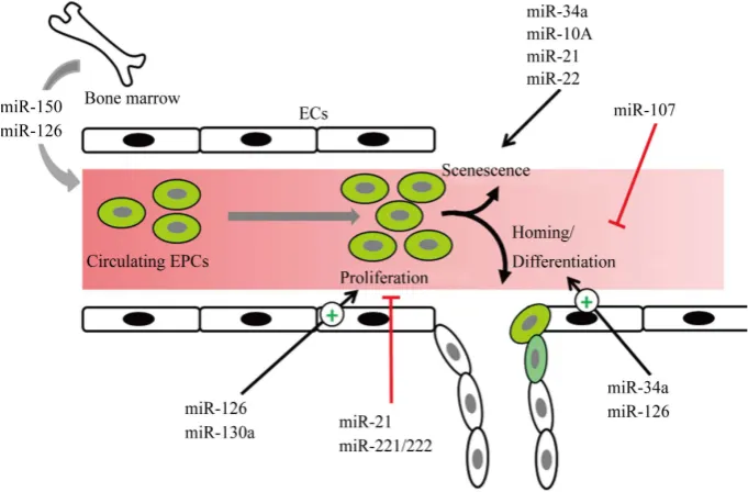
6. miRs Regulating Endothelial Progenitor Cell Functions
EPCs are the circulating cells that express different cell surface markers similar to those expressed by ECs, ad-here to endothelium at sites of injury, and participate in vasculogenesis [56]. Increasing body of evidence sug-gests an important role of miRs at different stages of EPCs differentiation to ECs and in their proper functioning.
Proliferation: The miR-221/222 family and miR-21 are reported to act as anti-proliferative miRs in EPCs. The expression of miR-221/222 is upregulated in patients with coronary artery disease (CAD) [57] which is weakly negatively correlated with proliferation of EPCs. Over expression of miR-221/222 family in healthy EPCs resulted in reduction in their proliferation rate [58][59]. Likewise, the expression of miR-21 is upregu-lated in EPCs from atherosclerotic patients accompanied by their reduced proliferation and migration [60]. The downregulation of miR-21 rescues this phenotype. Unlike miR-221/222 family and miR-21, miR-126 and miR- 130a have been reported as pro-proliferative miRs in EPCs. Both miR-126 and miR-130a are downregulated in EPCs from diabetic patients [61][62]. Furthermore, overexpression of miR-126 significantly increased the pro-liferation and migration while knockdown of miR-126 resulted in reduced propro-liferation of normal EPCs [61].
Senescence: Data about the role of miRs in the regulation of EPC senescence is emerging. Expression of miR-34a was found to be upregulated in EPCs from CAD patients [63], and overexpression of miR-34a in rat EPCs resulted in reduced expression of SIRT1 accompanied by reduced angiogenesis capacity and enhanced cellular senescence of EPCs [64]. The expression of miR-10A∗ and miR-21 is upregulated in EPCs from aged mice and knockdown of these miRs enhanced the proliferation of aged EPCs [65]. Likewise, miR-22 is upregu-lated in aged compared to young EPCs and over expression of miR-22 in young EPCs induced senescence in these cells via downregulation of Akt3 [66].
[74]. A graphical presentation of different miRs involved in different stages of EPCs differentiation to ECs is shown in Figure 2.
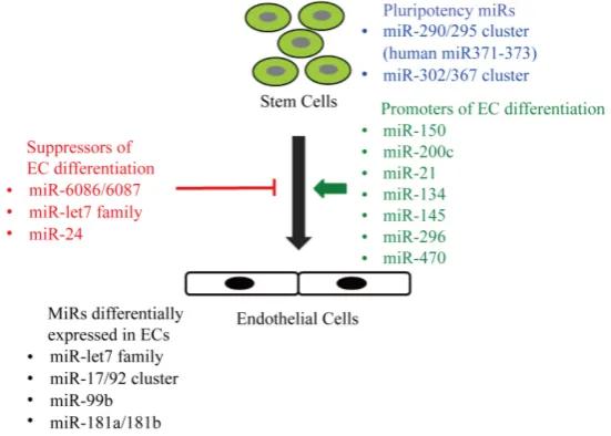
7. ESC Specific miRs Regulating Their Commitment to ECs
Embryonic stem cells (ESCs) are the pluripotent cells that originate from inner cell mass of a mammalian blas-tocyst and due to their ability to self-renewal and differentiate into various cell types hold the promise of clinical applications [75]. Several studies have identified ESC-specific miRs clusters which are preferentially expressed in ESCs and are downregulated during the process of differentiation [76][77]. The first evidence that miRs are required for self-renewal and differentiation of ESCs came from the study by Kanellopoulou et al. where authors demonstrated ESCs lacking Dicer lost the ability to proliferate or form the embryoid bodies [78]. The miR- 290/295 cluster accounts for ~50% of the miR contents of mouse ESCs and its expression is downregulated during differentiation [77]. Interestingly, over expression of three miRs (miR-291a-3p, miR-294, and miR-295) from this cluster are sufficient to promote induced pluripotency in somatic cells [79]. The human homolog of the mouse miR-290/295 cluster is miR371-373, specifically expressed in hESCs and is upregulated in some tumours
[80][81]. Second ESC-specific miR cluster is miR-302/367 which is expressed both in human and mouse ESCs
[82]. This miR cluster can be used to re-programme fibroblasts into induced pluripotent stem cells (iPSCs) [83]. Interestingly, all these miR clusters possess identical seed sequences (AAGUGCU) suggesting that they may be regulating similar pools of mRNAs [84]. During the process of differentiation certain set of miRs is upregulated which target the pluripotency genes Oct4, Sox2, and Nanog, suppressing pluripotency and thus assist ESCs to-wards differentiation. These include let-7 family of miRs, miR-21, miR-145, miR-134, miR-296, and miR-470
[image:5.595.142.483.468.692.2][85]-[87]. Additionally, numerous other miRs have been reported which are differentially expressed during the process of EC differentiation but their role in EC differentiation and vascular development needs to be estab-lished. Yoo et al., [88] reported two novel miRs, miR-6086 and miR-6087, which target vascular endothelial (VE-)cadherin and endoglin, respectively, and their expression is down regulated during EC differentiation [88]. Another study demonstrated that the expression of miR-99b, miR-181a, and miR-181b was increased in ECs during differentiation [89], which was reconfirmed by another group recently [90]. Interestingly, although over expression of these miRs promotes EC differentiation, their knockdown has no significant impact on EC differ-entiation of hESCs [89]. Likewise, Treguer et al., [91] showed miR-17/92a cluster is upregulated during EC differentiation of ESCs, however, forced knockdown of these miRs in ESCs during the process of differentiation had no effect on their EC differentiation [91]. A recent report shows an upregulation of miR-150 and miR-200c
during EC differentiation and antagomirs to these miRs lead to reduced vascularisation in chick embryos [92]. This finding is opposed by another report showing a downregulation of miR-200 family in mESCs during the process of EC differentiation and constitutive expression of miR-200 family members repressed EC differentia-tion [93]. A careful review of miRs regulating different transcription factors required for EC differentiation of ESCs may give a further insight into the miRs regulating trans-differentiation. For example, in an elegant study, Shi et al., [94] have demonstrated that GATA2 together with Etv2 regulates EC and haematopoietic cell differ-entiation of mESCs. Etv2 controls the vasculogenesis during zebrafish development [95] and its transcription is regulated by let-7 family of miRs. Ectopic expression of let-7 miRs leads to downregulation of Etv2 and vascu-lar defects in zebrafish [96]. Likewise, Gata2 expression in murine cardiac ECs is regulated by miR-24 and its ectopic expression leads to ECs apoptosis [96]. A graphical presentation of different miRs involved in ESCs differentiation to ECs is shown in Figure 3.
8. Induced Pluripotent SC-Specific miRs
[image:6.595.176.452.499.696.2]The clinical application of hESCs is limited by immune rejection and ethical concerns. In this regard, human induced pluripotent stem cells (hiPSCs) have recently emerged as promising alternative [97]. In this method somatic cells can be reprogrammed by forced expression of four pluripotent transcription factors (either Oct4- Sox2-Klf4-c-Myc or Oct4-Sox2-Nanog-Lin28) [98] [99]. Using Dgcr8 (miR processing protein) knockout mouse ESCs (mESCs) Wang et al., identified miR-290 cluster as regulators of mESC pluripotency [100]. Inter-estingly, forced expression of subsets of this miR cluster enhanced reprogramming efficiency of mouse embry-onic fibroblasts (MEFs) in the presence of four Yamanaka factors [79][101]. Likewise, over expression of hu-man orthologue, miR-371 cluster, and miR-302/367 clusters enhanced reprogramming of huhu-man fibroblasts by 10 - 15 fold [102]. Later Morrisey and colleagues demonstrated that expression of miR-302/367 cluster alone could successfully induce both mouse and human somatic cells in the presence of HDAC inhibitor without the need of external Yamanaka factors [83]. Recently, Deng et al., [103] demonstrated that transfection of miR- 302/367 cluster mimetic along with Oct4 and Sox2 could increase the pluripotency efficiency by 50-fold. On the other hand, knockdown of miR-302/367 cluster completely blocked iPSC generation [104] suggesting an im-portant role of miR-302/367 cluster in induction of pluripotency. The increased efficiency by miR-290 (miR-371 human homologue) is mediated by promoting a mesenchymal-to-epithelial (MET) transition, affecting the cell cycle, and inhibiting the TGF-β signalling [101], while miR-302 cluster targets epigenetic regulators responsible for DNA methylation [105]. Some recent reports indicate other miRs may also be contributing to somatic cell reprogramming. Rana and colleagues identified that c-Myc regulated miRs, miR-21 and miR-29a, are highly expressed in MEFs and depletion of these miRs enhanced reprogramming efficiency of MEFs [106]. Likewise, miR-34 deficient MEFs depicted higher reprogramming efficiency [107], suggesting these miRs act as negative
regulators of pluripotency.
The role of miRs in EC differentiation of iPSCs is emerging. As discussed earlier, Rana and colleagues dem-onstrated that depletion of miR-21 enhanced reprogramming efficiency of MEFs [106] and contrarily, re-ex- pression of miR-21 in iPSCs enhanced their EC differentiation capacity [108], suggesting miR-21 an important candidate for reprogramming of somatic cells into ECs. Recently, Chen and colleagues identified miR-199a and miR-199b as differentially regulated miRs during EC differentiation [109][110] but their role in EC differentia-tion needs to be verified. Likewise, Wang et al., [90] have performed an extensive miR profiling of hiPSCs and differentiated ECs and identified several differentially regulated miRs including miR-20a, miR-20b, miR-27b, miR-100, miR-125a-5p, miR-137, miR-149, miR-181a, miR-210, miR-222, and miR-296-5p, however, their role in EC differentiation of iPSCs is not verified yet.
9. Future Applications
Recent studies provide new strategies to improve reprogramming process for vascular regeneration. Almost all of these strategies require an over-expression of two or more pluripotent transcription factors. However, miR- induced pluripotency provides a new tool which has the potential to totally substitute for all transcription factors in reprogramming of somatic cells and their EC differentiation. Thus miRs may prove to be a promising and safe strategy to create high quality patient-specific iPSCs. Numerous miR clusters and individual miRs have been identified which have the potential to be used in the induction of pluripotency as well as their EC differentiation. In this regard the miR-290 and miR-302/367 clusters have been identified for the induction and maintenance of pluripotency and miR-21 as promotor of their EC differentiation. A potential clinical avenue is combining the miR modulation with cell-therapy strategies. For example, transplantation of EPCs or pluripotent cells into the site of injury with modulated expression of miRs may promote a faster healing and organ recovery via paracrine mechanisms.
10. Future Research Aspects
As miR-targeted therapies are being established to enter the clinical practice, a detailed understanding of their role in EC differentiation of stem/progenitor cells and their deregulation in cardiovascular disease is required. By detailed understanding of the role played by specific miRs in regulating endogenous vascular repair res-ponses during injury/disease may lead to discovery of novel therapeutic strategies to activate regenerative poten-tial of progenitor cells and thereby promote vascular repair. Although a large number of miRs have been identi-fied which are differentially expressed in pluripotent and ECs, little data are available confirming their role in endothelial differentiation. Therefore, there is still a need to identify specific miRs actually involved in the process of endothelial differentiation.
A big challenge in miR-therapeutics would be the local delivery of specific miR at the site of injury. In this regard a combination of cell-therapy with miR expression can be exploited. Progenitor cells programmed to over express a specific set of miRs may be injected at the site of injury to locally release these endogenous miRs synthesized by these progenitor cells.
Funding
The study was supported by the University of Giessen Anschubsfinanzierung grant to M. Aslam.
Conflict of Interest
None declared.References
[1] Mehta, D. and Malik, A.B. (2006) Signaling Mechanisms Regulating Endothelial Permeability. Physiological Reviews,
86, 279-367. http://dx.doi.org/10.1152/physrev.00012.2005
[2] Gaengel, K., Genove, G., Armulik, A. and Betsholtz, C. (2009) Endothelial-Mural Cell Signaling in Vascular Develop- ment and Angiogenesis. Arteriosclerosis, Thrombosis, and Vascular Biology, 29, 630-638.
http://dx.doi.org/10.1161/ATVBAHA.107.161521
Circulation Research, 112, 1272-1287. http://dx.doi.org/10.1161/CIRCRESAHA.113.300506
[4] Fish, J.E. and Wythe, J.D. (2015) The Molecular Regulation of Arteriovenous Specification and Maintenance. Deve-lopmental Dynamics, 244, 391-409. http://dx.doi.org/10.1002/dvdy.24252
[5] Fadini, G.P., Losordo, D. and Dimmeler, S. (2012) Critical Reevaluation of Endothelial Progenitor Cell Phenotypes for Therapeutic and Diagnostic Use. Circulation Research, 110, 624-637.
http://dx.doi.org/10.1161/CIRCRESAHA.111.243386
[6] Zhou, Y., Yang, F., Yang, M., Xiao, Q. and Zhang, L. (2014) MicroRNAs in Endothelial Development and Differen-tiation. Stem Cell Research & Therapy, 4, 1000191.
[7] Lewis, B.P., Burge, C.B. and Bartel, D.P. (2005) Conserved Seed Pairing, Often Flanked by Adenosines, Indicates That Thousands of Human Genes Are microRNA Targets. Cell, 120, 15-20.
http://dx.doi.org/10.1016/j.cell.2004.12.035
[8] He, L. and Hannon, G.J. (2004) MicroRNAs: Small RNAs with a Big Role in Gene Regulation. Nature Reviews Ge-netics, 5, 522-531. http://dx.doi.org/10.1038/nrg1379
[9] Lee, Y., Jeon, K., Lee, J.T., Kim, S. and Kim, V.N. (2002) MicroRNA Maturation: Stepwise Processing and Subcellu-lar Localization. EMBO Journal, 21, 4663-4670. http://dx.doi.org/10.1093/emboj/cdf476
[10] Ha, M. and Kim, V.N. (2014) Regulation of microRNA Biogenesis. Nature Reviews Molecular Cell Biology, 15, 509- 524. http://dx.doi.org/10.1038/nrm3838
[11] Pratt, A.J. and MacRae, I.J. (2009) The RNA-Induced Silencing Complex: A Versatile Gene-Silencing Machine. The Journal of Biological Chemistry, 284, 17897-17901. http://dx.doi.org/10.1074/jbc.R900012200
[12] Verdel, A., Jia, S., Gerber, S., Sugiyama, T., Gygi, S., Grewal, S.I. and Moazed, D. (2004) RNAi-Mediated Targeting of Heterochromatin by the RITS Complex. Science, 303, 672-676. http://dx.doi.org/10.1126/science.1093686
[13] Buhler, M., Verdel, A. and Moazed, D. (2006) Tethering RITS to a Nascent Transcript Initiates RNAi- and Heteroch-romatin-Dependent Gene Silencing. Cell, 125, 873-886. http://dx.doi.org/10.1016/j.cell.2006.04.025
[14] Kozomara, A. and Griffiths-Jones, S. (2014) miRBase: Annotating High Confidence microRNAs Using Deep Se-quencing Data. Nucleic Acids Research, 42, D68-D73. http://dx.doi.org/10.1093/nar/gkt1181
[15] Sun, G. and Gerecht, S. (2009) Vascular Regeneration: Engineering the Stem Cell Microenvironment. Regenerative Medicine, 4, 435-447. http://dx.doi.org/10.2217/rme.09.1
[16] Yang, Z. and Wu, J. (2007) MicroRNAs and Regenerative Medicine. DNA and Cell Biology, 26, 257-264.
http://dx.doi.org/10.1089/dna.2006.0548
[17] Isner, J.M. and Asahara, T. (1999) Angiogenesis and Vasculogenesis as Therapeutic Strategies for Postnatal Neovas-cularization. Journal of Clinical Investigation, 103, 1231-1236. http://dx.doi.org/10.1172/JCI6889
[18] Zhang, L. and Xu, Q. (2014) Stem/Progenitor Cells in Vascular Regeneration. Arteriosclerosis, Thrombosis, and Vas-cular Biology, 34, 1114-1119. http://dx.doi.org/10.1161/ATVBAHA.114.303809
[19] Guiducci, S., Distler, O., Distler, J.H. and Matucci-Cerinic, M. (2008) Mechanisms of Vascular Damage in SSc— Implications for Vascular Treatment Strategies. Rheumatology (Oxford), 47, v18-v20.
http://dx.doi.org/10.1093/rheumatology/ken267
[20] Ang, Y.S., Tsai, S.Y., Lee, D.F., Monk, J., Su, J., Ratnakumar, K., Ding, J., Ge, Y., Darr, H., Chang, B., Wang, J., Rendl, M., Bernstein, E., Schaniel, C. and Lemischka, I.R. (2011) Wdr5 Mediates Self-Renewal and Reprogramming via the Embryonic Stem Cell Core Transcriptional Network. Cell, 145, 183-197.
http://dx.doi.org/10.1016/j.cell.2011.03.003
[21] Bar-Nur, O., Russ, H.A., Efrat, S. and Benvenisty, N. (2011) Epigenetic Memory and Preferential Lineage-Specific Differentiation in Induced Pluripotent Stem Cells Derived from Human Pancreatic Islet Beta Cells. Cell Stem Cell, 9, 17-23. http://dx.doi.org/10.1016/j.stem.2011.06.007
[22] Hirai, H., Tani, T., Katoku-Kikyo, N., Kellner, S., Karian, P., Firpo, M. and Kikyo, N. (2011) Radical Acceleration of Nuclear Reprogramming by Chromatin Remodeling with the Transactivation Domain of MyoD. Stem Cells, 29, 1349- 1361. http://dx.doi.org/10.1002/stem.684
[23] Wang, S. and Olson, E.N. (2009) AngiomiRs—Key Regulators of Angiogenesis. Current Opinion in Genetics & De-velopment, 19, 205-211. http://dx.doi.org/10.1016/j.gde.2009.04.002
[24] Yang, W.J., Yang, D.D., Na, S., Sandusky, G.E., Zhang, Q. and Zhao, G. (2005) Dicer Is Required for Embryonic An-giogenesis during Mouse Development. The Journal of Biological Chemistry, 280, 9330-9335.
http://dx.doi.org/10.1074/jbc.M413394200
[25] Suárez, Y., Fernandez-Hernando, C., Pober, J.S. and Sessa, W.C. (2007) Dicer Dependent microRNAs Regulate Gene Expression and Functions in Human Endothelial Cells. Circulation Research, 100, 1164-1173.
[26] Kuehbacher, A., Urbich, C., Zeiher, A.M. and Dimmeler, S. (2007) Role of Dicer and Drosha for Endothelial micro-RNA Expression and Angiogenesis. Circulation Research, 101, 59-68.
http://dx.doi.org/10.1161/CIRCRESAHA.107.153916
[27] Suárez, Y., Fernandez-Hernando, C., Yu, J., Gerber, S.A., Harrison, K.D., Pober, J.S., Iruela-Arispe, M.L., Merken-schlager, M. and Sessa, W.C. (2008) Dicer-Dependent Endothelial microRNAs Are Necessary for Postnatal Angioge-nesis. Proceedings of the National Academy of Sciences of the United States of America, 105, 14082-14087.
http://dx.doi.org/10.1073/pnas.0804597105
[28] Wang, S., Aurora, A.B., Johnson, B.A., Qi, X., McAnally, J., Hill, J.A., Richardson, J.A., Bassel-Duby, R. and Olson, E.N. (2008) The Endothelial-Specific microRNA miR-126 Governs Vascular Integrity and Angiogenesis. Develop-mental Cell, 15, 261-271. http://dx.doi.org/10.1016/j.devcel.2008.07.002
[29] Fish, J.E., Santoro, M.M., Morton, S.U., Yu, S., Yeh, R.F., Wythe, J.D., Ivey, K.N., Bruneau, B.G., Stainier, D.Y. and Srivastava, D. (2008) miR-126 Regulates Angiogenic Signaling and Vascular Integrity. Developmental Cell, 15, 272- 284. http://dx.doi.org/10.1016/j.devcel.2008.07.008
[30] Santulli, G., Wronska, A., Uryu, K., Diacovo, T.G., Gao, M., Marx, S.O., Kitajewski, J., Chilton, J.M., Akat, K.M., Tuschl, T., Marks, A.R. and Totary-Jain, H. (2014) A Selective microRNA-Based Strategy Inhibits Restenosis While Preserving Endothelial Function. Journal of Clinical Investigation, 124, 4102-4114.
http://dx.doi.org/10.1172/JCI76069
[31] Yan, T., Cui, K., Huang, X., Ding, S., Zheng, Y., Luo, Q., Liu, X. and Zou, L. (2014) Assessment of Therapeutic Effi-cacy of miR-126 with Contrast-Enhanced Ultrasound in Preeclampsia Rats. Placenta, 35, 23-29.
http://dx.doi.org/10.1016/j.placenta.2013.10.017
[32] Sessa, R., Seano, G., di Blasio, L., Gagliardi, P.A., Isella, C., Medico, E., Cotelli, F., Bussolino, F. and Primo, L. (2012) The miR-126 Regulates Angiopoietin-1 Signaling and Vessel Maturation by Targeting p85β. Biochimica et Biophysica Acta, 1823, 1925-1935. http://dx.doi.org/10.1016/j.bbamcr.2012.07.011
[33] Mogilyansky, E. and Rigoutsos, I. (2013) The miR-17/92 Cluster: A Comprehensive Update on Its Genomics, Genetics, Functions and Increasingly Important and Numerous Roles in Health and Disease. Cell Death & Differentiation, 20, 1603-1614. http://dx.doi.org/10.1038/cdd.2013.125
[34] Mendell, J.T. (2008) miRiad Roles for the miR-17-92 Cluster in Development and Disease. Cell, 133, 217-222.
http://dx.doi.org/10.1016/j.cell.2008.04.001
[35] Dews, M., Homayouni, A., Yu, D., Murphy, D., Sevignani, C., Wentzel, E., Furth, E.E., Lee, W.M., Enders, G.H., Mendell, J.T. and Thomas-Tikhonenko, A. (2006) Augmentation of Tumor Angiogenesis by a Myc-Activated micro-RNA Cluster. Nature Genetics, 38, 1060-1065. http://dx.doi.org/10.1038/ng1855
[36] Doebele, C., Bonauer, A., Fischer, A., Scholz, A., Reiss, Y., Urbich, C., Hofmann, W.K., Zeiher, A.M. and Dimmeler, S. (2010) Members of the microRNA-17-92 Cluster Exhibit a Cell-Intrinsic Antiangiogenic Function in Endothelial Cells. Blood, 115, 4944-4950. http://dx.doi.org/10.1182/blood-2010-01-264812
[37] Kaluza, D., Kroll, J., Gesierich, S., Manavski, Y., Boeckel, J.N., Doebele, C., Zelent, A., Rossig, L., Zeiher, A.M., Augustin, H.G., Urbich, C. and Dimmeler, S. (2013) Histone Deacetylase 9 Promotes Angiogenesis by Targeting the Antiangiogenic microRNA-17-92 Cluster in Endothelial Cells. Arteriosclerosis, Thrombosis, and Vascular Biology, 33, 533-543. http://dx.doi.org/10.1161/ATVBAHA.112.300415
[38] Yin, K.J., Olsen, K., Hamblin, M., Zhang, J., Schwendeman, S.P. and Chen, Y.E. (2012) Vascular Endothelial Cell- Specific microRNA-15a Inhibits Angiogenesis in Hindlimb Ischemia. The Journal of Biological Chemistry, 287, 27055-27064. http://dx.doi.org/10.1074/jbc.M112.364414
[39] Spinetti, G., Fortunato, O., Caporali, A., Shantikumar, S., Marchetti, M., Meloni, M., Descamps, B., Floris, I., Sangalli, E., Vono, R., Faglia, E., Specchia, C., Pintus, G., Madeddu, P. and Emanueli, C. (2013) MicroRNA-15a and micro-RNA-16 Impair Human Circulating Proangiogenic Cell Functions and Are Increased in the Proangiogenic Cells and Serum of Patients with Critical Limb Ischemia. Circulation Research, 112, 335-346.
http://dx.doi.org/10.1161/CIRCRESAHA.111.300418
[40] Chen, Y. and Gorski, D.H. (2008) Regulation of Angiogenesis through a microRNA (miR-130a) That Down-Regulates Antiangiogenic Homeobox Genes GAX and HOXA5. Blood, 111, 1217-1226.
http://dx.doi.org/10.1182/blood-2007-07-104133
[41] Richart, A., Loyer, X., Neri, T., Howangyin, K., Guerin, C.L., Ngkelo, A., Bakker, W., Zlatanova, I., Rouanet, M., Vi-lar, J., Levy, B., Rothenberg, M., Mallat, Z., Puceat, M. and Silvestre, J.S. (2014) MicroRNA-21 Coordinates Human Multipotent Cardiovascular Progenitors Therapeutic Potential. Stem Cells, 32, 2908-2922.
http://dx.doi.org/10.1002/stem.1789
[42] Xu, X., Kriegel, A.J., Jiao, X., Liu, H., Bai, X., Olson, J., Liang, M. and Ding, X. (2014) miR-21 in Ischemia/Reperfusion Injury: A Double-Edged Sword? Physiological Genomics, 46, 789-797.
[43] Schaper, W. (2009) Collateral Circulation: Past and Present. Basic Research in Cardiology, 104, 5-21.
http://dx.doi.org/10.1007/s00395-008-0760-x
[44] Hans, F.P., Moser, M., Bode, C. and Grundmann, S. (2010) MicroRNA Regulation of Angiogenesis and Arteriogenesis. Trends in Cardiovascular Medicine, 20, 253-262. http://dx.doi.org/10.1016/j.tcm.2011.12.001
[45] Weber, M., Baker, M.B., Moore, J.P. and Searles, C.D. (2010) MiR-21 Is Induced in Endothelial Cells by Shear Stress and Modulates Apoptosis and eNOS Activity. Biochemical and Biophysical Research Communications, 393, 643-648.
http://dx.doi.org/10.1016/j.bbrc.2010.02.045
[46] Ji, R., Cheng, Y., Yue, J., Yang, J., Liu, X., Chen, H., Dean, D.B. and Zhang, C. (2007) MicroRNA Expression Signa-ture and Antisense-Mediated Depletion Reveal an Essential Role of MicroRNA in Vascular Neointimal Lesion Forma-tion. Circulation Research, 100, 1579-1588. http://dx.doi.org/10.1161/CIRCRESAHA.106.141986
[47] Meng, F., Henson, R., Wehbe-Janek, H., Ghoshal, K., Jacob, S.T. and Patel, T. (2007) MicroRNA-21 Regulates Ex-pression of the PTEN Tumor Suppressor Gene in Human Hepatocellular Cancer. Gastroenterology, 133, 647-658.
http://dx.doi.org/10.1053/j.gastro.2007.05.022
[48] Hutcheson, R., Chaplin, J., Hutcheson, B., Borthwick, F., Proctor, S., Gebb, S., Jadhav, R., Smith, E., Russell, J.C. and Rocic, P. (2014) miR-21 Normalizes Vascular Smooth Muscle Proliferation and Improves Coronary Collateral Growth in Metabolic Syndrome. The FASEB Journal, 28, 4088-4099. http://dx.doi.org/10.1096/fj.14-251223
[49] Landskroner-Eiger, S., Qiu, C., Perrotta, P., Siragusa, M., Lee, M.Y., Ulrich, V., Luciano, A.K., Zhuang, Z.W., Corti, F., Simons, M., Montgomery, R.L., Wu, D., Yu, J. and Sessa, W.C. (2015) Endothelial miR-17 Approximately 92 Cluster Negatively Regulates Arteriogenesis via miRNA-19 Repression of WNT Signaling. Proceedings of the Na-tional Academy of Sciences of the United States of America, 112, 12812-12817.
http://dx.doi.org/10.1073/pnas.1507094112
[50] Pankratz, F., Bemtgen, X., Zeiser, R., Leonhardt, F., Kreuzaler, S., Hilgendorf, I., Smolka, C., Helbing, T., Hoefer, I., Esser, J.S., Kustermann, M., Moser, M., Bode, C. and Grundmann, S. (2015) MicroRNA-155 Exerts Cell-Specific An-tiangiogenic but Proarteriogenic Effects during Adaptive Neovascularization. Circulation, 131, 1575-1589.
http://dx.doi.org/10.1161/CIRCULATIONAHA.114.014579
[51] Eisenhardt, S.U., Weiss, J.B., Smolka, C., Maxeiner, J., Pankratz, F., Bemtgen, X., Kustermann, M., Thiele, J.R., Schmidt, Y., Bjoern Stark, G., Moser, M., Bode, C. and Grundmann, S. (2015) MicroRNA-155 Aggravates Ischemia- Reperfusion Injury by Modulation of Inflammatory Cell Recruitment and the Respiratory Oxidative Burst. Basic Re-search in Cardiology, 110, 32. http://dx.doi.org/10.1007/s00395-015-0490-9
[52] Grundmann, S., Hans, F.P., Kinniry, S., Heinke, J., Helbing, T., Bluhm, F., Sluijter, J.P., Hoefer, I., Pasterkamp, G., Bode, C. and Moser, M. (2011) MicroRNA-100 Regulates Neovascularization by Suppression of Mammalian Target of Rapamycin in Endothelial and Vascular Smooth Muscle Cells. Circulation, 123, 999-1009.
http://dx.doi.org/10.1161/CIRCULATIONAHA.110.000323
[53] Leonhardt, F., Grundmann, S., Behe, M., Bluhm, F., Dumont, R.A., Braun, F., Fani, M., Riesner, K., Prinz, G., He-chinger, A.K., Gerlach, U.V., Dierbach, H., Penack, O., Schmitt-Graff, A., Finke, J., Weber, W.A. and Zeiser, R. (2013) Inflammatory Neovascularization during Graft-versus-Host Disease Is Regulated by Alphav Integrin and miR-100. Blood, 121, 3307-3318. http://dx.doi.org/10.1182/blood-2012-07-442665
[54] Welten, S.M., Bastiaansen, A.J., de Jong, R.C., de Vries, M.R., Peters, E.A., Boonstra, M.C., Sheikh, S.P., La Monica, N., Kandimalla, E.R., Quax, P.H. and Nossent, A.Y. (2014) Inhibition of 14q32 MicroRNAs miR-329, miR-487b, miR-494, and miR-495 Increases Neovascularization and Blood Flow Recovery after Ischemia. Circulation Research,
115, 696-708. http://dx.doi.org/10.1161/CIRCRESAHA.114.304747
[55] Lei, Z., van Mil, A., Brandt, M.M., Grundmann, S., Hoefer, I., Smits, M., El Azzouzi, H., Fukao, T., Cheng, C., Doe-vendans, P.A. and Sluijter, J.P. (2015) MicroRNA-132/212 Family Enhances Arteriogenesis after Hindlimb Ischaemia through Modulation of the Ras-MAPK Pathway. Journal of Cellular and Molecular Medicine, 19, 1994-2005.
http://dx.doi.org/10.1111/jcmm.12586
[56] Asahara, T., Murohara, T., Sullivan, A., Silver, M., van der Zee, R., Li, T., Witzenbichler, B., Schatteman, G. and Isner, J.M. (1997) Isolation of Putative Progenitor Endothelial Cells for Angiogenesis. Science, 275, 964-967.
http://dx.doi.org/10.1126/science.275.5302.964
[57] Minami, Y., Satoh, M., Maesawa, C., Takahashi, Y., Tabuchi, T., Itoh, T. and Nakamura, M. (2009) Effect of Atorvas-tatin on microRNA 221/222 Expression in Endothelial Progenitor Cells Obtained from Patients with Coronary Artery Disease. European Journal of Clinical Investigation, 39, 359-367. http://dx.doi.org/10.1111/j.1365-2362.2009.02110.x
[58] Chang, T.Y., Huang, T.S., Wang, H.W., Chang, S.J., Lo, H.H., Chiu, Y.L., Wang, Y.L., Hsiao, C.D., Tsai, C.H., Chan, C.H., You, R.I., Wu, C.H., Tsai, T.N., Cheng, S.M. and Cheng, C.C. (2014) miRNome Traits Analysis on Endothelial Lineage Cells Discloses Biomarker Potential Circulating microRNAs Which Affect Progenitor Activities. BMC Ge-nomics, 15, 802. http://dx.doi.org/10.1186/1471-2164-15-802
In-hibits PAK1 in Endothelial Progenitor Cells and Impairs Its Function via c-Raf/MEK/ERK Pathway. Biochemical and Biophysical Research Communications, 431, 404-408. http://dx.doi.org/10.1016/j.bbrc.2012.12.157
[60] Zuo, K., Li, M., Zhang, X., Lu, C., Wang, S., Zhi, K. and He, B. (2015) MiR-21 Suppresses Endothelial Progenitor Cell Proliferation by Activating the TGFbeta Signaling Pathway via Down-Regulation of WWP1. International Jour-nal of Clinical and Experimental Pathology, 8, 414-422.
[61] Meng, S., Cao, J.T., Zhang, B., Zhou, Q., Shen, C.X. and Wang, C.Q. (2012) Down-Regulation of microRNA-126 in Endothelial Progenitor Cells from Diabetes Patients, Impairs Their Functional Properties, via Target Gene Spred-1. Journal of Molecular and Cellular Cardiology, 53, 64-72. http://dx.doi.org/10.1016/j.yjmcc.2012.04.003
[62] Ye, M., Li, D., Yang, J., Xie, J., Yu, F., Ma, Y., Zhu, X., Zhao, J. and Lv, Z. (2015) MicroRNA-130a Targets MAP3K12 to Modulate Diabetic Endothelial Progenitor Cell Function. Cellular Physiology and Biochemistry, 36, 712- 726. http://dx.doi.org/10.1159/000430132
[63] Tabuchi, T., Satoh, M., Itoh, T. and Nakamura, M. (2012) MicroRNA-34a Regulates the Longevity-Associated Protein SIRT1 in Coronary Artery Disease: Effect of Statins on SIRT1 and microRNA-34a Expression. Clinical Science, 123, 161-171. http://dx.doi.org/10.1042/CS20110563
[64] Zhao, T., Li, J. and Chen, A.F. (2010) MicroRNA-34a Induces Endothelial Progenitor Cell Senescence and Impedes Its Angiogenesis via Suppressing Silent Information Regulator 1. American Journal of Physiology—Endocrinology and Metabolism, 299, E110-E116. http://dx.doi.org/10.1152/ajpendo.00192.2010
[65] Zhu, S., Deng, S., Ma, Q., Zhang, T., Jia, C., Zhuo, D., Yang, F., Wei, J., Wang, L., Dykxhoorn, D.M., Hare, J.M., Goldschmidt-Clermont, P.J. and Dong, C. (2013) MicroRNA-10A* and MicroRNA-21 Modulate Endothelial Progeni-tor Cell Senescence via Suppressing High-Mobility Group A2. Circulation Research, 112, 152-164.
http://dx.doi.org/10.1161/CIRCRESAHA.112.280016
[66] Zheng, Y. and Xu, Z. (2014) MicroRNA-22 Induces Endothelial Progenitor Cell Senescence by Targeting AKT3. Cel-lular Physiology and Biochemistry, 34, 1547-1555. http://dx.doi.org/10.1159/000366358
[67] Boyette, L.B., Creasey, O.A., Guzik, L., Lozito, T. and Tuan, R.S. (2014) Human Bone Marrow-Derived Mesenchym-al Stem Cells Display Enhanced Clonogenicity but Impaired Differentiation with Hypoxic Preconditioning. Stem Cells Translational Medicine, 3, 241-254. http://dx.doi.org/10.5966/sctm.2013-0079
[68] Meng, S., Cao, J., Wang, L., Zhou, Q., Li, Y., Shen, C., Zhang, X. and Wang, C. (2012) MicroRNA 107 Partly Inhibits Endothelial Progenitor Cells Differentiation via HIF-1beta. PLoS ONE, 7, e40323.
http://dx.doi.org/10.1371/journal.pone.0040323
[69] Goretti, E., Rolland-Turner, M., Leonard, F., Zhang, L., Wagner, D.R. and Devaux, Y. (2013) MicroRNA-16 Affects Key Functions of Human Endothelial Progenitor Cells. Journal of Leukocyte Biology, 93, 645-655.
http://dx.doi.org/10.1189/jlb.1012511
[70] Obi, S., Yamamoto, K. and Ando, J. (2014) Effects of Shear Stress on Endothelial Progenitor Cells. Journal of Bio-medical Nanotechnology, 10, 2586-2597. http://dx.doi.org/10.1166/jbn.2014.2014
[71] Cheng, B.B., Qu, M.J., Wu, L.L., Shen, Y., Yan, Z.Q., Zhang, P., Qi, Y.X. and Jiang, Z.L. (2014) MicroRNA-34a Targets Forkhead Box j2 to Modulate Differentiation of Endothelial Progenitor Cells in Response to Shear Stress. Journal of Molecular and Cellular Cardiology, 74, 4-12. http://dx.doi.org/10.1016/j.yjmcc.2014.04.016
[72] Qiang, L., Hong, L., Ningfu, W., Huaihong, C. and Jing, W. (2013) Expression of miR-126 and miR-508-5p in Endo-thelial Progenitor Cells Is Associated with the Prognosis of Chronic Heart Failure Patients. International Journal of Cardiology, 168, 2082-2088. http://dx.doi.org/10.1016/j.ijcard.2013.01.160
[73] Goerke, S.M., Kiefer, L.S., Stark, G.B., Simunovic, F. and Finkenzeller, G. (2015) miR-126 Modulates Angiogenic Growth Parameters of Peripheral Blood Endothelial Progenitor Cells. The Journal of Biological Chemistry, 396, 245- 252. http://dx.doi.org/10.1515/hsz-2014-0259
[74] Meng, Q., Wang, W., Yu, X., Li, W., Kong, L., Qian, A., Li, C. and Li, X. (2015) Upregulation of MicroRNA-126 Contributes to Endothelial Progenitor Cell Function in Deep Vein Thrombosis via Its Target PIK3R2. Journal of Cel-lular Biochemistry, 116, 1613-1623. http://dx.doi.org/10.1002/jcb.25115
[75] Thomson, J.A., Itskovitz-Eldor, J., Shapiro, S.S., Waknitz, M.A., Swiergiel, J.J., Marshall, V.S. and Jones, J.M. (1998) Embryonic Stem Cell Lines Derived from Human Blastocysts. Science, 282, 1145-1147.
http://dx.doi.org/10.1126/science.282.5391.1145
[76] Houbaviy, H.B., Murray, M.F. and Sharp, P.A. (2003) Embryonic Stem Cell-Specific MicroRNAs. Developmental Cell, 5, 351-358. http://dx.doi.org/10.1016/S1534-5807(03)00227-2
[77] Gruber, A.J., Grandy, W.A., Balwierz, P.J., Dimitrova, Y.A., Pachkov, M., Ciaudo, C., Nimwegen, E. and Zavolan, M. (2014) Embryonic Stem Cell-Specific microRNAs Contribute to Pluripotency by Inhibiting Regulators of Multiple Differentiation Pathways. Nucleic Acids Research, 42, 9313-9326. http://dx.doi.org/10.1093/nar/gku544
K. (2005) Dicer-Deficient Mouse Embryonic Stem Cells Are Defective in Differentiation and Centromeric Silencing. Genes & Development, 19, 489-501. http://dx.doi.org/10.1101/gad.1248505
[79] Judson, R.L., Babiarz, J.E., Venere, M. and Blelloch, R. (2009) Embryonic Stem Cell-Specific microRNAs Promote Induced Pluripotency. Nature Biotechnology, 27, 459-461. http://dx.doi.org/10.1038/nbt.1535
[80] Stadler, B., Ivanovska, I., Mehta, K., Song, S., Nelson, A., Tan, Y., Mathieu, J., Darby, C., Blau, C.A., Ware, C., Peters, G., Miller, D.G., Shen, L., Cleary, M.A. and Ruohola-Baker, H. (2010) Characterization of microRNAs Involved in Embryonic Stem Cell States. Stem Cells and Development, 19, 935-950. http://dx.doi.org/10.1089/scd.2009.0426
[81] Jia, W., Chen, W. and Kang, J. (2013) The Functions of microRNAs and Long Non-Coding RNAs in Embryonic and Induced Pluripotent Stem Cells. Genomics Proteomics Bioinformatics, 11, 275-283.
http://dx.doi.org/10.1016/j.gpb.2013.09.004
[82] Card, D.A., Hebbar, P.B., Li, L., Trotter, K.W., Komatsu, Y., Mishina, Y. and Archer, T.K. (2008) Oct4/Sox2-Regulated miR-302 Targets Cyclin D1 in Human Embryonic Stem Cells. Molecular and Cellular Biology, 28, 6426-6438.
http://dx.doi.org/10.1128/MCB.00359-08
[83] Anokye-Danso, F., Trivedi, C.M., Juhr, D., Gupta, M., Cui, Z., Tian, Y., Zhang, Y., Yang, W., Gruber, P.J., Epstein, J.A. and Morrisey, E.E. (2011) Highly Efficient miRNA-Mediated Reprogramming of Mouse and Human Somatic Cells to Pluripotency. Cell Stem Cell, 8, 376-388. http://dx.doi.org/10.1016/j.stem.2011.03.001
[84] Li, M.A. and He, L. (2012) microRNAs as Novel Regulators of Stem Cell Pluripotency and Somatic Cell Reprogram-ming. Bioessays, 34, 670-680. http://dx.doi.org/10.1002/bies.201200019
[85] Tay, Y., Zhang, J., Thomson, A.M., Lim, B. and Rigoutsos, I. (2008) MicroRNAs to Nanog, Oct4 and Sox2 Coding Regions Modulate Embryonic Stem Cell Differentiation. Nature, 455, 1124-1128.
http://dx.doi.org/10.1038/nature07299
[86] Singh, S.K., Kagalwala, M.N., Parker-Thornburg, J., Adams, H. and Majumder, S. (2008) REST Maintains Self- Renewal and Pluripotency of Embryonic Stem Cells. Nature, 453, 223-227. http://dx.doi.org/10.1038/nature06863
[87] Xu, N., Papagiannakopoulos, T., Pan, G., Thomson, J.A. and Kosik, K.S. (2009) MicroRNA-145 Regulates OCT4, SOX2, and KLF4 and Represses Pluripotency in Human Embryonic Stem Cells. Cell, 137, 647-658.
http://dx.doi.org/10.1016/j.cell.2009.02.038
[88] Yoo, J.K., Kim, J., Choi, S.J., Noh, H.M., Kwon, Y.D., Yoo, H., Yi, H.S., Chung, H.M. and Kim, J.K. (2012) Discov-ery and Characterization of Novel microRNAs during Endothelial Differentiation of Human Embryonic Stem Cells. Stem Cells and Development, 21, 2049-2057. http://dx.doi.org/10.1089/scd.2011.0500
[89] Kane, N.M., Howard, L., Descamps, B., Meloni, M., McClure, J., Lu, R., McCahill, A., Breen, C., Mackenzie, R.M., Delles, C., Mountford, J.C., Milligan, G., Emanueli, C. and Baker, A.H. (2012) Role of microRNAs 99b, 181a, and 181b in the Differentiation of Human Embryonic Stem Cells to Vascular Endothelial Cells. Stem Cells, 30, 643-654.
http://dx.doi.org/10.1002/stem.1026
[90] Wang, L., Su, W., Du, W., Xu, Y., Wang, L., Kong, D., Han, Z., Zheng, G. and Li, Z. (2015) Gene and MicroRNA Profiling of Human Induced Pluripotent Stem Cell-Derived Endothelial Cells. Stem Cell Reviews and Reports, 11, 219- 227. http://dx.doi.org/10.1007/s12015-014-9582-4
[91] Treguer, K., Heinrich, E.M., Ohtani, K., Bonauer, A. and Dimmeler, S. (2012) Role of the microRNA-17-92 Cluster in the Endothelial Differentiation of Stem Cells. Journal of Vascular Research, 49, 447-460.
http://dx.doi.org/10.1159/000339429
[92] Luo, Z., Wen, G., Wang, G., Pu, X., Ye, S., Xu, Q., Wang, W. and Xiao, Q. (2013) MicroRNA-200C and -150 Play an Important Role in Endothelial Cell Differentiation and Vasculogenesis by Targeting Transcription Repressor ZEB1. Stem Cells, 31, 1749-1762. http://dx.doi.org/10.1002/stem.1448
[93] Gill, J.G., Langer, E.M., Lindsley, R.C., Cai, M., Murphy, T.L. and Murphy, K.M. (2012) Snail Promotes the Cell-Autonomous Generation of Flk1+ Endothelial Cells through the Repression of the microRNA-200 Family. Stem Cells and Development, 21, 167-176. http://dx.doi.org/10.1089/scd.2011.0194
[94] Shi, X., Richard, J., Zirbes, K.M., Gong, W., Lin, G., Kyba, M., Thomson, J.A., Koyano-Nakagawa, N. and Garry, D.J. (2014) Cooperative Interaction of Etv2 and Gata2 Regulates the Development of Endothelial and Hematopoietic Li-neages. Developmental Biology, 389, 208-218. http://dx.doi.org/10.1016/j.ydbio.2014.02.018
[95] Moore, J.C., Sheppard-Tindell, S., Shestopalov, I.A., Yamazoe, S., Chen, J.K. and Lawson, N.D. (2013) Post- Transcriptional Mechanisms Contribute to Etv2 Repression during Vascular Development. Developmental Biology,
384, 128-140. http://dx.doi.org/10.1016/j.ydbio.2013.08.028
[97] Robinton, D.A. and Daley, G.Q. (2012) The Promise of Induced Pluripotent Stem Cells in Research and Therapy. Na-ture, 481, 295-305. http://dx.doi.org/10.1038/nature10761
[98] Takahashi, K. and Yamanaka, S. (2006) Induction of Pluripotent Stem Cells from Mouse Embryonic and Adult Fi-broblast Cultures by Defined Factors. Cell, 126, 663-676. http://dx.doi.org/10.1016/j.cell.2006.07.024
[99] Yu, J., Vodyanik, M.A., Smuga-Otto, K., Antosiewicz-Bourget, J., Frane, J.L., Tian, S., Nie, J., Jonsdottir, G.A., Ruotti, V., Stewart, R., Slukvin, I.I. and Thomson, J.A. (2007) Induced Pluripotent Stem Cell Lines Derived from Human So-matic Cells. Science, 318, 1917-1920. http://dx.doi.org/10.1126/science.1151526
[100]Wang, Y., Baskerville, S., Shenoy, A., Babiarz, J.E., Baehner, L. and Blelloch, R. (2008) Embryonic Stem Cell-Specific microRNAs Regulate the G1-S Transition and Promote Rapid Proliferation. Nature Genetics, 40, 1478-1483.
http://dx.doi.org/10.1038/ng.250
[101]Li, Z., Yang, C.S., Nakashima, K. and Rana, T.M. (2011) Small RNA-Mediated Regulation of iPS Cell Generation. The EMBO Journal, 30, 823-834. http://dx.doi.org/10.1038/emboj.2011.2
[102]Subramanyam, D., Lamouille, S., Judson, R.L., Liu, J.Y., Bucay, N., Derynck, R. and Blelloch, R. (2011) Multiple Targets of miR-302 and miR-372 Promote Reprogramming of Human Fibroblasts to Induced Pluripotent Stem Cells. Nature Biotechnology, 29, 443-448. http://dx.doi.org/10.1038/nbt.1862
[103]Deng, W., Cao, X., Chen, J., Zhang, Z., Yu, Q., Wang, Y., Shao, G., Zhou, J., Gao, X., Yu, J. and Xu, X. (2015) Mi-croRNA Replacing Oncogenic Klf4 and c-Myc for Generating iPS Cells via Cationized Pleurotus eryngii Polysaccha-ride-Based Nanotransfection. ACS Applied Materials & Interfaces, 7, 18957-18966.
http://dx.doi.org/10.1021/acsami.5b06768
[104]Zhang, Z., Xiang, D., Heriyanto, F., Gao, Y., Qian, Z. and Wu, W.S. (2013) Dissecting the Roles of miR-302/367 Cluster in Cellular Reprogramming Using TALE-Based Repressor and TALEN. Stem Cell Reports, 1, 218-225.
http://dx.doi.org/10.1016/j.stemcr.2013.07.002
[105]Lin, S.L., Chang, D.C., Lin, C.H., Ying, S.Y., Leu, D. and Wu, D.T. (2011) Regulation of Somatic Cell Reprogram-ming through Inducible mir-302 Expression. Nucleic Acids Research, 39, 1054-1065.
http://dx.doi.org/10.1093/nar/gkq850
[106]Yang, C.S., Li, Z. and Rana, T.M. (2011) microRNAs Modulate iPS Cell Generation. RNA, 17, 1451-1460.
http://dx.doi.org/10.1261/rna.2664111
[107]Choi, Y.J., Lin, C.P., Ho, J.J., He, X., Okada, N., Bu, P., Zhong, Y., Kim, S.Y., Bennett, M.J., Chen, C., Ozturk, A., Hicks, G.G., Hannon, G.J. and He, L. (2011) miR-34 miRNAs Provide a Barrier for Somatic Cell Reprogramming. Nature Cell Biology, 13, 1353-1360. http://dx.doi.org/10.1038/ncb2366
[108]Di Bernardini, E., Campagnolo, P., Margariti, A., Zampetaki, A., Karamariti, E., Hu, Y. and Xu, Q. (2014) Endothelial Lineage Differentiation from Induced Pluripotent Stem Cells Is Regulated by microRNA-21 and Transforming Growth Factor beta2 (TGF-beta2) Pathways. The Journal of Biological Chemistry, 289, 3383-3393.
http://dx.doi.org/10.1074/jbc.M113.495531
[109]Li, Z., Margariti, A., Wu, Y., Yang, F., Hu, J., Zhang, L. and Chen, T. (2015) MicroRNA-199a Induces Differentiation of Induced Pluripotent Stem Cells into Endothelial Cells by Targeting Sirtuin 1. Molecular Medicine Reports, 12, 3711-3717.
Abbreviations
AGO: argonauteCAD: coronary artery disease ECs: endothelial cells ESCs: embryonic stem cells
hAHFSCs: human amniotic fluid stem cells HDAC9: histone deacetylase 9
HOXA5: homeobox A5
iPSCs: induced pluripotent stem cells MEFs: mouse embryonic fibroblasts MET: mesenchymal-to-epithelial miR: micro RNA
MSCs: mesenchymal stem cells pri-miRs: primary micro RNA