Prognostic implication of the coronary microvascular
dysfunction in patients with isolated left bundle branch
block
Francisco J. Rodríguez Rodrigo, Juan Medina Peralta, Eddy Velásquez Arias, Ana Alegría Barrero, Teresa San Agustín Lascorz, Elena Pérez Pereira, Adriana Rodríguez Chaverri
Servicio Cardiología, Hospital Universitario Madrid Montepríncipe, Madrid, Spain Email:[email protected]
Received 25 November 2013; revised 29 December 2013; accepted 7 January 2014
Copyright © 2014 Francisco J. Rodríguez Rodrigo et al. This is an open access article distributed under the Creative Commons Attribu-tion License, which permits unrestricted use, distribuAttribu-tion, and reproducAttribu-tion in any medium, provided the original work is properly cited. In accordance of the Creative Commons Attribution License all Copyrights © 2014 are reserved for SCIRP and the owner of the intellectual property Francisco J. Rodríguez Rodrigo et al. All Copyright © 2014 are guarded by law and by SCIRP as a guardian.
ABSTRACT
The present study aims to determine the influence of microvascular dysfunction (MVD) in the prognosis of patients presenting isolated left bundle branch block (LBBB). Methods: We studied 30 patients (pts), 22 males, 8 females, mean age 57 ± 4 years, with isolated LBBB, with a mean follow up of 48 ± 6 months. The control group consisted of 20 healthy individuals, 12 males, mean age 52 ± 10 years. Both groups were screened for cardiovascular risk factors (RF); they also had an echocardiogram and Coronary CT Scan, ruling out both structural heart disease and obstruc-tive lesions of the epicardial coronary arteries. A myo-cardial perfusion study was then performed, with two groups emerging according to these results: Group A, 8 pts (26%), with reversible perfusion defects, in which the diagnosis of MVD was suspected, and Group B, 22 pts (74%), with either normal perfusion or minor septal/apical reversible defects (related to LBBB). All Group A pts, and 9 of the Group B pts, underwent coronary arteriography, with intracoronary acetyl-choline and nitroglycerine infusion, thus evaluating vasomotor response as endothelium dependent (ace-tylcholine) or endothelium independent (nitroglycer-ine). During follow up, we reviewed functional class, 12 lead ECG and echocardiogram on a yearly basis. Results: All Group A patients had an abnormal ace-tylcholine response; only three of them had abnormal response to nitroglycerine infusion, suggesting endo-thelium dependent MVD. Of those in Group B, only one patient had abnormal acetylcholine response. At the end of the follow up period, 3 pts (37%) in Group A, showed functional class decrease vs 5 pts (22%) of
those in Group B. In Group A, a significant increase of End Diastolic Left Ventricle Diameter (EDLVD) was found (51.6 ± 3.6 vs 59.3 ± 6.8 mm; p < 0.05) with significant decrease in LVEF (62 ± 4.8 vs 46% ± 3.7%, p < 0.01); both controls and Group B showed no variation. In neither group major complications (death, heart failure admissions) were found. Conclusion: We confirm the association between MVD and a worse clinical prognosis in isolated LBBB patients. Repeated ischemia and myocardial fibrosis are highlighted as possible physiopathological mechanisms, precluding a progressive left ventricular function decrease, with a higher mortality and arrhythmia risk. Endothelial function preserving strategies, both preventive and therapeutic, might be useful in improving LBBB with MVD patient’s prognosis.
KEYWORDS
Microvascular Dysfunction; Isolated Left Bundle Branch Block; Left Ventricular Dysfunction
1. INTRODUCTION
Previous studies regarding prognostic value of isolated LBBB yielded unhomogeneous results, due to different populations and study designs. Usually considered as a benign condition, in some cases, a progressive deteriora-tion of the left ventricular funcdeteriora-tion is noted, with a higher than expected mortality; the causal mechanisms and prog-nostic factors remain unknown.
Among those causes possibly related with a poor out-come, MVD was pointed out a few years back [4]. This hypothesis was highlighted after the X Syndrome (angor pectoris with normal coronary arteries) description; this was first suggested by Olser in 1910, in a patient with a long period recurrent angina with normal coronary arter-ies in necropsy [5]. A progressive deterioration of left ventricular contractility and, subsequently, a worse prog-nosis were described in some patients with either per-manent LBBB or with frequency dependent LBBB [6]. Previous studies seem to underline an alteration of the coronary artery dilation capability (MVD) as a main fac-tor in this poor outcome [7,8].
This paper aims to investigate the correlation between coronary arteries micro vascular dysfunction and a worse clinical prognosis in patients with isolated LBBB who develop left ventricular dysfunction during follow up.
2. METHODS
The study group included 30 patients (22 male, 8 female), with a mean age of 57 ± 4 years, with isolated LBBB diagnosed in the six months before inclusion, with a
mean follow up period of 48 ± 6 months. Structural heart disease was ruled out by echocardiography, and major epicardial coronary arteries were studied by means of Coronary CT Scan. The control group consisted in 20 normal subjects without LBBB (12 male, 8 female, 52 ± 10 years) (Table 1).
The exclusion criteria were pregnancy, previous heart disease (congenital, valvular, ischemic or hypertensive cardiopathies), primary cardiomyopathies and connective tissue disorders. No previous history of pacemaker, atrial fibrillation and heart surgery was found both in patients and control subjects. Patients undergoing therapy sus-ceptible of depressing heart contractility or affecting the conduction system were also excluded. Inclusion and exclusion criteria are summarized in Table 2.
We screened for major cardiovascular risk factors, spe-cifically smoking history, high blood pressure, type 2 diabetes and dyslipidemias. All patients in the study group were in sinus rhythm, with a LBBB pattern in the ECG with a QRS complex duration ≥140 ms in men and ≥130 ms in women, QS or rS in V1 and V2 precordial leads, and end QRS slurring in ≥2 leads (V1, V2, V5, V6, I and aVl).
The study was approved by our institution ethics committee, and informed consent was obtained in all patients.
2.1. Coronary CT Scan
[image:2.595.57.545.467.546.2]A coronary CT scan was performed as a means to rule
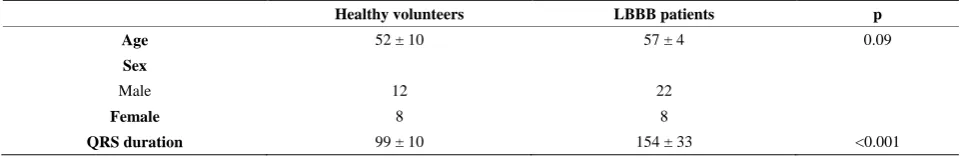
Table 1. Clinical data.
Healthy volunteers LBBB patients p
Age 52 ± 10 57 ± 4 0.09
Sex
Male 12 22
Female 8 8
[image:2.595.58.543.543.732.2]QRS duration 99 ± 10 154 ± 33 <0.001
Table 2.Exclusion and inclusion criterias.
Admission criteria
- Both sexes. - Age 18 - 65 years.
- Left Bundle Branch Block diagnosed the previous six months. - Sinus rhythm.
- Normal epicardial coronary arteries (Coronary CT Scan) - No current structural cardiopathy (Echocardiogram).
Exclusion criteria
- Pregnancy.
- Congenital heart disease. - Valvular heart disease. - Hypertensive cardiopathy. - Epicardial coronary arteries lesions. - Other cause myocardiopathies. - Connective tissue disorders. - Active antineoplasic therapy
- Therapy susceptible of depressing heart contractility or affecting the conduction system. - Atrial fibrillation.
out epicardial coronary artery stenosis. The study was obtained with a 64 slice tomographic scanner, with a temporal resolution of 165 ms and a spatial resolution of 0.4 mm3 (Siemens). When heart rate was higher than 65 bpm, intravenous metoprolol (5 mg up to a maximum of 10 mg) was administered. With antecubital venous ac-cess, 82 - 105 ml of non-ionic contrast (Iodixanol) was then infused, followed by 50 ml saline infusion at a 5 ml per second rate.
2.2. Echocardiographic Study
Transthoracic echocardiographic studies were performed with commercially available ultrasound machines (Sonos 5000, Philips Ultrasound; Vivid System 4, GE/Vingmed), equipped with a multifrequency phased-array sector scan 3 mHz probe. M mode measurements (in mm) of the left ventricular walls, end diastolic diameter (EDD) and end systolic diameter (ESD) were obtained according to stan-dardized protocols. 2D mode protocol included end dia-stolic volume index (EDVI, ml/m2), end systolic volume index (ESVI, ml/m2) and left ventricular ejection fraction (LVEF, %) measurements as per Simpson’s method.
2.3. Gammagraphic Study
A Bruce protocol stress test was performed (CASE 16, GE Healthcare tread mill), with continuous ECG and blood pressure monitoring. Electrocardiographic response was considered non evaluable due to LBBB. Functional class was measured in METs.
At the final stage 27 mCi (1.000 MBq) of 99 m Tc- sestamibi were infused, and single photonic emission computerized tomography (SPECT) (Siemens) was ob-tained both immediately and in a 6 - 8 hours interval from injection of the isotope. A multiple spindle gamma camera, with high resolution and low energy collimators, acquired cardiac perfusion images. A 64 × 64 matrix with 40 s per image acquisition time were used.
Trans-axial slices reconstruction was performed with a specific dedicated computer program. Polar maps (“bull’s eye”) of the relative distribution in the left ventricle were rendered by using a volumetric sampling tool. Each polar map was normalized to its individual maximum. SPECT polar maps quantitative analysis allowed the calculation of the activity profile in every single short axis slice, from apex to base. The global values of each slice were represented in a color scale proportional to the activity, the center of this representation being occupied by the ventricular apex and the periphery by the basal segments. Contractility and systolic thickening of the left ventricle were evaluated employing ECG synchronized (gating) tomographic study. Fat oral intake was recommended to favor hepatobiliar radio tracer elimination, thus decreas-ing interference with the image acquisition.
According to this gammagraphic study, two groups were established:
Group A: 8 people (26%), with perfusion reversible defects, either single or multiple, in which micro vascu-lar dysfunction was suspected (Figure 1).
Group B: 22 people (74%), with either normal perfu-sion tests or with minor isolated septal or apical defects, considered secondary to phasic coronary flow alteration induced by interventricular septum paradoxical movement, in itself secondary to LBBB.
2.4. Endothelial Function Study
All patients in Group A, and 9 patients in Group B, un-derwent coronary arteriography with intra coronary ace-tylcholine and nitroglycerine infusion as per usual pro-tocol, in order to evaluate vasomotor response, either endothelium dependent or independent respectively. All vaso-active medications were stopped 48 hours before angiography. In all procedures femoral artery access was granted. An 8 Fr angioplasty guide catheter was placed in the left coronary artery ostium. Through this guide catheter, an infusion catheter (Cordis 3/2,5 Fr) was ad-vanced over an angioplasty guide wire until it was placed in the proximal artery segment subject of study (left an-terior descending in all cases). To avoid vasospasm in-duction, the guide wire was retrieved once the infusion catheter was properly placed.
Endothelial dependent vasodilatation response was analyzed in the first place by selective intracoronary acetylcholine infusion. Assuming an 80 ml/m coronary flow, we prepared 106, 105 and 104 mol/l concentrations, estimating and intracoronary 108, 107 and 106 mol/l ace-tylcholine concentrations. A basal 5% glucose mechani-cal injection served as control, with further successively increasing concentration infusions of acetylcholine dur-ing two minutes (106, 105 and 104 mol/l). A manual in-jection coronariography was performed after each infu-sion.
Endothelial independent vasodilatation response was then studied, with 200 µg nitroglycerine infusion and a further angiographic injection.
Blood pressure, heart rate end ECG were monitored continuously and registered immediately before each an-giographic injection. For reproducibility allowances, dur-ing basal study the angiography angle, rotation, height and catheter type were registered.
2.5. Quantitative Coronary Angiography
[image:4.595.57.291.80.559.2]
Figure 1. Septal and inferior defects (white arrow).
observer using automatic border detection software and the guide catheter as reference. For each acetylcholine dose, percentual luminal diameter variation from basal measurement was determined at three different arterial segments (proximal, medial and distal). In each patient the mean percentual variation in every segment was cal-culated for further analysis. Finally, mean percentual lu-minal diameter variation was measured between acetyl-choline and nitroglycerine perfusion (after analyzing the two distal segments mean variation).
Intra observer method variability was analyzed by repetition of measurements in 20 angiographies after at
least 3 months delay (yielding a total number of 200 measurements). The observed differences (mean ± 2 stan-dard deviations) in the percentual luminal diameter varia-tion were: 2.0% ± 3.9% for basal values; 1.8% ± 4.0% for the maximum acetylcholine dose and 1.7% ± 3.8% after nitroglycerine.
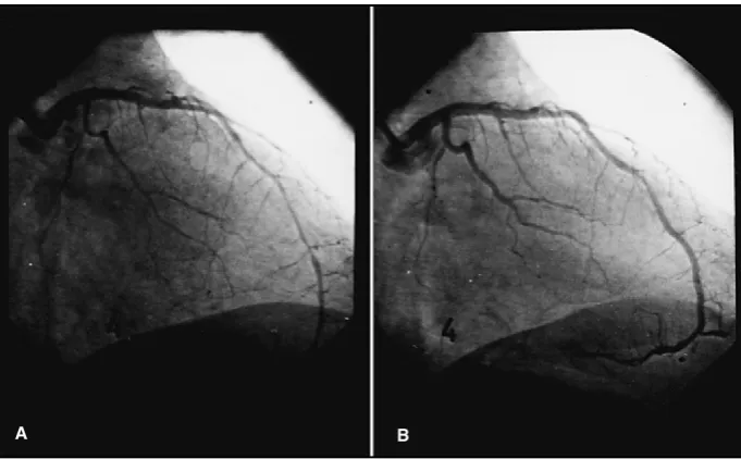
Figure 2. Abnormal vasomotor response in the anterior descending artery alter maximum dose acetylcholine infusion (10−4 mol/l) (A) with ulterior recovery of the luminal diameter (B).
2.6. Follow Up
Patient mean follow up was of 48 ± 6 months. Functional class, echocardiogram and 12 lead ECG were performed on a yearly basis. At the end of follow up a coronary CT scan was obtained to rule out new coronary epicardial obstructive lesions in those patients with obvious ven-tricular function deterioration.
2.7. Statistical Analysis
Quantitative variables were expressed as mean ± stan-dard deviation. Paired data t Student method was used for quantitative variable comparison. Pearson correlation coefficient was used when determining relation between quantitative variables. A p < 0.05 value was considered statistically significant.
3. RESULTS
More Group A patients had more that 2 RF: 6 pts (75%) vs 10 pts (45%) (p < 0.05), while only 2 pts (10%) in the control group showed this risk profile (p < 0.001). Clini-cal data of the three groups are summarized in Table 3.
All patients were asymptomatic at inclusion (NYHA functional class I), with a left ventricle ejection fraction (LVEF) in the normal range and a slightly higher (though non significant) left ventricular end diastolic diameter (LVEDD) in group A pts, as summarized in Table 4.
To determine coronary vasomotor response to intra-coronary acetylcholine infusion (endothelium mediated vasodilator response) and to nitroglycerine (non endothe-lium mediated vasodilator response) all Group A pts and 9 of the Group B pts underwent coronary arteriography. In Group A all 8 pts showed abnormal response to
ace-tylcholine (arterial vasoconstriction) while only 3 of them had abnormal response to nitroglycerine infusion (no increase in luminal diameters); these results were com-patible with microvascular endothelium dependent dys-function. In Group B, just one patient showed abnormal response to acetylcholine while every patient in this group showed significant increase of the luminal diame-ter afdiame-ter intracoronary nitroglycerine infusion, as sum-marized in Table 5.
At one year follow up functional class, 12 lead ECG and echocardiographic measurements were recorded. No atrioventricular conduction defects or QRS duration in-creases were found. At the end of follow up, 3 pts (37%) in Group A showed a 1 - 2 grades worsening of their functional class, while only 5 pts (22%) of those in Group B. LVEDD showed significant increase (51.6 ± 3.6 vs 59.3 ± 6.8 mm; p < 0.05) with a significant de-crease in LVEF 62 ± 4.8 vs 46% ± 3.7%, p < 0.01) in Group A patients, while these values remained unaltered in both Group B pts and controls. No deaths or heart failure hospital admissions were recorded Table 6.
Functional class deterioration, ejection fraction de-crease, or both, were found in eleven patients (7 in group A, 4 in Group B). All of them had a new coronary CT scan, but no new epicardial coronary lesions were found.
4. DISCUSSION
Table 3. Clínical profile of Group A, Group B and controls.
Cardiovascular risk factors (RF)
Age Sex Smoker Hyperthension Diabetes Dislipemia >2RF
GROUP A (n = 8 patients) 57 ± 4 6M, 2F 6 2 1 4 6*
GROUP B n = 22 57 ± 4 16M, 6F 8 3 2 6 10*
CONTROL n = 20 52 ± 10 12M, 8F 3 0 0 4 2†
[image:6.595.59.541.217.268.2]Group A, multiple perfusion defects; Group B, normal or isolated interventricular septum perfusion defect gammagraphy. Values expressed as mean ± SD. Significant statistical difference: *p < 0.05, †p < 0.001.
Table 4. Functional class and echocardiopgraphic measurements at admission.
FC I EDD (mm) EDVI (ml/m2) ESD (mm) ESVI (ml/m2) EF (%)
GROUP A (n = 8 patients) 8 51.6 ± 3.6 75 ± 22.8 23 ± 0.8 22 ± 8.1 62 ± 4.8
GROUP B (n = 22 patients) 22 51.0 ± 3.3 74 ± 19.3 22 ± 0.7 21 ± 11.3 64 ± 6.8
HEALTHY CONTROLS (n = 20 patients) 20 51.1 ± 3.2 74 ± 14.2 21 ± 0.8 21 ± 10.6 67 ± 6.2
[image:6.595.59.539.318.357.2]FC, functional class; EDD, end diastolic diameter; EDVI end diastolic volume index; ESD, end systolic diameter; ESDVI end systolic volume index; EF, left ventricular ejection fraction. Significant statistical difference: *p < 0.05, †p < 0.001.
Table 5. Arterial vasomotor response to acetylcholine and nitroglycerine.
Abnormal response to AC Abnormal response to NTG
Group A (n = 8 patients) 8 patients (100%) 3 patients (37%)
Group B (n = 9 patients) 1 patient (11%) 0 (0%)
[image:6.595.57.539.397.449.2]AC: Acetylcholine. NTG: Nitroglycerine.
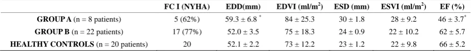
Table 6. Functional class and echocardiopgraphic measurements at end of follow up.
FC I (NYHA) EDD(mm) EDVI (ml/m2) ESD (mm) ESVI (ml/m2) EF (%)
GROUP A (n = 8 patients) 5 (62%) 59.3 ± 6.8 * 84 ± 25.3 30 ± 1.8 28 ± 9.2 46 ± 3.7*
GROUP B (n = 22 patients) 17 (77%) 52.0 ± 3.5 75 ± 18.3 24 ± 0.9 22 ± 10.2 62 ± 5.7
HEALTHY CONTROLS (n = 20 patients) 20 52.1 ± 2.2 73 ± 12.2 23 ± 1.2 22 ± 9.8 66 ± 5.2
FC, functional class; EDD, end diastolic diameter; EDVI end diastolic volume index ; ESD, end systolic diameter; ESDVI end systolic volume index; EF, left ventricular ejection fraction. Significant statistical difference: *p < 0.05, †p < 0.001.
work we found a worse clinical outcome in isolated LBBB patients with associated MVD data.
Since Wiggers’s first description [9], other studies suggested that an abnormal ventricular activation pattern caused by LBBB or right ventricular apical stimulation is linked to left ventricular function deterioration, structural changes and regional myocardial perfusion differences. An anomalous inverse activation sequence is found in LBBB patients, leading to interventricular asynchrony with interventricular septum anomalous movement and subsequently reducing septal contribution to left ven-tricular systolic performance [10]. Isolated LBBB related interventricular and intraventricular asynchrony by itself would not account for the divergent progression of left ventricular anatomical and functional parameters found in different subsets of patients.
Coronary microvascular dysfunction hypothesis as ex-planatory cause of the left ventricular function worsening found in isolated LBBB patients emerges after the X Syndrome (angor pectoris with normal coronary arteries) description. Cannon RO et al. [11] described a poor
We studied 30 patients with isolated LBBB and nor-mal epicardial coronary arteries. According to stress gam-magraphy results, two patient groups were established: Group A, with 8 patients (26%) with single or multiple reversible perfusion defects, in which MVD was accepted, and Group B (22 patients, 74%) with either normal or with isolated septal defects, secondary to paradoxical movement of the interventricular septum related to the LBBB itself.
The clinical profile was homogenous in both groups but for a higher presence of RF in Group A.
Huge epidemiologic studies [17,18] have defined those factors related to coronary disease and its complications. This RF may act precociously, causing vascular damage well before clinical disease becomes evident. In arterio-sclerotic affected adults, endothelial dysfunction has been described [19,20], but it is indeed a remarkable early event in atherogenesis [21,22]. The cause of the altera-tion of the endothelium dependent vasodilataaltera-tion in risk factors prone subjects remains unclear, but decreased liberation or synthesis, or both, of the endothelial derived relaxing factor (RFED) have been strongly suggested [23-25] to be related. Zeiher et al. [26] showed an in-creased coronary blood flow after intracoronary acetyl-choline infusion in a group of healthy subjects with no cardiovascular risk factors and normal epicardial coro-nary arteries. Those subjects with hypercholesterolemia and high plasmatic LDL concentrations, even with an-giographycallly normal arteries, had a selective coronary dysfunction with vasoconstriction and decreasing coro-nary blood flow after acetylcholine infusion.
As expected, coronary vasomotor response to intra-coronary acetylcholine infusion (endothelium derived vasodilatation) and to nitroglycerine (non endothelium dependent vasodilatation) diverged in both groups. All 8 group A patients showed an abnormal response to ace-tylcholine (arterial vasoconstriction) and only 3 of them an abnormal response to the nitroglycerine bolus (ab-sence of any increase in the arterial diameter), which is compatible with endothelium dependent micro vascular dysfunction. Of those in Group B just one patient showed abnormal acetylcholine response while all of them re-acted with intra arterial diameter significant increase after intracoronary nitroglycerine infusion, consistent with a preserved coronary reserve. Considering a definite rela-tion being established between cardiovascular risk fac-tors and microvascular dysfunction in the absence of epicardial coronary arteries stenosis, the additional effect of isolated LBBB on the progressive deterioration of the left ventricular function remains questionable.
All patients at inclusion presented a diskinetic move-ment of the interventricular septum as only sing of ven-tricular asynchrony, with well in the normal range LVEF and ventricular measurements. Patients were reviewed on
a yearly basis, with current functional class assessment and new ECG and echocardiograms. Neither QRS com-plex prolongation nor atrioventricular conduction defects were observed.
At the end of the follow up period a greater functional class deterioration was found in Group A patient vs Group B (37% vs 22%). Compared to inclusion values, Group A patients showed an increase in end diastolic left ventricular diameter (EDLVD) (51.6 ± 3.6 vs 59.3 ± 6.8 mm; p < 0.05) and a LVEF drop (62 ± 4.8 vs 46% ± 3.7%, p < 0.01) while both values remained unaltered in Group B. No deaths were recorded in any group.
The role of MVD in those LBBB patients who pre-sented left ventricular function deterioration at follow up might be related to myocardial fibrosis. One study [27] describes SPECT images in 21 dilated cardiomyopathy patients, 10 min and 2 hours after basal 111 MBq201Tl injections. Myocardial uptake patterns were related to endomyocardial biopsy results, and an inverse relation between isotopic uptake percentage and the fibrosis/ normal tissue ratio was found. Fibrosis found in the area of a focal perfusion defect is probably linked to intraven-tricular conduction delay, venintraven-tricular dyssynchrony and left ventricular systolic function deterioration. Left ven-tricular diastolic function might as well be affected through prolonged systolic intervals, asynchronous relaxation, and both metabolic and energetic deficits [28,29]. Myo-cardial fibrosis related focal left ventricular perfusion de-fects might be an arrythmogenic substrate with a risk for ventricular arrhythmias [30].
Group A patients in our study, with perfusion defects and microvascular dysfunction sings in the coronary ar-teriography, might show myocite destruction and repair-ing fibrosis, as some autopsy studies have shown both in humans [31,32] and experimental models [33,34] These fibrotic areas might be responsible of the functional class deterioration, the increase in LVEDD and the LVEF drop found in Group A pts, worsening the clinical prognosis compared to those in Group B.
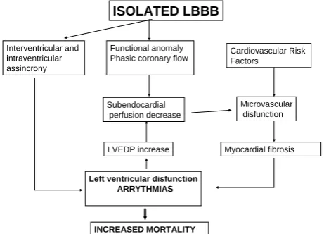
Figure 3.Physiological mechanisms involved.
their clinical progression.
REFERENCES
[1] Jain, A.C. and Mehta, M.C. (2003) Etiologies of left bun-dle branch block and correlations to hemodynamic and an- giographic findings. American Heart Journal, 91, 1375- 1382.
[2] Miller, W.L., Ballman, K.V., Hodge, D.O., et al. (2005) Risk factor implications of incidentally discovered un-complicated bundle branch block. Mayo Clinic Proceed-ings, 80, 1585-1896.
http://dx.doi.org/10.4065/80.12.1585
[3] Eriksson, P., Wilhelmson, L. and Rosengren, A. (2005) Bundle-branch block in middle—Aged men: Risk of complications and death over 28 years. European Heart Journal, 26, 2300-2316.
http://dx.doi.org/10.1093/eurheartj/ehi580
[4] Opherk, D., Zebe, H., Weihe, E., Mall, G., Durr, C., Gra- vert, B., Mehmel, H.C., Schwarz, F. and Kubler, W. (1981) Reduced coronary dilatory capacity and ultra-structural changes of the myocardium in patients with an-gina pectoris but normal coronary arteriograms. Circula-tion, 63, 817-825.
http://dx.doi.org/10.1161/01.CIR.63.4.817
[5] Osler, W. (1910) The Lumleian Lectures on angina pec-toris. Lancet, 1, 839.
http://dx.doi.org/10.1016/S0140-6736(00)51244-6
[6] Cannon, R.O., Wattson, R.M., Rosing, D.R. and Epstein, S.E. (1983) Angina caused by reduced vasodilator reserve of the small coronary arteries. American College of Car- diology Foundation, 1, 1359-1373.
http://dx.doi.org/10.1016/S0735-1097(83)80037-0
[7] Cannon, R.O., Bonow, R.O., Bacharach, S.L., Green, M.V., Rosing, D.R., Leon, M.B., Watson, R.M. and Ep-stein, S.E. (1985) Left ventricular dysfunction in patients with angina pectoris, normal epicardial coronary arteries and abnormal vasodilator reserve. Circulation, 71, 218- 226. http://dx.doi.org/10.1161/01.CIR.71.2.218
[8] Fahy, G.J., Pinski, S.L., Miller, D.P., et al. (1996) Natural
history of isolated bundle branch block. American Jour-nal of Cardiology, 77, 1185-1190.
http://dx.doi.org/10.1016/S0002-9149(96)00160-9
[9] Wiggers, C.J. (1925) The muscular reactions of the mam- maliam ventricles to artificial surface stimuli. American Journal of Physiology, 73, 346-378.
[10] Grines, C.L., Bashore, T.M., Boudoulas, H., et al. (1989) Functional abnormalities in isolated left bundle branch block. The effect of interventricular asynchrony. Circula-tion, 79, 845-853.
http://dx.doi.org/10.1161/01.CIR.79.4.845
[11] Cannon, R.O., Bonow, R.O., Bacharach, S.L., Green, M.V., Rosing, D.R., Leon, M.B., Watson, R.M. and Ep-stein, S.E. (1985) Left ventricular dysfunction in patients with angina pectoris, normal epicardial coronary arteries and abnormal vasodilator reserve. Circulation, 71, 218- 226. http://dx.doi.org/10.1161/01.CIR.71.2.218
[12] Cannon, R.O., Wattson, R.M., Rosing, D.R. and Epstein, S.E. (1983) Angina caused by reduced vasodilator reserve of the small coronary arteries. Journal of the American College of Cardiology, 1, 1359-1373.
http://dx.doi.org/10.1016/S0735-1097(83)80037-0
[13] Cannon, R.O., Schenke, W.H., Leon, M.B., Rosing, D.R., Urquart, J. and Epstein, S.E. (1987) Limited coronary flow reserve after dipyridamole in patients with ergo- novine-induced coronary vasoconstriction. Circulation,
75, 163-170. http://dx.doi.org/10.1161/01.CIR.75.1.163
[14] Greenberg, M.A., Grose, R.M., Strain, J.E., McGuinis, J. and Cohen, M.V. (1984) Decreased coronary dilatory re-sponse in syndrome X (abstract). Circulation, 70, II-21. [15] Legrand, V., Hodgson, J.M., Bates, E.R., Aueron, F.M.,
Mancini, G.B.J., Smith, J.S., Gross, M.D. and Vogel, R.A. (1985) Abnormal coronary flow reserve and abnormal ra- dionuclide exercise tests in patients with normal coronary angiograms. Journal of the American College of Cardi-ology, 6, 1245-1251.
http://dx.doi.org/10.1016/S0735-1097(85)80209-6
[16] Schmidt, D.H., Hendrix, L., Lassar, T., Ray, G. and Port, S. (1984) Ergonovine/dipyridamole-induced changes in regional myocardial perfusion in patients with angina and normal coronary arteries (abstract). Circulation, 70, II- 274.
[17] Castelli, W.P. (1984) Epidemiology of coronary heart di- sease. The framingham study. American Journal of Me- dicine, 76, 4-12.
http://dx.doi.org/10.1016/0002-9343(84)90952-5
[18] Lipid Research Clinics Program. (1984) The Lipid Re-search Clinics Primary Prevention Trial results. Journal of the American Medical Association, 251, 351-364.
http://dx.doi.org/10.1001/jama.1984.03340270029025
[19] Ludmer, P.L., Selwyn, A.P., Shook, T.L., et al. (1986) Paradoxical vasoconstriction induced by acetylcholine in atherosclerotic coronary arteries. The New England Jour- nal of Medicine, 315, 1046-1051.
http://dx.doi.org/10.1056/NEJM198610233151702
[20] Nabel, E.G., Selwyn, A.P. and Ganz, P. (1990) Large co- ronary arteries in humans are responsive to changing blood flow an endothelium-dependent mechanism thal fails in patients with atherosclerosis. Journal of the Ame-
ISOLATED LBBB
Cardiovascular Risk Factors
Microvascular disfunction
Myocardial fibrosis Functional anomaly
Phasic coronary flow Interventricular and
intraventricular assincrony
Left ventricular disfunction ARRYTHMIAS
INCREASED MORTALITY
LVEDP increase Subendocardial
rican College of Cardiology, 16, 349-356.
http://dx.doi.org/10.1016/0735-1097(90)90584-C
[21] Ross, R. (1986) The pathogenesis of atherosclerotic—An update. The New England Journal of Medicine, 8, 488- 500. http://dx.doi.org/10.1056/NEJM198602203140806
[22] Celermajer, D.S., Sorensen, K.E., Gooch, V.M., et al. (1992) Non-invasive detection of endothelial dysfunction in children and adults at risk of atherosclerosis. Lancet,
340, 1111-1115.
http://dx.doi.org/10.1016/0140-6736(92)93147-F
[23] Verbeuren, T.J., Jordaens, F.H., Zonuckeyn, L.I., Van Howe, C.E. and Herman, A.O. (1986) Effect of hyper- cholesterolemia on vascular reactivity in the rabbit I. En-dothelium-dependent and endothelium independent con-tractions and relaxations in isolated arteries o f control and hypercholesterolemic rabbits. Circulation Research,
58, 552-564. http://dx.doi.org/10.1161/01.RES.58.4.552
[24] Bossaller, C., Habbib, G.B., Yamamoto, H., Williams, C., Wells, S. and Henry, P.D. (1987) Impaired muscarinic endothelium-dependent relaxation and cyclic guanosine 5’-monophosphate formation in atherosclerotic human coronary artery and rabbit aorts. The Journal of Clinical Investigation, 79, 170-174.
http://dx.doi.org/10.1172/JCI112779
[25] Egashira, K., Inco, T., Hirooka, Y., et al. (1993) Effects of age on endothelium-dependent vasodilation of resis- tance coronary artery by acethylcholine in humans. Cir- culation, 88, 77-81.
http://dx.doi.org/10.1161/01.CIR.88.1.77
[26] Zeiher, A.M., Drexler, H., Wollschlager, H., Just, H. (1991) Modulation of coronary vasomotor tone in hu- mans. Progressive endothelial dysfunction with different early stages of coronary atherosclerosis. Circulation, 83, 391-401.
http://dx.doi.org/10.1161/01.CIR.83.2.391
[27] Watanabe, M., Gotoh, K., Nagashima, K., Uno, Y., Noda, T., Nishigaki, K., Takemura, G., Kanoh, M., Yasuda, N., Ohno, Y., Minatoguchi, S. and Fujiwara, H. (2001) Rela- tionship between thallium-201 myocardial SPECT and findings of endomyocardial biopsy specimens in dilated cardiomyopathy.Annals of Nuclear Medicine, 15, 13-19.
http://dx.doi.org/10.1007/BF03012125
[28] Nowak, B., Sinha, A.M., Schaefer, W.M., Koch, K.C., Kaiser, H.J., Hanrath, P., Buell, U. and Stellbrink, C. (2003) Cardiac resynchronization therapy homogenizes myocardial glucose metabolism and perfusion in dilated cardiomyopathy and left bundle branch block. Journal of the American College of Cardiology, 41, 1523-1528.
http://dx.doi.org/10.1016/S0735-1097(03)00257-2
[29] Sundell, J., Engblom, E., Koistinen, J., Ylitalo, A., Naum, A., Stolen, K.Q., Kalliokoski, R., Nekolla, S.G., Aira- ksinen, K.E., Bax, J.J. and Knuuti, J. (2004) The effects of cardiac resynchronization therapy on left ventricular function, myocardial energetics, and metabolic reserve in patients with dilated cardiomyopathy and heart failure.
Journal of the American College of Cardiology, 43, 1027-1033. http://dx.doi.org/10.1016/j.jacc.2003.10.044
[30] Soejima, K., Stevenson, W.G., Sapp, J.L., Selwyn, A.P., Couper, G. and Epstein, L.M. (2004) Endocardial and epicardial radiofrequency ablation of ventricular tachy- cardia associated with dilated cardiomyopathy: The im- portance of low-voltage scars. Journal of the American College of Cardiology, 43, 1834-1842.
http://dx.doi.org/10.1016/j.jacc.2004.01.029
[31] Torres, C.M. (1958) Arteriosclerosis of the fine arterial branches of the myocardium (Chagas coronaritis ) and focal myocytolysis in chronic Chagas’heart disease. Hos- pital, 54, 19-34.
[32] Koberle, F. (1974) Pathogenesis of Chagas’disease. Ciba Foundation symposium, 20, 137-158.
[33] Rossi, M.A., Gonzalves, S. and Ribeiro-dos-Santos, R. (1984) experimental Tripanosoma cruzi cardiomyopathy in BALB/c mice: The potencial role of intravascular platelet aggregation in its genesis. American Journal of Pathology, 114, 209-216.
[34] Morris, S.A., Weiss, L.M., Factor, S., Bilezikian, J.P., Tanowitz, H. and Wittner, M. (1989) Verapamil ame- liorates clinical, pathologic and biochemical manifesta- tions of experimental chagasic cardiomyopathy in mice.
Journal of the American College of Cardiology, 14, 782- 789. http://dx.doi.org/10.1016/0735-1097(89)90126-5
ACRONYMS
ECG: Electrocardiogram.
CT: Computerized Axial Tomography.
LBBB: Left bundle branch block.
MVD: Coronary micro vascular dysfunction.
LVEDD: Left ventricle end diastolic diameter (mm).
LVESD: Left ventricle end systolic diameter (mm).
EDVI: End diastolic volume index (ml/m2).
ESVI: End systolic volume index (ml/m2).
LVEF: Left ventricle ejection fraction (%).
RF: Risk factors.
SPECT: Single photon emission computerized tomo-graphy.
EDRF: Endothelial derived relaxing factor.
NO: Nitric oxide.