Journal of Chemical and Pharmaceutical Research, 2014, 6(4):984-998
Research Article
CODEN(USA) : JCPRC5
ISSN : 0975-7384
Ameliorative effect of selenium and curcumin on sodium fluoride
induced hepatotoxicity and oxidative stress in male mice
Mohammad S. AL-Harbi
a, Reham Z. Hamza
a,b*and Afaf A. Dwary
aa
Biology Department, Faculty of Science, Taif University, Taif, Saudi Arabia
b
Zoology Department, Faculty of Science, Zagazig University, Zagazig, Egypt
_____________________________________________________________________________________________
ABSTRACT
Sodium fluoride is the most commonly used compound in oral caries prevention in the form of fluorinated drinking water, salts or milk, tooth pastes, mouth washes and fluoride tablets that adversely affects liver functions parameters. This study evaluated the effects of Sodium fluoride on liver function parameters and also assessed the ameliorating effects of selenium and Curcumin extract. Mature male mice (weighing 35-45 g and each group of ten animals) were given sodium fluoride (10.3 mg/Kg bw) and/or Selenium (0.5 mg/Kg) + Curcumin extract (60 mg/Kg) daily intraperitoneally (I.P) for 4 weeks. In the present study, Sodium Fluoride exposure resulted in an increase in the ALT, AST, Total Protein and LDH levels with respect to the control. Further, Light microscopic investigation revealed that Sodium fluoride exposure induced histopathological alterations in the liver tissues. Supplementations of Curcumin and/or selenium to Sodium fluoride -induced groups increased liver enzymes activities. While some histopathological changes were observed in animal Co-treated with Curcumin extract and /or selenium supplementation to Sodium fluoride treated mice. As a result, Sodium fluoride induced hepatotoxicity is reduced by Curcumin extract and/or selenium to great extent by the entire restoration of the histological structures.
Key words: Sodium fluoride, Selenium, Curcumin extract, Hepatic tissues.
_____________________________________________________________________________________________
INTRODUCTION
Sodium fluoride is the most commonly used compound in oral caries prevention in the form of fluorinated drinking water, salts or milk, tooth pastes, mouth washes and fluoride tablets (Dabrowska et al., 2006). (Barot., 1998 and
Ortiz et al., 2003) stated that there are about 21 countries that had problems with endemic fluorosis where the main
pathway of fluoride exposure is the ingestion of tap water from contaminated ground water sources.
Fluoride (F) anions are widely distributed in the environment in different forms and their compounds are extensively used. Fluoride anions are naturally present in water sources and drinking water as they are released from the run off of F-containing rocks and soils and leach into groundwater (ATSDR, 2003). In some areas drinking water is artificially fluoridated, therefore water consumption is typically the largest contributor to daily F intake. Furthermore, F anions are incorporated in various insecticide formulations, fluoridated foodstuffs, dentifrices, drugs, vapors emitted from industries using fluoride containing compounds (NRC, 2006).
musculoskeletal, dental systems (El-lethey and Kamel.,2011), kidney (Nuscheler et al.,1996), liver (Chinoy NJ and
Patel.,1999) and brain (Shivarajashankara et al.,2002).
Recent studies revealed that fluoride induces excessive production of oxygen free radicals, and might cause the depletion in biological activities of some antioxidant enzymes (Chlubek, 2003).Toxic effects of fluoride on various biochemical parameters are known (Singh, 1984; Chlubek, 2003). Increased free radical generation and lipid peroxidation are proposed to mediate the toxic effects of fluoride on soft tissues (Rzeuski et al., 1998).
Shivarajashankara et al. (2001a,b) reported increased lipid peroxidation and disturbed antioxidant defense systems
in brain, erythrocytes and liver of rats exposed to fluoride.
The naturally occurring element selenium (Se) is essential for a wide variety of biological processes in mammals
(Schomburg et al., 2004). Its beneficial role in human health is due to low molecular weight selenium compounds,
as well as to its presence within at least 25 proteins, named selenoproteins, in the form of the amino acid selenocysteine, that is incorporated during translation and is directly involved in redox catalysis (Driscoll and
Copeland, 2003; Romero et al., 2005).
Liao et al. (2008) reported that selenium played a beneficial role for prevention of cisplatin hepatotoxicity in mice. Jihen et al. (2008) mentioned that selenium has a cooperative effect in the protection against cadmium-induced
structural damage in the liver of rat.
Several investigators have demonstrated protection by selenium supplementation against metal induced hepatic and renaltoxicity. Moreover, in few recent studies by us we observed a beneficial role of essential trace element (selenium) supplementation during the course of chelation treatment of inorganic mercury intoxication (Saricaoglu
et al., 2005).
Natural herbal constituents are extensively studied for their ability to protect cells from miscellaneous damages. Currently, the use of phytochemicals as a therapy in diseases related to oxidative stress has gained immense interest for their ability to quench free radicals by electron or proton donation and their capability to protect body tissues against oxidative stress (Nabavi et al., 2012).
Curcumin (1,7-bis(4-hydroxy-3-methoxyphenyl)-1,6-heptadiene 3,5-dione), the active portion of turmeric, has been shown to have significant antioxidant activity, both in vitro and in vivo (Joe et al., 2004), curcumin is a potent scavenger of reactive oxygen and nitrogen species such as hydroxyl radicals and nitrogen dioxide radicals (Reddy
and Lokesh, 1994).
The antihepatotoxic effects of curcumin against chemically induced hepatic damage are well documented, and they have been attributed to its intrinsic antioxidant properties (Farombi et al., 2008). Thus, curcumin has shown to protect liver against hepatic injury and fibrogenesis by suppressing hepatic inflammation (Nanji, et al., 2003), attenuating hepatic oxidative stress (Yousef et al., 2010).
EXPERIMENTAL SECTION
2.1. Animals
2.2. Chemicals 2.2.1 Sodium Fluoride
Sodium fluoride (NaF) was purchased from Sigma Chemical Co., St. Louis, Mo., USA. The tested dose of NaF (10.3 mg/kg b.wt) was chosen based on the previous studies of Zabulyte et al. (2007). A stock solution was prepared by dissolving of 100 g of NaF in 1000 ml of distilled water. The dose schedule was so adjusted that the amount of NaF administration per animal was as per their respective weight.
2.2.2 Selenium
Selenium was purchased from BDH Chemicals Ltd., England. The tested dose of selenium (0.5 mg/kg) was chosen based on the previous studies of Ibtissem et al.(2011).
2.2.3. Curcumin extract
Fresh Curcumin was obtained from local market (Cairo, Egypt), then washed and was soaked in water for 24 hours and after that it was dried then homogenized by using electrical mixer and then the dose was prepared (60 mg/Kg) and this dose was chosen according to Abdul-Hamid and Moustafa . (2013).
2.3. Experimental protocols
The study was performed on 70 mature male mice, divided into 7 main groups; each group was consisted of 10 rats. The 1st Control group: Animal’s received 1ml of distilled water orally daily for 30 successive days. The 2nd Sodium Fluoride (NaF) treated group: Animals were daily received NaF (10.3 mg/Kg) for 30 successive days intraperitoneally (I.P).The 3rd Selenium treated group: Animals were received selenium (0.5 mg/Kg) for 30 successive days intraperitoneally (I.P).The 4th Curcumin extract treated group: Animals were received Curcumin extract (60mg/kg) for 30 successive intraperitoneally (I.P).The 5th NaF + Selenium treated group: Animals were given sodium fluoride (NaF) (10.3 mg/Kg) for 30 successive days and then co-administered by selenium (0.5 mg/Kg) intraperitoneally (I.P). The 6th NaF + Curcumin treated group: Animals were given sodium fluoride (NaF) (10.3 mg/Kg) for 30 successive days and then co-administered Curcumin extract (60mg/Kg) for 30 successive days intraperitoneally (I.P). The 7th NaF+ Selenium+ Curcumin extract treated group: Animals were given NaF (10.3mg/Kg) and then co-administered with selenium (0.5mg/Kg) and then followed by Curcumin extract (60 mg/Kg) for 30 successive days (I.P).The substances were administered in the morning (between 09.30 and 10.30 h) to non fasted rats. The first day the animals were treated was considered experimental day 0.At the end of the 30 days of treatment, all animals were scarified and dissected. The testis tissues were quickly excised to light microscope investigations and biochemical examinations.
2.4. Blood samples collection
Blood samples were collected after the end of the experiment from the retro-orbital vein, which is a simple, convenient and successful procedure that allows bleeding of the same animal more than one time with minimal stress (Scherners, 1967).
After the end of 4th week, individual blood samples were drawn by orbital puncture (from eye plexus) using microhematocrit capillary tubes (Lancer, Athy, County-Kildare, Republic of Ireland), Serum was harvested from blood without EDTA and then serum samples were transferred into eppendorf tubes and subsequently used for the determination of (Total protein, Aspartate amino transferase (AST), Alanine amino transferase (ALT), Lactate dehydrogenase (LDH), Catalase, Superoxide dismutase and Malondialdhyde. Plasma was harvested from blood with EDTA and subsequently used for the determination of reduced Glutathione, Glutathione peroxidase.
2.5. Preparation of liver Homogenate:
2.6. Antioxidant enzyme determination
TBARS content was evaluated using the thiobarbituric acid (TBA) test as described by Ohkawa et al. (1979). After incubation of testis homogenate with TBA at 95 ˚C, TBARS reacts to form a colored complex. Absorbance was measured spectrophotometrically at 532 nm to determine the TBARS content. The specific activity is expressed as nmol/mg protein protein.
2.6.1. Measurement of superoxide dismutase (SOD)
SOD activity was measured according to the method described by Marklund and Marklund (1974) by assaying the auto oxidation of pyrogallol at 440 nm for 3 min. One unit of SOD activity was calculated as the amount of protein that caused 50% pyrogallol autooxidation inhibition. A blank without homogenate was used as a control for non-enzymatic oxidation of pyrogallol in Tris–EDTA buffer (50 Mm Tris, 10 mM EDTA, pH 8.2). The SOD activity is expressed as U/mg protein.
2.6.2. Measurement of catalase (CAT)
CAT activity was measured determined according to the method described by Aebi (1984) by assaying the hydrolysis of H2O2 and the resulting decrease in absorbance at 240 nm over a 3 min period at 25˚C. Before determination of the CAT activity, samples were diluted 1:9 with 1% (v/v) Triton X-100. CAT activity is expressed as mmol/mg protein.
2.6.3. Measurement of glutathione peroxidase (GPx)
GPx activity was measured using H2O2 as substrate according to the method described by Paglia and Valentine (1967). The reaction was monitored indirectly as the oxidation rate of NADPH at 240 nm for 3 min. A blank without homogenate was used as a control for non-enzymatic oxidation of NADPH upon addition of hydrogen peroxide in 0.1 M Tris buffer, pH 8.0. Enzyme activity was expressed as nmol/mg protein.
2.6.4. Liver function biomarkers in the serum
Alanine aminotransferase (ALT) and aspartate aminotransferase (AST) were determined colorimetrically by measuring the amount of pyruvate or oxaloacetate produced by forming 2,4-dinitrophenylhydrazine, the color of which was measured at 546 nm (Retiman and Frankel, 1957). Lactate dehydrogenase was determined according to the method of Vassault (1983) which depends on the oxidation of lactate to pyruvate with the simultaneous conversion of the cosubstrate NADH to NAD. The decrease in absorbance (measured at 340 nm) due to this conversion is directly proportional to LDH activity.
2.7. Histopathology
For histopathological examination, liver tissues were dissected and fixed in neutral buffered formalin solution. Then samples were processed using a graded ethanol series, and embedded in paraffin. The paraffin sections were cut into 5 µ-thick slices and stained with hematoxylin and eosin for histological examination (Carleton., 1967).
2.8. Statistical analysis:
Data were collected, arranged and reported as mean ± standard error of mean (S.E.M) of nine groups (Each group was considered as one experimental unit), summarized and then analyzed using the computer program SPSS/ version 15.0) The statistical method was one way analyzes of variance ANOVA test (F-test), and if significant differences between means were found, Duncan’s multiple range test (Whose significant level was defined as (P<0.05) was used according to (Snedecor and Cochran.,1982) to estimate the effect of different treated groups.
RESULTS
3.1. Evaluation of biochemical parameters (Liver function parameters):
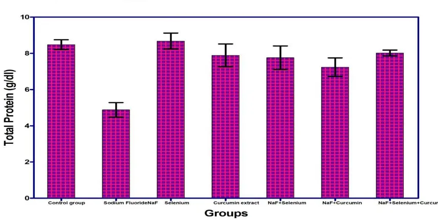
3.1.1. Effect of Sodium Fluoride, Selenium, Curcumin extract and their combinations on serum total protein: The administration of Sodium Fluoride in it’s recommended dose for successive 30 days into normal rats
group.However, the combinations of the test plant extract (Curcumin) and selenium appear to ameliorate the hypoproteinaemia produced by sodium fluoride alone.
Table (1): Effect of Sodium fluoride (10. 5 mg/kg), Selenium (0.5 mg/ Kg), Curcumin extract (60 mg/Kg) and their combinations on Liver function in male mice (mean ± SE). (N = 7)
Groups Total Protein(g/dl) AST(U/mI) ALT(U/mI) LDH(µIU/ml)
Control group 8.48±0.27bc 13.20±0.58g 12.00±0.70g 371.66±47.21f
Sodium fluoride 4.88±0.40g 197.80±7.39a 81.00±1.18a 1828.40±35.37a
Selenium 8.68±0.44 ab 13.80±6.02fg 12.80±1.82fg 370.30±33.34f
Curcumin extract 7.89±0.63d 14.10±1.01 ef 14.20±2.10e 405.32±14.06e
Sodium fluoride + Selenium 7.76±0.65e 45.00±1.67 c 23.20±0.86c 820.00±30.37c
Sodium fluoride + Curcumin extract 7.24±0.51f 56.40±3.12b 25.40±0.50b 862.66±50.63bc
Sodium fluoride + Selenium + Curcumin extract 8.02±0.16 c 24.20±4.61d 20.60±2.73d 520.31±51.26d
Means within the same column in each category carrying different litters are significant at (P ≤ 0.05) using Duncan's multiple range test, where the highest mean value has symbol (a) and decreasing in value were assigned alphabetically.
Fig.(1): Effect of Sodium fluoride (10.3 mg/kg), Selenium (0.5 mg/ Kg), Curcumin extract (60 mg/Kg) and their combinations on Total protein (g/dl) in male mice
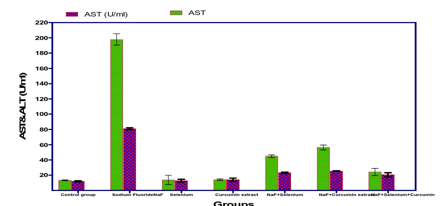
3.1.2. Effect of Sodium Fluoride, Selenium, Curcumin extract and their combinations on serum GOT and GPT (AST & ALT): Serum transferases (AST &ALT) levels were markedly elevated after 4th week post Sodium
Fluoride administration to normal rats when compared with normal control group. Meanwhile significant elevation (P<0.05) in serum AST&ALT were recorded after 4th week in normal rats in response to administration of Curcumin extract and non significant increase in response to administration of sodium fluoride , mean while their combinations with Sodium Fluoride afforded significant elevation in serum , but this effect was less intense than that produced by sodium fluoride alone as sodium fluoride combination with either selenium and/or Curcumin extract still more better than sodium fluoride alone as these combinations exhibited significant decrease in ALT and AST serum values when compared with sodium fluoride treated group , group treated with selenium & curcumin extract alone which revealed non significant change in serum ALT when compared with normal control group (Table 1 and Fig.1).
3.1.3. Effect of Sodium Fluoride, Selenium, Curcumin extract and their combinations on serum Lactic dehydrogenase enzyme (LDH) activity: Treatment of normal rats with Sodium Fluoride for 30 successive days in
[image:5.595.87.515.286.501.2]Control group Sodium FluorideNaF Selenium Curcumin extract NaF+Selenium NaF+Curcumin extractNaF+Selenium+Curcumin 20 40 60 80 100 120 140 160 180 200 220
AST (U/ml) AST
Groups A S T & A L T ( U /m l)
Fig. (2): Effect of Sodium fluoride (10.3 mg/kg), Selenium (0.5 mg/ Kg), Curcumin extract (60 mg/Kg) and their combinations on AST & ALT (U/ml) in male mice
Control group Sodium Fluoride NaF Selenium Curcumin extract NaF+Selenium NaF+Curcumin NaF+Selenium+Curcumin
0 500 1000 1500 2000 LDH (µIU/ml) Groups L D H ( µ IU /m l)
Fig. (3): Effect of Sodium fluoride (10.3 mg/kg), Selenium (0.5 mg/ Kg), Curcumin extract (60 mg/Kg) and their combinations on LDH enzyme (μIU/ml) in male mice
Table (2): Effect of Sodium fluoride (10.3 mg/kg), Selenium (0.5 mg/ Kg), Curcumin extract (60 mg/Kg) and their combinations on Catalase in male mice (mean ± SE). (N = 7)
Groups Liver Catalase (U/g) Liver SOD (U/g) Liver MDA (nmol/g) Liver Glutathione reductase (U/g) Liver Glutathione Peroxidase (U /g)
Control group 4.29±0.01b 23.32±0.87ab 1.74±0.10f 6.91±0.75a 8.04±0.74b
Sodium fluoride 1.72±0.03g 9.61±1.52g 9.35±0.26a 1.12±0.48e 1.99±0.51f
Selenium 4.43±0.03ab 22.14±2.98b 1.50±0.66g 6.99±0.58a 8.64±0.44ab
Curcumin extract 4.03±0.04c 18.82±0.91d 2.81±0.12e 5.30±0.94c 6.50±1.34c
Sodium fluoride +Selenium 2.57±0.04f 14.94±1.66e 7.30±0.25c 3.01±0.82d 4.89±0.82e
Sodium fluoride +Curcumin extract 2.72±0.05ef 12.92±1.59f 7.38±0.38bc 3.03±1.56d 4.83±1.00e
Sodium fluoride + Selenium+Curcumin extract 3.95±0.02d 19.91±0.88cd 4.04±0.26d 5.77±1.19bc 5.70±1.13d
[image:6.595.75.517.102.308.2] [image:6.595.80.518.358.547.2]Fig. (4): Effect of Sodium fluoride (10.3 mg/kg), Selenium (0.5 mg/ Kg), Curcumin extract (60 mg/Kg) and their combinations on Catalase activity (in tissue homogenates) in male mice
Fig.(5): Effect of Sodium fluoride (10.3 mg/kg), Selenium (0.5 mg/ Kg), Curcumin extract (60 mg/Kg) and their combinations on SOD activity (in tissue homogenates)in male mice
Fig. (6): Effect of Sodium fluoride (10.3 mg/kg), Selenium (0.5 mg/ Kg), Curcumin extract (60 mg/Kg) and their combinations on Malondialdhyde (MDA) (in tissue homogenates) in male mice
[image:7.595.73.537.87.244.2] [image:7.595.86.522.273.386.2] [image:7.595.85.515.418.523.2] [image:7.595.86.529.552.685.2]Fig (8):Effect of Sodium fluoride (10.3 mg/kg), Selenium (0.5 mg/ Kg), Curcumin extract (60 mg/Kg) and their combinations on Glutathione peroxidase (in tissue homogenates) in male mice
Fig. (9): Cross section of control mice liver of (Group 1) formed of small central vein surrounded by cords of hepatocytes showing central vesicular nuclei and eosinophilic cytoplasm (H and E x 200) (CV: central vein, EN: esoinophilic nuclei)
Fig. (10): Cross section of mice liver of group (2) treated with Sodium Fluoride (10.3 mg/kg) showing severe haemorage and congestion (H and E x 200) (HG: Hydropic degeneration)
3.2. Antioxidant activities:
3.2.1. Effect of Sodium Fluoride, selenium, Curcumin extract and their combinations on Catalase activity:
Regarding the effect of Sodium Fluoride on catalase activity of normal rats, Sodium Fluoride afforded a marked decrease (P<0.05) in plasma, liver and kidney catalase after the end of the study when compared with control group, whereas, non significant changes in the enzyme activity was recorded in selenium treated group. Treatment of normal rats with selenium alone exhibited non significant changes in Catalase of liver ,kidney and plasma after the
C.V
[image:8.595.75.537.78.229.2] [image:8.595.170.430.256.430.2] [image:8.595.175.423.464.634.2]end of the experiment when compared with control group , Whereas , a significant decrease was reported in liver , plamsa and kidney tissues respectively when administered Curcumin extract only compared with control group (Table 2 and Figs.3).While combinations of Sodium Fluoride with either Selenium or curcumin extract exhibited a significant decrease in Catalase activity of liver, kidney and plasma after the end of the study as compared with normal control group.
3.2.2. Effect of Sodium Fluoride, selenium, Curcumin extract and their combinations on Superoxide dismutase (SOD) activity: The results of the study revealed that treatment of normal rats with Sodium Fluoride
elicited a highly significant decrease (P<0.05) in plasma and liver SOD level after the end of the study together with a marked decrease in SOD activity of kidney and brain when compared with control group. Treatment of normal rats with either selenium or curcumin extract for 4 weeks elicited a non significant decrease in SOD activity of the liver, Kidney and plasma after the end of the study except with curcumin extract which showed a slight significant decrease in SOD activity compared with control group. Whereas, the combinations of the curcumin extract and/or selenium with sodium fluoride afforded a slight decrease (P<0.05) in SOD activity of the liver, kidney and plasma respectively compared with normal control group (Table 2 and Figs.4). Meanwhile combination of sodium fluoride with Curcumin extract and selenium afforded slight decrease in SOD activity but the effect was much better than group treated with sodium fluoride only and other treatment combinations.
3.2.3. Effect of Sodium Fluoride, selenium, Curcumin extract and their combinations on Malondialdhyde (MDA) activity: The MDA content of the serum, liver and kidney were significantly elevated (P<0.05) in response
to treatment of normal male rats with Sodium Fluoride for 4 weeks compared with normal control group. The same previous response was reported with selenium, curcumin extract combinations with Sodium Fluoride compared with control group (Table 3and Figs. 5 ) but the effect was much less intense. Meanwhile, groups treated with either selenium or curcumin extract induced non significant changes in serum MDA level as compared to normal control group while selenium treated group afforded non significant changes in MDA activity in kidney homogenates while afforded a significant decrease in MDA activity in liver homogenates as compared with normal control group. At the same time, Curcumin extract afforded slight significant increase in MDA activities in Liver and kidney homogenates as compared to normal control group.
Fig. (11)Cross section of mice liver of group (3) treated with selenium(0.5 mg/ Kg) showing normal liver tissue formed of control vein surrounded by cords of hepatocytes with normal vesicular nuclei and eosinophilic cytoplasm (H and E x 200) (EN: Eosinophilic nuclei,
CV: Central vein)
3.2.4.Effect of Sodium Fluoride, selenium, Curcumin extract and their combinations on Glutathione reductase (GR) activity: It was apparent from Table (9) and (Figs.32,24) that treatment of rats with Sodium
Fluoride, alone afforded a significant decrease (P<0.05) in serum, liver and kidney reduced glutathione after the end of the study when compared with normal control group. On the other hand, the results revealed that Selenium induced a non significant change in reduced Glutathione content of the kidney, liver and serum together as compared to normal control group with slight significant increase in the reduced glutathione content of serum, liver and kidney homogenates in Curcumin extract treated group compared with control group.
EN
[image:9.595.169.432.430.584.2]Fig. (12) :Cross section of mice liver of group (4) treated with curcumin extract (60 mg/ Kg) showing central vein surrounded by cords
of hepatocytes (H and E x 200) (CV: Central vein)
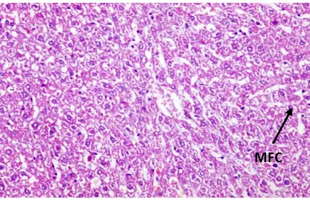
Fig. (13): Cross section of mice liver of group (5) treated with (Sodium Fluoride +Selenium) (10.3 mg/kg) & (0.5 mg/kg) respectively showing tissue cords of hepatocytes with mild fatty change in the form of central nuclei surrounded by vaculated cytoplasm (↑) (H and E
x 200) (MFC: Mild fatty change)
Fig. (14): Cross section of mice liver of group (6) treated with (Sodium Fluoride +curcumin extract) (10.3 mg/kg) & (60 mg/kg) respectively showing cords of hepatocytes with mild fatty change in the form of central nuclei surrounded by vaculated cytoplasm (↑) (H
and E x 200) (MFC: Mild fatty change)
3.2.5. Effect Sodium Fluoride, selenium, Curcumin extract and their combinations on Glutathione Peroxidase activity: The plasma Glutathione peroxidase level was significantly reduced (P<0.05) in all groups treated with
CV
[image:10.595.104.499.77.275.2] [image:10.595.180.420.308.456.2] [image:10.595.181.430.500.664.2]Sodium Fluoride alone and in combination with selenium, curcumin extract for successive 30 days when compared with normal control group. Whereas, non significant increase (P<0.05) was recorded in the enzyme activity of the kidney, Plasma and liver homogenates of group treated with selenium when compared with control group. Together with a significant decrease in the enzyme activity in Plasma, Kidney and liver homogenates in response to treatment with Curcumin extract as compared with normal control group. The enzyme activity in the plasma was markedly decreased in response to treatment with either Sodium Fluoride or it’s combinations with selenium and/or curcumin extract compared with control group. Beside a non significant increase in response to treatments with selenium. Whereas, a significant decrease was reported in response to treatments with all combinations used except combination of Sodium Fluoride with selenium and curcumin which showed a slight decrease compared with normal control group (Table 3 and Figs.6).
3.3. Histopathology investigations of liver tissues:
Control group: Normal liver tissue formed of small central vein surrounded by cords of hepatocytes showing central vesicular nuclei and eosinophilic cytoplasm (Fig. 9).Sodium Fluoride treated group: Liver tissue showing markedly dilated central vein filled by large number of red blood cells and surrounded by hepatic cords and showing severe fatty change of hepatocytes and markedly congested central vein (Fig. 10) and showing liver cells necrosis in the form of pyknotic nuclei (↑) and more eosinophilia of the cytoplasm was also seen (Figs.32&33).Selenium treated group: Normal liver tissue formed of central vein surrounded by cords of hepatocytes showing central vesicular nuclei and eosinophilic cytoplasm (Fig. 11).Curcumin extract treated group: The liver tissues of this group are formed of central vein surrounded by cords of hepatocytes (Fig. 12).Sodium Fluoride+ Selenium treated group: cords of hepatocytes showing mild fatty change in the form of central nuclei surrounded by vaculated cytoplasm (↑) (Fig.13).Sodium Fluoride+ Curcumin extract treated group: cords of hepatocytes showing mild fatty change in the form of central nuclei surrounded by vacuolated cytoplasm (↑), (Fig.14).Sodium Fluoride + Selenium + Curcumin extract treated group: liver tissue shows moderate fatty change in the hepatocytes (Fig.15) and liver tissue cords of hepatocytes showing mild fatty change in the form of central nuclei surrounded by vacuolated cytoplasm.
Fig. (15): Cross section of mice liver of group (7) treated with (NaF +selenium+ Curcumin extract) (10.3mg/kg),(0.5 mg/kg) & (60 mg/Kg) respectively showing moderate fatty changes in the hepatocytes (H and E x 200) (MFC: Moderate fatty change)
DISCUSSION
Sufficient evidence has demonstrated that fluoride produces deleterious effects in skeletal, dental and soft tissues. There are documents concerned with the mechanism of bone and dental fluorosis. But it is not clear of how fluoride interferes with soft tissues. However, some of the following viewpoints may be very helpful for us to understand the mechanism of fluorosis in soft tissues. Firstly, studies have shown that fluoride can induce excessive production of oxygen free radicals, and cause the decrease in biological activities of some substances, such as catalase (CAT), superoxide dismutase (SOD), xanthine oxidase (XOD), and glutathione peroxidase (GSH-Px) which play important roles in antioxidation and eliminating free radicals (Zhang and Ji, 1996).
Secondly, fluoride can also disturb the metabolism of proteins. It is evidently indicated that fluoride can impair the activities of a series of enzymes such as alkaline phosphatase, cholinesterase (Zabulyte et al., 2007). Thirdly, fluoride can interfere with the metabolism of carbohydrate, lipid and nucleic acids, injure immune system, and damage the parts of the body (Liu et al., 2003; Zabulyte et al., 2007).
[image:11.595.187.410.395.540.2]Increased generation of reactive oxygen species (ROS) is implicated in the pathogenesis of many diseases and in the toxicity of a wide range of compounds (Halliwell and Gutteridge, 1985). Lipid peroxidation represents one of the most frequent reactions resulting from free radical’s attack on biological structures (Stohs, 1995). The present study revealed increased levels of TBARS, the marker of extent of lipid peroxidation, in the liver of sodium fluoride-treated rats. And our findings are greatly reinforced by earlier studies have recorded increased free radicals levels in the erythrocytes of fluorotic humans (Shivarajashankara et al., 2001a), and in the erythrocytes, liver, kidney, brain and ovary of experimental animals (Halliwell and Gutteridge, 1985; Sharma and Chinoy, 1998; Shivarajashankara
et al., 2002).
Levels of reduced glutathione (GSH) and total antioxidant capacity (TAC) as well as the activities of superoxide dismutase (SOD) and catalase(CAT) decreased with NaF testament. These results are in accordance with
Shanthakumari et al. (2004) who found decrease in the levels of glutathione with an increase in glutathione
peroxidase (GSH-Px) activity in the brain, erythrocytes and liver of rats exposed to fluoride. They reported that the decrease in glutathione level with an increase in malondialdehyed (MDA) level and GSHPx activity indicates utilization of GSH for GSH-Px catalyzed scavenging of H2O2 or lipid hydroperoxides generated. Thus, this degree of toxicity of fluoride resulted in reduction in the level of the free radical scavenger glutathione.
The decreased GSH along with an increased activity of GSH-Px in the fluoride-treated group suggests increased conversion of GSH to GSSG to combat lipid hydroperoxides or H2O2. Previous studies in this field reported decreased GSH and GSH-Px in various tissues of experimental animals subjected to chronic fluoride toxicity
(Sharma and Chinoy, 1998).
Qujeq et al. (2002) reported that food and/or water contaminated with a much higher concentration of sodium
fluoride produced characteristic signs of biochemical defects. Biochemical changes in the composition of bone, urine, and plasma, and some hormonal changes in fluorosis have been reported (reviewed in Das., 1996).Higher concentrations of fluoride are known to affect collagen synthesis and bone mineralization (Harrison et al., 1990). In addition, fluoride has been shown to inhibit many enzymes such as those involved in the pentose pathway, antioxidant defense system, and the myosin-ATPase path. Furthermore, fluoride is also known to cross the cell membrane and enter soft tissues. Impairment of soft tissue function has been demonstrated in fluorideintoxicated animals (Vani and Reddy, 2000).
On serum total protein, The administration of Sodium Fluoride in its recommended dose for successive 30 days into normal
rats elicited highly significant decrease (P<0.05) in serum total protein level compared with normal control group. Whereas, non significant changes were observed in the group treated with Selenium and a slight decrease in group treated with Curcumin extract after 4th week when compared with normal control group, Whereas combinations of Sodium Fluoride with either Selenium or Curcumin extract afforded slight significant decrease when compared with control group.
On serum GOT and GPT (AST & ALT), serum transferases levels Serum transferases (AST &ALT) levels were
markedly elevated after 4th week post Sodium Fluoride administration to normal rats when compared with normal control group. Meanwhile significant elevation (P<0.05) in serum AST&ALT were recorded after 4th week in normal rats in response to administration of Curcumin extract and non significant increase in response to administration of sodium fluoride. On serum lactic dehydrogenase enzyme (LDH) activity, Treatment of normal rats with Sodium Fluoride for 30 successive days in their recommended doses elicited a marked elevation in serum LDH activity after the end of the study when compared with normal control group.
Transaminases (AST and ALT) enzymes are a common mean of detecting liver damage. Alterations in these enzymes are reported in hepatic disease and in myocardial infarction. Fluoride toxicosis caused elevated in the activities of transaminases (Shanthakumari et al., 2004). The alterations in transaminases could be expected to occur associated with pathology involving necrosis of the liver.
Guo-Ving et al. (2003) found also an increase in the activities of AST and ALT in plasma of rats after 3 months
treated with 50, 100 and 150 mg NaF. Transaminases (ALT, AST), a rise in blood transaminase activities is a sensitive indicator of damage to cytoplasmic and/or mitochondrial membranes. Plasma enzyme activities rise when the membranes of only very few cells are damaged.
Liver cells contain more AST than ALT, but ALT is confined to the cytoplasm in which its concentration is higher than that of AST. In inflammatory or infective conditions: the cytoplasmic membrane sustains the main damage; leakage of cytoplasmic contents causes a relatively greater increase in plasma ALT than AST. In infiltrative disorders, in which there is damage to both mitochondrial and cytoplasmic membranes, there is a proportionally greater increase in AST activity than ALT (Akila et al., 1998).The activities of lactate dehydrogenase (LDH) and creatine kinase (CK) were significantly increased in rats treated with sodium fluoride. This may be attributed to a generalized increase in membrane permeability and is particularly useful in the diagnosis of muscular dystrophy
(Kaczor et al., 2005).
Also, it has been reported that the increased levels of LDH result from superoxide anions and hydroxyl radicals in the presence of transition metal ions which cause oxidative damage to the cell membrane (Yadav et al., 1997). So the observed increases of both TBARS and NO free radicals in fluoride intoxicated rats are more likely to explain the obtained elevation of enzymes activity.
Increased utilization of medicinal plants became a World Health Organization (WHO) policy in 1970. Plants and herbs are chemical factories that directly provide about 25% of currently used drugs and another 25% of drugs comprise chemically altered natural products (Desmet, 1997).
Curcumin, a phenolic compound, exhibits protective effects against oxidative damage and it is considered to be a potent cancer chemopreventive agent (Duvoix et al., 2005). In agreement with Pari and Amali (2005), curcumin alone significantly decreased the levels of TBARS. Kalpana and Menon (2004) suggested that curcumin exerts its protective effect by modulating the biochemical marker enzymes, lipid peroxidation and augmenting antioxidant defense system. More specifically, curcumin was significantly decrease the levels of free radicals and this protective effect of curcumin attributed to its free radical scavenging activity, induction of detoxification enzymes and provides protection against degenerative diseases (Manikandana et al., 2004).
Our results are greatly reinforced by Liao et al. (2008) who reported that selenium played a beneficial role for prevention of cisplatin hepatotoxicity in mice. Jihen et al. (2008) mentioned that selenium has a cooperative effect in the protection against cadmium-induced structural damage in the liver of rat. El-Shenawy and Hassan (2008) reported that selenium has a protective effect against liver and kidney damage induced by mercury chloride in rats.
REFERENCES
[1] E Dabrowska; R Letko; Balunowska, M, 2006. Advances in Medical Sciences.,51(1): 91- 95. [2] V Barot, 1998. Bull. Environ. Contam. Toxicol., 61(3): 303-310.
[3] P Ortiz; M Rodriguez ; M Martinez ; H Borja; J Castelo; F Diaz, 2003. Environ. Res., 93(1): 20-30.
[4] Agency for Toxic Substances and Disease Registry (ATSDR), 2003. Toxicological Profile for Fluorides, Hydrogen Fluoride, and Fluorine. US Department of Health and Human Services, Atlanta, US.
[5] National Research Council (NRC), 2006. Fluoride in Drinking-Water. A scientific review of EPA’s standards, Washington.
[6] G Aydin; I Ekrem; A Mehmet; G Osman, 2003. J. Appl. Toxicol ., (23): 437–446. [7] HS El-lethey;MM Kamel , 2011. Journal of American Science., 7(4): 243-254. [8] NJ Chinoy; TN Patel, 1999. Fluoride., 32: 215–229.
[9] YM Shivarajashankara; AR Shivashankara; PG Bhat; SM Rao; SH Rao, 2002. Fluoride., 35(1):12-21. [10] D Chlubek, 2003. Fluoride., 36: 217-228.
[11] YM Shivarajashankara; AR Shivashankara; P Gopalakrishna Bhat; S Hanumanth Rao, 2001a. Fluoride., 34:108–113.
[12] YM Shivarajashankara; AR Shivashankara; S Hanumanth Rao; P Gopalakrishna Bhat, 2001b,.Fluoride., 34:103–107.
[13] M Singh, 1984..Fluoride., 17, 81–93.
[15] DM Driscoll; PR Copeland, 2003, Annu. Rev. Nutr., 23, 17–40.
[16] H Romero; Y Zhang; VN Gladyshev; G Salinas, 2005. Genome Biol ., 6, 66. [17] Y Liao; X Lu; C Lu; G Li; Y Jin; H Tang, 2008. Pharmacol Res., 57(2):125–31.
[18] EH Jihen; M Imed; H Fatima; K Abdelhamid, 2008. Food Chem Toxicol., 46(11):3522–7.
[19] F Saricaoglu; D Dal; AE Salman; OA Atay; MN Doral; MA Salman; K Kilinc; U Aypar, 2005. Acta
Anaesthesiol Scand .,49:847–51.
[20] B Joe; M Vijaykumar; BR Lokesh, 2004. Crit. Rev. Food Sci. Nutr ., 44, 97. [21] AC Reddy; BR Lokesh, 1994. Mol.Cell. Biochem., 137, 1–8.
[22] SF Nabavi; SM Nabavi; F Abolhasani; AH Moghaddam; S Eslami, 2012. Bull Environ Contam
Toxicol.,88:486-90.
[23] EO Farombi; S Shrotriya; HK Na; SH Kim; YJ Surh, 2008. Food and Chemical Toxicology., 46(4): 1279–1287. [24] AA Nanji; K Jokelainen; GL Tipoe; A Rahemtulla; P Thomas; AJ Dannenberg, 2003. American Journal of
Physiology., vol. 284, no. 2, pp. G321–G327.
[25] MI Yousef; SA M Omar; MI El-Guendi; LA Abdelmegid,2010, Food and Chemical Toxicology., vol. 48, no.
11, pp. 3246– 3261.
[26] D Zabulyte; S Uleckiene; J Kalibatas; A Paltanaviciene; N Jascaniniene; M Stosik, 2007. Bull. Vet. Inst. Pulawy.,51, 79–82.
[27] M Abdul-Hamid; N Moustafa, 2013. Protective effect of curcumin on histopathology and ultra structure of pancreas in the alloxan treated rats for induction of diabetes. The Journal of Basic & Applied Zoology., (In press). [28] BA Ibtissem; N NejlaSoudan; S AfefTroudi; B Hanen; B Tahia; N NajibaZegha,2011. Ecotoxicology and
Environmental Safety., 74 (4): 811-819.
[29] W Habig; M Pabast; WJ Jakoby, 1974. Biol. Chem., 249: 7130 - 7139. [30] H Ohkawa; W Ohishi; K Yagi, 1979. Anal Biochem., 95 (2): 351 - 358.
[31] S Scherners.1967, The blood morphology of laboratory animals. Blackwell Scientific Publication (3rd Ed); 20: 22. Sci., 72: 363 - 368.
[32] H Aebi, 1984, Method Enzymol., Catalase in vitro., 105: 121 – 126. [33] DE Paglia; WN Valentine,1967. J. Lab. Clin. Med.,70: 158 - 169. [34] S Retiman; S Frankel, 1957. Am. J. Clin. Pathol., 28, 56–63.
[35] A Vassault, 1983. Lactate dehydrogenase. UV-method with pyruvate and NADH. In: Bergmeyer, J., Grabl, M.
(Eds.), Methods of Enzymatic Analysis. Verlag-Chemie, Deerfield Beach, Florida, pp. 119–126.
[36] HM Carleton,1967. Carleton’s Histological Technique. (4th Ed), Pub. London, New York, Toronto, Oxford University press.
[37] GW Snedecor; WG Cochran, 1982. Statistical Methods (8thEd), Ames Iowa State University. [38] Y Zhang; RD Ji, 1996. Chin. J. Hygiene Res.,4, 217–221.
[39] G Liu; C Chai; L Cui, 2003. Environ.Toxicol. Pharmacol., 13, 199–204. [40] B Halliwell; JMC Gutteridge, 1985. Trends Neurosci ., 8, 22–26.
[41] SJ Stohs, 1995. Synthetic pro-oxidants: drugs, pesticides and other environmental pollutants. In: Ahmad, S. (Ed.), Oxidative Stress.
[42] A Sharma; NJ Chinoy, 1998. Fluoride., 31,S26.
[43] D Shanthakumari; S Srinivasalu; S Subramanian, 2004. Toxicology., 204, 214–228.
[44] D Qujeq; B Laghaie; A Gholipour; N Solimani; S Hassenzadeh, 2002. Biomed. Pharmacol., 56, 169–172.
[45] ML Vani; KP Reddy, 2000. Fluoride., 33:17–26.
[46] AA Das,1996.In: Bamji, M.S., Rao, N.P., Reddy, V. (Eds.), Text Book of Human Nutrition. Oxford and IBH Publishing, New Delhi, Fluorosis, pp. 424– 440.
[47] JE Harrison; AJ Hitchman; A Hitchman; ME Haltrope, 1990. J. Bone Min. Res., 5 (Suppl. 1), S81–S85. [48] X Guo-Ving; G Sun; Y Shun, 2003. Fluoride 36, 25–29.
[49] G Akila; V Rajakrishnan; P Viswanathan; KN Rajashekaran; VP Menon, 1998. Hepatol. Res., 11, 147–157.
[50] JJ Kaczor; W Ziolkowski; J Popinigis; M Tarnopolsky,2005. Pediatr. Res., 57, 331–335. [51] P Yadav; S Sarkar; D Rhatnagar, 1997. Pharm. Res., 36, 221–228.
[52] PA Desmet, 1997. Drugs., 54: 801 - 840.
[53] A Duvoix; R Blasius; S Delhalle; M Schnekenburger; F Morceau; E Henry; M Dicato; M Diederich, 2005.
Cancer Letter., 223, 181–190.
[54] C Kalpana; VP Menon, 2004. Italian Journal of Biochemistry., 53, 82–86.
[55] L Pari; DR Amali, 2005. Journal of Pharmacology and Pharmaceutical Science., 8, 115–123.
[56] P Manikandana; M Sumitra; S Aishwarya; BM Manohar; B Lokanadam; R Puvanakrishnan, 2004. International
[57] F Saricaoglu; D Dal; AE Salman; OA Atay; MN Doral; MA Salman; K Kilinc; U Aypar,2005. Acta
Anaesthesiol Scand., 49:847–51.