R E S E A R C H A R T I C L E
Open Access
Triptolide attenuates
lipopolysaccharide-induced inflammatory responses in human
endothelial cells: involvement of NF-
κ
B
pathway
Chundong Song
†, Youping Wang
*†, Lin Cui, Fengna Yan and Si Shen
Abstract
Background:Endothelial cell inflammation is a central event in the pathogenesis of numerous cardiovascular diseases, including sepsis and atherosclerosis. Triptolide, a principal bioactive ingredient of Traditional Chinese MedicineTripterygium wilfordiiHook.F., displays anti-inflammatory actions in vivo. However, the mechanisms underlying these beneficial effects remain undetermined. The present study investigated the effects and possible mechanisms of triptolide on lipopolysaccharide (LPS)-induced inflammatory responses in human umbilical vein endothelial cells (HUVECs).
Methods:The effects of triptolide on the LPS-induced production and expression of inflammatory molecules, monocyte adhesion and activation of nuclear factor (NF)-κB pathway were examined in cultured HUVECs.
Results:In cultured HUVECs, pre-treatment with triptolide dose-dependently attenuated LPS-induced cytokine and chemokine production, adhesion molecule expression and monocyte adhesion. Mechanistically, triptolide was found to dose-dependently inhibit the LPS-induced increases in the DNA binding activity of NF-κB p65 associated with attenuating IκBαphosphorylation and its degradation. Additionally, the present study revealed that triptolide inhibited LPS-triggered NF-κB transcriptional activation in a dose-dependent manner.
Conclusions:The results of the present study indicated that triptolide suppresses the inflammatory response of endothelial cells possibly via inhibition of NF-κB activation.
Keywords:Triptolide, Nuclear factor-κB, Endothelial cells, Inflammation, Lipopolysaccharide
Background
It is well accepted that inflammation is involved in the de-velopment of cardiovascular diseases [1,2]. Inflammatory response is the most common characteristic of numerous cardiovascular diseases, including atherosclerosis and hypertension [1–3]. Endothelial dysfunction is a major contributor to the development of various pathological in-flammatory conditions [3, 4]. Under these pathological conditions, pro-inflammatory cytokines and chemokines are produced, and adhesion molecules are expressed in
endothelial cells, resulting in leukocyte recruitment [5]. Excessive leukocyte accumulation can accelerate the de-velopment of inflammatory damage.
Tripterygium wilfordii Hook.F. (TWHF, Leigongteng Family Celastraceae), commonly known as thunder god vine in China, and its preparations are widely used to treat
inflammatory and autoimmune diseases, including
rheumatoid arthritis, systemic lupus, psoriatic arthritis and nephritis [6–8]. Triptolide, extracted from the roots of the Chinese herb TWHF, is a primary bioactive ingredi-ent of this plant [9,10], and it possesses a broad-spectrum therapeutic potential due to its immunosuppressive, anti-inflammatory and anti-tumor properties [11, 12]. Al-though considerable efforts have been made to investigate the effects of triptolide on inflammatory responses in vivo
© The Author(s). 2019Open Access This article is distributed under the terms of the Creative Commons Attribution 4.0 International License (http://creativecommons.org/licenses/by/4.0/), which permits unrestricted use, distribution, and reproduction in any medium, provided you give appropriate credit to the original author(s) and the source, provide a link to the Creative Commons license, and indicate if changes were made. The Creative Commons Public Domain Dedication waiver (http://creativecommons.org/publicdomain/zero/1.0/) applies to the data made available in this article, unless otherwise stated.
* Correspondence:[email protected]
†Chundong Song and Youping Wang contributed equally to this work.
[13, 14], only limited data are available that define the mechanism for triptolide-mediated anti-inflammation.
Nuclear factor (NF)-κB is a pleiotropic transcriptional regulator that has been shown to regulate numerous genes. In addition to cell survival and apoptosis, NF-κB is also involved in the transcriptional regulation of pro-in-flammatory cytokines, chemokines, adhesion molecules and numerous other genes associated with inflammation [15, 16]. There is accumulating evidence demonstrating that triptolide inhibited the NF-κB pathway in several cell culture systems [17–19]. However, no data are available that define the effects of triptolide on the NF-κB signaling pathway in the development of inflammation in endothe-lial cells.
Based on the possibility that certain drugs exert their anti-inflammatory effects by targeting endothelial cells, we hypothesized that triptolide-mediated anti-inflammatory effects are due to its inhibition of endothelial cell-associ-ated inflammatory responses. The present study aimed to determine whether triptolide attenuates endothelial cell-associated inflammatory responses via suppression of the NF-κB signaling pathway in vitro in human umbilical vein endothelial cells (HUVECs).
Methods
Chemicals and reagents
Triptolide (molecular weight 360.4, purity ≥98% by
HPLC), lipopolysaccharide (LPS), 3-(4,5-dimethylthiazol-2-yl)-2,5-diphenyltetrazolium bromide (MTT), dimethyl sulfoxide (DMSO) and protease inhibitor solution were obtained from Sigma Chemical (St. Louis, MO, USA). RPMI-1640 medium, fetal bovine serum (FBS), penicil-lin, streptomycin, LipofectAMINE 2000 and carboxyflu-orescein diacetate succinimidyl ester were purchased from Invitrogen (Camarillo, CA, USA). The cytokine and chemokine ELISA assay kits were obtained from R&D Systems (Minneapolis, MN, USA). RIPA was sup-plied by Thermo Scientific (Waltham, MA, USA). A protein assay kit and polyvinylidene difluoride (PVDF) membranes were obtained from Bio-Rad Laboratories (Hercules, CA, USA).
Media and cell lines
HUVECs were purchased from Lonza (Catalogue No: C2517A, Allendale, NJ, USA) and cultured in EGM-2 Bul-let kit medium supplied with growth supplements (Lonza, Allendale, NJ, USA) in a humidified atmosphere at 37 °C with 5% CO2. HUVECs were cultured to 80–90% conflu-ence and starved in endothelial basal medium-2 (EBM-2) with no growth supplements for 4 h before the experiment was initiated. Cells used in the experiments were between 3 and 7 passages.
Triptolide treatment
Triptolide was dissolved in DMSO to a stock concentra-tion of 1 mg/ml. For the experiments, cells were cultured in growth supplement-containing medium. After 24 h of incubation, cells were pre-treated with triptolide at con-centrations of 25, 50 or 100 nM in growth supplement-free medium at 37 °C for 1 h, and then incubated with LPS for the indicated times. Cells treated with equal di-lutions of DMSO alone served as a control.
Cell viability assay
Cell viability was evaluated using an MTT colorimetric assay. In brief, HUVECs were cultured in 96-well plates (1 × 10⁴cells per well) and allowed to adhere overnight. Following treatment with LPS or triptolide at various concentrations for 6, 12 or 24 h, the MTT reagent was added to a final concentration of 0.5 mg/ml and incu-bated for 4 h. Next, the culture medium with MTT re-agent was removed, the water-insoluble formazan crystals were dissolved in DMSO and the absorbance was assayed at 570 nm with a SpectraMax microplate reader (Molecular Devices, Sunnyvale, CA, USA). Cell viability was presented as the change relative to control.
Cytokine/chemokine assay
HUVECs were cultured in 24-well plates (1 × 10⁵ cells/ ml in each well) and treated with LPS, in the presence or absence of triptolide at the indicated concentrations for 6 h. The cell-free supernatant fractions were col-lected, and the levels of TNF-α, IL-6 and MCP-1 in the culture supernatant were measured by the use of the corresponding ELISA kit, according to the manufac-turer’s protocol. The levels of cytokines and chemokines are expressed as pg/ml of culture supernatants.
Cell-based ELISA assay
density of each well was determined by the use of a Spec-traMax microplate reader (Molecular Devices, Sunnyvale, CA, USA) at 485/520 nm. The optical density of the nega-tive control was subtracted as background from that of each well.
Monocyte adhesion assay
The monocyte adhesion assay was performed as previ-ously described [20]. In brief, HUVECs were cultured in 96-well plates (1 × 10⁴cells per well) for 12 h and subse-quently stimulated with LPS in the presence or absence of triptolide at the indicated concentrations for 6 h. THP-1 cells, a human monocytic cell line (Catalogue No: ATCCTIB-202, ATCC, Manassas, VA, USA), were labeled with 1μM carboxyfluorescein diacetate succini-midyl ester at 37 °C for 30 min prior to adding the THP-1 to the stimulated HUVECs. The fluorescence-labeled THP-1 cells were rinsed 3–5 times with PBS and resus-pended in serum-free RPMI-1640 medium for addition to the stimulated HUVECs. Subsequently, the pre-la-beled THP-1 cells (1 × 10⁶ cells per well) were co-cul-tured with stimulated HUVECs and allowed to attach at 37 °C for 30 min. Next, the cells were rinsed 3–5 times with PBS and incubated with PBS containing 2% Triton X-100 to lyse the THP-1 cells. Cell lysates were analyzed to determine the total fluorescence level by the use of a SpectraMax microplate reader (Molecular Devices, Sun-nyvale, CA, USA) at 485/520 nm. In another set of experiments, HUVECs were cultured in 24-well plates (0.5 × 10⁵ cells per well). The fluorescence staining of monocyte adhesion to HUVECs was performed as de-scribed earlier. Finally, non-adherent pre-labeled THP-1 cells were removed, and residual cells were rinsed 3 times with PBS and viewed under a fluorescence micro-scope (DMI 3000B, Leica, Wetzlar, Germany) at 485/ 535 nm.
Western blot analysis
HUVECs were lysed in RIPA buffer containing protease inhibitor solution following treatment, and samples were centrifuged at 11,000×g for 20 min to collect the super-natants. The protein concentration was quantified using
the Bio-Rad protein assay. The protein samples (20–
40μg/lane) were resolved by electrophoresis on 10% so-dium dodecyl sulfate-polyacrylamide gels prior to being transferred to a PVDF membrane as previously de-scribed [20]. Blots were blocked by incubation with 5% skimmed dry milk in TBST (50 mM Tris·HCl, 100 mM NaCl and 0.1% Tween 20 at pH 7.5) for 1 h at room temperature. Subsequently, blots were incubated
over-night at 4 °C with rabbit anti-phosphorylated IκBα
(Ser32) and rabbit anti-IκBα(dilution, 1:800–1,000; Cell Signaling Technology, Inc., Danvers, MA, USA) in blocking solution. Following incubation with the primary
antibodies, blots were washed and incubated with bovine anti-rabbit horseradish peroxidase-conjugated secondary antibody (dilution, 1:1,000-2,000; Santa Cruz Biotechnol-ogy, Santa Cruz, CA, USA) for 1 h at room temperature.
Detection was accomplished using an enhanced
chemiluminescence Western blot test kit (Amersham Biosciences, Piscataway, NJ, USA). Band intensity was densitometrically assayed.β-Actin was used to normalize protein loaded on blots.
NF-κB p65 DNA binding activity assay
A total of 30 min after treatment, nuclear protein was isolated from the cells using a nuclear extract kit (Active Motif, Carlsbad, CA, USA), according to the
manufac-turer’s protocol. Protein concentrations of nuclear
extract were determined using a protein assay kit. The DNA binding activity of NF-κB p65 was evaluated using
a non-radioactive, ELISA-based TransAM NF-κB p65
assay kit (Active Motif, Carlsbad, CA, USA), according to the manufacturer’s protocol. The DNA binding activ-ity of NF-κB p65 was normalized to the concentration of nuclear protein.
Dual-luciferase reporter assay for NF-κB
To determine NF-κB transcriptional activity, HUVECs were transfected with a cis-reporter plasmid containing 5 copies of consensus NF-κB sequences linked to a minimal E1B promoter-luciferase gene (pNF-κB Luc; Stratagene, LaJolla, CA, USA) using LipofectAMINE 2000, according to the manufacturer’s protocol. pRL-SV40Renilla luciferase con-trol reporter vector (Promega Corporation, Madison, WI, USA) was co-transfected as an internal control. A total of 24 h after transfection, the cells were treated with LPS in the presence or absence of triptolide at the indicated doses. After 1 h, the cells were harvested and lysed with cell lysis buffer (Cell Signaling Technology, Inc., Danvers, MA, USA). Firefly luciferase activity was measured using a Dual-Luciferase Reporter Assay System (Promega Corporation, Madison, WI, USA) with a TD-20/20 luminometer (Turner BioSystems, Sunnyvale, CA, USA). Relative luciferase activ-ity is presented as a ratio of firefly luciferase activactiv-ity to
Renillaluciferase activity.
Statistical analysis
Results
Triptolide decreases the viability of HUVECs
Triptolide decreased the viability of HUVECs in a
dose- and time-dependent manner (Fig. 1). Compared
with the control group, the viability of HUVECs was decreased after 12 or 24 h of exposure to triptolide at a concentration of 100 or 50 nM, respectively. How-ever, the cell viability did not change when HUVECs were exposed to triptolide at concentrations of 25, 50 or 100 nM for 6 h. In order to analyze the events more fully, triptolide was used at the doses of 25, 50 and 100 nM for 6 h in the present study. In addition, previous studies by our laboratory have demonstrated
that treatment with LPS (1μg/ml) for 6 h had no
ef-fect on the viability of HUVECs [20].
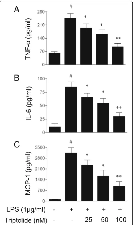
Triptolide attenuates LPS-induced production of pro-inflammatory cytokines and chemokines in HUVECs To assess the possible impact of triptolide on endothelial cell-associated inflammatory responses, the present study first determined the effects of triptolide on the LPS-induced production of inflammatory mediators in endothelial cells. As demonstrated in Fig. 2, treatment with LPS (1μg/ml) for 6 h increased the production of pro-inflammatory cytokines and chemokines, including
TNF-α, IL-6 and MCP-1 in HUVECs. The LPS-induced
effects on pro-inflammatory cytokines and chemokines were dose-dependently inhibited by pre-treatment with triptolide 1 h prior to LPS treatment. Taken together, the results of the present study demonstrated that triptolide suppresses inflammatory responses in endothelial cells.
Triptolide attenuates LPS-induced expression of adhesion molecules in HUVECs
It has been recognized that adhesion molecules are responsible for the pathological process of leukocyte infiltration. Therefore, it was subsequently examined whether triptolide treatment inhibits the LPS-induced expression of adhesion molecules in endothelial cells.
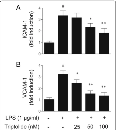
As illustrated in Fig. 3, LPS (1μg/ml) treatment
evoked an increase in ICMA-1 and VCAM-1 expres-sion in HUVECs, and pre-treatment with triptolide was revealed to attenuate the LPS-induced expression
of ICAM-1 and VCAM-1 in a dose-dependent
Fig. 1The effects of triptolide on the viability of human umbilical vein endothelial cells (HUVECs). HUVECs were treated with triptolide (25, 50 or 100 nM) for the indicated times. Cell viability was determined by an MTT assay. Data are expressed as the mean ± standard error of the mean. *p< 0.05 and **p< 0.01, compared with the corresponding control group
A
B
C
[image:4.595.306.539.87.479.2] [image:4.595.57.292.494.664.2]manner. Consequently, the diminished adhesion mol-ecule expression induced by triptolide may contribute to attenuating the adhesion of leukocytes to endothe-lial cells.
Triptolide attenuates LPS-induced monocyte adhesion to HUVECs
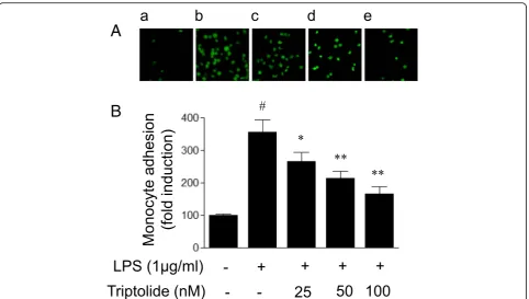
Since attachment of leukocytes to endothelial cells via adhesion molecules is a central step in the development of inflammation, the present study determined whether triptolide inhibits leukocyte adhesion to endothelial cells. As demonstrated in Fig. 4, LPS (1μg/ml) treatment for 6 h increased the number of monocytes that attached to HUVECs. Pretreatment with triptolide dose-dependently inhibited LPS-induced monocyte adhesion. Taken to-gether, these results suggested that triptolide inhibits LPS-induced leukocyte adhesion to endothelial cell pos-sibly via suppression of adhesion molecule expression.
Triptolide attenuates LPS-induced activation of NF-κB pathway in HUVECs
NF-κB is a crucial transcriptional factor for the expres-sion of inflammatory mediators. The phosphorylation of IκBα (Ser32) and its degradation are required for the
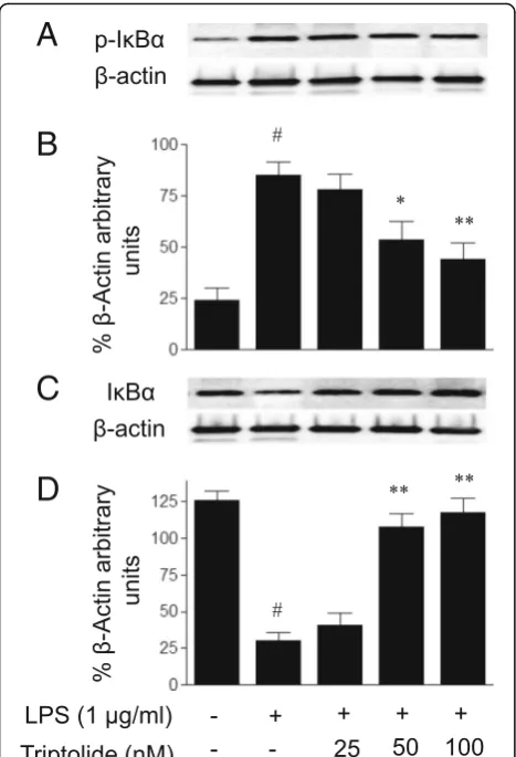
binding of NF-κB to DNA. Therefore, the present study examined the effect of triptolide on LPS-induced NF-κB activation by measuring IκBα (Ser32) phosphorylation. As demonstrated in Fig.5, LPS treatment for 30 min led to the phosphorylation of IκBα in HUVECs, and the re-sponses were attenuated by pre-treatment with tripto-lide. Additionally, the LPS-induced degradation of IκBα was attenuated by triptolide pre-treatment. Full-length Western blots for the protein expression of IκBα phos-phorylation and IκBα are presented in Additional file1: Figures S1 and S2. Since the phosphorylation of IκBα and its degradation are followed by nuclear NF-κB DNA binding and transcriptional activation, the effect of
trip-tolide on LPS-induced NF-κB activation was
subse-quently determined by assaying the DNA binding activity of NF-κB p65 and NF-κB transcriptional activity, respectively. As demonstrated in Fig. 6, the DNA bind-ing activity of NF-κB p65 was markedly increased fol-lowing LPS treatment in HUVECs, and the LPS-induced binding of NF-κB to DNA was attenuated by pre-treat-ment with triptolide in a dose-dependent manner.
Fur-thermore, treatment with LPS led to increased NF-κB
reporter activity in HUVECs and this response was dose-dependently inhibited by pre-treatment with tripto-lide (Fig.7).
Taken together, the results of the present study showed that triptolide treatment suppressed LPS-induced NF-κB transcriptional activation with the inhibition of IκBα phos-phorylation and its degradation, and subsequent NF-κB DNA binding.
Discussion
The present study provides the first characterization of the anti-inflammatory actions of triptolide in endothelial cells. To begin with, it was demonstrated that triptolide, a major bioactive component of the Chinese herb TWHF, leads to an anticipated inhibition of endothelial inflamma-tory response induced by LPS. The anti-inflammainflamma-tory ef-fects include decreased release of pro-inflammatory cytokines and chemokines, and decreased expression of adhesion molecules that contribute to modulating inflam-matory cell adhesion to endothelial cells. Next, mechanis-tic analysis revealed that the anti-inflammatory effect of triptolide is mediated possibly via suppression of NF-κB activation. Therefore, it is the first study to show that triptolide attenuates endothelial cell-associated inflamma-tory responses possibly via inhibition of NF-κB signaling pathway.
Triptolide is a major bioactive compound in Trad-itional Chinese Medicine TWHF, which has been widely used as a therapeutic agent for hundreds of years in China [6–8, 21]. Despite considerable advances in in vivo research for triptolide in the field of anti-inflamma-tion, it is unclear whether it has anti-inflammatory
A
B
[image:5.595.57.291.87.349.2]effects on vascular endothelial cells. Given that endothe-lial cells serve a pivotal role in inflammation by the re-lease and expression of inflammatory mediators and adhesion molecules that recruit leukocytes from blood to tissues [22], the present study assessed the ability of triptolide to modulate the release of inflammatory medi-ators and activation of the NF-κB signaling pathway in LPS-stimulated HUVECs. The results of the present study demonstrated that triptolide attenuated LPS-in-duced endothelial inflammatory responses. Notably, these effects were associated with suppression of NF-κB activation. Taken together, the results suggested that triptolide attenuates inflammatory responses possibly via inhibition of NF-κB activation.
The integrated function of endothelial cells serves a crucial role in the maintenance of cardiovascular homeostasis [23]. Pro-inflammatory stimuli, including LPS and TNF-α, usually cause endothelial dysfunction, leading to the initiation of inflammatory responses in numerous cardiovascular diseases [3, 4, 20]. Inflamma-tory responses occurring in endothelial cells is featured by the overproduction of inflammatory mediators,
in-cluding pro-inflammatory cytokines (TNF-α and IL-6),
chemokines (MCP-1) and adhesion molecules (ICAM-1 and VCAM-1). Activation of the NF-κB signaling path-way contributes to the expression of inflammatory genes in endothelial cells [24]. Therefore, it is well accepted that modulation of the processes is an effective strategy for the treatment of the diseases characterized with inflammation.
TNF-α, IL-6 and MCP-1 are important pro-inflammatory cytokines or chemokines. They are well characterized in-flammatory mediators, and are involved in the initiation and amplification of inflammatory responses. The present study selected TNF-α, IL-6 and MCP-1 as biomarkers of endothelial inflammatory responses, since there is a strong correlation between the release and expression of these in-flammatory mediators and cardiovascular morbidity and mortality [25, 26]. In addition, there is accumulating evi-dence demonstrating that these inflammatory mediators serve a key role in the development of cardiovascular dis-eases [27–29]. Although we only determined the effects of triptolide on these inflammatory mediators, it is reasonable to propose that triptolide may also work on other inflam-matory mediators. Therefore, further studies are required to clarify this issue.
A
B
[image:6.595.58.541.86.359.2]Inflammation is the body’s protective mechanism against infection or injury. However, when the inflam-matory response becomes severe or prolonged, it leads to a pathological state such as sepsis and atherosclerosis. In addition to the production of inflammatory media-tors, inflammation is characterized by massive
recruit-ment of leukocytes from the blood into the
inflammatory sites. The infiltration of leukocytes in-volves the interaction between adhesion molecules on endothelial cell surfaces and its counter receptors on the surface of leukocytes [22]. Chemokines (e.g., MCP-1) are also involved in recruiting leukocytes to sites of inflam-mation. The expression of the inflammatory mediators is
under the tight control of the inflammatory transcription factor NF-κB [24].
A characterization of the effects of triptolide on NF-κB activation in endothelial cells was determined. It is well known that NF-κB is activated upon phosphorylation of its cytoplasmic inhibitor IκBα by IκB kinase (IKK) complex [30, 31]. IKK-induced phosphorylation triggers the ubiquitination-mediated degradation of IκBαand
sub-sequent nuclear NF-κB DNA binding, leading to the
A
B
C
D
Fig. 5The effects of triptolide on lipopolysaccharide (LPS)-induced changes in the protein levels of (aandb) phosphorylated IκBα (p-IκBα) and (candd) IκBαin human umbilical vein endothelial cells (HUVECs). Full-length blots for p-IκBαand IκBαare presented in Additional file Data. HUVECs were treated with LPS (1μg/ml) in the presence or absence of triptolide (25, 50 or 100 nM) for 30 min. Cell lysates were subjected to immunoblot analysis using the antibodies against p-IκBα(Ser32) and IκBα. The data are presented as the relative protein expression normalized toβ-actin expression. Data are expressed as the mean ± standard error of the mean. *p< 0.05 and **p< 0.01, compared with LPS treatment alone. #p< 0.05, compared with the control group
Fig. 6The effects of triptolide on lipopolysaccharide (LPS)-induced increases in NF-κB p65 DNA binding activity in human umbilical vein endothelial cells (HUVECs). HUVECs were treated with LPS (1μg/ml) in the presence or absence of triptolide (25, 50 or 100 nM) for 30 min. The DNA binding activity of NF-κB p65 in HUVECs was determined using a non-radioactive, ELISA-based assay kit. Data are expressed as the mean ± standard error of the mean. *p< 0.05 and **p< 0.01, compared with LPS treatment alone. #p< 0.05, compared with the control group
[image:7.595.57.291.87.430.2] [image:7.595.304.541.89.234.2] [image:7.595.304.539.475.622.2]transcription of inflammatory genes. To delineate the mechanisms by which triptolide regulates NF-κB activity, unrelated methodological approaches were used. As ex-pected, the present study demonstrated that triptolide, in addition to inhibiting NF-κB transcriptional activation as measured with an NF-κB luciferase reporter gene assay, also suppressed the events upstream of NF-κB transcrip-tional activation, including IκBα phosphorylation and its
degradation, and NF-κB DNA binding. The results
im-plied that triptolide attenuates LPS-induced NF-κB tran-scriptional activation at a cytosolic and nuclear site.
Limitations of this study include the focus of our experi-ments in specific endothelial cell lines. The characterization of these responses to primary endothelial cells from differ-ent tissues remains to be determined. Furthermore, in addition to LPS, other pro-inflammatory stimuli, including oxidized low-density lipoprotein (ox-LDL) and TNF-α, also cause endothelial dysfunction, resulting in the initiation of inflammatory responses in numerous cardiovascular dis-eases [3,4,32]. Therefore, the effects of triptolide on endo-thelial cells exposed to other pro-inflammatory stimuli (e.g., ox-LDL) will be determined in the future studies.
Conclusions
In summary, the results of the present study demonstrated that triptolide possesses anti-inflammatory effects on in-flammatory responses in endothelial cells. Moreover, to the best of our knowledge, the present study was the first to show that the mechanisms for suppression of endothe-lial cell-associated inflammatory responses by triptolide may involve inhibition of the NF-κB signaling pathway.
Inflammation is the most common characteristic of a number of diseases, including atherosclerosis and hyper-tension, and there is a clear requirement for effective therapies. The results of the present study provided insight into the mechanisms for inhibition of endothelial cell-associated inflammatory responses by triptolide, a major bioactive component of the Chinese herb TWHF, and will contribute to our understanding of the mecha-nisms by which TWHF and its preparations attenuate in-flammatory responses.
Additional file
Additional file 1:Supplementary Data.Figure S1.Full-length Western blots for Figure 5A, the effects of triptolide on lipopolysaccharide (LPS)-induced changes in the protein levels of phosphorylated IκBα(p-IκBα) in human umbilical vein endothelial cells (HUVECs). The blots show p-IκBα (A) with blots stripped and reprobed to detectβ-actin (B). The blots are presented individually for each staining, intact without splicing.Figure S2.Full-length Western blots for Figure 5C, the effects of triptolide on lipopolysaccharide (LPS)-induced changes in the protein levels of IκBαin human umbilical vein endothelial cells (HUVECs). The blots show IκBα(A) with blots stripped and reprobed to detectβ-actin (B). The blots are presented individually for each staining, intact without splicing. (DOCX 705 kb)
Abbreviations
DMSO:dimethyl sulfoxide; EBM-21: endothelial basal medium-2; FBS: fetal bovine serum; HUVECs: human umbilical vein endothelial cells;; IKK: IκB kinase; LPS: lipopolysaccharide; MTT: 3-(4,5-dimethylthiazol-2-yl)-2,5-diphenyltetrazolium bromide; NF-κB: nuclear factor-κB; ox-LDL: oxidized low-density lipoprotein; PVDF: polyvinylidene difluoride; TWHF:Tripterygium wilfordiiHook.F.
Acknowledgments
Not applicable.
Authors’contributions
CS and YW: conception and design of research; LC, FY, and SS performed experiments; CS, YW, LC, FY, and SS analyzed data and interpreted results of experiments; CS and YW drafted manuscript; CS and YW edited and revised manuscript. All authors read and approved the final manuscript.
Funding
This study was founded by the grants from the National Nature Science Foundation of China (No. 81673734 & 81170243), and Henan Provincial Science and Technique Foundation for International Cooperation Program (No. 162102410048) to YW. This study was also founded in part by the Science and Technique Foundation from the Education Department of Henan Province (No. 2010B360012) to CS. The funding body provided financial support and had no role in the design of the study, collection, analysis, interpretation of data, or writing the manuscript.
Availability of data and materials
The datasets used and/or analyzed during the current study are available from the corresponding author on reasonable request.
Ethics approval and consent to participate
Not applicable.
Consent for publication
Not applicable.
Competing interests
The authors declare that they have no competing interests.
Received: 5 May 2019 Accepted: 23 July 2019
References
1. Santos-Gallego CG, Picatoste B, Badimón JJ. Pathophysiology of acute coronary syndrome. Curr Atheroscler Rep. 2014;16:401.
2. McMaster WG, Kirabo A, Madhur MS, Harrison DG. Inflammation, immunity, and hypertensive end-organ damage. Circ Res. 2015;116:1022–33. 3. Boisramé-Helms J, Kremer H, Schini-Kerth V, Meziani F. Endothelial
dysfunction in sepsis. Curr Vasc Pharmacol. 2013;11:150–60.
4. Xiao L, Liu Y, Wang N. New paradigms in inflammatory signaling in vascular endothelial cells. Am J Physiol Heart Circ Physiol. 2014;306:H317–25. 5. Shiraki A, Oyama J, Komoda H, Asaka M, Komatsu A, Sakuma M, Kodama K,
Sakamoto Y, Kotooka N, Hirase T, Node K. The glucagon-like peptide 1 analog liraglutide reduces TNF-alpha-induced oxidative stress and inflammation in endothelial cells. Atherosclerosis. 2012;221:375–82. 6. Lipsky PE, Tao XL. A potential new treatment for rheumatoid arthritis:
thunder god vine. Semin Arthritis Rheum. 1997;26:713–23.
7. Zhu B, Wang Y, Jardine M, Jun M, Lv JC, Cass A, Liyanage T, Chen HY, Wang YJ, Perkovic V.Tripterygiumpreparations for the treatment of CKD: a systemic review and meta-analysis. Am J Kidney Dis. 2013;62:515–30. 8. Marcus DM. Comparison of Tripterygium wilfordii hook F with methotrexate
in the treatment of rheumatoid arthritis. Ann Rheum Dis. 2014;73:e56. 9. Kupchan SM, Court WA, Dailey RG, Gilmore CJ Jr, Bryan RF. Triptolide and
tripdiolide, novel antileukemic diterpenoid triepoxides from Tripterygium wilfordii. J Am Chem Soc. 1972;94:7194–5.
10. Gu WZ, Chen R, Brandwein S, McAlpine J, Burres N. Isolation, purification, and characterization of immunosuppressive compounds from tripterygium: triptolide and tripdiolide. Int J Immunopharmacol. 1995;17:351–6. 11. Liu Q. Triptolide and its expanding multiple pharmacological functions. Int
12. Kumar A, Corey C, Scott I, Shiva S, D’Cunha J. Minnelide/triptolide impairs mitochondrial function by regulating SIRT3 in p53-dependent manner in non-small cell lung cancer. PLoS One. 2016;11:e0160783.
13. Hoyle GW, Hoyle CI, Chen J, Chang W, Williams RW, Rando RJ. Identification of triptolide, a natural diterpenoid compound, as an inhibitor of lung inflammation. Am J Physiol Lung Cell Mol Physiol. 2010;298:L830–6. 14. Wei D, Huang Z. Anti-inflammatory effects of triptolide in LPS-induced
acute lung injury in mice. Inflammation. 2014;37:1307–16.
15. Kondylis V, Kumari S, Vlantis K, Pasparakis M. The interplay of IKK, NF-κB and RIPK1 signaling in the regulation of cell death, tissue homeostasis and inflammation. Immunol Rev. 2017;277:113–27.
16. Afonina IS, Zhong Z, Karin M, Beyaert R. Limiting inflammation-the negative regulation of NF-κB and the NLRP3 inflammasome. Nat Immunol. 2017;18:861–9.
17. Krosch TC, Sangwan V, Banerjee S, Mujumdar N, Dudeja V, Saluja AK, Vickers SM. Triptolide-mediated cell death in neuroblastoma occurs by both apoptosis and autophagy pathways and results in inhibition of nuclear factor-kappa B activity. Am J Surg. 2013;205:387–96.
18. Xu B, Guo X, Mathew S, Armesilla AL, Cassidy J, Darling JL, Wang W. Triptolide simultaneously induces reactive oxygen species, inhibits NF-kappaB activity and sensitizes 5-fluorouracil in colorectal cancer cell lines. Cancer Lett. 2010;291:200–8.
19. Chen M, Shi JT, Lv ZQ, Huang LJ, Lin XL, Zhang W, Liang RY, Li YQ, Jiang SP. Triptolide inhibits TGF-β1 induced proliferation and migration of rat airway smooth muscle cells by suppressing NF-κB but not ERK1/2. Immunology. 2015;144:486–94.
20. Wang Y, Cui L, Xu H, Liu S, Zhu F, Yan F, Shen S, Zhu M. TRPV1 agonism inhibits endothelial cell inflammation via activation of eNOS/NO pathway. Atherosclerosis. 2017;260:13–9.
21. Ge Y, Xie H, Li S, Jin B, Hou J, Zhang H, Shi M, Liu Z. Treatment of diabetic nephropathy with Tripterygium wilfordii hook F extract: a prospective, randomized, controlled clinical trial. J Transl Med. 2013;11:134. 22. Gerhardt T, Ley K. Monocyte trafficking across the vessel wall. Cardiovasc
Res. 2015;107:321–30.
23. Ross R. Atherosclerosis-an inflammatory disease. N Engl J Med. 1999; 340:115–26.
24. Cheng HS, Njock MS, Khyzha N, Dang LT, Fish JE. Noncoding RNAs regulate NF-kB signaling to modulate blood vessel inflammation. Front Genet. 2014;5:422.
25. Dessein PH, Joffe BI, Singh S. Biomarkers of endothelial dysfunction, cardiovascular risk factors and atherosclerosis in rheumatoid arthritis. Arthritis Res Ther. 2005;7:R634–43.
26. Narizhneva NV, Razorenova OV, Podrez EA, Chen J, Chandrasekharan UM, DiCorleto PE, Plow EF, Topol EJ, Byzova TV. Thrombospondin-1 up-regulates expression of cell adhesion molecules and promotes monocyte binding to endothelium. FASEB J. 2005;19:1158–60.
27. Wang Y, Wang DH. Aggravated renal inflammatory responses in TRPV1 gene knockout mice subjected to DOCA-salt hypertension. Am J Physiol Renal Physiol. 2009;297:F1550–9.
28. Elmarakby AA, Quigley JE, Imig JD, Pollock JS, Pollock DM. TNF-alpha inhibition reduces renal injury in DOCA-salt hypertensive rats. Am J Physiol Regul Integr Comp Physiol. 2008;294:R76–83.
29. Wang Y, Zhu M, Xu H, Cui L, Liu W, Wang X, Shen S, Wang DH. Role of the monocyte chemoattractant protein-1/C-C chemokine receptor 2 signaling pathway in transient receptor potential vanilloid type 1 ablation-induced renal injury in salt-sensitive hypertension. Exp Biol Med. 2015;240:1223–34. 30. Solt LA, May MJ. The IkappaB kinase complex: master regulator of
NF-kappaB signaling. Immunol Res. 2008;42:3–18.
31. De Falco F, Di Giovanni C, Cerchia C, De Stefano D, Capuozzo A, Irace C, Iuvone T, Santamaria R, Carnuccio R, Lavecchia A. Novel non-peptide small molecules preventing IKKβ/NEMO association inhibit NF-κB activation in LPS-stimulated J774 macrophages. Biochem Pharmacol. 2016;104:83–94. 32. Jiang JX, Zhang SJ, Liu YN, Lin XX, Sun YH, Shen HJ, Yan XF, Xie QM. EETs
alleviate ox-LDL-induced inflammation by inhibiting LOX-1 receptor expression in rat pulmonary arterial endothelial cells. Eur J Pharmacol. 2014; 727:43–51.
Publisher’s Note