Rheumatoid arthritis (RA) is traditionally regarded as an archetypal disease of the synovial tissue. Th e synovial membrane indeed undergoes early infl ammatory changes, which include increased vascularity as well as intimal lining layer hyperplasia and accumulation of macro-phages, plasma cells, T cells, B cells, dendritic cells, natural killer cells and mast cells in the sublining, among other changes [1]. Collectively, localization of infl amma-tion to the synovium is primarily responsible for the dysregulated cellular and molecular mechanisms that
ultimately lead to the typical signs and symptoms of RA, including joint pain, stiff ness, swelling and structural changes. Although the pathogenic and clinical impor-tance of synovitis is beyond question, it is becoming increasingly apparent that a ‘synovio-centric’ model of RA could be limiting. Indeed, other anatomic compart-ments appear to be involved at all stages of the disease. Th e most important example comes from the recognition that clinical arthritis and subclinical synovitis are antici-pated by a pre-articular immunologic phase possibly developing in lymphoid tissues as well as in the lungs [2,3]. Full-blown joint disease itself spreads well beyond synovial tissue infl ammation. Established arthritis can be characterized by the involvement of at least two other compartments that are in direct contact with the joint space. Th ese include the draining lymph nodes (LNs) [4,5] and, of special relevance, the subchondral bone marrow (BM) [6].
As dysregulated B cell responses are central pathogenic events in RA and B-cell autoreactivity originates, at least in part, from defective checkpoints within the BM (a primary lymphoid organ) [7], the role of this compart-ment in the immunopathogenesis of RA has attracted great attention over the years. Data supporting the existence of BM abnormalities were mainly derived from studies on marrow aspirates/biopsies from the sternum and the iliac crest, which are primary hematopoietic and immunologic sites in adult life. Th e role that the ‘red marrow’ might play in the immunologic disturbances of RA thus appears interesting but not completely un-expected. Less predictably, evidence has accumulated supporting the additional involvement of the BM adjacent to infl amed joints at peripheral sites. Th is is normally a fat-rich tissue (‘yellow marrow’) where hematopoiesis is not prominent. In RA, the fat can be replaced by a vascularized, cell-rich infl ammatory tissue. Combined with imaging data coming from magnetic resonance imaging (MRI) studies, histopathologic changes described within the subchondral BM have been postu-lated to be intimately involved in the pathological processes producing local infl ammation and tissue
Abstract
The synovial tissue stands at the epicenter of joint pathology in rheumatoid arthritis (RA). As a primary target of the disease, studies on the synovium have provided invaluable insights into the mechanisms involved in disease pathogenesis. Recent work has, however, revealed the importance of a previously unseen anatomic compartment in direct contact with the joint space, namely the subchondral bone marrow. Bone marrow edema (BME) visible on magnetic resonance imaging (MRI) is clinically meaningful in both early and late RA as it associates with future development of bone erosions and poor functional outcomes. Although the histopathologic correlates of MRI-based BME in early RA remain obscure, studies in advanced disease are consistent in describing lymphocytic infl ammatory infi ltrates within the subchondral marrow cavity of aff ected joints. In this review, we discuss the nature of bone marrow lesions in patients with RA, analyze their relationship with synovitis, and explore their potential contribution to the pathological processes of the disease.
© 2010 BioMed Central Ltd
Infl ammatory lesions in the bone marrow of
rheumatoid arthritis patients: a morphological
perspective
Serena Bugatti*, Antonio Manzo, Roberto Caporali and Carlomaurizio Montecucco
R E V I E W
*Correspondence: serena.bugatti@unipv.it
Division and Laboratory of Rheumatology, University of Pavia School of Medicine, IRCCS Policlinico San Matteo Foundation; Piazzale Golgi, 2, 27100 Pavia, Italy
remodeling in RA joints [6,8-10]. Here we will discuss the most relevant fi ndings related to BM alterations in RA, focusing on their morphological characteristics and patho logic signifi cance. In view of the clinical relevance of MRI fi ndings, special emphasis is given to the subchondral BM.
Histopathology of the bone marrow in rheumatoid arthritis
Systemic bone marrow
Alongside its hematopoietic function and its role in the early selection of lymphocytes, the BM is an immune regulatory organ involved in migration, selective retain-ment and function of innate and adaptive immune cells [11]. A variety of morphological, immunophenotypic and functional abnormalities in BM cells in RA have been detected as a consequence of augmented local production of infl ammatory cytokines and cell-cell interactions. Both myeloid and lymphoid lineage cells appear aff ected at some stage of the disease.
Th e absolute number of mononuclear cells was found markedly increased in iliac BM aspirates in RA patients compared to controls [12]. In addition, the spontaneous generation of CD14+ myeloid cells from BM progenitors
in vitro as well as their maturation towards HLA-DR+ cells were accelerated in RA patients, indicating that alterations in the regenerative potential of the BM may occur [13,14]. Th e fi nding that levels of IL-6 and IL-8 were markedly elevated in BM aspirates from RA patients (compared with controls) [15] is suggestive of ongoing pathology in the BM region. DNA microarray analysis also demonstrated that BM cells from RA patients have abnormal functional networks in immune response and cell cycle when compared to those from osteoarthritis patients, with overexpression of genes that take part in the antigen presentation pathway and interferon signal-ing [16]. Studies in experimental arthritis have confi rmed a generalized pattern of red marrow conversion with enhanced myelopoiesis in conjunction with either increased IL-1 and IL-6 activity [17] or TNF overexpres-sion [18]. Interestingly, high cellularity throughout the marrow seen on histologic examination in TNF-trans-genic mice appears associated with a diff use bone marrow edema (BME) pattern on MRI, distinct from sub chondral BME coexisting with focal erosions [18]. Together with the observed alterations in the myeloid compartment, there is evidence that the BM environment in patients with RA may harbor aberrant B- and T-cell immune responses. Marrow plasmacytosis and hyper-globulinemia were already described in the 1950s in a number of independent studies (reviewed by Duthie and colleagues [19]). B cells derived from RA BM were later found to be capable of producing RA-associated auto-antibodies, such as all rheumatoid factor (RF) isotypes
[20] as well as IgM anti-citrullinated peptide antibodies (ACPAs) [21]. Supporting a pathogenic role of T cells in this disease, an abnormal accumulation of HLA-DR+CD8+ and recently activated CD3+CD4+ T lymphocytes has been detected in the iliac BM of RA patients (a site remote from infl amed joints) and also in femoral heads [12,22,23].
From a morphological perspective, it is worth noting that, in chronic infl ammatory conditions such as RA, the expanded lymphoid component of the BM may form follicle-like structures and undergo a localized process of ectopic lymphoid neogenesis. In a series of 65 marrow trephines, Engels and colleagues [24] reported morpho-logically benign lymphoid aggregates in 15 cases, 5 of which were diagnosed with RA or other autoimmune disorders. More recently, Kuca-Warnawin and colleagues [23] confi rmed a follicular pattern of infi ltrating lympho-cytes in 9 of 15 BM trephine biopsy specimens from RA patients. Of these, fi ve exhibited features of compart-mentalized lymphoid follicles, with a core of CD20+ B cells surrounded by CD4+ T cells and histologic evidence of germinal center (GC)-like structures. Function ality has yet to be demonstrated at these sites, but B-cell clonality was recognized in 9 of 15 patients (4 with an autoimmune disorder) in the series described by Engels and colleagues [24]. Local expression of the activation marker CD69 on follicular cells as well as increased levels of the T-cell growth factor IL-15 [23] are further clues that local immune cell activation may take place.
local expression of anti-apoptotic molecules, patient-related diff erences, or others. Further investigations on the mode of action of rituximab in the BM and in other compartments of the immune system are awaited in order to gain better understanding of the pathogenetic and clinical variability of RA.
Subchondral bone marrow
Infl ammatory changes occurring within the juxta-articular BM in RA were reported over three decades ago. Barrie [29] observed cartilage damage, reparative changes and fi brovascular tissue within the subchondral marrow of surgically removed metatarsal and metacarpal heads in advanced RA. In similar surgical material, Wyllie [30] further described proliferation of fi broblasts, deposition of collagen, formation of thin-walled vascular channels, infi ltrates of macrophages, lymphocytes and plasma cells. In RA knee joints, Bromley and colleagues [31] observed the presence of blood vessels and infl ammatory cell infi ltrates including mononuclear/macrophage cells in subchondral regions remote from the synovial pannus-cartilage junction. Th ese cellular aggregates were adjacent to erosions of the articular cartilage. In a similar study of RA subchondral bone removed from a patient under-going knee replacement surgery, Watson and colleagues [32] described a local infl ammatory reaction with nodular lymphocytic infi ltrates accompanied by immuno globulin deposits near areas of destroyed cartilage. More recently, in a series of 164 hip arthroplasties performed for various joint diseases, O’Connell and colleagues [33] found a total of ten cases (six with RA) with subchondral infl am-mation variably characterized by diff use and nodular distribution. Altogether, descriptions of a subchondral infl ammatory reaction in damaged RA joints gave rise at the end of the 1980s to the hypothesis of a ‘bi-compart-mental’ model of RA, in which infl ammation of the BM of aff ected joints actively contributed to the establish-ment of local damage [8,31].
Th e almost complete inaccessibility of the BM com part-ment to either clinical examination or traditional imaging (radiography) has contributed to a lack of interest in local marrow pathology until recent years. It has only been since the advent of MRI that the subchondral bone has gained recognition as a site of pivotal importance when considering the nature of the infl ammatory process in RA. MRI-based BM involvement in the form of BME is a common feature in late and early RA, where it associates with disease activity and, most importantly, with future development of bone erosions and poor functional out-comes (for a review on BME in RA, see [9,10] and the manuscript by FM McQueen in this edition of Arthritis Research and Th erapy).
While the clinical importance of MRI-based BM involvement has been demonstrated consistently, its
of B-cell markers revealed an abundance of CD27+ mature B cells. Plasma cells located at the periphery of marrow aggregates contributed 8 ± 4% of total cells [39]. A follicular organization of the lymphocytic infi ltrate with histologic evidence of GC-like structures has been recently confi rmed in three out of six subchondral bone specimens obtained from hip replacement surgery in patients with advanced RA [23].
Although no direct comparison is available with the histologic pattern of BME in other joint diseases, some morphological peculiarities of RA osteitis could refl ect disease-specifi c mechanisms. BME in the spondylo-arthritides equally corresponds to a mononuclear infi l-trate possibly organized into follicles. Here, however, T cells largely predominate [40-43], in keeping with the limited role attributable to B cells in these conditions [44]. On the other hand, the marrow infl ammatory tissue in osteoarthritis is mainly fi brovascular with scarce cellular infi ltration [45-48]. Th is pattern could result from biomechanical (excessive loading) rather than infl ammatory alterations [49].
In summary, notwithstanding the relatively small case series analyzed and the unavailability of data on early arthritis, it appears indisputable that the subchondral aspect of the RA joint may harbor regions of bone infl am-mation (osteitis) dominated by lymphocytes. Whether this process is quantitatively or qualitatively comparable to synovial infl ammation (synovitis) and to what extent it aff ects specifi c disease outcomes will be discussed in the next two paragraphs. Table 1 summarizes the main
fi ndings of modern studies addressing the histologic picture of subchondral BM involvement in RA.
Relationship between subchondral bone marrow infl ammation and synovitis
Morphological relationship
A detailed description of the histopathology of the infl amed synovium in RA goes beyond the scope of this review. Some specifi c aspects of the lymphoid infl am ma-tory infi ltrate, however, deserve brief discussion in light of the similarities to BM features.
[image:4.612.66.547.89.288.2]for an oligoclonal repertoire with highly mutated V regions, compatible with a local antigen-driven GC-like reaction [54]. Furthermore, a large amount of plasma cells infi ltrating the synovial tissue appeared to be generated by locally activated B cells [55]. Th ese fi ndings are consistent with the observed local expression of AID (activation-induced cytidine deaminase), an enzyme critically involved in somatic hypermutation and class-switching of the immunoglobulin genes within conven-tional GCs of secondary lymphoid tissues [56]. Synovial lymphoid follicles may represent an histologic feature associated with disease severity and target organ damage [57]. Ectopic lymphoid aggregates are indeed associated with more severe synovial and systemic infl ammation [58,59] and B-cell-rich infi ltrates appear a marker of erosive disease [60-62]. Whether these relationships are linked causally or represent bystander events remains unknown. Supporting a direct link between B-cell autoimmunity and tissue damage, human autoantibodies against citrullinated vimentin have been recently shown to induce osteoclastogenesis and bone loss [63].
Although studies directly comparing marrow and synovial follicles are limited [39], data presented herein indicate that the overall morphological picture mainly coincides. Slight diff erences in cellular composition may be related to higher proportions of B cells and plasma cells and less macrophages in subchondral BM aggregates [39]. Consistent with the considerable heterogeneity observed at the synovial tissue level [52], characteristics of the lymphoid-like features may vary considerably among diff erent BM samples. Th us, subchondral BM CD21+ FDC networks and histological features of GC-like structures have been described but appear rare [23,32,38] or even absent [39]. Similarly, vascular addres-sins are found in a variable proportion of specimens
[38,39]. Other relevant aspects, such as the local organi-zation of T-cell area fi broblastic reticular networks, have been assessed at the synovial tissue level [64] but their eventual correlates within the BM are so far unknown. Functionality at these sites has yet to be demonstrated. Th e possibility of a link between subchondral BM follicles and the pathological processes of the disease is suggested by the fi nding that these structures are more prominent in patients with large numbers of swollen joints, a high acute phase response and high titers of RF [39]. Histologically, enrichment in marrow follicles has been consistently observed in areas of cortical bone destruction [38,39,57].
Hierarchical relationship
A critical aspect for our understanding of the actual signi fi cance of marrow pathology in RA is the identi-fi cation of the hierarchical relationship between synovitis and subchondral osteitis. Two separate scenarios can theoretically be hypothesized: one is that RA is a disease starting in the synovial tissue, which then invades the subchondral bone cavity through established bone erosions; the other is that of a primary BM disease, which then spreads to the synovial membrane. In the absence of histopathologic data in the very early and possibly pre-clinical phases of the disease, both theories are based on assumptions rather than facts.
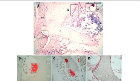
[image:5.612.65.548.111.297.2]Analyses of tissue samples obtained from patients with long-standing erosive RA have consistently shown that marrow follicles are preferentially located in superfi cial areas of the subchondral bone at sites of synovial tissue penetration through cortical bone erosions (Figure 1A) [38,39]. Such a picture appears strongly evocative of an ‘outside-in’ model in which marrow infl ammation origi-nates from propagation of adjacent synovial
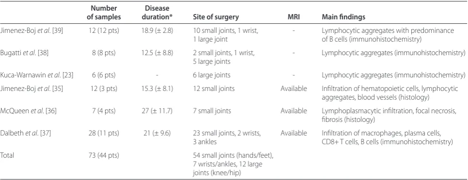
Table 1. Main studies addressing the histopathologic picture of subchondral bone marrow involvement in rheumatoid arthritis
Number Disease
of samples duration* Site of surgery MRI Main fi ndings
Jimenez-Boj et al. [39] 12 (12 pts) 18.9 (± 2.8) 10 small joints, 1 wrist, - Lymphocytic aggregates with predominance
1 large joint of B cells (immunohistochemistry)
Bugatti et al. [38] 8 (8 pts) 12.5 (± 8.8) 2 small joints, 1 wrist, - Lymphocytic aggregates (immunohistochemistry) 5 large joints
Kuca-Warnawin et al. [23] 6 (6 pts) - 6 large joints - Lymphocytic aggregates (immunohistochemistry)
Jimenez-Boj et al. [35] 12 (3 pts) 15.3 (± 8.1) 12 small joints Available Infi ltration of hematopoietic cells, lymphocytic aggregates, blood vessels (histology)
McQueen et al. [36] 7 (4 pts) 27 (± 11.7) 7 small joints Available Lymphoplasmacytic infi ltration, focal necrosis,
fi brosis (histology)
Dalbeth et al. [37] 28 (11 pts) 21 (± 9.6) 23 small joints, 2 wrists, Available Infi ltration of macrophages, plasma cells,
3 ankles CD8+ T cells, B cells (immunohistochemistry)
Total 73 (44 pts) 54 small joints (hands/feet),
7 wrists/ankles, 12 large
joints (knee/hip)
infl amma tion. On the other hand, although no clues at present indicate that early osteitis morphologically corresponds to the osteitis of advanced disease, MRI studies in the earliest stages of RA reveal BM alterations before any obvious communication with the synovium is detectable [65]. Extensive BME has been also documented in patients with undiff erentiated arthritis later developing RA [66] as well as in patients with ACPA-positive palindromic rheumatism, a proposed model of pre-clinical RA [67]. Histologically, mild infl ammatory infi ltrates have been described also in deeper areas of the subchondral bone remote from the synovial-marrow junction [38,39]. More intriguingly, lymphoid reactions occurring at the synovial-marrow boundary are conspicuous whilst the corresponding synovial tissue penetrating through cor tical erosions is mainly fi brous [38]. Altogether, these data would suggest the autonomy (at least in part) of osteitis from synovitis. Th e picture is, however, further complicated by recent evidence demonstrating that the synovium and the BM can physiologically communicate through microscopic bone canals (<0.5 mm in width or depth) visible using high-resolution computed tomo graphy [68]. Th ese canals could allow the transduction of infl ammation from the outside (synovium) to the inside (BM) and vice versa. Both routes are theoretically viable. In collagen-induced arthritis, mesenchymal cells ori ginat ing from the juxta-articular BM have been shown to travel to the synovium through enlarged bone canals in the pre-arthritic phase [69]. On the other hand, in the same experimental model, expression of infl ammatory cytokines and osteoclastogenic factors has been almost exclusively detected at the synovial tissue level, suggesting that subchondral BM reactions are in fact driven by signals propagating from the synovium [70]. Exactly how the fi ndings obtained in animal models fi t into the spectrum of human RA awaits further defi nition.
Th e ‘inside-out’ concept may be more applicable in other forms of arthritis, such as the spondyloarthritides. In anterior spondylitis, infl ammation dominates the BM in areas with no adjacent synovium [41,42]. In contrast, the centrality of synovitis in RA remains incontestable. Imaging and histologic studies have, however, added another layer of complexity to models of pathogenesis in the infl ammatory arthritides, by demonstrating that joint infl ammation is not exclusive to the synovial membrane but also extends to the neighboring BM.
Are additional structures involved in the local
infl ammatory process of RA? The possible contribution of the draining lymph nodes
As long as our perspective on joint infl ammation in RA has expanded from the synovial compartment to the subchondral BM, the draining LN has emerged as an
additional player involved in multiple aspects of the disease. Th ese include the generation of local immuno-logical responses as well as the control of cell effl ux from the joint.
LN involvement in experimental models appears early and possibly antedates clinical arthritis. An increase in the percentage of B lymphocytes as well as a high pro-liferation of CD8+ cells were observed in regional LNs in the latency period of adjuvant arthritis [71]. In the K/ BxN model of spontaneous autoimmunity, the LNs drain-ing the distal joints were found to be essential for the amplifi cation of the arthritogenic B-cell response [72]. Similarly, changes in the popliteal LN have been reported prior to disease onset in TNF-transgenic mice [73,74], with accumulation of CD23+CD21highCD1high B cells [75-77]. Such a cell population was recently shown to diff erentiate locally, to display an enhanced ability to capture and process antigens and to exhibit a GC pheno-type during T-cell-dependent immune responses [78]. Th e specifi c relevance of these fi ndings to autoimmune responses in RA is currently unknown. Also, early LN involvement in humans remains to be demonstrated. Former histologic studies on LN biopsies from diff erent anatomic sites in established RA described follicular hyperplasia and interfollicular plasmacytosis [79] as well as increased GCs with high B-cell activity [80]. In RF and/or ACPA-positive individuals at risk of developing RA and in early arthritis patients, an increase of activated CD69+ cells and a signifi cant change in the CD4/CD8 distribution was reported in inguinal LNs [81]. Ultrasound-guided biopsy of inguinal LNs appears feasible and safe [82] and promises to yield important information in the near future.
Alongside their potential role in modulating auto-immunity, the draining LNs may be also critically asso-ciated with the severity of joint involvement. Longitudinal studies in TNF-transgenic mice demonstrated a negative correlation between LN contrast enhancement and LN volume on MRI and local progression of synovitis, suggest ing that reduced LN drainage capacity may deter-mine worst arthritis outcomes [73,74]. A similar relation-ship between local disease activity in the joints and ultra-sonographic signs of axillary LN involvement has been recently reported in humans [5]. Histologically, arthritic fl ares in experimental models are associated with ipsilateral LN collapse due to lymphatic obstruction and diminished lymphatic fl ow [77]. Accordingly, inhibition of lymphatic drainage increases the severity of joint infl ammation in TNF-transgenic mice [75].
Relationship between subchondral bone marrow infl ammation and bone remodeling
an association implies a causative link remains to be determined.
Chondroclasts and osteoclasts were morphologically described in the subchondral bone of RA knee joints over 30 years ago [83]. More recently, using specifi c immuno-staining we demonstrated tartrate-resistant acid phos-pha tase (TRAP)- and cathepsin K-positive multi nucleated osteoclasts on the marrow side of RA samples, which were associated (in terms of density) with the extent of subchondral marrow infl ammation [38]. Others have later extended these fi ndings by providing evidence of local expression of molecules involved in osteoclasto-genesis and tissue destruction, such as receptor activator of NF-κB ligand (RANKL), cathepsins and metallo-proteinases [37,84]. Together with MRI studies, these data would suggest an active participation of the sub-chondral compartment to joint remodeling processes. RA erosions, however, anatomically and radiologically remain ‘outside-in’ processes. Th is pattern could be ascribed to diff erences in the local balance of erosive and reparative mechanisms between the synovial and the subchondral side of RA joints. Whilst bone-resorbing osteoclasts can be detected on both sides, bone-forming
osteoblasts and osteoid deposition are recognized only on bone surfaces adjacent to the marrow (Figure 2), as suggested by studies in experimental arthritis and human RA [38,39,85,86]. Th e role of the BM in RA might thus not be entirely negative. Accordingly, it has been recently shown that repair of bone erosions in RA patients treated with TNF inhibitors, although rare, is based on bone apposition at the base of erosion and probably involves the BM [87].
Conclusion
[image:7.612.71.545.88.361.2]central tolerance and as a survival niche for long-lived plasma cells, the systemic BM is a plausible candidate, although its involvement in the very early, pre-clinical phases of the disease remains speculative. Fascinating but still merely hypothetical is the role of secondary lymphoid organs, including LNs. Besides their potential role in systemic autoimmunity, the same compartments (BM and LNs) could be also involved in local pathology at the sites of joint infl ammation. From this perspective, the subchondral BM of aff ected joints appears tightly linked to the processes of local infl ammation and tissue remodeling, as suggested by (many) imaging studies and (few) histopathologic data. Analogous evidence is emerg-ing for the drainemerg-ing LNs, whose drainage capacity appears inversely related to local arthritis severity in experi mental models.
We have just started unraveling the mystery of a ‘multi-compartmental’ model of RA, but preliminary results encourage further research aimed at identifying novel pathogenic and clinical targets of the disease that may go beyond the well established synovial tissue immuno-pathologic environment.
Abbreviations
ACPA, anti-citrullinated protein antibody; BM, bone marrow; BME, bone marrow edema; FDC, follicular dendritic cell; GC, germinal center; IL, interleukin; LN, lymph node; MRI, magnetic resonance imaging; RA, rheumatoid arthritis; RF, rheumatoid factor; TNF, tumor necrosis factor; TRAP, tartrate resistant acid phosphatase.
Competing interests
The authors declare that they have no competing interests.
Acknowledgements
This study was supported in part by funding from the Italian Ministry of Health (grant 058/GR-2009-1608032 to AM).
Published: 27 December 2012
References
1. Hitchon CA, El-Gabalawy HS: The synovium in rheumatoid arthritis.Open
Rheumatol J 2011, 5:107-114.
2. Klareskog L, Rönnelid J, Lundberg K, Padyukov L, Alfredsson L: Immunity to citrullinated proteins in rheumatoid arthritis.Annu Rev Immunol 2008,
26:651-675.
3. van de Sande MG, de Hair MJ, van der Leij C, Klarenbeek PL, Bos WH, Smith MD, Maas M, de Vries N, van Schaardenburg D, Dijkmans BA, Gerlag DM, Tak PP: Diff erent stages of rheumatoid arthritis: features of the synovium in the preclinical phase. Ann Rheum Dis 2011, 70:772-777.
4. Manzo A, Bombardieri M, Humby F, Pitzalis C: Secondary and ectopic lymphoid tissue responses in rheumatoid arthritis: from infl ammation to autoimmunity and tissue damage/remodeling.Immunol Rev 2010, 233:267-285.
5. Manzo A, Caporali R, Vitolo B, Alessi S, Benaglio F, Todoerti M, Bugatti S, Calliada F, Montecucco C: Subclinical remodelling of draining lymph node structure in early and established rheumatoid arthritis assessed by power Doppler ultrasonography.Rheumatology 2011, 50:1395-1400.
6. Schett G, Firestein GS: Mr Outside and Mr Inside: classic and alternative views on the pathogenesis of rheumatoid arthritis.Ann Rheum Dis 2010,
69:787-789.
7. Samuels J, Ng YS, Coupillaud C, Paget D, Meff re E: Impaired early B cell tolerance in patients with rheumatoid arthritis.J Exp Med 2005,
201:1659-1667.
8. Fujii K, Tsuji M, Tajima M: Rheumatoid arthritis: a synovial disease?Ann
Rheum Dis 1999, 58:727-730.
9. McQueen FM: A vital clue to deciphering bone pathology: MRI bone oedema in rheumatoid arthritis and osteoarthritis.Ann Rheum Dis 2007,
66:1549-1552.
10. Schett G: Bone marrow edema.Ann N Y Acad Sci 2009, 1154:35-40. 11. Woodland DL, Blackman MA: Immunity: it’s in our bones.Immunity 2005,
22:143-144.
12. Tomita T, Kashiwagi N, Shimaoka Y, Ikawa T, Tanabe M, Nakagawa S, Kawamura S, Denno K, Owaki H, Ochi T: Phenotypic characteristics of bone marrow cells in patients with rheumatoid arthritis.J Rheumatol 1994,
21:1608-1614.
13. Tomita T, Shimaoka Y, Kashiwagi N, Hashimoto H, Kawamura S, Lee SB, Nakagawa S, Shiho O, Hayashida K, Ochi T: Enhanced expression of CD14 antigen on myeloid lineage cells derived from the bone marrow of patients with severe rheumatoid arthritis.J Rheumatol 1997, 24:465-469. 14. Hirohata S, Yanagida T, Itoh K, Nakamura H, Yoshino S, Tomita T, Ochi T:
Accelerated generation of CD14+ monocyte-lineage cells from the bone marrow of rheumatoid arthritis patients.Arthritis Rheum 1996, 39:836-843. 15. Tanabe M, Ochi T, Tomita T, Suzuki R, Sakata T, Shimaoka Y, Nakagawa S, Ono K: Remarkable elevation of interleukin 6 and interleukin 8 levels in the bone marrow serum of patients with rheumatoid arthritis.J Rheumatol
1994, 21:830-835.
16. Lee HM, Sugino H, Aoki C, Shimaoka Y, Suzuki R, Ochi K, Ochi T, Nishimoto N:
Abnormal networks of immune response-related molecules in bone marrow cells from patients with rheumatoid arthritis as revealed by DNA microarray analysis.Arthritis Res Ther 2011, 13:R89.
17. Hayashida K, Ochi T, Fujimoto M, Owaki H, Shimaoka Y, Ono K, Matsumoto K:
Bone marrow changes in adjuvant-induced and collagen-induced arthritis. Interleukin-1 and interleukin-6 activity and abnormal myelopoiesis. Arthritis Rheum 1992, 35:241-245.
18. Proulx ST, Kwok E, You Z, Papuga MO, Beck CA, Shealy DJ, Calvi LM, Ritchlin CT, Awad HA, Boyce BF, Xing L, Schwarz EM: Elucidating bone marrow edema and myelopoiesis in murine arthritis using contrast-enhanced magnetic resonance imaging.Arthritis Rheum 2008, 58:2019-2029. 19. Duthie JJ,Gardner DL,Richmond J, Roy LM: Nature of anaemia in
rheumatoid arthritis. III. Changes in the bone marrow and their relation to other features of the disease. Ann Rheum Dis 1956, 15:217–226.
20. Otten HG, Daha MR, Dolhain RJ, de Rooy HH, Breedveld FC: Rheumatoid factor production by mononuclear cells derived from diff erent sites of patients with rheumatoid arthritis.Clin Exp Immunol 1993, 94:236-240. 21. Reparon-Schuijt CC, van Esch WJ, van Kooten C, Schellekens GA, de Jong BA,
van Venrooij WJ, Breedveld FC, Verweij CL: Secretion of anti-citrulline-containing peptide antibody by B lymphocytes in rheumatoid arthritis.
Arthritis Rheum 2001, 44:41-47.
22. Doita M, Maeda S, Kawai K, Hirohata K, Sugiyama T: Analysis of lymphocyte subsets of bone marrow in patients with rheumatoid arthritis by two colour immunofl uorescence and fl ow cytometry.Ann Rheum Dis 1990,
49:168-171.
23. Kuca-Warnawin E, Burakowski T, Kurowska W, Prochorec-Sobieszek M, Radzikowska A, Chorazy-Massalska M, Maldyk P, Kontny E, Maslinski W:
Elevated number of recently activated T cells in bone marrow of patients with rheumatoid arthritis: a role for interleukin 15?Ann Rheum Dis 2011,
70:227-233.
24. Engels K, Oeschger S, Hansmann ML, Hillebrand M, Kriener S: Bone marrow trephines containing lymphoid aggregates from patients with rheumatoid and other autoimmune disorders frequently show clonal B-cell infi ltrates.Hum Pathol 2007, 38:1402-1411.
25. Leandro MJ, Cooper N, Cambridge G, Ehrenstein MR, Edwards JC: Bone marrow B-lineage cells in patients with rheumatoid arthritis following rituximab therapy.Rheumatology 2007, 46:29-36.
26. Teng YK, Levarht EW, Hashemi M, Bajema IM, Toes RE, Huizinga TW, van Laar JM: Immunohistochemical analysis as a means to predict responsiveness to rituximab treatment.Arthritis Rheum 2007, 56:3909-3918.
27. Rehnberg M, Amu S, Tarkowski A, Bokarewa MI, Brisslert M: Short- and long-term eff ects of anti-CD20 treatment on B cell ontogeny in bone marrow of patients with rheumatoid arthritis.Arthritis Res Ther 2009, 11:R123. 28. Nakou M, Katsikas G, Sidiropoulos P, Bertsias G, Papadimitraki E, Raptopoulou This article is part of the series on Is rheumatoid arthritis a bone
A, Koutala H, Papadaki HA, Kritikos H, Boumpas DT: Rituximab therapy reduces activated B cells in both the peripheral blood and bone marrow of patients with rheumatoid arthritis: depletion of memory B cells correlates with clinical response.Arthritis Res Ther 2009, 11:R131.
29. Barrie HJ: Histologic changes in rheumatoid disease of the metacarpal and metatarsal heads as seen in surgical material.J Rheumatol 1981, 8:246-257. 30. Wyllie JC: Histopathology of the subchondral bone lesion in rheumatoid
arthritis.J Rheumatol Suppl 1983, 11:26-28.
31. Bromley M, Bertfi eld H, Evanson JM, Woolley DE: Bidirectional erosion of cartilage in the rheumatoid knee joint.Ann Rheum Dis 1985, 44:676-681. 32. Watson WC, Tooms RE, Carnesale PG, Dutkowsky JP: A case of germinal
center formation by CD45RO T and CD20 B lymphocytes in rheumatoid arthritic subchondral bone: proposal for a two-compartment model of immune-mediated disease with implications for immunotherapeutic strategies. Clin Immunol Immunopathol 1994, 73:27-37.
33. O’Connell JX, Nielsen GP, Rosenberg AE: Subchondral acute infl ammation in severe arthritis: a sterile osteomyelitis?Am J Surg Pathol 1999, 23:192-197. 34. Scirè CA, Epis O, Codullo V, Humby F, Morbini P, Manzo A, Caporali R, Pitzalis C,
Montecucco C: Immunohistological assessment of the synovial tissue in small joints in rheumatoid arthritis: validation of a minimally invasive ultrasound-guided synovial biopsy procedure.Arthritis Res Ther 2007,
9:R101.
35. Jimenez-Boj E, Nöbauer-Huhmann I, Hanslik-Schnabel B, Dorotka R, Wanivenhaus AH, Kainberger F, Trattnig S, Axmann R, Tsuji W, Hermann S, Smolen J, Schett G: Bone erosions and bone marrow edema as defi ned by magnetic resonance imaging refl ect true bone marrow infl ammation in rheumatoid arthritis.Arthritis Rheum 2007, 56:1118-1124.
36. McQueen FM, Gao A, Ostergaard M, King A, Shalley G, Robinson E, Doyle A, Clark B, Dalbeth N: High-grade MRI bone oedema is common within the surgical fi eld in rheumatoid arthritis patients undergoing joint replacement and is associated with osteitis in subchondral bone.Ann
Rheum Dis 2007, 66:1581-1587.
37. Dalbeth N, Smith T, Gray S, Doyle A, Antill P, Lobo M, Robinson E, King A, Cornish J, Shalley G, Gao A, McQueen FM: Cellular characterisation of magnetic resonance imaging bone oedema in rheumatoid arthritis; implications for pathogenesis of erosive disease. Ann Rheum Dis 2009,
68:279-282.
38. Bugatti S, Caporali R, Manzo A, Vitolo B, Pitzalis C, Montecucco C:
Involvement of subchondral bone marrow in rheumatoid arthritis: lymphoid neogenesis and in situ relationship to subchondral bone marrow osteoclast recruitment.Arthritis Rheum 2005, 52:3448-3459. 39. Jimenez-Boj E, Redlich K, Türk B, Hanslik-Schnabel B, Wanivenhaus A, Chott A,
Smolen JS, Schett G: Interaction between synovial infl ammatory tissue and bone marrow in rheumatoid arthritis.J Immunol 2005, 175:2579-2588. 40. Bollow M, Fischer T, Reisshauer H, Backhaus M, Sieper J, Hamm B, Braun J:
Quantitative analyses of sacroiliac biopsies in spondyloarthropathies: T cells and macrophages predominate in early and active sacroiliitis- cellularity correlates with the degree of enhancement detected by magnetic resonance imaging. Ann Rheum Dis 2000, 59:135-140.
41. Appel H, Loddenkemper C, Grozdanovic Z, Ebhardt H, Dreimann M, Hempfi ng A, Stein H, Metz-Stavenhagen P, Rudwaleit M, Sieper J: Correlation of histopathological fi ndings and magnetic resonance imaging in the spine of patients with ankylosing spondylitis.Arthritis Res Ther 2006, 8:R143. 42. Appel H, Kuhne M, Spiekermann S, Ebhardt H, Grozdanovic Z, Köhler D,
Dreimann M, Hempfi ng A, Rudwaleit M, Stein H, Metz-Stavenhagen P, Sieper J, Loddenkemper C: Immunohistologic analysis of zygapophyseal joints in patients with ankylosing spondylitis. Arthritis Rheum 2006, 54:2845-2851. 43. Appel H, Kuhne M, Spiekermann S, Köhler D, Zacher J, Stein H, Sieper J,
Loddenkemper C: Immunohistochemical analysis of hip arthritis in ankylosing spondylitis: evaluation of the bone-cartilage interface and subchondral bone marrow.Arthritis Rheum 2006, 54:1805-1813. 44. Baeten D, Kruithof E, Breban M, Tak PP: Spondylarthritis in the absence of
B lymphocytes.Arthritis Rheum 2008, 58:730-733.
45. Zanetti M, Bruder E, Romero J, Hodler J: Bone marrow edema pattern in osteoarthritic knees: correlation between MR imaging and histologic fi ndings.Radiology 2000, 215:835-840.
46. Saadat E, Jobke B, Chu B, Lu Y, Cheng J, Li X, Ries MD, Majumdar S, Link TM:
Diagnostic performance of in vivo 3-T MRI for articular cartilage abnormalities in human osteoarthritic knees using histology as standard of reference.Eur Radiol 2008, 18:2292-22302.
47. Taljanovic MS, Graham AR, Benjamin JB, Gmitro AF, Krupinski EA, Schwartz SA,
Hunter TB, Resnick DL: Bone marrow edema pattern in advanced hip osteoarthritis: quantitative assessment with magnetic resonance imaging and correlation with clinical examination, radiographic fi ndings, and histopathology.Skeletal Radiol 2008, 37:423-431.
48. Leydet-Quilici H, Le Corroller T, Bouvier C, Giorgi R, Argenson JN, Champsaur P, Pham T, de Paula AM, Laff orgue P: Advanced hip osteoarthritis: magnetic resonance imaging aspects and histopathology correlations.Osteoarthritis
Cartilage 2010, 18:1429-1435.
49. Sun HB: Mechanical loading, cartilage degradation, and arthritis.Ann N Y
Acad Sci 2010, 1211:37-50.
50. Weyand CM, Goronzy JJ: Ectopic germinal center formation in rheumatoid synovitis.Ann N Y Acad Sci 2003, 987:140-149.
51. Bugatti S, Manzo A, Bombardieri M, Vitolo B, Humby F, Kelly S, Montecucco C, Pitzalis C: Synovial tissue heterogeneity and peripheral blood biomarkers.
Curr Rheumatol Rep 2011, 13:440-448.
52. Manzo A, Paoletti S, Carulli M, Blades MC, Barone F, Yanni G, Fitzgerald O, Bresnihan B, Caporali R, Montecucco C, Uguccioni M, Pitzalis C: Systematic microanatomical analysis of CXCL13 and CCL21 in situ production and progressive lymphoid organization in rheumatoid synovitis.Eur J Immunol
2005, 35:1347-1359.
53. Corsiero E, Bombardieri M, Manzo A, Bugatti S, Uguccioni M, Pitzalis C: Role of lymphoid chemokines in the development of functional ectopic lymphoid structures in rheumatic autoimmune diseases.Immunol Lett 2012,
145:62-67.
54. Schröder AE, Greiner A, Seyfert C, Berek C: Diff erentiation of B cells in the nonlymphoid tissue of the synovial membrane of patients with rheumatoid arthritis.Proc Natl Acad SciU S A 1996, 93:221-225. 55. Scheel T, Gursche A, Zacher J, Häupl T, Berek C: V-region gene analysis of
locally defi ned synovial B and plasma cells reveals selected B cell expansion and accumulation of plasma cell clones in rheumatoid arthritis.
Arthritis Rheum 2011, 63:63-72.
56. Humby F, Bombardieri M, Manzo A, Kelly S, Blades MC, Kirkham B, Spencer J, Pitzalis C: Ectopic lymphoid structures support ongoing production of class-switched autoantibodies in rheumatoid synovium.PLoS Med 2009,
6:e1.
57. McQueen F, Elliott B: B cell lymphoproliferation and organ-directed self-recognition to explain autoimmunity: back to the past.Med Hypotheses
2010, 75:328-333.
58. Thurlings RM, Wijbrandts CA, Mebius RE, Cantaert T, Dinant HJ, van der Pouw-Kraan TC, Verweij CL, Baeten D, Tak PP: Synovial lymphoid neogenesis does not defi ne a specifi c clinical rheumatoid arthritis phenotype.Arthritis
Rheum 2008, 58:1582-1589.
59. Cantaert T, Kolln J, Timmer T, van der Pouw Kraan TC, Vandooren B, Thurlings RM, Cañete JD, Catrina AI, Out T, Verweij CL, Zhang Y, Tak PP, Baeten D:
B lymphocyte autoimmunity in rheumatoid synovitis is independent of ectopic lymphoid neogenesis.J Immunol 2008, 181:785-794.
60. Bugatti S, Manzo A, Vitolo B, Fusetti C, Humby F, Caporali R, Pitzalis C, Montecucco C: B cell distribution and activation-induced cytidine deaminase expression in rheumatoid synovitis: clinical and bio-molecular correlates.Ann Rheum Dis 2011, 70 (Suppl 2):A55.
61. Mo YQ, Dai L, Zheng DH, Zhu LJ, Wei XN, Pessler F, Shen J, Zhang BY: Synovial infi ltration with CD79a-positive B cells, but not other B cell lineage markers, correlates with joint destruction in rheumatoid arthritis.
J Rheumatol 2011, 38:2301-2308.
62. Lanfant-Weybel K, Michot C, Daveau R, Milliez PY, Auquit-Auckbur I, Fardellone P, Brazier M, Mejjad O, Daragon A, Krzanowska K, Jouen F, Tron F, Le Loarer F, Le Loët X, Vittecoq O: Synovium CD20 expression is a potential new predictor of bone erosion progression in very-early arthritis treated by sequential DMARDs monotherapy - A pilot study from the VErA cohort.
Joint Bone Spine 2012, 79:574-580.
63. Harre U, Georgess D, Bang H, Bozec A, Axmann R, Ossipova E, Jakobsson PJ, Baum W, Nimmerjahn F, Szarka E, Sarmay G, Krumbholz G, Neumann E, Toes R, Scherer HU, Catrina AI, Klareskog L, Jurdic P, Schett G: Induction of osteoclastogenesis and bone loss by human autoantibodies against citrullinated vimentin.J Clin Invest 2012, 122:1791-1802.
64. Manzo A, Bugatti S, Caporali R, Prevo R, Jackson DG, Uguccioni M, Buckley CD, Montecucco C, Pitzalis C: CCL21 expression pattern of human secondary lymphoid organ stroma is conserved in infl ammatory lesions with lymphoid neogenesis.Am J Pathol 2007, 171:1549-1562.
bone marrow oedema predicts erosive progression.Ann Rheum Dis 2008,
67:794-800.
66. Duer-Jensen A, Hørslev-Petersen K, Hetland ML, Bak L, Ejbjerg BJ, Hansen MS, Johansen JS, Lindegaard HM, Vinterberg H, Møller JM, Østergaard M: Bone edema on magnetic resonance imaging is an independent predictor of rheumatoid arthritis development in patients with early undiff erentiated arthritis.Arthritis Rheum 2011, 63:2192-2202.
67. Bugatti S, Caporali R, Manzo A, Sakellariou G, Rossi S, Montecucco C:
Ultrasonographic and MRI characterisation of the palindromic phase of rheumatoid arthritis.Ann Rheum Dis 2012, 71:625-626.
68. Stach CM, Bäuerle M, Englbrecht M, Kronke G, Engelke K, Manger B, Schett G:
Periarticular bone structure in rheumatoid arthritis patients and healthy individuals assessed by high-resolution computed tomography.Arthritis
Rheum 2010, 62:330-339.
69. Marinova-Mutafchieva L, Williams RO, Funa K, Maini RN, Zvaifl er NJ:
Infl ammation is preceded by tumor necrosis factor-dependent infi ltration of mesenchymal cells in experimental arthritis.Arthritis Rheum 2002,
46:507-513.
70. Kishimoto Y, Fukumoto S, Nishihara S, Mizumura H, Hirai K, Teshima R: Gene expression relevant to osteoclastogenesis in the synovium and bone marrow of mature rats with collagen-induced arthritis.Rheumatology 2004,
43:1496-1503.
71. Rodríguez-Palmero M, Pelegrí C, Ferri MJ, Castell M, Franch A, Castellote C:
Alterations of lymphocyte populations in lymph nodes but not in spleen during the latency period of adjuvant arthritis. Infl ammation 1999,
23:153-165.
72. Mandik-Nayak L, Wipke BT, Shih FF, Unanue ER, Allen PM: Despite ubiquitous autoantigen expression, arthritogenic autoantibody response initiates in the local lymph node.Proc Natl Acad SciU S A 2002, 99:14368-14373. 73. Proulx ST, Kwok E, You Z, Beck CA, Shealy DJ, Ritchlin CT, Boyce BF, Xing L,
Schwarz EM: MRI and quantifi cation of draining lymph node function in infl ammatory arthritis.Ann N Y Acad Sci 2007, 1117:106-123.
74. Proulx ST, Kwok E, You Z, Papuga MO, Beck CA, Shealy DJ, Ritchlin CT, Awad HA, Boyce BF, Xing L, Schwarz EM: Longitudinal assessment of synovial, lymph node, and bone volumes in infl ammatory arthritis in mice by in vivo magnetic resonance imaging and microfocal computed tomography.
Arthritis Rheum 2007, 56:4024-4037.
75. Guo R, Zhou Q, Proulx ST, Wood R, Ji RC, Ritchlin CT, Pytowski B, Zhu Z, Wang YJ, Schwarz EM, Xing L: Inhibition of lymphangiogenesis and lymphatic drainage via vascular endothelial growth factor receptor 3 blockade increases the severity of infl ammation in a mouse model of chronic infl ammatory arthritis.Arthritis Rheum 2009, 60:2666-2676.
76. Li J, Kuzin I, Moshkani S, Proulx ST, Xing L, Skrombolas D, Dunn R, Sanz I, Schwarz EM, Bottaro A: Expanded CD23(+)/CD21(hi) B cells in infl amed lymph nodes are associated with the onset of infl ammatory-erosive arthritis in TNF-transgenic mice and are targets of anti-CD20 therapy.
J Immunol 2010, 184:6142-6150.
77. Li J, Zhou Q, Wood RW, Kuzin I, Bottaro A, Ritchlin CT, Xing L, Schwarz EM:
CD23(+)/CD21(hi) B-cell translocation and ipsilateral lymph node collapse is associated with asymmetric arthritic fl are in TNF-Tg mice.Arthritis Res Ther 2011, 13:R138.
78. Moshkani S, Kuzin II, Adewale F, Jansson J, Sanz I, Schwarz EM, Bottaro A:
CD23+ CD21(high) CD1d(high) B cells in infl amed lymph nodes are a locally diff erentiated population with increased antigen capture and activation potential.J Immunol 2012, 188:5944-5953.
79. Nosanchuk JS, Schnitzer B: Follicular hyperplasia in lymph nodes from patients with rheumatoid arthritis. A clinicopathologic study.Cancer 1969,
24:243-254.
80. Willkens RF, Roth GF, Husby G, Williams RC Jr: Immunocytological studies of lymph nodes in rheumatoid arthritis and malignant lymphoma.Ann
Rheum Dis 1980, 39:147-151.
81. van Baarsen LGM, de Hair MJH, Ramwadhdoebe TH, van de Sande M, Zijlstra IJAJ, Maas M, Gerlag DM, Tak PP: Investigating the cellular composition of lymph nodes in preclinical and early infl ammatory arthritis: a feasibility study.Ann Rheum Dis 2012, 71 Suppl 1:A20.
82. de Hair MJ, Zijlstra IA, Boumans MJ, van de Sande MG, Maas M, Gerlag DM, Tak PP: Hunting for the pathogenesis of rheumatoid arthritis: core-needle biopsy of inguinal lymph nodes as a new research tool.Ann Rheum Dis
2012, 71:1911-1912.
83. Bromley M, Woolley DE: Chondroclasts and osteoclasts at subchondral sites of erosion in the rheumatoid joint.Arthritis Rheum 1984, 27:968-975. 84. Kaneko M, Tomita T, Nakase T, Ohsawa Y, Seki H, Takeuchi E, Takano H, Shi K,
Takahi K, Kominami E, Uchiyama Y, Yoshikawa H, Ochi T: Expression of proteinases and infl ammatory cytokines in subchondral bone regions in the destructive joint of rheumatoid arthritis.Rheumatology 2001,
40:247-255.
85. Görtz B, Hayer S, Redlich K, Zwerina J, Tohidast-Akrad M, Tuerk B, Hartmann C, Kollias G, Steiner G, Smolen JS, Schett G: Arthritis induces lymphocytic bone marrow infl ammation and endosteal bone formation.J Bone Miner Res
2004, 19:990-998.
86. Walsh NC, Reinwald S, Manning CA, Condon KW, Iwata K, Burr DB, Gravallese EM: Osteoblast function is compromised at sites of focal bone erosion in infl ammatory arthritis.J Bone Miner Res 2009, 24:1572-1585.
87. Finzel S, Rech J, Schmidt S, Engelke K, Englbrecht M, Stach C, Schett G: Repair of bone erosions in rheumatoid arthritis treated with tumour necrosis factor inhibitors is based on bone apposition at the base of the erosion.
Ann Rheum Dis 2011, 70:1587-1593.
88. Nielen MM, van Schaardenburg D, Reesink HW, van de Stadt RJ, van der Horst-Bruinsma IE, de Koning MH, Habibuw MR, Vandenbroucke JP, Dijkmans BA: Specifi c autoantibodies precede the symptoms of rheumatoid arthritis: a study of serial measurements in blood donors.Arthritis Rheum
2004, 50:380-386.
89. Bobbio-Pallavicini F, Caporali R, Bugatti S, Montecucco C: What can we learn from treatment-induced changes in rheumatoid factor and anti-citrullinated peptide antibodies?J Rheumatol 2008, 35:1903-1905.
doi:10.1186/ar4115
Cite this article as: Bugatti S, et al.: Infl ammatory lesions in the bone marrow of rheumatoid arthritis patients: a morphological perspective.