R E S E A R C H A R T I C L E
Open Access
High sensitivity and negative predictive
value of the DETECT algorithm for an early
diagnosis of pulmonary arterial
hypertension in systemic sclerosis:
application in a single center
Alfredo Guillén-Del Castillo
1*, Eduardo L. Callejas-Moraga
1, Gabriela García
2, José F. Rodríguez-Palomares
2,
Antonio Román
3, Cristina Berastegui
3, Manuel López-Meseguer
3, Enric Domingo
2, Vicente Fonollosa-Plá
1and Carmen Pilar Simeón-Aznar
1Abstract
Background:Pulmonary arterial hypertension (PAH) is one of the most relevant causes of death in systemic sclerosis. The aims of this study were to analyse the recently published DETECT algorithm comparing it with European Society of Cardiology/European Respiratory Society (ESC/ERS) 2009 guidelines: as screening of PAH; (2) identifying median pulmonary arterial pressure (mPAP)≥21 mmHg; and (3) determining any group of pulmonary hypertension (PH). Methods:Eighty-three patients fulfilling LeRoy’s systemic sclerosis diagnostic criteria with at least right heart
catheterization were studied retrospectively. Clinical data, serological biomarkers, echocardiographic and hemodynamic features were collected. SPSS 20.0 was used for statistical analysis.
Results:According to right heart catheterization findings, 35 patients with PAH and 28 with no PH met the standards for DETECT algorithm analysis: 27.0% of patients presented with functional class III/IV. Applying DETECT, the sensitivity was 100%, specificity 42.9%, the positive predictive value 68.6% and the negative predictive value 100%, whereas employing the ESC/ERS guidelines these were 91.4%, 85.7%, 88.9% and 89.3%, respectively. There were no missed diagnoses of PAH using DETECT compared with three patients missed (8.5%) using ESC/ERS guidelines. The DETECT algorithm also showed greater sensitivity and negative predictive value to identify patients with mPAP≥21 mmHg or with any type of PH.
Conclusions:The DETECT algorithm is confirmed as an excellent screening method due to its high sensitivity
and negative predictive value, minimizing missed diagnosis of PAH. DETECT would be accurate either for early diagnosis of borderline mPAP or any group of PH.
Keywords:Systemic sclerosis, Pulmonary arterial hypertension, Echocardiography, Screening tools
* Correspondence:[email protected];[email protected]
1Department of Systemic Autoimmune Diseases, Hospital Universitari Vall d’Hebron, Universitat Autònoma de Barcelona, Passeig Vall d’Hebron 119-129, PC: 08035 Barcelona, Spain
Full list of author information is available at the end of the article
Background
Systemic sclerosis (SSc) is an autoimmune disease charac-terized by inflammation and autoimmunity, aberrant tissue reparation with excessive extracellular matrix deposition and altered vascular regeneration and endothe-lial injury [1, 2]. Pulmonary arterial hypertension (PAH) is one of the most severe complications in patients with SSc, in whom the prevalence is 10% as diagnosed by right heart catheterization (RHC) [3, 4]. PAH has a major negative impact on survival and has become one of the leading causes of SSc-related deaths [5–7]. The outcome of this disease unfortunately is poor, with a 51% survival rate at 5 years of diagnosis even after the introduction of potent vasodilatator agents [8]. Despite SSc being a well-known risk factor for developing PAH, there are still delays in diagnosing PAH and consequently more than 80% of patients present with World Health Organization (WHO) functional class III or IV at this point [8].
A French nationwide prospective multicentre study aimed to compare early PAH diagnosis using Doppler echocardiography prior to referral for RHC compared with routine clinical practice [9]. Later, the authors dem-onstrated prompt PAH identification in patients with lower functional classes of disease and subsequently a fourfold superior survival ratio [10]. The European Society of Cardiology and the European Respiratory Society (ESC/ERS) guidelines published in 2009 [11] rec-ommended echocardiography in symptomatic patients. RHC was carried out if the tricuspid regurgitant velocity (TRV) was ≥3.4 m/s, TRV between 2.8-3.4 m/s with symptoms or TRV ≤2.8 m/s with symptoms plus add-itional echocardiographic variables suggestive of PH.
Recently, Coghlan et al. reported the first evidence-based algorithm for the screening of PAH in SSc (DETECT) [12]. The DETECT algorithm is a tool to iden-tify patients with PAH in the asymptomatic stages, through the study of clinical variables, pulmonary function tests, immunological, biological, electrocardiographic and finally echocardiographic parameters. The authors demon-strated higher sensitivity of PAH detection than that achieved using the ESC/ERS 2009 guidelines.
The main aim of this study was to determine the value of the DETECT algorithm in a different population of patients with SSc from that studied in the original work and to compare it with the ESC/ERS 2009 guidelines. Other objectives were to ascertain the ability of DETECT to predict median pulmonary arterial pressure (mPAP)≥21 mmHg or to predict patients with pulmon-ary hypertension (PH) versus patients with no PH.
Methods
Patients
Eighty-three patients with SSc who had undergone at least one RHC were studied retrospectively. The RHC
was indicated due to suspicion of PAH after undergoing an annual complete pulmonary function test and annual echocardiography as routine monitoring of SSc. The rea-sons for carrying out the RHC in our population were the presence of right ventricular systolic pressure (RVSP) >36 mmHg plus a forced vital capacity/diffusing capacity for carbon monoxide (FVC/DLCO) ratio >1.6 in 35 pa-tients, 34 patients had RVSP >36 mmHg, 8 patients had a FVC/DLCO ratio >1.6 and 6 patients suffered exclu-sively from progressive unexplained dyspnoea. All patients fulfilled LeRoy’s SSc diagnostic criteria [13], and 80 patients also met the American College of Rheumatology/European League Against Rheumatism (ACR/EULAR) 2013 classification criteria [14]. The dur-ation of disease was more than 3 years from the first non-Raynaud’s phenomenon symptom in all cases. For validation of the DETECT algorithm, four patients were excluded as they presented with a FVC lower than the 40% predicted value, and another patient was excluded who did not meet the inclusion criteria due to DLCO ≥60%.
Comparisons of the DETECT algorithm and the ESC/ERS 2009 guidelines
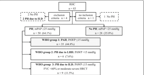
Three different scenarios were conducted for the com-parison of the DETECT algorithm with the ESC/ERS 2009 guidelines: First, 35 patients diagnosed with PAH and 28 with no PH after undergoing RHC were selected for the application of the DETECT algorithm (see Fig. 1). Second, these 63 patients were used to explore the cap-acity of both screening tests to identify mPAP ≥21 mmHg (both borderline mPAP and PAH). Third, with neither inclusion nor exclusion criteria 52 patients diagnosed with PH and 31 with no PH were selected to investigate the detection of any sort of PH.
Data collection
Data from the first available RHC of each patient were revised. PH was defined as mPAP ≥25 mmHg in RHC, and was classified in the following groups: WHO group 1 or PAH was defined if pulmonary artery wedge pres-sure (PAWP) was ≤15 mmHg and pulmonary vascular resistance (PVR) was >3 Wood units in RHC [15]; WHO group 2 or PH due to left heart disease, if PAWP was >15 mmHg; WHO group 3 or PH due to interstitial lung disease (ILD) if PAWP was ≤15 mmHg and pre-dicted FVC <60% or if the extent of interstitial disease was identified as moderate-severe on high-resolution computed tomography (HRCT) [12]. Borderline mPAP (BoPAP) was considered if mPAP was between 21 and 24 mmHg, with PAWP≤15 mmHg and if there was no ILD or the FVC was >60% [16].
thickening of the skin was distal and proximal to the elbows or knees, limited cutaneous SSc (lcSSc) if the affected skin did not exceed this limit proximally and sine scleroderma systemic sclerosis (ssSSc) if no skin thickening was present [17]. The disease onset was described as the date of the first symptom attributable to SSc, including Raynaud’s phenomenon (RP), although the time from the first symptom excluding RP also was analysed. All data were collected at the time of RHC.
The presence of telangiectasias, past or current digital ulcers (DU) and past history of scleroderma renal crisis (SRC) was recorded. ILD was defined as radiological evi-dence of interstitial disease on HRCT. Cardiac involve-ment was defined as current or past history of pericardial effusion, left ventricular ejection fraction (LVEF) <50% and ischaemic heart disease with no cardiovascular risk factor or conduction abnormalities.
Antinuclear antibodies (ANA) were evaluated by indir-ect immunofluorescence (IIF) assay using HEp-cell line 2. Anticentromere antibodies (ACA) were described by IIF or commercial line blot assay to centromere proteins A and B (EUROLINE Systemic Sclerosis (Nucleoli) Pro-file (IgG), Euroimmun, Germany). Anti-topoisomerase I antibodies were determined by enzyme-linked immuno-sorbent assay.
Demographics, cardiovascular risk factors, anthropo-metrical data, complete pulmonary function test (PFT), 6-minute walking distance (6MWD), WHO-functional class, laboratory data including serum urate, N-terminal pro-brain natriuretic peptide (NT-proBNP) and right axis deviation in electrocardiography (ECG) were regis-tered. Echocardiography included the determination of left ventricular ejection fraction (LVEF), right atrium (RA) area, tricuspid annular plane systolic excursion (TAPSE), tricuspid regurgitation velocity (TRV) and RVSP.
RHC details consisted of mPAP, PAWP, right atrial pressure (RAP), transpulmonary pressure gradient (TPG), cardiac output (CO), pulmonary vascular resist-ance (PVR), systemic vascular resistresist-ance (SVR), mixed venous oxygen saturation (SvO2) measured in pulmon-ary artery and arterial oxygen saturation (SaO2) mea-sured in arterial blood gas analysis.
The application of the DETECT algorithm was con-ducted in two steps through the website PAH risk calcu-lator (http://detect-pah.com) [18], the first step for referring the patient for echocardiography and the sec-ond for carrying out RHC [12]. The indication of RHC following ESC/ERS 2009 guidelines was considered if: (1) TRV was >3.4 m/s; or (2) TRV was >2.8 to≤3.4 m/s,
[image:3.595.59.540.88.338.2]with symptoms (undue dyspnoea or fatigue, chest pain, near syncope or signs of right heart failure); or (3) TRV was ≤2.8 m/s, with symptoms and additional echocar-diographic variables suggestive of PH.
Statistical analysis
Qualitative data were expressed as the mean and stand-ard deviation (SD) after checking the data using the normal distribution test, and non-normally distributed qualitative variables were described as the median and interquartile range (IQR). To assess whether there were statistically significant differences, Student’s t test or the Mann-Whitney U test were used according to the result of the normal distribution test. Categorical variables were analysed by the chi-square test and Fisher’s exact test. The 95% confidence intervals (CIs) were calculated for sensitivity, specificity, positive pre-dictive value (PPV) and negative prepre-dictive value (NPV). Apvalue <0.05 was considered significant. Stat-istical analysis was conducted using SPSS 20.0 for Win-dows (SPSS Inc., Chicago, IL, USA).
Results
Baseline characteristics
Comparing the indications for RHC in 83 patients, the proportions of patients diagnosed with PH were 24/35 patients (68.5%) with RVSP >36 mmHg plus FVC/DLCO ratio >1.6, 23/34 patients (67.6%) with RVSP >36 mmHg, 4/8 patients (50.0%) with FVC/DLCO ratio >1.6 and 1/6 patients (16.6%) with progressive unexplained dyspnoea. Two the four patients who were excluded had PH due to ILD. Following the RHC results in the 78 patients who fulfilled the inclusion criteria and did not meet the exclusion criteria, PH was identified in 50 patients (64.1%) and PH was ruled out in the other 28 subjects (35.8%), although 14 of them had BoPAP with mPAP be-tween 21 and 24 mmHg (Fig. 1). According to PH classi-fication, 35 patients (44.8%) were diagnosed with PAH, 6 (7.6%) had PH due to left heart disease and 9 (11.5%) had PH due to ILD. Sixty-three patients with PAH and without PH were selected for DETECT algorithm analysis.
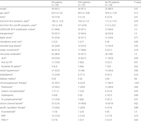
The baseline characteristics of the selected patients with SSc are summarized in Table 1. There were 59 women and 4 men, the mean ± SD age at RHC was 62.4 ± 11.6 years. Based on skin involvement, lcSSc was the most frequent cutaneous subtype in 45 patients (71.4%), 10 patients (15.9%) had dcSSc and 8 patients (12.7%) had ssSSc. The mean time from the first SSc symptom including RP was 18.6 ± 12.3 years, while for the time from first non-RP it was 10.5 ± 8.9 years. Almost the whole study population (n= 61 (96.8%)) fulfilled the ACR/EULAR 2013 classification criteria. There were no statistical differences in the presence of telangiectasias,
past or current DU, history of SRC, ILD or cardiac in-volvement, when comparing the two groups. Although there were no statistical differences in positivity to anti-nuclear antibodies or the observation of a nucleolar pat-tern in the indirect immunofluorescence, patients with PAH more often had ACA (n= 23, 65.7% vs n= 11, 39.3%, p= 0.03), but lower positivity against anti-topoisomerase I antibodies (n= 3, 8.6% vs n= 9, 32.1%,
p= 0.01). Arterial hypertension was the most frequent cardiovascular risk factor in the whole group, being identified in 27 patients (42.9%), followed by dyslipidae-mia in 15 patients (23.8%); however, there were no pa-tients with diabetes mellitus. No statistical differences were identified in the anthropometrical variables. The patients with PAH received less immunosuppressant therapy than the control group, and less treatment with calcium channel blockers. However, there were no statis-tical differences in the use of specific vasodilator ther-apy, for which the indication was RP in all cases.
Regarding respiratory parameters, the PAH group had higher FVC percent predicted/DLCO percent predicted than controls (2.0 ± 0.7% vs 1.5 ± 0.4%, p< 0.01) and shorter 6MWD (232.8 ± 76.7 m vs 322.4 ± 105.0 m,
p= 0.01) (Table 2). Advanced WHO functional classes III/IV were more common in patients with PAH (n= 15 (42.9%)), than in the group with no PH (n= 2 (7.1%)) (p< 0.01). There were higher NT-proBNP and serum urate levels and higher prevalence of right axis devi-ation on ECG in the PAH group. In reldevi-ation to echocardio-graphic data, patients with PAH had a greater RA area; moreover, they presented with higher TRV and RVSP than patients with no PH. Therefore, there were statistically sig-nificant differences between PAH and without PH patients in seven out of eight variables considered in DETECT algorithm.
RHC in the PAH group was characterized by superior mean PAP (Table 3). Specifically, the median (IQR) of mPAP was 42.0 (33.0−50.0) mmHg in patients with PAH, while it was 20.5 (17.0 to 23.0) mmHg in the group with no PH (p< 0.001). Furthermore, TPG was in-creased in patients with PAH, but CO was lower in those patients. The PAH group had higher pulmonary vascular resistance, with lower SvO2 and SaO2 during the RHC, compared with the group with no PH.
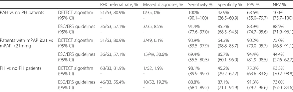
Comparisons of the DETECT algorithm and the ESC/ERS 2009 guidelines
subjects (57.1%) in patients with no PH (p< 0.001). However, based on the ESC/ERS 2009 guidelines, 32 patients with PAH (91.4%) and 4 patients in the group without PH (14.3%) would have been referred for RHC. The RHC referral ratio was higher using DETECT than it was using the ESC/ERS guidelines. The main reasons for not referring these patients for RHC using the ESC/ERS 2009 guidelines would have been that 12 patients had TRV >2.8 to ≤3.4 m/s but with no symptoms, 5 patients presented with TRV ≤2.8 m/s with symptoms but with no additional echo-cardiographic signs of PH, and another 10 patients had TRV ≤2.8 m/s with no symptoms and no add-itional echocardiographic signs of PH. There would
[image:5.595.60.542.98.513.2]have been no missed diagnosis of PAH with DETECT, but using the ESC/ERS guidelines it would have reached 8.5% (Table 5). The sensitivity (95% CI) of the DETECT algorithm was 100% (90.1–100) com-pared to sensitivity of 91.4% (77.6–97.0) using the ESC/ERS 2009 guidelines. The specificity was lower with DETECT at 42.9% (26.5–60.9) whereas it was 85.7% (68.2–94.3) using the ESC/ERS 2009 guidelines. No statistical differences were found in PPV, which was 68.6% (55.0–79.7) using DETECT compared to 88.9% (74.7–95.6) using the ESC/ERS 2009 guidelines. However, NPV reached 100% (75.7–100) using the DETECT algorithm, while using the ESC/ERS 2009 guidelines it was 88.9% (71.9–96.1).
Table 1Demographic data and baseline characteristics
All patients (n= 63)
PAH patients (n= 35)
No PH patients
(n= 28) P
value
Femalea 59 (93.7) 34 (97.1) 25 (89.3) 0.31
Age, yearsb 62.4 (±11.6) 64.4 (± 10.8) 59.9 (± 12.4) 0.13
DcSSca 10 (15.9) 4 (11.4) 6 (21.4) 0.31
Time from first symptom, yearsb 18.6 (± 12.3) 19.6 (±11.2) 17.2 (± 13.7) 0.29
Time from first non-RP symptom, yearsb 10.5 (± 8.9) 9.7 (±9.4) 11.5 (± 8.2) 0.21
ACR/EULAR 2013 classification criteriaa 61 (96.8) 34 (97.1) 27 (96.4) 1.0
Telangiectasiasa 59 (93.7) 33 (94.3) 26 (92.9) 1.0
Digital ulcersa 35 (55.6) 20 (57.1) 15 (53.6) 0.77
Scleroderma renal crisisa 2 (3.2) 2 (5.7) 0 (0) 0.49
Interstitial lung diseasea 32 (50.8) 19 (54.3) 12 (42.9) 0.45
Cardiac involvementa 26 (41.3) 17 (48.6) 9 (32.1) 0.18
Antinuclear antibodiesa 62 (98.4) 34 (97.1) 28 (100) 1.0
ACAa 34 (54.0) 23 (65.7) 11 (39.3) 0.03
Anti-Scl-70a 12 (19.0) 3 (8.6) 9 (32.1) 0.01
Nucleolar IIF patterna 4 (6.3) 3 (8.6) 1 (3.6) 0.62
Arterial hypertensiona 27 (42.9) 14 (40) 13 (43.4) 0.60
Dyslipidaemiaa 15 (23.8) 6 (17.1) 9 (32.1) 0.16
Diabetes mellitusa 0 (0) 0 (0) 0 (0) NA
Immunosuppressant therapya 25 (39.7) 8 (22.9) 17 (60.7) <0.01
Prednisonea 19 (30.2) 7 (20.0) 12 (40.9) 0.04
Sodium mycophenolatea 7 (11.1) 1 (2.9) 6 (21.4) 0.03
Azathioprine 3 (4.8) 0 (0) 3 (10.7) 0.08
IV cyclophosphamidea 12 (19.0) 3 (8.6) 9 (32.1) 0.01
Calcium channel blockera 33 (52.4) 14 (40.0) 19 (67.9) 0.02
Specific vasodilator therapya 13 (20.6) 7 (20.0) 6 (21.4) 0.88
IV prostanoidsa 1 (1.6) 1 (2.9) 0 (0) 1.0
ERAa 10 (15.9) 5 (14.3) 5 (17.9) 0.74
PDE5 Ia 5 (7.9) 2 (5.7) 3 (10.7) 0.64
PAHpulmonary arterial hypertension,PHpulmonary hypertension,dcSScdiffuse cutaneous systemic sclerosis,ACR/EULARAmerican College of Rheumatology/ European League Against Rheumatism,RPRaynaud’s phenomenon,ACAanticentromere antibodies,IIFindirect immunofluorescence,NAnot applicable,IV intravenous,ERAendothelin receptor antagonist,PDE5 Iphosphodiesterase type-5 inhibitor.a
Data are shown as number (%) for categorical variables.b
As a strategy to identify mPAP ≥21 mmHg (both BoPAP and PAH), the DETECT algorithm had superior sensitivity of 93.9% (83.5–97.9) and NPV of 75.0% (46.8–91.1) compared to ESC/ERS guidelines for which the sensitivity and NPV was 69.4% (55.5–80.5) and 44.4% (27.6–62.7), respectively. Furthermore, DETECT missed 6.1% of diagnoses of mPAP ≥21, whereas using the ESC/ERS 2009 guidelines the percentage was in-creased to 30.6%. In fact, analysing only patients with BoPAP, there were 3 out of 14 (21.1%) missed diagnoses using DETECT, whereas there were 12 out of 14 (85.7%)
missed using the ESC/ERS 2009 guidelines. However, specificity and PPV were slightly lower using the DETECT algorithm, at 64.3% and 90.2% compared to 85.7% and 94.4%, respectively, using the ESC/ERS guidelines.
[image:6.595.57.541.99.346.2]To investigate the capacity for the detection of any type of PH, the whole cohort of patients (n= 83) with a first RHC were studied, regardless of whether they met the inclusion or exclusion criteria for application of the DETECT algorithm: 1.9% of diagnoses of PH were missed using the DETECT algorithm compared with Table 2Respiratory, laboratory and echocardiography data
All patients (n= 63)
PAH patients (n= 35)
No PH patients
(n= 28) P
value
Pulmonary function tests
FVC, % predictedb 75.2 (± 16.4) 75.6 (± 15.5) 74.7 (± 17.8) 0.86
DLCO, % predictedb 46.3 (± 14.2) 41.5 (± 13.9) 51.0 (± 13.1) 0.01
FVC%/DLCO%b 1.8 (± 0.6) 2.0 (± 0.7) 1.5 (± 0.4) <0.01
6MWD, mb 256.5 (± 92.5) 232.8 (± 76.7) 322.4 (± 105.0) 0.01
WHO functional class III/IVa 17 (27.0) 15 (42.9) 2 (7.1) <0.01
Laboratory
NT-proBNP, pg/mLc 198 (41 to 647) 1271 (580 to 3154) 87 (32 to 202) <0.001
Serum urate, mg/dLb 5.6 (± 1.9) 6.8 (± 1.9) 4.6 (± 1.0) <0.001
Right axis in ECGa 18 (28.6) 10 (28.6) 1 (3.6) 0.01
Echocardiography
LVEF, %c 60.0 (59.0 to 65.0) 60.0 (58.5 to 65.0) 62.0 (59.7 to 65.0) 0.43
TAPSE, mmb 19.4 (± 3.9) 19.1 (± 4.6) 19.8 (± 3.0) 0.55
RA area, cm2c 16.0 (13.0 to 21.0) 17.0 (15.0 to 22.7) 14.0 (12.0 to 16.0) <0.01
TRV, m/sb 3.5 (± 0.7) 4.0 (± 0.6) 2.8 (± 0.3) <0.001
RVSP, mmHgc 59.5 (44.0 to 76.2) 73.0 (61.0 to 85.0) 44.0 (37.0 to 51.0) <0.001
PAHpulmonary arterial hypertension,PHpulmonary hypertension,FVCforced vital capacity,DLCOdiffusing capacity for carbon monoxide,6MWD6-minute walking distance,WHOWorld Health Organization,NT-proBNPN-terminal pro-brain natriuretic peptide,ECGelectrocardiography,LVEFleft ventricular ejection fraction,TAPSEtricuspid annular plane systolic excursion,RAright atrium,TRVtricuspid regurgitant velocity,RVSPright ventricular systolic pressure.a
Data are shown as number (%) for categorical variables.b
Data are shown as mean (± SD).c
Data are shown as median (IQR)
Table 3Haemodynamic characteristics on right heart catheterization
All patients (n= 63)
PAH patients (n= 35)
No PH patients (n= 28)
Pvalue
Mean PAP, mmHgb 31.0 (21.0 to 44.0) 42.0 (33.0 to 50.0) 20.5 (17.0 to 23.0) <0.001
PAWP, mmHga 9.6 (± 4.0) 9.7 (± 4.8) 9.5 (± 3.4) 0.90
RAP, mmHgb 5.0 (2.5 to 8.5) 5.0 (3.0 to 10.0) 4.5 (2.0 to 7.0) 0.14
TPG, mmHga 20.0 (± 13.4) 31.4 (± 11.8) 10.6 (± 3.9) <0.001
Cardiac output, L/minb 3.5 (2.9 to 4.5) 3.4 (2.8 to 4.0) 3.9 (2.9 to 5.1) 0.05
PVR, WUa 6.6 (± 4.8) 9.9 (± 4.6) 3.2 (± 1.6) <0.001
SVR, WUa 26.9 (± 10.3) 28.6 (± 9.7) 25.2 (± 11.0) 0.42
SvO2, %a 66.3 (± 8.1) 61.8 (± 8.3) 71.0 (± 4,7) <0.001
SaO2, %a 93.5 (± 4.0) 91.2 (± 3.7) 96.1 (± 2.5) <0.001
[image:6.595.56.542.558.707.2]19.2% missed using the ESC/ERS 2009 guidelines. More-over, sensitivity and NPV were greater at 98.1% and 93.3% using the DETECT method, compared with 80.8% and 73.0%, respectively, using the ESC/ERS guidelines. Nevertheless, specificity and PPV were again slightly in-ferior at 45.2% and 75.0% when applying the DETECT algorithm, whereas the values were 87.1% and 91.3%, respectively, using in the ESC/ERS guidelines.
Discussion
The present retrospective study compared the applica-tion of the DETECT algorithm with the ESC/ERS 2009 guidelines in patients with PAH and patients without PH, who had undergone RHC. The DETECT programme had greater sensitivity, the RHC would have been recommended in all patients suffering from PAH, and also superior NPV compared to the ESC/ERS guide-lines. Nevertheless, the ESC/ERS 2009 guidelines had higher specificity and PPV than the DETECT algorithm. Furthermore, the DETECT algorithm had higher per-centage sensitivity and NPV both for the detection of
patients with mPAP ≥21 and for patients with any type of PH, although specificity and PPV were lower.
[image:7.595.57.542.99.242.2]For the application of the DETECT algorithm it is ne-cessary to meet the inclusion criteria, so patients must present with a duration of SSc longer than 3 years from the first non-RP symptom and DLCO <60% predicted. This was to increase the specificity of the screening method, due to the fact that <10% of a cohort of 243 pa-tients with SSc-PH have been found to present with DLCO >60% [19]. Furthermore, patients could not fulfil the exclusion criteria such as previous PH diagnosed by RHC, forced vital capacity (FVC) <40% or the involve-ment of left heart disease [12]. Nevertheless, DETECT does not establish the periodicity of an assessment by this method, it has not been evaluated over a monitoring period in a cohort of patients and its ability to predict mPAP ≥21 mmHg or any type of PH have not been ex-plored. As a consequence of this work, the new ESC/ ESR 2015 guidelines were recently published, which strengthen the recommendation for annual screening of PH in this set of patients with SSc and introduce the Table 4Comparisons of the DETECT algorithm and the ESC/ERS 2009 guidelines
PAH vs no PH patients (n= 63)
Patients with mPAP≥21 vs mPAP <21mmg (n= 63)
PH vs no PH patients (n= 83)
PAH (n= 35)
No PH (n= 28)
Pvalue mPAP≥21 mmHg (n= 49)
mPAP <21mmg (n= 14)
Pvalue PH (n= 52)
No PH (n= 31)
Pvalue
DETECT algorithm
Score step 1b 330.0 (±25.3) 313.7 (±17.5) <0.01 326.9 (±22.8) 308.6 (±20.8) <0.01 327.9 (±23.6) 311.8 (±17.6) <0.01
Indication of Echoa 35 (100) 24 (85.7) 0.03 49 (100) 10 (71.4) <0.01 51 (98.1) 24 (77.4) <0.01
Score step 2b 60.2 (±11.3) 37.3 (±8.4) <0.001 54.7 (±13.3) 33.5 (±8.8) <0.001 56.4 (±12.4) 36.5 (±8.4) <0.001
RHC recommendationa 35 (100) 16 (57.1) <0.001 46 (93.9) 5 (35.7) <0.001 51 (98.1) 17 (54.8) <0.001
ESC/ERS 2009 guidelines
RHC recommendationa 32 (91.4) 4 (14.3) <0.001 34 (69.4) 2 (14.3) <0.001 42 (80.8) 4 (12.9) <0.001
PAHpulmonary arterial hypertension,PHpulmonary hypertension,mPAPmean pulmonary artery pressure,Echoechocardiography,RHCright heart catheterization, ESC/ERSEuropean Society of Cardiology and the European Respiratory Society.a
Data are shown as number (%) for categorical variables.b
Data are shown as mean (± SD)
Table 5Observation of PAH detection programs
RHC referral rate, % Missed diagnoses, % Sensitivity % Specificity % PPV % NPV %
PAH vs no PH patients DETECT algorithm (95% CI) 51/63, 80.9% -0/35, 0% -100% (90.1–100)
42.9% (26.5–60.9)
68.6% (55.0–79.7)
100% (75.7–100)
ESC/ERS guidelines (95% CI) 36/63, 57.1% -3/35, 8.5% -91.4% (77.6–97.0)
85.7% (68.5–94.3)
88.9% (74.7–95.6)
88.9% (71.9–96.1)
Patients with mPAP≥21 vs mPAP <21mmg DETECT algorithm (95% CI) 51/63, 80.9% -3/49, 6.1% -93.9% (83.5–97.9)
64.3% (38.8–83.7)
90.2% (79.0–95.7)
75.0% (46.8–91.1)
ESC/ERS guidelines (95% CI) 36/63, 57.1% -15/49, 30.6% -69.4% (55.5–80.5)
85.7% (60.1–96.0)
94.4% (81.9–98.5)
44.4% (27.6–62.7)
PH vs no PH patients DETECT algorithm (95% CI) 68/83, 81.9% -1/52, 1.9% -98.1% (89.9–99.7)
45.2% (29.2–62.2)
75.0% (63.6–83.8)
93.3% (70.2–98.8)
ESC/ERS guidelines (95% CI) 46/83, 55.4% -10/52, 19.2% -80.8% (68.1–89.2)
87.1% (71.1–94.9)
91.3% (79.7–96.6)
73.0% (57.0–84.6)
[image:7.595.57.538.554.706.2]DETECT algorithm as a valid method to be performed in this group of patients based on the evidence demon-strated [15].
Compared to the study of Coghlan et al., patients in our cohort had a surprisingly higher prevalence of telan-giectasias and ACA antibodies. Almost 60% of patients with PAH presented with WHO functional class I or II and mild reduced 6MWD comparable to the detection cohorts of previous studies that have focused on early PAH diagnosis [10, 12]. This fact seems to reflect that patients in our study were referred promptly for RHC as a result of active and early suspicion of PAH during monitoring. In the present paper we describe similar sensitivity, specificity and NPV values, although with a higher RHC referral ratio and PPV using the DETECT algorithm than in the original work. However, regarding the application of ESC/ERS 2009 guidelines we report higher sensitivity, specificity and PPV values and a higher RHC referral ratio than Coghlan et al. This may be explained due to the fact that the main indication for performing RHC in our study was the presence of an ab-normal echocardiographic result, suggesting the pres-ence of PAH. Furthermore, the high RHC referral ratio may be justified because the study was conducted in a selected population of patients at high risk of PAH based on clinical, pulmonary function test and echocardio-graphic parameters.
Few groups have published on the application of the DETECT algorithm. Hao et al. analysed the DETECT al-gorithm in patients from the multicentre Australian Scleroderma Cohort Study (ASCS) along with the Australian Scleroderma Interest Group (ASIG) algorithm and the ESC/ERS 2009 guidelines [20, 21]. The authors found higher sensitivity and NPV using the DETECT or the ASIG algorithm than they did using the ESC/ERS guidelines, which we can confirm in our study. Surprisingly, the ASIG protocol had superior specificity to the DETECT algorithm and even to the ESC/ERS guidelines, which contrasts with the values of 48% and 69%, respectively, that were previously published [12]. The authors reported that nine patients did not meet the criterion of DLCO <60% or the criterion for disease duration, and this may explain the lower specificity of the DETECT algorithm [21]. Furthermore, the DETECT and ASIG screening methods had the highest sensitivity and specificity to identify both precapillary PH and any group of PH, results that we have corroborated in our work. In the present work the ASIG programme was not analysed due to the fact that this method is not based on evidence and has not been well-validated in European cohorts.
Recently, a Czech scleroderma group designed a modi-fied DETECT algorithm, in which a right ventricular outflow tract is evaluated instead of RA area [22]. Using
this modified DETECT programme the RHC referral ra-tio was 41.4% compared to 24.1% using the ESC/ERS guidelines. Interestingly, although only 11/58 patients (18.9%) had undergone RHC, in patients who were rec-ommended by the modified DETECT algorithm there were also other types of PH diagnosed, such as PH due to left heart disease and PH due to ILD. Therefore, this article suggests that the DETECT algorithm would be useful for recommending RHC for diagnosing other types of PH according to the WHO classification.
Although there are some discrepancies on the prog-nostic role of BoPAP, a study has been recently pub-lished evidencing a higher risk of mortality and hospitalization in US veteran patients with BoPAP (defined as 19–24 mmHg) [23]. Consequently, the study highlights the relevance of diagnosing BoPAP in selected groups of patients such as those with sclero-derma. In a post-hoc analysis of the DETECT study, the authors demonstrated that patients with BoPAP had a higher FVC/DLCO rate, and higher TRV and NT-proBNP levels than in patients with normal mPAP [16]. Although the paper does not evaluate the application of DETECT in both groups of patients, it would support the increment of the specificity of this programme identified in our work when studying patients with mPAP ≥21 versus mPAP <21mmg, compared to the PAH versus no PH strategy. In the set of patients with SSc, identifying BoPAP in the first RHC was demon-strated as a high risk factor for developing PH, with a hazard ratio of 3.7 during follow up compared to pa-tients with mPAP <21 mmHg [24]. In this line, in a multicentre prospective SSc cohort, the Pulmonary Hypertension Assessment and Recognition of Out-comes in Scleroderma (PHAROS) study group reported that 55% of patients with BoPAP progressed to PH, whereas in the group of patients with mPAP <21 mmHg this was 32%, over a mean 25.7-month period of moni-toring [25]. Having established the relevance of identi-fying patients with SSc and BoPAP for close supervision, this is the first paper that demonstrates the accuracy of the DETECT algorithm in the identification of patients with mPAP≥21.
The main limitation of the present study was the retrospective design. RHC was not performed in the whole cohort of patients with SSc, therefore some mild forms of PAH may have been undiagnosed. In the same line, although the periodicity of performing the screen-ing methods is not well established, these were not carried out in the whole cohort, which may not reflect the real referral rate for RHC.
Conclusions
diagnosis has demonstrated increased survival rates. The present study supports the DETECT algorithm as an ex-cellent screening method due to its high sensitivity and NPV, which minimizes the number of missed diagnoses of previous recommendations. Furthermore, our data suggest the DETECT algorithm is also accurate either for an early diagnosis of borderline mPAP, patients who are at highest risk of developing PAH, or the diagnosis of any type of PH.
Abbreviations
6MWD:6-Minute walking distance; ACA: Anticentromere antibodies; ACR: American College of Rheumatology; ANA: Antinuclear antibodies; ASCS: Australian Scleroderma Cohort Study; ASIG: Australian Scleroderma Interest Group; BoPAP: Borderline median pulmonary arterial pressure; CI: Confidence interval; CO: Cardiac output; dcSSc: Diffuse cutaneous systemic sclerosis; DLCO: Diffusing capacity for carbon monoxide; DU: Digital ulcers; ECG: Electrocardiography; ERA: Endothelin receptor antagonist; ESC/ ERS: European Society of Cardiology and the European Respiratory Society; EULAR: European League Against Rheumatism; FVC: Forced vital capacity; HR: Hazard ratio; HRCT: High-resolution computed tomography; IIF: Indirect immunofluorescence; ILD: Interstitial lung disease; IQR: Interquartile range; IV: Intravenous; lcSSc: limited cutaneous systemic sclerosis; LHD: Left heart disease; LVEF: Left ventricular ejection fraction; mPAP: Median pulmonary arterial pressure; NPV: Negative predictive value; NT-proBNP: N-terminal pro-brain natriuretic peptide; PAH: Pulmonary arterial hypertension; PAWP: Pulmonary artery wedge pressure; PDE5 I: Phosphodiesterase type-5 inhibitor; PFT: Pulmonary function test; PH: Pulmonary hypertension; PHAROS: Pulmonary Hypertension Assessment and Recognition of Outcomes in Scleroderma; PPV: Positive predictive value; PVR: Pulmonary vascular resistance; RA: Right atrium; RAP: Right atrial pressure; RHC: Right heart catheterization; RP: Raynaud’s phenomenon; RVSP: Right ventricular systolic pressure; SaO2: Arterial oxygen saturation; SD: Standard deviation; SRC: Scleroderma renal crisis; SSc: Systemic sclerosis; ssSSc: Sine scleroderma systemic sclerosis; SvO2: Mixed venous oxygen saturation; SVR: Systemic vascular resistance; TAPSE: Tricuspid annular plane systolic excursion; TPG: Transpulmonary pressure gradient; TRV: Tricuspid regurgitant velocity; vs: Versus; WHO: World Health Organization; WU: Wood units
Acknowledgements
The authors thank the patients for their participation.
Funding
This work was funded by Instituto de Salud Carlos III, grant PI16/02088 co-financed by the European Regional Development Fund (ERDF). This work was supported by an educational unrestricted scholarship granted by Actelion Pharmaceuticals. AGDC was supported by the Contratos Predoctorales de Formación en Investigación (PFIS) grant from Instituto de Salud Carlos III (FI14/00643).
Availability of data and materials
The datasets analysed during the current study are available from the corresponding author on reasonable request.
Authors’contributions
AGDC made substantial contributions to study conception and design, acquisition, analysis and interpretation of data, drafting the article and approval of the final manuscript. ELCM and GG made substantial
contributions to study conception and design, analysis and interpretation of data, revising the manuscript critically for important intellectual content and approval of the final manuscript. JFRP, AR, CB, MLM, ED and VFP made substantial contributions to study conception and design, revising the manuscript critically for important intellectual content and approval of the final manuscript. CPSA made substantial contributions to study conception and design, interpretation of data, revising the manuscript critically for important intellectual content and approval of the final manuscript. AGDC and ELCM had full access to all of the data in the study and they both take responsibility for the integrity and accuracy of the data. Funders had no access to the data, were not involved in study design, acquisition, analysis or
interpretation of data or writing the manuscript. All authors read and approved the final manuscript.
Competing interests
The authors declare that they have no competing interests.
Consent for publication Not applicable.
Ethics approval and consent to participate
This study was approved by the hospital Ethics Committee for Clinical Research and all subjects consented to use of their clinical and laboratory data.
Publisher’s Note
Springer Nature remains neutral with regard to jurisdictional claims in published maps and institutional affiliations.
Author details 1
Department of Systemic Autoimmune Diseases, Hospital Universitari Vall d’Hebron, Universitat Autònoma de Barcelona, Passeig Vall d’Hebron 119-129, PC: 08035 Barcelona, Spain.2Department of Cardiology, Hospital Universitari Vall d’Hebron, Universitat Autònoma de Barcelona, Barcelona, Spain. 3
Department of Pneumology, Hospital Universitari Vall d’Hebron, Universitat Autònoma de Barcelona, Barcelona, Spain.
Received: 25 January 2017 Accepted: 11 May 2017
References
1. Denton CP, Ong VH. Targeted therapies for systemic sclerosis. Nat Rev Rheumatol. 2013;9:451–64.
2. Allanore Y, Simms R, Distler O, Trojanowska M, Pope J, Denton CP, et al. Systemic sclerosis. Nat Rev Dis Primers. 2015;1:15002.
3. Mukerjee D, St George D, Coleiro B, Knight C, Denton CP, Davar J, et al. Prevalence and outcome in systemic sclerosis associated pulmonary arterial hypertension: application of a registry approach. Ann Rheum Dis. 2003;62: 1088–93.
4. Avouac J, Airo P, Meune C, Beretta L, Dieude P, Caramaschi P, et al. Prevalence of pulmonary hypertension in systemic sclerosis in European Caucasians and meta analysis of 5 studies. J Rheumatol. 2010;37:2290–8. 5. Steen VD, Medsger TA. Changes in causes of death in systemic sclerosis,
1972-2002. Ann Rheum Dis. 2007;66:940–4.
6. Nihtyanova SI, Schreiber BE, Ong VH, Rosenberg D, Moinzadeh P, Coghlan JG, et al. Prediction of pulmonary complications and long-term survival in systemic sclerosis. Arthritis Rheumatol. 2014;66:1625–35.
7. Simeon-Aznar CP, Fonollosa-Pla V, Tolosa-Vilella C, Espinosa-Garriga G, Campillo-Grau M, Ramos-Casals M, et al. Registry of the Spanish Network for Systemic Sclerosis: survival, prognostic factors, and causes of death. Medicine (Baltimore). 2015;94:e1728.
8. Rubenfire M, Huffman MD, Krishnan S, Seibold JR, Schiopu E, McLaughlin VV. Survival in systemic sclerosis with pulmonary arterial hypertension has not improved in the modern era. Chest. 2013;144:1282–90.
9. Hachulla E, Gressin V, Guillevin L, Carpentier P, Diot E, Sibilia J, et al. Early detection of pulmonary arterial hypertension in systemic sclerosis: a French nationwide prospective multicenter study. Arthritis Rheum. 2005; 52:3792–800.
10. Humbert M, Yaici A, de Groote P, Montani D, Sitbon O, Launay D, et al. Screening for pulmonary arterial hypertension in patients with systemic sclerosis: clinical characteristics at diagnosis and long-term survival. Arthritis Rheum. 2011;63:3522–30.
11. Galie N, Hoeper MM, Humbert M, Torbicki A, Vachiery JL, Barbera JA, et al. Guidelines for the diagnosis and treatment of pulmonary hypertension: the Task Force for the Diagnosis and Treatment of Pulmonary Hypertension of the European Society of Cardiology (ESC) and the European Respiratory Society (ERS), endorsed by the International Society of Heart and Lung Transplantation (ISHLT). Eur Heart J. 2009;30:2493–537.
13. LeRoy EC, Black C, Fleischmajer R, Jablonska S, Krieg T, Medsger Jr TA, et al. Scleroderma (systemic sclerosis): classification, subsets and pathogenesis. J Rheumatol. 1988;15:202–5.
14. van den Hoogen F, Khanna D, Fransen J, Johnson SR, Baron M, Tyndall A, et al. 2013 classification criteria for systemic sclerosis: an American College of Rheumatology/European League against Rheumatism collaborative initiative. Arthritis Rheum. 2013;65:2737–47.
15. Galie N, Humbert M, Vachiery JL, Gibbs S, Lang I, Torbicki A, et al. 2015 ESC/ ERS Guidelines for the diagnosis and treatment of pulmonary hypertension: The Joint Task Force for the Diagnosis and Treatment of Pulmonary Hypertension of the European Society of Cardiology (ESC) and the European Respiratory Society (ERS): endorsed by: Association for European Paediatric and Congenital Cardiology (AEPC), International Society for Heart and Lung Transplantation (ISHLT). Eur Heart J. 2016;37:67–119.
16. Visovatti SH, Distler O, Coghlan JG, Denton CP, Grunig E, Bonderman D, et al. Borderline pulmonary arterial pressure in systemic sclerosis patients: a post-hoc analysis of the DETECT study. Arthritis Res Ther. 2014;16:493. 17. Simeon-Aznar CP, Tolosa-Vilella C, Gabarro-Julia L, Campillo-Grau M, Guillen
Del Castillo A, Fonollosa-Pla V, et al. Systemic sclerosis sine scleroderma and limited cutaneous systemic sclerosis: similarities and differences. Clin Exp Rheumatol. 2014;32:S-33–40.
18. DETECT - DETECTion of PAH in SSC. http://detect-pah.com. Accessed 25 Apr 2016. 19. Schwaiger JP, Khanna D, Gerry Coghlan J. Screening patients with
scleroderma for pulmonary arterial hypertension and implications for other at-risk populations. Eur Respir Rev. 2013;22:515–25.
20. Thakkar V, Stevens W, Prior D, Youssef P, Liew D, Gabbay E, et al. The inclusion of N-terminal pro-brain natriuretic peptide in a sensitive screening strategy for systemic sclerosis-related pulmonary arterial hypertension: a cohort study. Arthritis Res Ther. 2013;15:R193.
21. Hao Y, Thakkar V, Stevens W, Morrisroe K, Prior D, Rabusa C, et al. A comparison of the predictive accuracy of three screening models for pulmonary arterial hypertension in systemic sclerosis. Arthritis Res Ther. 2015;17:7.
22. Soukup T, Pudil R, Kubinova K, Hromadkova L, Dusek J, Tosovsky M, et al. Application of the DETECT algorithm for detection of risk of pulmonary arterial hypertension in systemic sclerosis: data from a Czech tertiary centre. Rheumatology (Oxford). 2016;55:109–14.
23. Maron BA, Hess E, Maddox TM, Opotowsky AR, Tedford RJ, Lahm T, et al. Association of borderline pulmonary hypertension with mortality and hospitalization in a large patient cohort: insights from the Veterans Affairs Clinical Assessment, Reporting, and Tracking Program. Circulation. 2016; 133:1240–8.
24. Valerio CJ, Schreiber BE, Handler CE, Denton CP, Coghlan JG. Borderline mean pulmonary artery pressure in patients with systemic sclerosis: transpulmonary gradient predicts risk of developing pulmonary hypertension. Arthritis Rheum. 2013;65:1074–84.
25. Bae S, Saggar R, Bolster MB, Chung L, Csuka ME, Derk C, et al. Baseline characteristics and follow-up in patients with normal haemodynamics versus borderline mean pulmonary arterial pressure in systemic sclerosis: results from the PHAROS registry. Ann Rheum Dis. 2012;71:1335–42.
• We accept pre-submission inquiries
• Our selector tool helps you to find the most relevant journal • We provide round the clock customer support
• Convenient online submission • Thorough peer review
• Inclusion in PubMed and all major indexing services • Maximum visibility for your research
Submit your manuscript at www.biomedcentral.com/submit