Identification and preliminary characterization of novel
B3-type metallo-
β
-lactamases
Manfredi Miraula1,2
, Conor S. Brunton1, Gerhard Schenk2, Nataša Mitić1
1
Department of Chemistry, National University of Ireland—Maynooth, Maynooth, Co., Kildare, Ireland
2
School of Chemistry and Molecular Biosciences, The University of Queensland, Brisbane, Australia Email: [email protected], [email protected]
Received 30 May 2013; revised 23 June 2013; accepted 9 July 2013
Copyright © 2013 Manfredi Miraula et al. This is an open access article distributed under the Creative Commons Attribution License, which permits unrestricted use, distribution, and reproduction in any medium, provided the original work is properly cited.
ABSTRACT
Antibiotic resistance has emerged as a major global threat to human health. Among the strategies em- ployed by pathogens to acquire resistance the use of metallo-β-lactamases (MBLs), a family of dinuclear metalloenzymes, is among the most potent. MBLs are subdivided into three groups (i.e. B1, B2 and B3) with most of the virulence factors belonging to the B1 group. The recent discovery of AIM-1, a B3-type MBL, however, has illustrated the potential health threat of this group of MBLs. Here, we employed a bioinformatics approach to identify and characterize novel B3-type MBLs from Novosphingobium pentaro-
mativorans and Simiduia agarivorans. These enzymes may not yet pose a direct risk to human health, but their structures and function may provide important insight into the design and synthesis of a still elusive universal MBL inhibitor.
Keywords:Antibiotic Resistance; β-Lactam Antibiotics; Metallo-β-Lactamases; Sequence Homology;
Novosphingobium Pentaromativorans; Simiduia Agarivorans
1. INTRODUCTION
The introduction of β-lactam antibiotics (Figure 1) in the 1940s has been considered as a breakthrough, if not the most significant breakthrough in the history of medicine. However, only a few years after introducing penicillin resistance was observed in Staphylococcus aureus and meanwhile a large and increasing number of pathogens have acquired resistance to the most commonly used antibiotics [1,2], triggering some experts to liken antibi-otic resistance to terrorism in terms of its global impact.
http://www.bbc.co.uk/news/health-21737844
One of the most frightening forms of antibiotic resis-
(a) (b)
[image:1.595.330.516.285.421.2](c) (d)
Figure 1. Representatives for the most common β- lactam antibiotic families. (a) Penicillin; (b) carba- penem; (c) cephalosporin; and (d) monobactam.
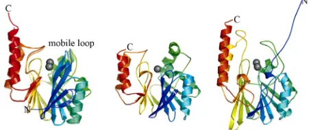
tance occurs through the action of metallo-β-lactamases (MBLs), and enzymes are capable of breaking down most widely used β-lactam antibiotics [1-5]. These Zn2+-dependent enzymes are not susceptible to any known drugs [5]. MBLs have been divided into three subgroups (i.e. B1, B2 and B3) based on their sequences and metal requirements [1,4,5]. Despite only low levels of sequence similarity these subgroups show homology at the level of structure; they all exhibit α/β/β/α folded with two metal binding sites located between the two central β sheets (Figure 2). The key active site residues responsible for binding the metal ions in the three sub-groups show variations that result in differences in metal requirements and catalytic mechanisms [5]. Most of the known MBL virulence factors belong to subgroup B1 and include BCII from Bacillus cereus [6], CcrA from
Bacteroides fragilis [7], as well as IMP-1 and SPM-1,
Figure 2. Representative MBL structures. Left: B1-type CcrA from B. fragilis; center: B2-type CphA from A.
hydrophila; right: B3-type L1 from S. maltophilia (only one subunit shown). The protein main chains are color-ramped from the N-(blue) to the C-terminus (red). Zinc ions are rendered as gray spheres.
mology with B1-type MBLs, hydrolyze exclusively car-bapenems (e.g. meropenem and imipenem) and require only one metal ion for catalysis [5,11]. Representative B2-type MBLs are CphA from Aeromonas hydrophila [12], ImiS from A. veronii [13] and Sfh-1 from Serratia fonticola [14]. Subgroup B3 is closer related to B1-type MBLs rather than B2-type MBLs, requiring two bound metal ions for catalysis (Figure 2). The most studied representative is the tetrameric L1 from Stenotropho-
monas maltophilia [15]. Other members include FEZ-1
from Legionella gormanii [16], GOB-1 from Eliza- bethkingia meningoseptica (of which to date 18 variants have been reported) [17] and SMB-1 from Serratia marcesens [18], the most recently identified MBL, which has a higher hydrolytic activity against a wide range of
β-lactams than other B3-type MBLs [18]. Of clinical re- levance is in particular the enzyme AIM-1 from Pseu-
domonas aeruginosa, which has been identified re-
cently in multi-drug resistant isolating in a hospital in Adelaide, Australia (hence, the nomenclature of “Ade-laide Imipenemase-1”—AIM-1) [19].
Despite rather modest homology across their full length amino acid sequences MBLs share considerable similarities in their active site structures [5]. All MBLs provide binding sites for two closely spaced metal ions that are invariably Zn2+ in vivo (i.e. Zn1 and Zn2). Some variations are observed among the ligands that bind to the zinc ions in the active site. B1- and B3-type MBLs have three histidines (His) on the Zn1 site. In the B2-type MBLs one of these histidines (His116) is replaced by an asparagine (Asn). For B1- and B2-type MBLs the second metal binding site is conserved with an aspartate (Asp), a histidine and a cysteine (Cys) coordinating the metal ion. In the B3 subclass the cysteine is replaced by another histidine (Figure 3). A standard numbering scheme for residues in MBLs has been developed based on sequence alignments for B1, B2 and B3 MBLs, and is used throughout this work to simplify comparisons between different MBLs [4,20,21].
In light of the rapid spread of antibiotic resistance and
Figure 3. Active site structures of representative MBLs. Left: B1-type BcII from B. cereus; center: B2-type CphA from A.
hydrophila; right: B3-type L1 from S. maltophilia. Zinc ions are rendered as grey spheres, and water molecules are shown as red spheres. Coordination bonds are shown as solid lines.
the increasing emergence of novel virulence factors (ex- emplified by NDM-1 and AIM-1). It is essential to iden- tify novel putative MBLs, ideally before they become a threat to health care. Furthermore, novel MBLs may also provide essential insight into the structure and/or func- tional aspects relevant to the design and synthesis of universal inhibitors that may be clinically useful to com- bat antibiotic resistance. Here, our focus was on the B3 subgroup because they appear to be less known than their counterparts in the other two subgroups, but they are a danger to be reckoned as the recent emergence of AIM-1 and SMB-1 illustrated.
2. METHODS
2.1. Selection of the Query Sequence and Protein Database Search Using BLAST
The B3-type AIM-1 from P. aeruginosa was used as query sequence for the protein database search. The pro- tein sequence of AIM-1 was obtained from the Protein Data Bank (PDB; accession code: 4AWY), and the Basic Local Alignment Search Tool (BLAST;
http://blast.ncbi.nlm.nih.gov/Blast.cgi) was used to iden-
tify homologues. The two most promising candidates, from Novosphingobiumpentaromativorans and Simiduia
agarivorans were selected for multiple sequence com-
parisons including known B3-type MBLs.
2.2. Multiple Sequence Alignments
Multiple sequence alignments including the novel B3- type MBLs and well known members of this group of enzymes (i.e. AIM-1 [19], L1 [15] and SMB-1 [18]) were carried out using ClustalW2, a multiple sequence alignment program, available via The European Bioin- formatics Institute website.
http://www.ebi.ac.uk /Tools/msa/clustalw2/
3. RESULTS AND DISCUSSION
3.1. Protein Database Search, Nomenclature and Classification of Novel MBLs
[image:2.595.58.285.88.183.2]query two promising candidate sequences were retrieved, i.e. MBL-like sequences from N.pentaromativorans (ac- cession code: ZP_09194167.1) and S. agarivorans (ac- cession code: YP_006917856.1). These microorganisms are both Gram-negative. N.pentaromativorans is a poly- cyclic aromatic hydrocarbon-degrading bacterium [22], while S. agarivorans is a heterotrophic marine bacterium [23]. None of these organisms poses a direct current threat to human health, however, the observation that they harbor a potential MBL may not only foreshadow a future problem, they also could represent a genetic pool from which horizontal genetic transfer can occur and they could lead to the design and development of univer- sally applicable inhibitors against MBLs. It is thus essen- tial to investigate the properties of these novel MBL-like proteins and compare them with those of well-known MBLs (e.g. AIM-1, L1 or SMB-1).
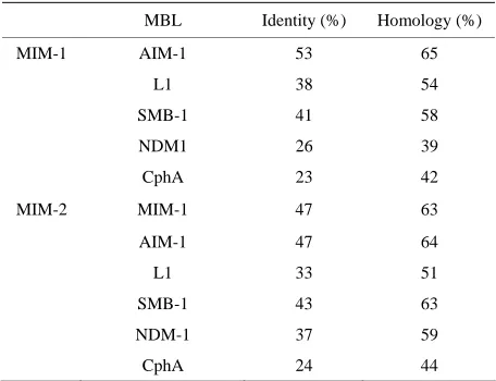
As a step towards their characterization, the sequences of the N. pentaromativorans and S. agarivorans MBL- like proteins were compared with those of well-charac- terized MBLs from the B3 subgroup, i.e. AIM-1 [19], L1 [15] and SMB-1 [18]. The results from pairwise se- quence comparisons are summarized in Table 1. Not sur- prisingly, the two sequences are most closely related to AIM-1. The MBL-like protein from N. pentaromativo- rans shares 53% sequence identity and 65% homology (including conserved amino acid substitutions) with AIM-1. The sequence identity/homology to the other two well characterized B3-type MBLs, L1 and SMB-1, is smaller (38%/54% and 41%/58%, respectively) but still strongly indicative that the N. pentaromativorans protein is indeed a B3-type MBL. In comparison, pairwise se- quence comparisons with the B1-type NDM-1 and B2- type CphA indicate only 26%/39% and 23%/42%, re- spectively. A similar conclusion can be drawn for the MBL-like sequence from S. agarivorans. Its similar- ity/homology with AIM-1 (47%/64%) is less than that of
the N. pentaromativorans MBL, but it appears to be
closer related to SMB-1 instead (Table 1). The two MBL-like proteins share 47%/63% identity/homology in a direct pairwise sequence comparison. In summary, these pairwise comparisons strongly support the classifi- cation of these novel proteins sequences from N. pen- taromativorans and S.agarivorans as MBLs from the B3 subgroup. In accordance with frequently applied no- menclature procedures (e.g. “Adelaide Imipenemase-1” or AIM-1) the MBL-like sequences from N. pentaroma- tivorans and S.agarivorans are labeled here “Maynooth Imipenemase-1” (MIM-1) and “Maynooth Imipenemase- 2” (MIM-2), respectively.
3.2. Important Amino Acid Residues
[image:3.595.309.537.142.317.2]The above discussion demonstrated that the MBL-like sequences in the genomes of N. pentaromativorans and
Table 1. Pairwise sequence comparisons between MBL-like sequences from N. pentaromativorans (MIM-1; see text for details) and S. agarivorans (MIM-2) and selected MBLs from the B1 (NDM-1), B2 (CphA) and B3 (AIM-1, L1, SMB-1) subgroups.
MBL Identity (%) Homology (%)
MIM-1 AIM-1 53 65
L1 38 54
SMB-1 41 58
NDM1 26 39
CphA 23 42
MIM-2 MIM-1 47 63
AIM-1 47 64
L1 33 51
SMB-1 43 63
NDM-1 37 59
CphA 24 44
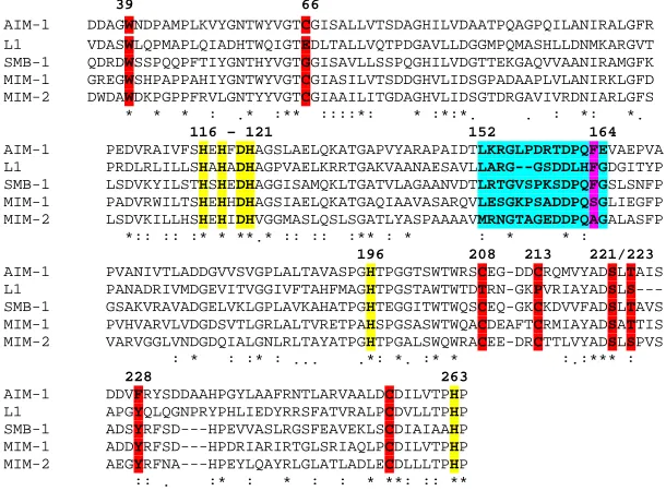
S.agarivorans are likely members of the B3 subgroup in the MBL family. However, in order to substantiate this interpretation it is essential to ascertain that amino acid residues that are essential for MBL function are con- served in the amino acid sequences of MIM-1 and MIM-2. The most relevant amino acid residues with re- spect to MBL function are those that form the metal binding site. Other residues in the proximity of the metal ion binding sites may be important for substrate or in- hibitor binding or both. In order to evaluate the function- ality of MIM-1 and MIM-2 a multiple sequence align- ment between these sequences and the structurally well-characterized AIM-1 [19], L1 [15] and SMB-1 [18] was carried out (Figure 4).
Importantly, the six amino acids that form the metal ion binding site are also invariant in both MIMs (i.e. His116, His118 and His196 in the Zn1 site and Asp120, His121 and His263 in the Zn2 site; see also Figure 3). Other amino acid side chains that were identified as im- portant in MBL function are well conserved, including those in positions 221 (Ser) and 223 (Ser/Thr) that line the pocket where the β-lactam substrate may bind [5]. Tyr228, a residue that aids the polarization of the β-lac- tam carbonyl oxygen as a means to increase the suscep- tibility of the carbonyl bond for a nucleophilic attack by the “bridging” hydroxide is conserved in all MBLs com-pared here except AIM-1 (Figure 4). Conserved is also Trp39, another residue that has been shown to play an important role in substrate binding [24]. Furthermore, in AIM-1 and to a large extent SMB-1 (but not L1) the structure of the enzyme is stabilized by the presence of three disulfide bridges (i.e. the pairs Cys32-Cys66, Cys208-Cys213 and Cys256-Cys290 [18,19]). These six
39 66
AIM-1 DDAGWNDPAMPLKVYGNTWYVGTCGISALLVTSDAGHILVDAATPQAGPQILANIRALGFR
L1 VDASWLQPMAPLQIADHTWQIGTEDLTALLVQTPDGAVLLDGGMPQMASHLLDNMKARGVT
SMB-1 QDRDWSSPQQPFTIYGNTHYVGTGGISAVLLSSPQGHILVDGTTEKGAQVVAANIRAMGFK
MIM-1 GREGWSHPAPPAHIYGNTWYVGTCGIASILVTSDDGHVLIDSGPADAAPLVLANIRKLGFD
MIM-2 DWDAWDKPGPPFRVLGNTYYVGTCGIAAILITGDAGHVLIDSGTDRGAVIVRDNIARLGFS
* * * : .* :** ::::*: * :*:*. . : *: *.
116 – 121 152 164
AIM-1 PEDVRAIVFSHEHFDHAGSLAELQKATGAPVYARAPAIDTLKRGLPDRTDPQFEVAEPVA
L1 PRDLRLILLSHAHADHAGPVAELKRRTGAKVAANAESAVLLARG--GSDDLHFGDGITYP
SMB-1 LSDVKYILSTHSHEDHAGGISAMQKLTGATVLAGAANVDTLRTGVSPKSDPQFGSLSNFP
MIM-1 PADVRWILTSHEHHDHAGSIAELQKATGAQIAAVASARQVLESGKPSADDPQSGLIEGFP
MIM-2 LSDVKILLHSHEHIDHVGGMASLQSLSGATLYASPAAAAVMRNGTAGEDDPQAGALASFP
*:: :: :* * **.* :: :: :** : * : * * :
196 208 213 221/223
AIM-1 PVANIVTLADDGVVSVGPLALTAVASPGHTPGGTSWTWRSCEG-DDCRQMVYADSLTAIS
L1 PANADRIVMDGEVITVGGIVFTAHFMAGHTPGSTAWTWTDTRN-GKPVRIAYADSLS---
SMB-1 GSAKVRAVADGELVKLGPLAVKAHATPGHTEGGITWTWQSCEQ-GKCKDVVFADSLTAVS
MIM-1 PVHVARVLVDGDSVTLGRLALTVRETPAHSPGSASWTWQACDEAFTCRMIAYADSATTIS
MIM-2 VARVGGLVNDGDQIALGNLRLTAYATPGHTPGALSWQWRACEE-DRCTTLVYADSLSPVS
: * : :* : ... .*: *. :* * :.:*** :
228 263
AIM-1 DDVFRYSDDAAHPGYLAAFRNTLARVAALDCDILVTPHP
L1 APGYQLQGNPRYPHLIEDYRRSFATVRALPCDVLLTPHP
SMB-1 ADSYRFSD---HPEVVASLRGSFEAVEKLSCDIAIAAHP
MIM-1 ADDYRFSD---HPDRIARIRTGLSRIAQLPCDILVTPHP
MIM-2 AEGYRFNA---HPEYLQAYRLGLATLADLECDLLLTPHP
[image:4.595.145.450.88.313.2]:: . :* : * : : * **: :: **
Figure 4. Multiple sequence alignment between known (AIM-1, L1 and SMB-1) and putative (MIM-1 and MIM-2) B3-type MBLs. Amino acid side chains involved in Zn2+ binding are shown in yellow. Other relevant residues are also indicated in color and described in the text.
It is now essential to characterize the properties of these novel B3-type MBLs to determine which of these variations are functionally relevant and in particular which of those may affect the mode and magnitude of inhibition by known inhibitors (such as MCR). Ulti-mately, it is anticipated that the functional and structural comparison of a multitude of related MBLs will identify residues that are suitable targets to develop universally applicable inhibitors that may be resistant to frequent mutational changes characteristic of this family of en- zymes.
MIM-2, suggesting that these enzymes possess a similar overall fold as AIM-1 and SMB-1.
Some observed sequence variations may, however, de- serve mentioning here as they may be significant for dif- ferences in substrate preference, inhibitor binding and/or catalysis. The region between residues 152 and 164 forms a flexible loop that may clamp down on the bound substrate and thus assist catalysis [5]. In this loop, the degree of sequence conservation is low (Figure 4) which may indicate variations in substrate selection, catalytic efficiency and possibly also interactions with potential enzyme inhibitors.
While there is currently no clinically useful MBL in- hibitor available, mercaptoacetates (MCRs) are known to be potent in vitro inhibitors of some MBLs [25]. A recent crystallographic study with SMB-1 has shown that MCR interacts with active site residues Ser221 and Thr223 and has a Ki = 9.4 ± 0.4 µM (Figure 4) [18]. MIM-1 and
AIM-1 have a sequence identical to that of SMB-1 in this so-called “MCR binding” region, whereas L1 and MIM- 2 have a Ser instead of Thr. Although this represents a conserved substitution, it may nonetheless be of signifi- cance as the different sizes of these side chains may af- fect the modes of substrate/inhibitor binding. Of particu- lar interest may also be the residue in position 162, oc- cupied by the bulky and nonpolar Phe residue in AIM-1, L1 and SMB-1, but by the small and polar Ser in MIM-1 and the small and nonpolar Ala in MIM-2. This residue lies within the flexible loop mentioned in the previous paragraph and may thus play an essential role in sub- strate and inhibitor binding.
3.3. Conclusion
The main finding of this study is the identification of two novel MBLs from N. pentaromativorans (MIM-1) and S.
agarivorans (MIM-2) that belong to the B3 sub-group of
their recombinant expression and purification, as well as their catalytic and structural characterization are cur-rently in progress. Importantly, we and others have de-veloped an arsenal of in vitro inhibitors, mainly against B1-type MBLs [26], and these compounds will be tested for their effects against MIM-1 and MIM-2. It is hoped that in due course a universal inhibitor may emerge from these and related studies to combat successfully the threat of antibiotic resistance which poses an immediate threat to global health.
4. ACKNOWLEDGEMENTS
N. M. thanks the Science Foundation Ireland (SFI) for financial support in form of a President of Ireland Young Researcher Award (PIYRA) and G. S. acknowledges the award of a Future Fellowship from the Australian Research Council (FT120100694) and is grateful to the National Health and Medical Research Council of Australia for fund- ing.
REFERENCES
[1] Fisher, J.F., Meroueh, S.O. and Mobashery, S. (2005) Bacterial resistance to beta-lactam antibiotics: Compel- ling opportunism, compelling opportunity. Chemical Re-
views, 105, 395-424. http://dx.doi.org/10.1021/cr030102i
[2] Walsh, T.R., Toleman, M.A., Poirel, L. and Nordmann, P. (2005) Metallo-beta-lactamases: The quiet before the storm? Clinical Microbiology Reviews, 182, 306-325.
http://dx.doi.org/10.1128/CMR.18.2.306-325.2005
[3] Cornaglia, G., Giamarellou, H. and Rossolini, G.M. (2011) Metallo-beta-lactamases: A last frontier for beta- lactams? The Lancet Infectious Diseases, 115, 381-393.
http://dx.doi.org/10.1016/S1473-30991170056-1
[4] Bebrone, C. (2007) Metallo-beta-lactamases classifica- tion, activity, genetic organization, structure, zinc coor-dination and their superfamily. Biochemical
Pharmacol-ogy, 7412,1686-1701.
http://dx.doi.org/10.1016/j.bcp.2007.05.021
[5] Crowder, M.W., Spencer, J. and Vila, A.J. (2006) Met- allo-beta-lactamases: Novel weaponry for antibiotic re-sistance in bacteria. Accounts of Chemical Research, 3910, 721-728. http://dx.doi.org/10.1021/ar0400241
[6] Carfi, A., Pares, S., Duee, E., Galleni, M., Duez, C., Frere, J. and Dideberg, O. (1995) The 3-D Structure of a Zinc Metallo-Beta-Lactamase from Bacillus-Cereus Reveals a New-Type of Protein Fold. The EMBO Journal, 1420, 4914-4921.
[7] Crowder, M.W., Wang, Z.G., Franklin, S.L., Zovinka, E. P. and Benkovic, S.J. (1996) Characterization of the metal-binding sites of the beta-lactamase from Bacte- roides fragilis.Biochemistry, 3537, 12126-12132.
http://dx.doi.org/10.1021/bi960976h
[8] Laraki, N., Franceschini, N., Rossolini, G.M., Santucci, P., Meunier, C., de Pauw, E., Amicosante, G., Frere, J.M. and Galleni, M. (1999) Biochemical characterization of the Pseudomonas aeruginosa 101/1477 metallo-beta-lac-
tamase IMP-1 produced by Escherichia coli.Antimicrob
Agents Chemother, 434,902-906.
[9] Murphy, T.A., Catto, L.E., Halford, S.E., Hadfield, A.T., Minor, W., Walsh, T.R. and Specer, J. (2006) Crystal structure of Pseudomonas aeruginosa SPM-1 provides in- sights into variable zinc affinity of metallo-beta-lac- tamases.Journal of Molecular Biology, 3573, 890-903.
http://dx.doi.org/10.1016/j.jmb.2006.01.003
[10] Thomas, P.W., Zheng, M., Wu, S.S., Guo, H., Liu, D.L., Xu, D.G. and Fast, W. (2011) Characterization of Puri- fied New Delhi Metallo-beta-lactamase-1.Biochemistry, 5046, 10102-10113. http://dx.doi.org/10.1021/bi201449r
[11] Valladares, M.H., Kiefer, M., Heinz, U., Soto, R.P., Meyer-Klaucke, W., Nolting, H.F., Zeppezauer, M., Gal- leni, M., Frere, J.M., Rossolini, G.M., Amicosante, G. and Adolph, H.W. (2000) Kinetic and spectroscopic characterization of native and metal-substituted beta-lac- tamase from Aeromonas hydrophila AE036. FEBS
Let-ters, 4773, 285-290.
http://dx.doi.org/10.1016/S0014-57930001761-0
[12] Villadares, M.H., Galleni, M., Frere, J.M., Felici, A., Perilli, M., Franceschini, N., Rossolini, G.M., Oratore, A., Amicosante, G. (1996) Overproduction and purification of the Aeromonas hydrophila CphA metallo-beta-lac- tamase expressed in Escherichia coli. Microbial Drug
Resistance-Mechanisms Epidemiology and Disease, 22, 253-256. http://dx.doi.org/10.1089/mdr.1996.2.253
[13] Crawford, P.A., Sharma, N., Chandrasecar, S., Sigdel, T., Walsh, T.R., Spencer, J. and Crowder, M.W. (2004) Over-expression, purification, and characterization of metallo-beta-lactamase ImiS from Aeromonas veronii bv. sobria. Protein Expression and Purification, 362, 272- 279. http://dx.doi.org/10.1016/j.pep.2004.04.017
[14] Saavedra, M.J., Peixe, L., Sousa, J.C., Henriques, I., Alves, A., Correia, A. (2003) Sfh-1, a subclass B2 met-allo-beta-lactamase from a Serratia fonticola environ-mental isolate.Antimicrobial Agents and Chemotherapy, 477, 2330-2333.
http://dx.doi.org/10.1128/AAC.47.7.2330-2333.2003
[15] Crowder, M.W., Walsh, T.R., Banovic, L., Pettit, M., and Spencer, J. (1998) Overexpression, purification, and characterization of the cloned metallo-beta-lactamase L1 from Stenotrophomonas maltophilia.Antimicrobial Agents
and Chemotherapy, 424, 921-926.
[16] Mercuri, P.S., Bouillenne, F., Boschi, L., Lamote-Bras- seur, J., Amicosante, G., Devreese, B., Van Beeumen, J., Free, J.M., Rossolini, G.M. and Galleni, M. (2001) Bio-chemical characterization of the FEZ-1 metallo-alpha-lac- tamase of Legionella gormanii ATCC 33297T produced in Escherichia coli. Antimicrobial Agents and
Chemo-therapy, 454, 1254-1262.
http://dx.doi.org/10.1128/AAC.45.4.1254-1262.2001
[17] Bellais, S., Poirel, L., Fortineau, N., Decousser, J.W. and Nordmann, P. (2001) Biochemical-genetic characteriza-tion of the chromosomally encoded extended-spectrum class a beta-lactamase from Rahnella aquatilis.
Antim-icrobial Agents and Chemotherapy, 4510, 2965-2968.
http://dx.doi.org/10.1128/AAC.45.10.2965-2968.2001
Arakawa, Y. and Shibayama, K. (2013) Structural in-sights into the subclass B3 metallo-beta-lactamase SMB-1 and the mode of inhibition by the common metallo- beta-lactamase inhibitor mercaptoacetate. Antimicrobial Agents and Chemotherapy, 571, 101-109.
http://dx.doi.org/10.1128/AAC.01264-12
[19] Leiros, H.K., Borra, P.S., Brandsdal, B.O., Edvardsen, K.S.W., Spencer, J., Walsh, T.R. and Samuelsen, O. (2012) Crystal structure of the mobile metallo-beta-lac- tamase AIM-1 from Pseudomonas aeruginosa: Insights into antibiotic binding and the role of Gln157. Antimicro-bial Agents and Chemotherapy, 568, 4341-4353.
http://dx.doi.org/10.1128/AAC.00448-12
[20] Galleni, M., Lamotte-Basseur, J., Rossolini, G.M., Spencer, J., Dideberg, O. and Frere, J.M. (2001) Standard
numbering scheme for class B beta-lactamases. Antim-icrobial Agents and Chemotherapy, 453,660-66 3.
http://dx.doi.org/10.1128/AAC.45.3.660-663.2001
[21] Jacoby, G.A. (2006) Beta-lactamase nomenclature. An-timicrobial Agents and Chemotherapy, 504, 1123-1139.
http://dx.doi.org/10.1128/AAC.50.4.1123-1129.2006
[22] Sohn, J.H., Kwon, K.K., Kang, J.H., Jun, H.B., Kim, S.J. (2004) Novosphingobium pentaromativorans sp nov., a high-molecular-mass polycyclic aromatic hydrocarbon- degrading bacterium isolated from estuarine sediment.
International Journal of Systematic and Evolutionary
Microbiology, 54 1483-1487.
http://dx.doi.org/10.1099/ijs.0.02945-0
[23] Shieh, W.Y., Liu, T.Y., Lin, S.Y., Jean, W.D. and Chen, J.S. (2008) Simiduia agarivorans gen. nov., sp nov., a marine, agarolytic bacterium isolated from shallow coastal water from Keelung, Taiwan.International Jour-
nal of Systematic and Evolutionary Microbiology, 58, 895-900. http://dx.doi.org/10.1099/ijs.0.65371-0
[24] Garrity, J.D., Pauff, J.M. and Crowder, M.W. (2004) Probing the dynamics of a mobile loop above the active site of L1, a metallo-beta-lactamase from
Stenotropho-monas maltophilia, via site-directed mutagenesis and stopped-flow fluorescence spectroscopy. The Journal of
Biological Chemistry, 279, 39663-39670.
http://dx.doi.org/10.1074/jbc.M406826200
[25] Yang, K.W. and Crowder, M.W. (1999) Inhibition studies on the metallo-beta-lactamase L1 from Stenotrophomo- nas maltophilia. Archives of Biochemistry and Biophysics, 3681, 1-6. http://dx.doi.org/10.1006/abbi.1999.1293
[26] Vella, P., Hussein, W.M., Leung, E.W.W., Clayton, D., Ollis, D.L., Mitic, N., Schenk, G. and McGeary, R.P. (2011) The identification of new metallo-beta-lactamase inhibitor leads from fragment-based screening.Bioorga-
nic & Medicinal Chemistry Letters, 2111, 3282-3285.