Single Stage Oncologic Resection and Reconstruction: A
Step toward Development of Sarcoma Service in Resource
Constrained Country
*
Haroon ur Rashid1, Kashif Abbas2#, Masood Umer1 1
The Aga Khan University Hospital, Karachi, Pakistan; 2Islam Medical and Dental College, Sialkot, Pakistan. Email: #[email protected]
Received February 28th, 2013; revised March 27th, 2013; accepted April 5th, 2013
Copyright © 2013 Haroon ur Rashid et al. This is an open access article distributed under the Creative Commons Attribution License, which permits unrestricted use, distribution, and reproduction in any medium, provided the original work is properly cited.
ABSTRACT
Tumor free-margin surgical resection remains the single most important treatment in the curative therapy of muscu-loskeletal tumor of limbs. Refinements in surgical techniques have led to increased function preservation and limb sal-vage. Patients and Methods: The records of patients (n = 24) who underwent microsurgical soft tissue reconstruction subsequent to resection of limb tumour during the period 2006 to 2011 were reviewed. Primary outcome i.e. uptake of the flap was evaluated. Perioperative morbidities were also noted including donor as well as recipient site complications. Assessment of Functional outcome (Musculoskeletal Tumor Society score, MSTS) local recurrence, free survival, and disease-specific survival was also made. Results: Twenty four patients (age range: 7 - 72 years) who have undergone tumor resection followed by flap coverage were identified. Lower limb reconstruction outnumbered upper limb by 6:1. Complications included, one complete failure of free vascularized iliac crest flap done for reconstruction of a heel de-fect. One of the patients had secondary hemorrhage 10 days after surgery. Another patient with internal hemipelvec-tomy for Ewing’s sarcoma had a dura puncture during resection of sacrum. Partial epidermal necrosis was evident in four cases. Eighty three percent of the patients remained alive (n = 20), 19 of whom currently have no evidence of dis-ease (NED) Disdis-ease recurrence was noted in three patients. Overall MSTS score was 73.5%. Conclusion: The micro-surgical repair of defects is a reliable option that, though not free of complications, is necessary in selected cases. The procedure enables both adequate oncosurgical resection and function preservation.
Keywords: Musculoskeletal Tumor; Reconstruction; Flap
1. Introduction
Survival rates after limb salvage surgery have improved greatly over the past 20 years, primarily because of new techniques in soft tissue reconstruction. The required surgical margin of 2 - 3 cm of tumor free tissue fre- quently causes large soft tissue defects. Local or free flaps are often required to achieve tension free wound closure or to reconstruct tissue defects.
The basic principles in soft tissue vascular anatomy and bony reconstruction have long been established. In the last two decades treatment of soft tissue defects has become commonly available and reliable. Today, using a combination of surgery and radiotherapy, better func- tional results are achieved with equal rates of local con-
trol.
A viable yet painful, stiff or insensate limb hardly serves the patient; the instinctive desire on both the pa- tient and physicians’ part to save a limb at all costs must be tempered by the expected long-term functional result. The most heroic and beautifully performed vascular and bony reconstructions are wasted without concomitant coverage of these repairs [1,2].
Modern treatment consists of multidisciplinary team approach; orthopedic oncologist resects the tumor and reconstructs the skeletal defect which is followed by the second team doing the soft tissue reconstruction work [3].
In advanced cases and delayed presentations, limb salvage is impossible, and sometimes amputation is un-avoidable [4].For very proximal shoulder or pelvic gir-dle resections, soft tissue reconstruction may not be
pos-*Disclosure: No benefits in any form have been received or will be
received related directly or indirectly to the subject of this article.
#
sible without a rotational or free flap [5].
The aim of the study is to evaluate the results of our cases of microvascular reconstruction done for extensive bone and soft tissue defects after tumor resection.
2. Materials and Methods
This is a retrospective review of twenty four patients who underwent reconstruction of oncologic defects by a sin-gle team of two surgeons at a sinsin-gle institution from 2006-2011. All patients with tumors of extremities re-quiring soft tissue reconstruction for wound closure were included. We excluded cases that required split or full thickness skin grafting as a sole means of wound cover-age. Medical record number are retrieved through surgi-cal team database and demographics and further details were reviewed through confidential files and hospital based software called Patient Care Inquiry (PCI), con-taining patient records of hospital visits. Primary out-come i.e. uptakes of the flap were evaluated. Periopera-tive morbidities were also noted including donor as well as recipient site complications. The Musculoskeletal tu-mor society (MSTS) score is a clinical scored system assessing pain, function, and emotional acceptance in patients for upper and lower extremities. Patients with lower extremity reconstructions were also evaluated with regard to walking ability, gait, and the use of walking aids. Patients with upper extremity reconstructions were evaluated for manual dexterity, hand positioning, and lifting ability.
Surgical Team Protocol
Our surgery team comprises of two surgeons each spe-cialized in tumor surgery and soft tissue reconstruction.
All surgeries were done under general anesthesia. Preoperative dose of Tranexamic acid 1 g (to reduce post-operative blood loss) and cefazolin 1 gm is a routine at the time of induction. Most of the surgeries below mid-thigh level were done with tourniquet. Reconstruc-tion followed immediately after tumor resecReconstruc-tion and was done by the second surgeon with new sets of instruments. Microsurgical aids were used where required. In three cases of vascularized fibula, the procedure (tumor resec-tion and reconstrucresec-tion) was started simultaneously with different set of instruments and scrubbed personnel to minimize overall surgical duration.
Postoperative flap monitoring was done on hourly ba-sis for initial 12 hours followed by 4 hourly monitoring. Initial dressing change is done after 3 days and patients are usually discharged after 5 days. Outpatient follow up is weekly for first 3 weeks followed by monthly visit for next 3 months. Patients are then followed up each quarter for next 5 years. Patient living in remote cities were fol-lowed on phone and mail.
3. Results
Twenty four patients were identified who have under-gone tumor resection followed by flap coverage. There were thirteen males (54%) and 11 females (46%). Mean age was 29 years (7 - 72 years), reflecting mix population of diverse age group. Pathologic diagnosis of osteosar-coma was present in 10 patients (42%) followed by 4 patients (17%) with soft tissue sarcoma, 3 patients (12%) with Ewing sarcoma and squamous cell carcinoma of extremities each, 2 (8%) with malignant melanoma of extremities, one (4%) each of chondrosarcoma and giant cell tumor. Tissue diagnosis was available in all patients preoperatively. Lower limb reconstruction (88%) out-numbered upper limb by 6:1. Eighty three percent of the patients remained alive (n = 20), 19 of whom currently have no evidence of disease (NED) Disease recurrence was noted in three patients (13%), two patients under-went wide margin resection for malignant melanoma, both of which had recurrence within 2 years. A more radical procedure in the form of forearm amputation was done in one and the second patient refused any further intervention and was subsequently lost to follow. An-other patient with pleomorphic sarcoma of the proximal tibia also had recurrence of disease, remote from the site of surgery within 6 months; he also lost to follow subse-quently. One patient died of metastatic disease and an-other patient with ewing sarcoma of pelvis died during the course of her treatment after 12 months of surgery (Table1).
Complications included, one complete failure of free vascularized iliac crest flap done for reconstruction of a heel defect. This was subsequently managed with vac-uum dressing and secondary wound closure. Another patient had secondary hemorrhage 10 days after surgery. Reconstruction involved in this case was coverage of post external hemipelvectomy wound defect with free flap harvested from amputated limb and anastomosed with external iliac vessels. Patient was rushed to opera-tive room due to expanding hematoma and drop in he-moglobin. Intraoperative finding were consistent with generalized ooze, thus wound was closed over drains; the flap survived without any further complication. Another patient with internal hemipelvectomy for Ewing’s sar-coma had a dura puncture during resection of sacrum for which a rectus abdominus flap and lumbar drain was placed, postoperative recovery was uneventful and the drain was removed after 5 days (Table2).
Partial epidermal necrosis was evident in four cases whereas wound infection was observed in three patients.
4. Discussion
Table 1. Patient demographics.
Gender Age
(years) Site Biopsy Surgery Flap
Follow up (months) Status at last follow up
Complication Recurrence Flap
dimensions MSTS
score
1 Male 65 Right foot Malignant
melanoma
Wide margin excision + lymph
node dissection Medial plantar artery flap 30 months Recurrence:
24 months Nil
Yes (nodule after 2 years— biopsy proven)
8 × 5 cm 72
2 Female 64 Right wrist Malignant
melanoma
Wide margin excision + lymph
node dissection Posterior interroseous artery flap 36 months Recurrence:
18 months Nil
Yes—tow years after first surgery
10 × 6 cm 60
3 Male 37 Left scapula metastatic from primary larynx Squamous cell carcinoma Scapulectomy with wide margins excision Latissimus dorsi flap + STSG 38 months Died Partial epidermal necrosis
No 18 × 10 cm 65
4 Male 28 Distal leg
Malignant fibrous histiocytoma Wide margin excision and tendo Achilles reconstruction Posterior tibial island flap 30
months NED Nil Nil 12 × 6 cm 78
5 Male 28 Left knee
Fibrosarcoma on the background of dermatomfi-brosarcoma protuberans Wide margin excision Sural artery flap 38
months NED Nil Nil 10 × 5 cm 82
6 Male 44 Right heel Squamous cell
carcinoma
Wide margin excision + tendo
Achilles reconstruction
Supramalleolar flap
20
months NED Nil Nil 8 × 4 cm 86
7 Male 40 Right neck
of femur
Osteogenic sarcoma
Hindquarter
amputation Fillet flap
34
months NED
Secondary
hemorrhage Nil 20 × 14 cm 68
8 Female 13
Left proximal tibia Osteogenic sarcoma Wide margin excision Sural artery flap 24
months NED Nil Nil 9 × 5 cm 84
9 Female 19 Right proximal tibia Osteogenic sarcoma Wide margin excision Tibialization + sural artery flap
24
months NED Nil Nil 10 × 5 cm 86
10 Male 37
Left popliteal mass Pleomorphic sarcoma Wide margin excision Gastrocnemius
flap + STSG 6 months
Recurrence: lost to follow up Nil Recurrence mid thigh region anteriorly
8 × 5 cm 78
11 Male 58
Left forearm mass Pleomorphic leiomyosarcoma Wide margin excision Free osteocutanous fibular flap 40
months NED Nil Nil 14 × 6 cm 80
12 Male 44 Right shoulder
mass
Chondrosarcoma Forequarter
amputation Fillet flap
36
months NED
Wound
infection Nil 14 × 8 cm 70
13 Female 10 Right ilium
mass Ewing sarcoma Internal hemipelvectomy Rectus abdominis muscle flap 12 months DIED Epidermal
Continued
14 Female 20 Calcaneum
mass
Osteogenic
sarcoma WME
Free iliac crest
flap 32 NED Flap failure Nil 6 × 4 cm 75
15 Female 20 Iliac crest
mass Ewing sarcoma Internal hemipelvectomy Rectus
abdominus flap 6 NED
Epidermal necrosis + dura puncture + sciatic palsy
8 × 6 cm 68
16 Male 14 Iliac Mass Ewing
Sarcoma
Internal hemipelvectomy
Rectus
abdominus flap 6 NED Nil 8 × 6 cm 72
17 Male 17 Right proximal
tibia
Osteosarcoma Wide margin excision Sural artery flap 14 NED nonuncion Infection/ 8 × 4 cm 82
18 Female 24
Right distal femur mass
Giant cell tumor Extra articular resection
Gastrocnemius
flap 24 NED
Flap failure/ sural flap/
free lattissimus
dorsi flap
6 × 4 cm (1st)
8 × 4 cm (2nd) 14 × 8 cm
(3rd)
62
19 Male 38 Right tibia
Squamous cell carcinoma of right leg Wide margin excision Free latissimus
dorsi flap 6 NED
STSG failure/ healed by secondary intention
16 × 10
cm 78
20 Female 20
Left Distal femur Mass
Osteosarcoma
Extraarticular resection of knee
mass
Gastrocnemius flap/free latissimus dorsi flap
20 NED Initial wound
dehiscence
7 × 4 cm (1st)
12 × 7 cm (2nd)
70
21 Female 14
Distal femur mass
Osteosarcoma Wide margin
excision
Vascularized
fibula 14 NED
No cutaneous
island 72
22 Female 15 Right mid
tibia Osteosarcoma
Wide margin resection
Vascularized
fibula 10 NED
No cutaneous
island 70
23 Female 13
Right mid femur
mass
Osteosarcoma Wide margin
resection
Vascularized
fibula 6 NED
No cutaneous
island 72
24 Male 12 Distal femur lesion
Osteosarcoma Wide margin
resection
Vascularized
fibula 10 NED
No cutaneous
[image:4.595.58.536.109.531.2]island 68
Table 2. Peripoperative complications (n = 27 flaps in 24 patients).
Death 0
Flap failure 3
Recipient site Morbidity
Wound breakdown 4
Seroma/hematoma 3
Hemorrhage 1
Infection 3
Poor graft uptake 1
Others
Dura puncture 1
ous tumor resection and soft tissue reconstruction have obvious functional benefits [6].
Selection of flaps varies with location of primary dis- ease and extent of resection. Pedicle based flaps are al- ways preferred over free flaps. Gastrocnemius muscle flap is a salvage option for soft tissue reconstruction around knee joint and is also helpful in providing pliable tissue cushion around neurovascular structures. The me- dial or lateral heads of the gastrocnemius muscle can be expended with little or no deficit when walking or in normal running [7].
index surgery due to excessive undermining of skin flaps for tumor resection. Free myocutaneous latissimus dorsi flap was done to cover the defect in first patient and had an uneventful postoperative recovery. In the second pa- tient, sural artery pedicle flap was done initially which failed gradually within 3 weeks; this was followed by a free latissimus dorsi flap, which proved to be the final procedure in this patient. Third gastrocnemius flap was done for coverage of wound defect in popliteal fossa to provide cushion around neurovascular structure. Wound healing was uneventful in this patient.
In a study by Liu T et al, group with the transposition of medial gastrocnemius muscle flap, local skin necrosis occurred in 2 (5.7%), and prosthesis deep infection oc- curred in 1 (2.9%). In the group without the transposition of medial gastrocnemius muscle flap, subctaneous he- matocele, and effusion occurred in 10.0%, wound infec- tion occurred in 4 (13.3%), 1 cured and the other 3 de- veloped prosthesis deep infection.There was significant difference in the rate of local complications (P < 0.05). There was significant difference in function assessment between the 2 groups (P < 0.05). Results of patients in our study was similar, although on a low scale, both of our patient with extra articular resection and prosthetic
reconstruction, epiderma necrosis did not result in pros- thetic deep infection due to cushion provided by gas- trocnemius flap [8].
For decades, rectus abdominis flap has been used to reconstruct breast defects, primarily as a pedicled flap based on superior epigastric artery. While using inferior epigastric artery as a pedicle, flap can be used to cover upper part of thigh especially in cases of hemipelvec- tomy where the flaps are so thinned out that wound de- hiscence is likely, resulting in direct exposure of under- lying neurovascular structures after tumour resection. The flap provides healthy muscle, with or without a skin paddle that can be used to replace soft tissue bulk and is easy to perform and does not require microsurgical tech- nique [9,10]. In our series only muscular portion of the flap were used for soft tissue cushion. Rectus abdominis flap was done in three patients who underwent internal hemipelvectomy for pelvic tumor; one of them developed wound dehiscence following epidermal necrosis. Her wound was managed conservatively with dressings and healed with secondary intention (Figure1).
The concept of spare part surgery is also prevalent in musculoskeletal oncology [11,12]. Reconstruction of the defects is done using amputated limbs, myocutaneous
(a) (b)
(c) (d)
[image:5.595.71.523.394.694.2]component is harvested over main vessels from the am- putated limb and is anastomosed with recipient vessel. We have used same type of reconstruction in two patients, one with forequarter amputation and another with exter- nal hemipelvectomy. Both patients had good take of flap in the recipient region.
Seven patients had soft tissue sarcomas for which all of them underwent wide margin excision followed by reconstruction with pedicled flaps in all except one in whom free lattissimus dorsi flap was done. Two patients among this group had malignant melanoma of upper and lower limb each. First patient was 65 years of age with heel tumor, cardiac arrythmias and ejection fraction of 25%. Flap failure was a concern due to age and circula-tory compromise but fortunately flap survived. In this case Breslow thickness of 13 mm was noted and a closest positive margin was 0.5 cm away. Recurrence noted in the form of a nodule away from the surgical site after two years. Excision of lesion with repeat biopsy was consis- tent with malignant melanoma. It is evident from the literature that melanoma of lower limb carries poor prog- nosis than upper limb disease [13]. In second patient with melanoma of wrist which was excised, axillary lymph
node clearance was done. Initial wound healing and re- covery was satisfactory. Resected mass was a margin positive at final histopathology with no nodal involve- ment. Re excision was offered which was denied. Dis- ease recurred at the surgical site in the form of small le- sion after one and half year and thus more radical proce- dure in the form of forearm amputation was done.
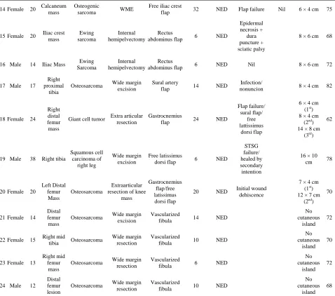
Vascularized fibula was done in six patients; five of them had osteogenic sarcoma and remaining one had pleomorphic sarcoma of forearm for which osteocutane- ous free fibular flap was done to reconstruct the radius. Four patients had onlay grafting of vascularized fibula to augment biological reconstruction with autograft and autoclaved bone. One patient had it done for intercalary defect reconstruction. Viability of flap was checked with bone scan after 2 weeks. Tracer uptake by fibula was evident in all 6 cases. Union is noticed in all cases. Three out of five patients with lower limb reconstruction are ambulating full weight bearing without support, where as remaining two are still using walking aid (Figure2).
Overall functional outcome was assessed using Mus- culoskeletal tumor society (MSTS) score. In our study mean score was 73.5% (Table 3). Reduced functional
(a) (b) (c)
(d) (e) (f)
(g) (h)
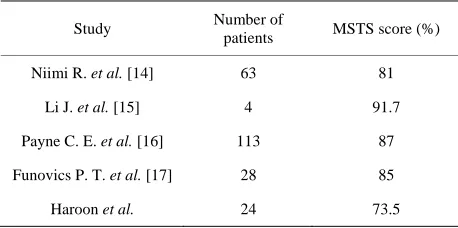
[image:6.595.145.454.384.689.2]Table 3. Comparision of our study and that of the other studies.
Study Number of patients MSTS score (%)
Niimi R. et al. [14] 63 81
Li J. et al. [15] 4 91.7
Payne C. E. et al. [16] 113 87
Funovics P. T. et al. [17] 28 85
Haroon et al. 24 73.5
score compared to international literature is possibly due to cases with diverse age groups, with different patholo-gies. So far no study has been published from our part of the world describing their progress in musculoskeltal- oncology. This may well be related to paucity of re-sources as well as specialized centers in the same field in our region.
5. Conclusion
Large soft tissue defects are the usual endpoint of wide surgical resections and coverage of those defects is es- sential. To overcome the fear of inadequate margins, one must not be afraid of the resultant wound size. It is im- perative to have multidisciplinary team approach for the treatment of musculoskeletal tumor, especially when aiming for limb salvage with resultant functional limb. Development of sarcoma service demands high quality centers equipped with trained staff and resources for the management of cases and postoperative rehabilitation.
REFERENCES
[1] S. A. Rosenberg, J. Tepper, E. Glatstein, et al., “The Treatment of Soft-Tissue Sarcomas of the Extremities: Prospective Randomized Evaluations of 1) Limb-Sparing Surgery plus Radiation Therapy Compared with Amputa- tion and 2) the Role of Adjuvant Chemotherapy,” Annals of Surgery, Vol. 196, No. 3, 1982, pp. 305-315.
http://dx.doi.org/10.1097/00000658-198209000-00009 [2] J. C. Yang, A. E. Chang, A. R. Baker, et al., “Random-
ized Prospective Study of the Benefit of Adjuvant Radio- therapy in the Treatment of Soft Tissue Sarcomas of the Extremity,” Journal of Clinical Oncology, Vol. 16, No. 1, 1998, pp. 197-203.
[3] M. A. Clark, C. Fisher, I. Judson and J. M. Thomas, “Soft-Tissue Sarcomas in Adults,” New England Journal of Medicine, Vol. 353, 2005, pp. 701-711.
http://dx.doi.org/10.1056/NEJMra041866
[4] M. A. Clark and J. M. Thomas, “Amputation for Soft- Tissue Sarcoma,” Lancet Oncology, Vol. 4, No. 6, 2003, pp. 335-342.
http://dx.doi.org/10.1016/S1470-2045(03)01113-6
[5] M. A. Clark and J. M. Thomas, “Major Amputation for Soft-Tissue Sarcoma,” British Journal of Surgery, Vol. 90, No. 1, 2003, pp. 102-107.
http://dx.doi.org/10.1002/bjs.4004
[6] J. M. Serletti, A. J. Carras, R. J. O’Keefe and R. N. Ros- ier, “Functional Outcome after Soft-Tissue Reconstruc- tion for Limb Salvage after Sarcoma Surgery,” Plastic and Reconstructive Surgery, Vol. 102, No. 5, 1998, pp. 1576-1583.
http://dx.doi.org/10.1097/00006534-199810000-00036 [7] I. A. Kramers-de Quervain, J. M. Läuffer, K. Käch, O.
Trentz and E. Stüssi, “Functional Donor-Site Morbidity during Level and Uphill Gait after a Gastrocnemius or Soleus Muscle-Flap Procedure,” The Journal of Bone and Joint Surgery (American Volume), Vol. 83, No. A2, 2001, pp. 239-246.
[8] T. Liu, Q. Zhang, X. Zhang, Z. Li, Y. Shen, X. Guo and L. Ling, “Medial Head Gastrocnemius Muscle Flap in the Limb-Salvage Operation for Proximal Tibial Osteosar-coma,” Journal of Central South University. Medical Sciences, Vol. 37, No. 12, 2012, pp. 1250-1254.
[9] S. V. Deo, K. S. Nootan, B. Niranjan and K. Dinesh, “Vertical Rectus Abdominis Myocutaneous Flap Cover for Lower Abdomen, Chest Wall, Groin and Thigh De- fects Following Resection of Malignant Tumours,” Indian Journal of Cancer, Vol. 38, No. 1, 2001, pp. 33-37. [10] B. S. Glatt, J. J. Disa, B. J. Mehrara, A. L. Pusic, P.
Boland and P. G. Cordeiro, “Reconstruction of Extensive Partial or Total Sacrectomy Defects with Transabdominal Vertical Rectus Abdomnis Myocutaneous Flap,” Annals of Plastic Surgery, Vol. 56, No. 5, 2006, pp. 526-530. http://dx.doi.org/10.1097/01.sap.0000205772.15061.39 [11] T. Morii, M. Susa, R. Nakayama, K. Kishi, H. Morioka
and H. Yabe, “Reconstruction Modality Based on the Spare Part Concept for Massive Soft Tissue Defects Fol- lowing Oncological Hemipelvectomy,” Journal of Or- thopaedic Science, Vol. 14, No. 2, 2009, pp. 192-197. http://dx.doi.org/10.1007/s00776-008-1316-5
[12] M. V. Küntscher, D. Erdmann, H. H. Homann, H. U. Steinau, S. L. Levin and G. Germann, “The Concept of Fillet Flaps: Classification, Indications, and Analysis of Their Clinical Value,” Plastic and Reconstructive Surgery, Vol. 108, No. 4, 2001, pp. 885-896.
http://dx.doi.org/10.1097/00006534-200109150-00011 [13] S. M. Walsh, S. G. Fisher and R. A. Sage, “Survival of
Patients with Primary Pedal Melanoma,” Journal of Foot and Ankle Surgery, Vol. 42, No. 4, 2003, pp. 193-198. http://dx.doi.org/10.1016/S1067-2516(03)70028-3 [14] R. Niimi, A. Matsumine, T. Hamaguchi, T. Nakamura, A.
Uchida and A. Sudo, “Prosthetic Limb Salvage Surgery for Bone and Soft Tissue Tumors around the Knee,” On- cology Reports, Vol. 28, No. 6, 2012, pp. 1984-1990.
[15] J. Li, Z. Wang, Z. Guo, M. Yang, G. Chen and G. Pei, “Composite Biological Reconstruction Following Total Calcanectomy of Primary Calcaneal Tumors,” Journal of Surgical Oncology, Vol. 105, No. 7, 2012, pp. 673-678. http://dx.doi.org/10.1002/jso.23022
Ferguson and J. S. Wunder, “Functional Outcome Fol- lowing upper Limb Soft Tissue Sarcoma Resection with Flap Reconstruction,” Journal of Plastic, Reconstructive & Aesthetic Surgery, Vol. 66, No. 5, 2013, pp. 601-607. http://dx.doi.org/10.1016/j.bjps.2013.01.034