muscular dystrophy in mice lacking Mkp5
Hao Shi, … , Richard A. Flavell, Anton M. Bennett
J Clin Invest.
2013;
123(5)
:2064-2077.
https://doi.org/10.1172/JCI64375
.
Duchenne muscular dystrophy (DMD) is a degenerative skeletal muscle disease caused by
mutations in dystrophin. The degree of functional deterioration in muscle stem cells
determines the severity of DMD. The mitogen-activated protein kinases (MAPKs), which are
inactivated by MAPK phosphatases (MKPs), represent a central signaling node in the
regulation of muscle stem cell function. Here we show that the dual-specificity protein
phosphatase DUSP10/5 negatively regulates muscle stem cell function in mice.
MKP-5 controlled JNK to coordinate muscle stem cell proliferation and p38 MAPK to control
differentiation. Genetic loss of
Mkp5
in mice improved regenerative myogenesis and
dystrophin-deficient
mdx
mice lacking
Mkp5
exhibited an attenuated dystrophic muscle
phenotype. Hence, enhanced promyogenic MAPK activity preserved muscle stem cell
function even in the absence of dystrophin and ultimately curtailed the pathogenesis
associated with DMD. These results identify MKP-5 as an essential negative regulator of
the promyogenic actions of the MAPKs and suggest that MKP-5 may serve as a target to
promote muscle stem cell function in the treatment of degenerative skeletal muscle
diseases.
Research Article
Muscle biology
Find the latest version:
Research article
Improved regenerative myogenesis and
muscular dystrophy in mice lacking
Mkp5
Hao Shi,1 Mayank Verma,1 Lei Zhang,1 Chen Dong,2 Richard A. Flavell,3 and Anton M. Bennett1,4
1Department of Pharmacology, Yale University School of Medicine, New Haven, Connecticut, USA. 2Department of Immunology,
The University of Texas MD Anderson Cancer Center, Houston, Texas, USA. 3Department of Immunobiology and Howard Hughes Medical Institute, and 4Program in Integrative Cell Signaling and Neurobiology of Metabolism, Yale University School of Medicine, New Haven, Connecticut, USA.
Duchenne muscular dystrophy (DMD) is a degenerative skeletal muscle disease caused by mutations in
dys-trophin. The degree of functional deterioration in muscle stem cells determines the severity of DMD. The
mitogen-activated protein kinases (MAPKs), which are inactivated by MAPK phosphatases (MKPs), represent
a central signaling node in the regulation of muscle stem cell function. Here we show that the
dual-specific-ity protein phosphatase DUSP10/MKP-5 negatively regulates muscle stem cell function in mice. MKP-5
con-trolled JNK to coordinate muscle stem cell proliferation and p38 MAPK to control differentiation. Genetic loss
of Mkp5 in mice improved regenerative myogenesis and dystrophin-deficient mdx mice lacking Mkp5 exhibited
an attenuated dystrophic muscle phenotype. Hence, enhanced promyogenic MAPK activity preserved muscle
stem cell function even in the absence of dystrophin and ultimately curtailed the pathogenesis associated
with DMD. These results identify MKP-5 as an essential negative regulator of the promyogenic actions of the
MAPKs and suggest that MKP-5 may serve as a target to promote muscle stem cell function in the treatment
of degenerative skeletal muscle diseases.
Introduction
Muscle precursor cells, known as satellite cells (SCs), govern the maintenance and repair of adult skeletal muscle (1–5). Located between the basal lamina and sarcolemma, SCs are quiescent in nature and respond to regenerative cues such as physical damage or exercise by undergoing cycles of proliferation before terminally differentiating into multinucleated myofibers (1, 2, 6, 7). SCs are the primary cell type through which adult regenerative myogenesis is mediated, and loss of SC function contributes to degenerative skeletal muscle diseases (5–12). Duchenne muscular dystrophy (DMD) is a devastating X-linked recessive genetic disorder affect-ing approximately 1 in 3,500 male births worldwide (13). DMD is caused by mutations in the dystrophin gene, which result in a lack of dystrophin protein expression (13, 14). In DMD, skeletal muscle regeneration is severely impaired due to the depletion of SCs as a result of a futile cycle of degeneration and regeneration. Ultimately, DMD patients lose muscle strength and mobility, and the disease often results in death. There is neither a cure, nor an effective treatment for DMD (9, 15). Because the progression of DMD is associated with deteriorating SC functionality (9), under-standing the mechanisms of SC control will provide insight into new therapeutic strategies for the treatment of DMD.
An important aspect of SC biology is the ability of SCs to prolif-erate in response to injury. Myoblasts from DMD patients exhibit impaired proliferation (16). More recently, mice lacking the expres-sion of dystrophin (mdx), which represents a mouse model of DMD, exhibit an exacerbated progression of muscle degeneration when the RNA component of telomerase is deleted (17). SCs derived from mdx
mice with the defective telomerase have an impaired regenerative capacity that is attributed, in part, to a reduced capacity of SCs from these mice to proliferate (17). Hence, the replicative capacity of SCs plays a critical role in the maintenance and repair of skeletal muscle.
The ability of SCs to proliferate and differentiate in order to effectively repair damaged skeletal muscle requires both coordi-nated activation and inactivation of signaling pathways. Although there has been much emphasis placed on the signals that initiate SC proliferation and differentiation, there is still relatively little known about the signaling pathways that promote the cessation of regenerative myogenesis. One of the more prominent signal-ing pathways involved in the initiation of regenerative myogen-esis is the mitogen-activated protein kinase (MAPK) pathway. Of the 3 major MAPK family members, the p38 MAPK subfamily, in particular p38 α/β MAPKs, referred to herein as p38 MAPK, have figured most prominently in promyogenic signaling to promote SC function (18–24). Although p38 MAPK is considered unequiv-ocally to be a promyogenic MAPK, the role of other MAPK family members, such as the extracellular signal-regulated kinases 1 and 2 (ERK1/2) and the c-Jun NH2 terminus kinases (JNKs), has been
less clear. ERK1/2 has been shown to exhibit both positive and negative regulatory roles in myogenesis (25–29), and JNK appears in some cases to be either dispensable or negative in myogenesis (30–32). Therefore, the role of ERK1/2 and JNK in regenerative myogenesis has yet to be conclusively resolved.
Despite the established contribution of the MAPKs, such as p38 MAPK, in SC promyogenic signaling and regenerative myo-genesis, it is unclear how MAPKs are temporally inactivated in order to terminate myogenesis. One mechanism likely involves the coordinated inactivation of the promyogenic MAPKs by the MAPK phosphatases (MKPs). The MKPs constitute a family of 10 dual-specificity phosphatases (DUSPs) that exhibit the capacity to specifically dephosphorylate MAPKs on the regulatory thre-onine and tyrosine residues (33–36). However, the identity of the physiologically relevant MKP that inactivates the promyogenic MAPKs in SCs, and then curtails regenerative myogenesis, has yet to be determined. Previous work has suggested a role for MKP-1 in regenerative myogenesis (20, 37), muscle growth (38–42), and skel-etal muscle regeneration (43). MKP-1–deficient SCs have impaired Conflict of interest: The authors have declared that no conflict of interest exists.
proliferation and differentiate precociously (43). Moreover, when crossed on to the mdx background, MKP-1–deficient mice exhibit an exacerbated muscular dystrophinopathy compared with mdx
mice (43). These findings suggest that MKP-1 promotes, rather than attenuates, SC function and regenerative myogenesis. In light of these results, it remains unclear how p38 MAPK and other MAPKs that are involved in regenerative myogenesis are maintained in a low state of activity, as would be the prediction in quiescent SCs. Moreover, how these MAPKs are coordinately inactivated once SC proliferation and differentiation have been completed remains to be determined.
In this study, we investigated the role of the dual-specificity phosphatase, DUSP10/MKP-5, in SC function and regener-ative myogenesis. MKP-5 is a cytosolic and nuclear MKP (34) that has been implicated in innate immune responses (44) and vascular inflammation (45) in mice. Here, we show that MKP-5 negatively regulates SC proliferation and differentiation by selec-tively dephosphorylating JNK and p38 MAPK, but not ERK1/2, in a temporal manner during regenerative myogenesis. Consis-tent with this, mice lacking MKP-5 expression exhibit improved regenerative myogenesis. Remarkably, mdx mice lacking MKP-5 are protected from progressive muscular dystrophinopathy and demonstrate preserved skeletal muscle function. Collectively,
these findings define MKP-5 as a critical negative regulator of the promyogenic MAPKs in SCs. These data suggest that MKP-5 may serve as a potential therapeutic target for the treatment of degen-erative muscle diseases.
Results
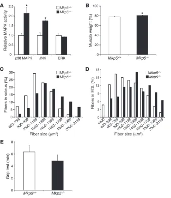
Increased MAPK activity, enhanced skeletal muscle mass, and regenerative myogenesis in MKP-5–deficient mice. MKP-5 is highly enriched in skel-etal muscle (46, 47), suggesting that it may play an important role in this tissue. In order to determine whether MKP-5 plays a role in skeletal muscle function, we examined the skeletal muscle pheno-type in mice lacking MKP-5 (Mkp5–/–) expression (44). We found
that uninjured Mkp5–/– mice exhibited increased skeletal muscle
JNK and p38 MAPK, but not ERK1/2, activity compared with
Mkp5+/+ mice (Figure 1A). Mkp5–/– mice exhibited increased skeletal
muscle mass and skeletal muscle fiber size compared with Mkp5+/+
mice (Figure 1, B–D, and Supplemental Figure 1, A–D; supple-mental material available online with this article; doi:10.1172/ JCI64375DS1). Although Mkp5–/– mice had increased skeletal
mus-cle mass, this did not result in an appreciable difference in musmus-cle strength since grip strength performance was comparable between
Mkp5–/– and Mkp5+/+ mice (Figure 1E). These results demonstrate
that MKP-5 plays a role in the negative regulation of skeletal
mus-Figure 1
[image:3.585.44.382.79.471.2]research article
Figure 2
cle p38 MAPK and JNK activity, but not ERK1/2 activity, in the suppression of skeletal muscle growth.
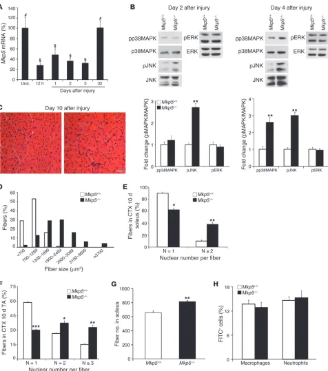
To investigate the role of MKP-5 in adult skeletal muscle regen-eration, we induced skeletal muscle damage by cardiotoxin (CTX) injection. MKP-5 expression was significantly reduced follow-ing muscle damage and returned to levels equivalent to those of undamaged muscle approximately 1 month after injury (Figure 2A). During skeletal muscle regeneration, MAPK hyperactiva-tion in skeletal muscles from Mkp5–/– mice was initially selective
for JNK, which had increased activity 2 days after damage, and by 4 days after injury, activity for both JNK and p38 MAPK was increased (Figure 2B). These results suggest that MKP-5 is
essen-tial for the negative regulation of JNK both early and late after injury. In contrast, p38 MAPK appears to be the predominant target of MKP-5 later on during regenerative myogenesis. No dif-ferences in ERK1/2 phosphorylation were observed at either 2 or 4 days after injury between Mkp5+/+ and Mkp5–/– mice (Figure 2B).
Newborn myofibers at 10 days after injury in Mkp5–/– mice were
larger in diameter (Figure 2, C and D) and contained more nuclei per myofiber compared with Mkp5+/+ mice at the same stage after
injury (Figure 2, E and F). The enhanced regenerative myogenesis observed in the skeletal muscle of Mkp5–/– mice was also
accom-panied by a significant increase in the total number of myofibers (Figure 2G). We interpret these results to suggest that MKP-5
neg-Figure 3
research article
atively regulates JNK and p38 MAPK in a temporal manner dur-ing regenerative myogenesis and that loss of MKP-5 results in an enhanced regenerative response following acute muscle injury.
MKP-5 has been reported to participate in the inflammatory response (44, 45), and our previous work suggested that MKPs, such as MKP-1, regulate inflammation during muscle repair (43). We tested whether MKP-5 behaved similarly. Neither macrophage nor neutrophil infiltration was found to be significantly different in the damaged areas of skeletal muscle between Mkp5+/+ and Mkp5–/–
mice (Figure 2H). Cytokine levels were not profoundly different, although IL-6 and IFN-γ were increased in the damaged skeletal muscles of Mkp5–/– mice (Supplemental Table 1). Therefore, in
skel-etal muscle, MKP-5 does not appear to profoundly influence the infiltration of hematopoietic cells into the damaged area.
Protection from dystrophic muscle disease in MKP-5–deficient mdx mice. Skeletal muscle in the mdx mouse displays a dystrophic
mus-cle phenotype, especially in the diaphragm (48). To investigate whether MKP-5 is also involved in exerting a negative effect on adult regenerative myogenesis in DMD, we intercrossed Mkp5–/–
mice into the mdx background in order to generate mdx/Mkp5+/+
and mdx/Mkp5–/– mice. As anticipated, mdx/Mkp5–/– mice exhibited
increased p38 MAPK and JNK phosphorylation, but not ERK1/2 phosphorylation, compared with mdx/Mkp5+/+ mice
(Supplemen-tal Figure 2, A and B). These results demonstrate that loss of dys-trophin does not interfere with the ability of MKP-5 to negatively regulate p38 MAPK and JNK. Improved skeletal muscle mor-phology and significantly reduced levels of degenerating muscle fibers were observed in the diaphragm and gastrocnemius of mdx/ Mkp5–/– mice (Figure 3A). Evans blue dye uptake, an indicator of
the loss of skeletal muscle fiber membrane integrity, was signifi-cantly reduced in mdx/Mkp5–/– mice compared with mdx/Mkp5+/+
[image:6.585.72.518.81.473.2]mice (Figure 3B). The onset of fibrosis, a hallmark of progression
Figure 4
of the dystrophic phenotype, was markedly reduced in the skel-etal muscles of mdx/Mkp5–/– mice compared with those of mdx/
Mkp5+/+ mice (Figure 3C). To quantify the extent of the damage,
we measured the areas that contained more than 10 degenerating myofibers and expressed this as a percentage of the total muscle cross-sectional area of the gastrocnemius muscle (49). We found that the damaged areas were greatly reduced in mdx/Mkp5–/–
mus-cle (Figure 3D). We also quantified damaged fibers by determining the areas of fibers that became permeable to Evans blue dye. We found that mdx/Mkp5–/– muscles had a significantly reduced area
of fibers that were Evans blue–positive (Figure 3E). The extent to which a lack of MKP-5 ameliorated the progression of the dys-trophic phenotype was further supported biochemically through measures of serum creatine kinase (CK) activity, which was signifi-cantly decreased in mdx/Mkp5–/– mice (Figure 3F). Together, these
findings demonstrate that a lack of MKP-5 ameliorates muscular dystrophinopathy in mdx mice.
To further examine whether the improved myopathy in MKP-5– deficient mdx mice reflects the recovery of skeletal muscle func-tion, we tested the muscle contractile properties in mdx/Mkp5+/+
and mdx/Mkp5–/– mice. No basal differences in gross muscle
strength were observed between Mkp5+/+ and Mkp5–/– mice
(Fig-ure 1E). Hence, Mkp5–/– mice were not developmentally distinct in
broad muscle contractile properties compared with Mkp5+/+ mice.
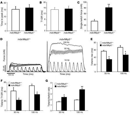
On the mdx background, MKP-5 deficiency revealed no detectable change in the velocities of contraction or relaxation in a single stimulation compared with wild-type mice (Figure 4, A and B). However, several parameters of muscle function were significantly improved in mdx/Mkp5–/– mice compared with mdx/Mkp5+/+ mice
(Figure 4, C–G). Extensor digitorum longus (EDL) muscle sin-gle-twitch force (Figure 4C) and tetanic force (Figure 4D) were sig-nificantly increased in the muscles of mdx/Mkp5–/– mice compared
with those of mdx/Mkp5+/+ mice. The time to rise to tetanus (Figure
4E) and the time to release after tetanus (Figure 4F) were markedly reduced, indicating that the responsiveness of mdx/Mkp5–/– mouse
EDL muscles to stimuli is significantly improved. The tetanic/ twitch force ratio, an indicator of muscle force buildup, increased significantly in mdx/Mkp5–/– EDL muscle compared with mdx/
Mkp5+/+ EDL muscle (Figure 4G). Taken together, these
parame-ters indicate that MKP-5 deficiency in the mdx model of
myopa-thy impairs the deterioration of muscle contractile function, an observation that is consistent with the improved skeletal muscle morphology in mdx/Mkp5–/– mice (Figure 3).
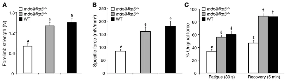
To further define the extent to which a lack of MKP-5 maintains skeletal muscle function, we compared mdx/Mkp5+/+ and mdx/
Mkp5–/– mice with wild-type C57BL/10 mice. Consistent with
the reduced severity of the dystrophic phenotype, mdx/Mkp5–/–
mice exhibited equivalent levels of skeletal muscle function, as determined by forelimb strength, when compared with wild-type (C57BL/10) mice (Figure 5A). Furthermore, the specific force of EDL muscle was preserved to levels equivalent to those of the wild-type (C57BL/10) mice (Figure 5B). Additionally, mdx/Mkp5–/–
mice showed resistance to repeated contraction-induced fatigue at levels equivalent to those of wild-type (C57BL/10) mice, whereas EDL muscles from mdx/Mkp5+/+ mice were significantly impaired
in their ability to recover (Figure 5C). These findings demonstrate that loss of MKP-5 ameliorates the progression of the dystrophic phenotype in mdx mice. Since SC dysfunction likely underlies the cellular basis of the dystrophic phenotype, these results strongly suggest that SCs represent a major site of MKP-5 action.
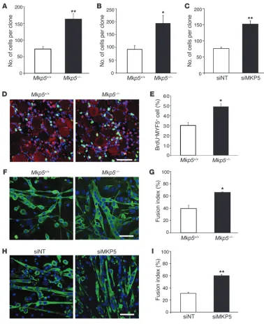
MKP-5 negatively regulates SC proliferation and differentiation. In order to elucidate the cause of the enhanced skeletal muscle regenerative capacity in Mkp5–/–, mice we measured SC
expan-sion in response to injury by enumerating PAX7+ SCs in muscle
cross sections 10 days after injury (50). In response to injury, the SC population expanded by 88.7% in Mkp5–/– mice compared
with 36.9% in Mkp5+/+ mice (Supplemental Figure 3, A–C). These
results suggest that MKP-5–deficient SCs are hyperproliferative. Consistent with this, SC-derived myoblasts from Mkp5–/– mice
exhibited increased proliferative capacity as determined using either a clonal assay in single myofiber explants (Figure 6A) or in FACS-sorted SC cultures (Figure 6B). In order to rule out the possibility that SC-derived myoblasts were hyperproliferative due to a developmentally acquired property of Mkp5–/– mice, we
[image:7.585.57.528.82.218.2]isolated SC-derived myoblasts from adult wild-type mice. These SC-derived myoblasts were then subjected to MKP-5 knockdown using siRNA (Figure 6C). Under these conditions, Mkp5 expres-sion was reduced by greater than 80% in nontargeted siRNA– treated SC-derived myoblasts (Supplemental Figure 4, A and B). MKP-5 siRNA–treated SC-derived myoblasts had increased
Figure 5
research article
proliferation levels compared with nontargeted siRNA–treated myoblasts (Figure 6C). To directly assess SC proliferation in vivo, skeletal muscle was damaged and BrdU was injected 24 hours after injury. The percentage of SCs (MYF5+) that were BrdU+ was
significantly increased in Mkp5–/– mice compared with Mkp5+/+
[image:8.585.102.480.81.545.2]mice (Figure 6, D and E). These results demonstrate that MKP-5 exerts autonomous negative regulation of postdevelopmental SC proliferation in adult mice.
Figure 6
Next, we evaluated the effects of MKP-5 on SC differentiation. SC-derived myoblasts from Mkp5–/– mice differentiated more
robustly with a significantly increased level of myoblasts fusing into multinucleated myotubes compared with that in Mkp5+/+ SCs
(Figure 6, F and G). Similarly, MKP-5 siRNA–treated SC-derived myoblasts from wild-type mice also had enhanced capacity to undergo fusion into multinucleated myotubes (Figure 6, H and I). These results demonstrate that MKP-5 negatively regulates both SC proliferation and differentiation, suggesting that loss of MKP-5 in SCs gives rise to enhanced SC function, which is likely causal to the improved regenerative capacity and ameliorative dys-trophic response observed in mice lacking MKP-5.
MKP-5 negatively regulates cyclin D3 expression in regenerating skeletal muscle. To examine the molecular mechanisms of MKP-5 regula-tion of regenerative myogenesis in mice, we assessed the expression of cell-cycle regulatory proteins in regenerating skeletal muscles from Mkp5+/+ and Mkp5–/– mice. Although SCs represent between
1% and 2% of total skeletal muscle mass, the population of SCs dramatically expands during regeneration. We hypothesized that changes in the expression of cell-cycle regulatory proteins in skel-etal muscle will likely reflect similar changes of these proteins in the population of proliferating SCs early on during regenerative myogenesis. We found that 2 days following muscle regeneration, cyclin D3 expression, but not D1 or D2 expression, was increased in Mkp5–/– skeletal muscle (Supplemental Figures 5 and 6).
Con-sistent with the observation of enhanced skeletal muscle regen-eration, it has been reported that cyclin D3 promotes myoblast proliferation, cell-cycle exit, and differentiation (51, 52). No appre-ciable differences in CDK4, CDK6, or p15INK4b levels were observed
(Supplemental Figures 5 and 6). In line with the increased cyclin D3 levels, Rb was phosphorylated to a greater extent in regener-ating skeletal muscle from Mkp5–/– mice compared with Mkp5+/+
mice (Supplemental Figures 5 and 6). In agreement with the upregulation of JNK, c-Jun phosphorylation at the JNK phos-phorylation site of Ser 63 was increased in the skeletal muscles of regenerating Mkp-5–/– mice (Supplemental Figures 5 and 6). The
expression of p53 increased in response to injury (Supplemental
Figure 7, A and B), and this was also elevated in regenerating skel-etal muscle derived from Mkp5–/– mice compared with that from
Mkp5+/+ mice (Supplemental Figure 7, C and D). Notably, other
major pathways involved in regenerative myogenesis, such as Akt and S6 kinase 1, were found to be unchanged (Supplemental Fig-ures 5 and 6). Therefore, MKP-5 exerts selective regulation on the cell cycle during skeletal muscle regeneration.
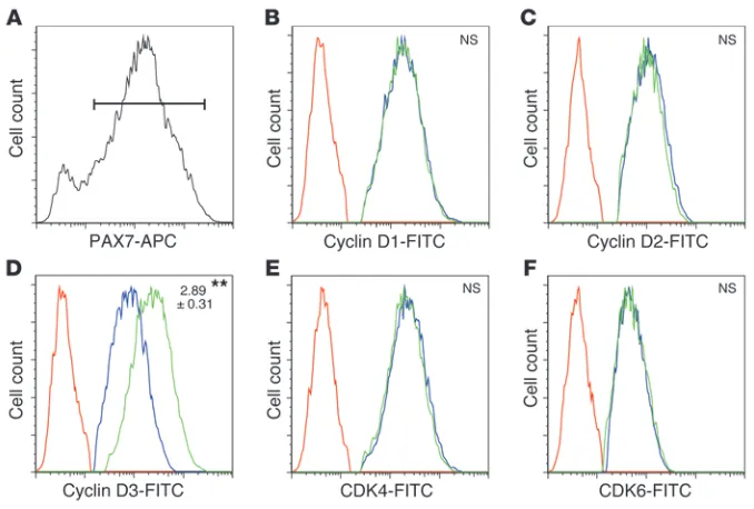
We next followed up on our observation that MKP-5 appears to negatively regulate cyclin D3 expression during regenerative myogenesis. To more directly examine the expression levels of the cyclins in the population of proliferating SCs, we isolated skele-tal muscle cells that had regenerated for 2 days from Mkp5+/+ and
Mkp5–/– mice. Consistent with the results obtained from
skele-tal muscle, we found that PAX7+ cells (Figure 7A) from Mkp5–/–
mice expressed significantly increased levels of cyclin D3, but not cyclin D1 or D2, compared with Mkp5+/+ mice (Figure 7, B–D).
The expression levels of both CDK4 and CDK6 also remained unchanged (Figure 7, E and F). These results identify cyclin D3 as a downstream target of MKP-5 and suggest that MKP-5 suppresses SC proliferation by negatively regulating cyclin D3.
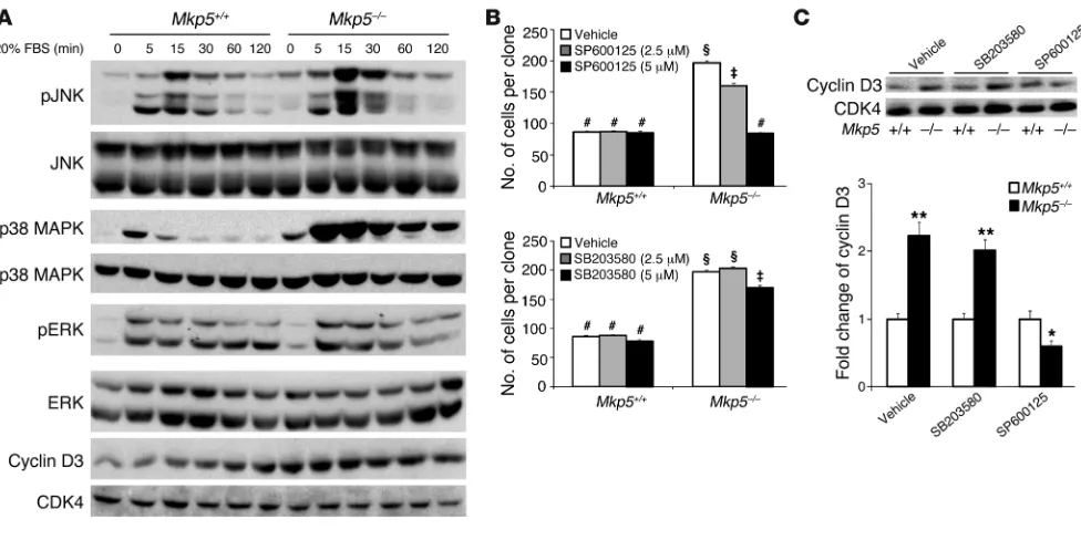
MKP-5 regulates SC proliferation through a JNK/cyclin D3–dependent pathway. To determine whether the effects of MKP-5 on MAPK signaling and cyclin D3 expression were SC autonomous, we measured MAPK activity and cyclin D3 levels in SC-derived myo-blasts. We found that Mkp5–/– SC–derived myoblasts exhibited
enhanced JNK and p38 MAPK, but not ERK1/2, phosphoryla-tion in response to serum and increased cyclin D3 levels (Figure 8A). To identify which of the MAPKs conveys MKP-5 effects on SC proliferation, we performed clonal proliferation assays using MAPK inhibitors. We found that inhibition of JNK, but not p38 MAPK, rescued the hyperproliferation of Mkp5–/– SC–derived
[image:9.585.41.380.81.310.2]myoblasts (Figure 8B). Similar approaches using pharmacolog-ical inhibitors of the MAPK pathway were used to demonstrate that JNK was responsible for the increased expression of cyclin D3 in SC-derived myoblasts (Figure 8C). Collectively, these results indicate that MKP-5 regulates SC proliferation through a JNK/ cyclin D3–dependent pathway.
Figure 7
research article
To evaluate whether the increased cyclin D3 expression levels were responsible for the enhanced SC proliferation, and subsequently if this was dependent upon MKP-5, the expression levels of MKP-5 (Figure 9A) and cyclin D3 in C2C12 myoblasts (Figure 9B) were reduced by siRNA knockdown, either alone or in combination, and their proliferation was assessed. Both p38 MAPK and JNK, but not ERK1/2, were enhanced in differentiating myoblasts in which MKP-5 had been knocked down by siRNA treatment (Supplemental Figure 8). Knockdown of MKP-5 increased myoblast proliferation, whereas knockdown of cyclin D3 reduced it (Figure 9, C and E). However, when both MKP-5 and cyclin D3 were knocked down, the increased proliferation induced by the loss of MKP-5 was blocked (Figure 9, C and E). These results imply that MKP-5 suppresses myoblast proliferation by impairing JNK-mediated expression of cyclin D3. Because cyclin D3 is also a regulator of muscle cell dif-ferentiation (51, 53), we determined whether cyclin D3 knockdown impairs the enhanced differentiation incurred by loss of MKP-5. Knockdown of MKP-5 enhanced myoblast differentiation, as mea-sured through the assessment of the myoblast fusion index (Fig-ure 9, D and F). In contrast, cyclin D3 knockdown alone blocked myoblast fusion (Figure 9, D and F). When both MKP-5 and cyclin D3 levels were knocked down, the impairment in differentiation induced upon loss of cyclin D3 was restored in the background of cells lacking MKP-5 (Figure 9, D and F). These results demonstrate that although cyclin D3 plays a significant role in promoting myo-blast differentiation, the suppressive effects of MKP-5 on myomyo-blast differentiation likely occurs through either a p38 MAPK- and/or JNK-dependent pathway that is cyclin D3 independent.
Discussion
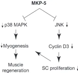
In this report, we have identified MKP-5 as an essential negative regulator of the promyogenic actions of MAPKs in mice. Based on these findings, we propose that MKP-5 maintains JNK and p38 MAPK in a dephosphorylated state, thereby maintaining SC qui-escence (Figure 10). An important finding uncovered in this study is the demonstration that loss of MKP-5 in the mdx background is sufficient to markedly ameliorate the progression of muscular dys-trophy. These results provide genetic evidence for a role of MKP-5 and the MAPK pathway in the progression of DMD and poten-tially other degenerative muscle diseases.
MKP-5–deficient mice exhibited enhanced MAPK activity in skeletal muscle and in SCs, with a rank order of dephosphoryla-tion toward p38 MAPK and JNK, but not ERK1/2. MKP-5 is highly enriched in skeletal muscle (46, 47), suggesting that it plays an important role in this tissue. This notion was supported by the observation that skeletal muscle mass was greater in Mkp5–/– mice
compared with that in wild-type mice, although this did not result in significant differences in the baseline strength properties of
Mkp5–/–mice. The increase in skeletal muscle mass is likely a
reflec-tion of increased myofiber size in Mkp5–/– mice. These results are
consistent with other observations that the MAPKs are involved in determining skeletal muscle mass (40).
MKP-5 expression becomes downregulated in response to skele-tal muscle damage, and JNK is hyperactivated in Mkp5–/– muscles,
[image:10.585.49.537.82.326.2]suggesting that MKP-5 downregulation facilitates the activation of JNK in order to trigger SC proliferation. Later on during regen-erative myogenesis, p38 MAPK was hyperactivated in the skeletal
Figure 8
muscles of Mkp5–/– mice, consistent with the established role of
p38 MAPK in promoting differentiation. The apparent effective-ness of MKP-5 to serve as an essential MKP for p38 MAPK and JNK during regeneration is also likely to be related to the expression status of other MKPs in skeletal muscle. For example, it has been
[image:11.585.87.494.83.547.2]shown that MKP-1 becomes transiently activated at the early stages of skeletal muscle regeneration (54). The induction of MKP-1, and possibly other MKPs, at this time point might account for the comparable levels of p38 MAPK phosphorylation at 2 days after injury in Mkp5–/– mice compared with wild-type mice. These
Figure 9
research article
mice. These results demonstrate that even in the absence of dystro-phin, augmenting the activity of the MAPKs through specific loss of MKP-5 can ameliorate the progressive degeneration of skeletal muscle function in mdx mice. Mice lacking MKP-5 exhibit com-parable levels of muscle strength compared with wild-type mice. Hence, the maintenance of muscle function in MKP-5–deficient
mdx mice stems from the postdevelopmental effects of MKP-5 on the progression of the disease rather than from those conferred during development. Along these lines, SC-derived myoblasts from adult wild-type mice also exhibit increased proliferation and dif-ferentiation when MKP-5 expression is acutely reduced by siRNA. Hence, the actions of MKP-5 on MAPK signaling in SCs and regen-erative myogenesis target processes that postdevelopmentally operate downstream of, and/or parallel to, dystrophin.
To support the supposition that MKP-5 was acting autonomously in SCs, we demonstrated that SCs derived from Mkp5–/– mice
exhibited an enhanced proliferative response following injury. Sev-eral lines of evidence support this. First, following injury, Mkp5–/–
SCs were found to expand nearly 3 times more rapidly than SCs from skeletal muscles of Mkp5+/+ mice. Second, in vivo proliferation
assays demonstrated that Mkp5–/– SCs proliferated more rapidly
than Mkp5+/+ SCs. Third, both clonal and single myofiber assays
showed that Mkp5–/– SC–derived myoblasts exhibited increased
pro-liferation compared with Mkp5+/+ SC–derived myoblasts. Finally,
acute depletion of MKP-5 by siRNA in SC-derived myoblasts from wild-type mice also enhanced cell proliferation, ruling out the possibility that MKP-5 loss during development was responsible for this effect. Since SC proliferation represents a major factor in the process of regenerative myogenesis, it is reasonable to surmise that the enhanced repair of skeletal muscle in both the acute and chronic paradigms of muscle injury stems from the increased levels of SC proliferation in mice lacking MKP-5.
We discovered that the expression of cyclin D3, but not cyclin D1 or cyclin D2, was enhanced in regenerating skeletal muscle from
Mkp5–/– mice. Isolation of MKP-5–deficient SC-derived myoblasts
confirmed that cyclin D3 was indeed upregulated in SCs, along with a concomitant upregulation of both p38 MAPK and JNK, but not ERK1/2. Thus, MKP-5 plays an essential role in negatively regulating SC proliferation through JNK-dependent regulation of cyclin D3. Cyclin D3 is induced during terminal differentiation, regulates myoblast cell-cycle exit, and is required for myogenic dif-ferentiation (51–53, 56, 57). In order to substantiate the relation-ship between MKP-5 and cyclin D3 in myoblast proliferation, we asked whether loss of cyclin D3 could rescue the enhanced prolifer-ation in MKP-5–deficient muscle cells. These experiments revealed that elimination of cyclin D3 expression in the background of MKP-5 loss rescues the enhanced proliferative effects in MKP-5– deficient muscle cells. Hence, cyclin D3 is a major downstream tar-get of MKP-5/JNK in the control of SC proliferation. Myoblasts deficient in p38 MAPK have been shown to reduce cyclin D3 levels, and loss of cyclin D3 inhibits myogenic gene activation (51, 56). In addition, cyclin D3 is transcriptionally activated through the AP-1/c-Jun module. Our data are consistent with MKP-5 regula-tion of SC proliferaregula-tion through JNK/c-Jun/cyclin D3, rather than through p38 MAPK. Despite the fact that cyclin D3 is required for differentiation, MKP-5–mediated suppression of the differentia-tion pathway appeared to occur even when cyclin D3 expression was eliminated. These results suggest that MKP-5 suppresses dif-ferentiation through pathways other than those mediated by cyclin D3, but is dependent on JNK and/or p38 MAPK.
results demonstrate that in response to skeletal muscle injury, MKP-5 temporally regulates the activities of both JNK and p38 MAPK in order to maintain the appropriate homeostatic control of these MAPKs, which coordinate regenerative myogenesis.
We show that loss of MKP-5 results in enhanced p38 MAPK and JNK activation, though only the inhibition of JNK appeared capa-ble of promoting SC quiescence by the suppression of cyclin D3. The role of JNK in SC function has remained less clear in contrast to that of p38 MAPK. However, JNK activity has been shown to be required for both myoblast proliferation and differentiation (21, 32, 55). The results presented here support the interpretation that JNK is required for SC proliferation. The ability of MKP-5 to inac-tivate p38 MAPK is also consistent with the promyogenic role of p38 MAPK in both SC proliferation and differentiation (21, 24). SC-derived myoblasts from Mkp5–/– mice or MKP-5 siRNA–treated
SC-derived myoblasts from wild-type mice exhibited increased dif-ferentiation. Therefore, these findings are supportive of the inter-pretation that MKP-5 plays an essential role in negatively regulat-ing the physiologically relevant pool of JNK and p38 MAPK in the control of SC function and regenerative myogenesis (Figure 10).
The immune response plays a major role in the efficacy of the repair process. Given that Mkp5–/– mice exhibited improved
mus-cle repair, it was important to assess the extent of the inflamma-tory response in damaged skeletal muscles from Mkp5–/– mice. We
observed equivalent levels of both macrophage and neutrophil infiltrates in the damaged areas of skeletal muscle from Mkp5+/+
and Mkp5–/– mice. This result was somewhat surprising, given
pre-vious findings of a negative regulatory role of MKP-5 in innate and adaptive immunity (44, 45). We cannot exclude the possibility that the repertoire of macrophage subpopulations is altered in a man-ner that promotes repair. Nonetheless, the inflammatory response in the context assessed here does not appear to play a deleterious role in the enhanced regenerative response in Mkp5–/– mice.
One of the most striking observations from this study is that the loss of MKP-5 is sufficient to markedly ameliorate the dystrophic phenotype of mdx mice. Although mdx mice succumb to a very mild dystrophic phenotype compared with humans, we were still able to observe an improvement in the dystrophic phenotype in
[image:12.585.98.229.83.206.2]mdx mice lacking MKP-5. Remarkably, mdx mice lacking MKP-5 expression demonstrated parameters of muscle function that were equivalent to those of wild-type C57BL/10 mice. Importantly, we demonstrated that in mdx mice lacking MKP-5, both p38 MAPK and JNK phosphorylation were enhanced compared with mdx Figure 10
ies were from Developmental Studies Hybridoma Bank. TO-PRO-3 iodide for nuclear staining was purchased from Invitrogen. See also Supplemental Methods for additional details on the antibodies.
Animal experiments. MKP-5 knockout mice were generated as described previously (44). To generate mdx/Mkp5–/– mice, male Mkp5–/– mice were
crossed with female mdx mice, the F1 males were backcrossed with female
mdx mice, and the resultant F2 offspring were then intercrossed.
Muscle damage analysis. Mice were anesthetized by administration of 10 mg/kg ketamine and 1 mg/kg xylazine. Muscle damage was induced by intramuscular injection of 50 μl CTX (Sigma-Aldrich, 0.1 mg/ml in PBS) into the tibialis anterior (TA) muscle longitudinally or 300 μl into the gastrocnemius/soleus muscles. At the indicated times (see Figure 2C), the damaged muscle was dissected and fixed in 10% neutral-buffered formalin overnight, washed twice in 70% ethanol, and paraffin embedded for H&E staining. For mdx muscle myopathy analyses, the degenerating area was counted as a combined total, with each area containing greater than or equal to 10 degenerating fibers, and the percentage of degenerating area was calculated as a percentage of the whole gastrocnemius muscle area. To evaluate mdx muscle fiber integrity, 1% Evans blue dye dissolved in sterile saline was administered by intraperitoneal injection at a dose of 1% of body weight. Twenty-four hours later, muscles were dissected and snap-frozen in isopentene precooled in liquid nitrogen. Muscle cryosections of 10-μm thickness were cut and Evans blue dye intake was evaluated using a Zeiss laser scanning confocal microscope (Carl Zeiss).
Muscle strength test. Grip tests were performed as previously described (43). To assess mouse forelimb strength, we used a Grip Strength Meter (Colum-bus Instruments) and allowed the mice to grip the horizontal bar attached to the front of the meter. Mice were pulled horizontally by the tail until the bar was released. Repetitions of this procedure were performed and the peak force was recorded for each mouse. To measure muscle-specific force and contraction-induced fatigue and recovery, the distal end of the EDL mus-cle was dissected and linked to a transducer (Kent Scientific). EDL musmus-cle was stimulated with an S48 square pulse stimulator (Grass Technologies) and the output signals were captured and analyzed using a PowerLab data acquisition system with LabChart Pro software (ADInstruments).
Immunohistochemical and immunocytochemical staining. Muscles were fixed in 10% neutral-buffered formalin overnight, washed in 70% ethanol, and par-affin embedded. Muscles were cut into 10-μm-thick cross sections. Before staining, muscle sections were de-paraffinized with 2 changes of xylene for 5 minutes each time, rehydrated in 2 changes of 100% ethanol for 3 minutes each, and followed by 95% and 80% ethanol for 1 minute each time before rinsing in distilled H2O. The sections were then immersed in sodium citrate
buffer preheated to approximately 100°C in a steamer and incubated for 30 minutes. After the steaming process, the sections were allowed to cool at room temperature in the same buffer for 20 minutes before rinsing 3 times in PBS for 5 minutes each time. The sections were blocked in 5% goat serum for 60 minutes at room temperature before application of the primary and secondary antibodies. For nuclear staining, TO-PRO-3 was applied to the reaction together with the secondary antibody.
Myosin heavy chain immunocytochemical staining was conducted as described previously (43). For BrdU staining, cells were fixed in ice-cold 70% ethanol for 5 minutes followed by incubation in 1.5 M HCl for 30 minutes at room temperature. Cells were rinsed and stained with anti-bodies against BrdU. PAX7 immunohistochemical staining was carried out as described in Supplemental Methods.
SC functional analyses. SC-derived myoblasts were isolated and cultured as previously described (43). For proliferation assays, fiber-derived or FACS-sorted SCs (based on surface marker expression; CD34+CD45–Sca-1–) (10)
were grown in F-10 medium containing 20% FBS, and the number of cells per clone was calculated at the indicated time. For in vivo BrdU
incorpo-We have shown previously that MKP-1–deficient mice are impaired in their ability to undergo regenerative myogenesis and that SCs derived from these mice proliferate at a significantly slower rate (43). Moreover, when MKP-1–deficient mice are inter-crossed with mdx mice, the dystrophic phenotype is exacerbated (43). These results suggest that despite the fact that MKP-1 also dephosphorylates JNK and p38 MAPK, it is unlikely to represent the physiologically relevant MKP that dephosphorylates the pool of MAPKs involved in maintaining SC quiescence. The mechanis-tic basis for why MKP-5, but not MKP-1, antagonizes SC prolifer-ation remains unclear, although there are several possible expla-nations. One explanation is that MKP-5 is expressed in both the nucleus and cytosol (58), whereas MKP-1 is expressed exclusively in the nucleus (59). These differences in localization may result in the regulation of functionally distinct pools of MAPKs between MKP-5 and MKP-1, and hence the regulation of downstream MAPK substrates. In addition, MKP-1 expression is regulated in a distinct manner during regenerative myogenesis (54) compared with MKP-5. Finally, MKP-1–deficient mice exhibit a profound immunological phenotype following muscle damage (43), whereas MKP-5–deficient mice do not show such effects compared with wild-type mice. Together, these differences could explain the dis-tinct phenotypes in SCs and regenerative myogenesis in MKP-1– and MKP-5–deficient mice. Interestingly, nonoverlapping func-tionalities between MKP-1 and MKP-5 in vascular injury have been reported previously (45). These observations highlight the com-plexities of physiological and pathophysiological MKP/MAPK– mediated signaling in vivo. Nonetheless, the results reported here demonstrate that MKP-5 controls the pool of MAPKs that are required collectively to regulate downstream phosphorylation of targets involved in the maintenance of SC quiescence and attenu-ation of regenerative myogenesis.
In DMD, the SC pool is depleted as a result of futile rounds of degeneration and regeneration. The severity of the DMD phe-notype has been linked to a loss of SC function. We suggest that the enhanced proliferation of MKP-5–deficient SCs is sufficient to counter the depletion of SCs in both the acute regenerative response and in mdx mice, thereby ameliorating the severity of the disease. Consistent with this notion, mdx mice lacking the RNA component of telomerase display impaired SC proliferation and a more severe phenotype resembling human DMD (17). The con-clusions derived here do not exclude other important factors that contribute to DMD, such as the loss of skeletal muscle structural integrity. Presumably, the collective repertoire of MKP-5/MAPK targets will need to be identified in order to fully define how MKP-5 functions in the maintenance of SC homeostasis.
In summary, these data identify MKP-5 as an essential negative regulator of SC proliferation and differentiation. The identifica-tion of MKP-5 as an essential negative regulator of SC funcidentifica-tion and DMD pathogenesis raises the possibility that pharmacological tar-geting of MKP-5 may represent a novel strategy for the treatment of DMD and possibly other skeletal muscle degenerative diseases.
Methods
antibod-research article
blocked in 7% nonfat dried milk for 1 hour at room temperature. After a brief wash in TBST, the membrane was incubated with primary antibody in 5% BSA overnight at 4°C. After three 5-minute washes in TBST, the membrane was incubated with an HRP-conjugated secondary antibody for 1 hour at room temperature. The membrane was developed using ECL reagents (GE Healthcare). See Supplemental Methods for additional details.
Kinase activity assays. MAPK activity assays were performed as described previously (42). Serum creatine kinase was assayed using the Enzy-Chrom Creatine Kinase Assay kit (BioAssay Systems) according to the manufacturer’s instructions.
Quantitative RT-PCR. Total RNA from cells or skeletal muscles was extracted using TRIzol reagent (Life Technologies), and genomic DNA was removed using a DNA-free kit (Applied Biosystems). cDNA was syn-thesized using a High Capacity cDNA Reverse Transcription kit (Applied Biosystems). For quantitative PCR, TaqMan MKP-5 and 18S rRNA primers were used in combination with the TaqMan Gene Expression Master Mix (Applied Biosystems) in a total reaction of 20 μl. A 7500 Fast-Real Time PCR System (Applied Biosystems) was used to quantify mRNA expression.
Mkp5 mRNA relative abundance was normalized to internal control 18S rRNA and expressed as percentage change in either the nondamaged mus-cle or nontargeted siRNA where indicated in Figures 2A and 9A.
Statistics. Statistical analysis was performed using the Student’s t test with 2-tailed distribution, assuming normal distribution and equal variance among different samples. Data are presented as the means ± SEM. Statisti-cal significance is signified as *P < 0.05, **P < 0.01, and ***P < 0.001. Where appropriate, symbols were used for comparison; data points with symbols that differ signify a statistical significance of P < 0.05, with the same symbol signifying no statistical significance between samples.
Study approval. The Yale University Institutional Animal Care and Use Committee approved all procedures.
Acknowledgments
We thank Dake Qi and Lawrence H. Young (Yale School of Medi-cine) for assistance with the muscle contraction experiments. We also thank Gerald I. Shulman (Department of Endocrinology/ Internal Medicine, Yale School of Medicine) for Minispec analysis of muscle mass. This work was supported by NIH grants (AR46504 and DK075776, to A.M. Bennett) and a Muscular Dystrophy Asso-ciation grant (MDA 186936, to H. Shi).
Received for publication April 17, 2012, and accepted in revised form January 31, 2013.
Address correspondence to: Anton M. Bennett, Yale Univer-sity School of Medicine, Department of Pharmacology, SHM B226D, 333 Cedar Street, New Haven, Connecticut 06520-8066, USA. Phone: 203.737.2441; Fax: 203.737.2738; E-mail: anton. [email protected].
ration assays, BrdU (100 mg/kg) was injected intraperitoneally 24 hours after the TA muscles were damaged. Muscles were collected 18 hours later to stain for both BrdU and MYF5. To induce SC differentiation, growth medium was withdrawn and cells were washed twice with PBS and cul-tured in differentiation medium (DMEM containing 4% horse serum). Differentiated SCs were immunocytochemically stained with the differ-entiation marker myosin heavy chain antibody (MF20). The fusion index was calculated as the percentage of nuclei within myotubes containing at least 2 nuclei as a ratio of total nuclei in the field. For MAPK phosphory-lation experiments, SCs were serum starved in F-10 containing 0.1% FBS overnight before switching to F-10 containing 20% FBS for the indicated time (see Figure 8A). For knockdown of MKP-5, SC-derived myoblasts were isolated from C57BL/6 muscles, preplated to enrich for myoblasts, and transfected with ON-TARGETplus nontargeting pool (siNT) and ON-TARGETplus SMART pool MKP-5 siRNA (siMKP-5) (Thermo Scien-tific) using Lipofectamine 2000 (Life Technologies) according to the man-ufacturer’s instructions.
C2C12 myoblast culture. C2C12 myoblasts were cultured in DMEM con-taining 10% FBS. For siRNA knockdown experiments, C2C12 myoblasts were transfected with ON-TARGETplus SMART pool cyclin D3 or MKP-5 siRNA (Thermo Scientific) using DharmaFECT 1 reagent (Thermo Scien-tific). For BrdU incorporation, transfected cells were pulse-chased 72 hours later with 10 μM BrdU for 3 hours. For differentiation assays, myoblasts were shifted to differentiation medium (DMEM containing 0.1% FBS, 5 μg/ml insulin, 5 μg/ml selenium) for the indicated times (see Figure 9, D and F). The fusion index was calculated as the percentage of nuclei within myotubes containing at least 2 nuclei as a ratio of total nuclei in the field.
Flow cytometry. To sort for SCs, hind limb muscles were minced and digested with collagenase B/dispase II, filtered through 45-μm nylon cell strainers, and pelleted at 350 g. PE-CD45 and FITC-Sca-1 antibodies were purchased from BD Biosciences; biotin-CD34 and APC-streptavidin were from eBioscience. For FACS analysis of macrophage and neutrophil infil-tration, muscles were damaged by CTX injection. Forty-two hours later, damaged muscles were isolated and digested with collagenase B/dispase II. The isolated cells were then stained with CD11b (Developmental Studies Hybridoma Bank) and 7/4 (AbD Serotec) antibodies, respectively. After a brief wash, FITC-conjugated secondary antibodies were applied and the cells were analyzed using a BD FACSCalibur flow cytometer (BD Biosci-ences). For FACS analysis of cell-cycle regulator protein levels, muscles were CTX-damaged for 2 days and then digested, and cells were isolated for staining. Rabbit anti-PAX7 antibody and mouse anti-cyclin D1, cyclin D2, and CDK4 were purchased from Abcam. Mouse anti–cyclin D3 was pur-chased from Thermo Scientific. Goat anti-CDK6 antibody was purpur-chased from Santa Cruz Biotechnology. Secondary APC-conjugated anti-rabbit and FITC-conjugated anti-mouse or anti-goat antibody were obtained from Jackson ImmunoResearch.
Biochemical analysis. For immunoblotting, proteins were separated by 10% SDS-PAGE and transferred to PVDF membrane. The membrane was
1. Hawke TJ, Garry DJ. Myogenic satellite cells: physiology to molecular biology. J Appl Physiol. 2001;91(2):534–551.
2. Seale P, Rudnicki MA. A new look at the origin, function, and “stem-cell” status of muscle satellite cells. Dev Biol. 2000;218(2):115–124.
3. Wagers AJ, Conboy IM. Cellular and molecular signatures of muscle regeneration: current con-cepts and controversies in adult myogenesis. Cell. 2005;122(5):659–667.
4. Shi X, Garry DJ. Muscle stem cells in develop-ment, regeneration, and disease. Genes Dev. 2006; 20(13):1692–1708.
5. Buckingham M, Montarras D. Skeletal muscle stem
cells. Curr Opin Genet Dev. 2008;18(4):330–336. 6. Kuang S, Kuroda K, Le Grand F, Rudnicki MA.
Asymmetric self-renewal and commitment of satel-lite stem cells in muscle. Cell. 2007;129(5):999–1010. 7. Bischoff R. Chemotaxis of skeletal muscle satellite
cells. Dev Dyn. 1997;208(4):505–517.
8. Rudnicki MA, Le Grand F, McKinnell I, Kuang S. The molecular regulation of muscle stem cell function. Cold Spring Harb Symp Quant Biol. 2008; 73:323–331.
9. Tedesco FS, Dellavalle A, Diaz-Manera J, Messina G, Cossu G. Repairing skeletal muscle: regenerative potential of skeletal muscle stem cells. J Clin Invest.
2010;120(1):11–19.
10. Montarras D, et al. Direct isolation of satellite cells for skeletal muscle regeneration. Science. 2005; 309(5743):2064–2067.
11. Nishino I, Ozawa E. Muscular dystrophies. Curr Opin Neurol. 2002;15(5):539–544.
12. Porter JD. Introduction to muscular dystrophy.
Microsc Res Tech. 2000;48(3–4):127–130. 13. Hoffman EP, Brown RH Jr, Kunkel LM.
Dystro-phin: the protein product of the Duchenne mus-cular dystrophy locus. Cell. 1987;51(6):919–928. 14. Petrof BJ, Shrager JB, Stedman HH, Kelly AM,
Sweeney HL. Dystrophin protects the sarcolemma from stresses developed during muscle contraction.
15. Bhatnagar S, Kumar A. Therapeutic targeting of signaling pathways in muscular dystrophy. J Mol Med (Berl). 2010;88(2):155–166.
16. Blau HM, Webster C, Pavlath GK. Defective myo-blasts identified in Duchenne muscular dystrophy.
Proc Natl Acad Sci U S A. 1983;80(15):4856–4860. 17. Sacco A, et al. Short telomeres and stem cell
exhaustion model Duchenne muscular dystrophy in mdx/mTR mice. Cell. 2010;143(7):1059–1071. 18. Cuenda A, Cohen P. Stress-activated protein
kinase-2/p38 and a rapamycin-sensitive pathway are required for C2C12 myogenesis. J Biol Chem. 1999; 274(7):4341–4346.
19. Weston AD, Sampaio AV, Ridgeway AG, Under-hill TM. Inhibition of p38 MAPK signaling pro-motes late stages of myogenesis. J Cell Sci. 2003; 116(pt 14):2885–2893.
20. Kondoh K, Sunadome K, Nishida E. Notch sig-naling suppresses p38 MAPK activity via induc-tion of MKP-1 in myogenesis. J Biol Chem. 2007; 282(5):3058–3065.
21. Perdiguero E, et al. Genetic analysis of p38 MAP kinases in myogenesis: fundamental role of p38-alpha in abrogating myoblast proliferation. EMBO J. 2007;26(5):1245–1256.
22. Lluis F, Perdiguero E, Nebreda AR, Munoz-Canoves P. Regulation of skeletal muscle gene expression by p38 MAP kinases. Trends Cell Biol. 2006;16(1):36–44. 23. Keren A, Tamir Y, Bengal E. The p38 MAPK signaling
pathway: a major regulator of skeletal muscle devel-opment. Mol Cell Endocrinol. 2006;252(1–2):224–230. 24. Jones NC, et al. The p38alpha/beta MAPK func-tions as a molecular switch to activate the quies-cent satellite cell. J Cell Biol. 2005;169(1):105–116. 25. Gredinger E, Gerber AN, Tamir Y, Tapscott SJ,
Ben-gal E. Mitogen-activated protein kinase pathway is involved in the differentiation of muscle cells. J Biol Chem. 1998;273(17):10436–10444.
26. Milasincic DJ, Calera MR, Farmer SR, Pilch PF. Stimulation of C2C12 myoblast growth by basic fibroblast growth factor and insulin-like growth factor 1 can occur via mitogen-activated protein kinase-dependent and -independent pathways. Mol Cell Biol. 1996;16(11):5964–5973.
27. Coolican SA, Samuel DS, Ewton DZ, McWade FJ, Florini JR. The mitogenic and myogenic actions of insulin-like growth factors utilize distinct signal-ing pathways. J Biol Chem. 1997;272(10):6653–6662. 28. Weyman CM, Ramocki MB, Taparowsky EJ,
Wolfman A. Distinct signaling pathways regulate transformation and inhibition of skeletal muscle differentiation by oncogenic Ras. Oncogene. 1997; 14(6):697–704.
29. Jones NC, Fedorov YV, Rosenthal RS, Olwin BB. ERK1/2 is required for myoblast proliferation but is dispensable for muscle gene expression and cell fusion. J Cell Physiol. 2001;186(1):104–115.
30. Gallo R, Serafini M, Castellani L, Falcone G, Alem S. Distinct effects of Rac1 on differentiation of primary avian myoblasts. Mol Biol Cell. 1999; 10(10):3137–3150.
31. Meriane M, Charrasse S, Comunale F, Gauthier-Rou-viere C. Transforming growth factor beta activates Rac1 and Cdc42Hs GTPases and the JNK pathway in skeletal muscle cells. Biol Cell. 2002;94(7–8):535–543. 32. Khurana A, Dey CS. Involvement of c-Jun N-termi-nal kinase activities in skeletal muscle differentia-tion. J Muscle Res Cell Motil. 2004;25(8):645–655. 33. Dickinson RJ, Keyse SM. Diverse physiological
functions for dual-specificity MAP kinase phos-phatases. J Cell Sci. 2006;119(pt 22):4607–4615. 34. Owens DM, Keyse SM. Differential regulation of
MAP kinase signalling by dual-specificity protein phosphatases. Oncogene. 2007;26(22):3203–3213. 35. Boutros T, Chevet E, Metrakos P.
Mitogen-acti-vated protein (MAP) kinase/MAP kinase phos-phatase regulation: roles in cell growth, death, and cancer. Pharmacol Rev. 2008;60(3):261–310. 36. Lawan A, Shi H, Gatzke F, Bennett AM. Diversity and
specificity of the mitogen-activated protein kinase phosphatase-1 functions. Cell Mol Life Sci. 2013; 70(2):223–237.
37. Bennett AM, Tonks NK. Regulation of distinct stages of skeletal muscle differentiation by mito-gen-activated protein kinases. Science. 1997; 278(5341):1288–1291.
38. Roth RJ, et al. MAPK phosphatase-1 facilitates the loss of oxidative myofibers associated with obesity in mice. J Clin Invest. 2009;119(12):3817–3829. 39. Shi H, et al. Modulation of skeletal muscle fiber
type by mitogen-activated protein kinase signaling.
FASEB J. 2008;22(8):2990–3000.
40. Shi H, et al. Mitogen-activated protein kinase sig-naling is necessary for the maintenance of skel-etal muscle mass. Am J Physiol Cell Physiol. 2009; 296(5):C1040–C1048.
41. Shi H, Zeng C, Ricome A, Hannon KM, Grant AL, Gerrard DE. Extracellular signal-regulated kinase pathway is differentially involved in beta-ago-nist-induced hypertrophy in slow and fast muscles.
Am J Physiol Cell Physiol. 2007;292(5):C1681–C1689. 42. Wu JJ, et al. Mice lacking MAP kinase phosphatase-1 have enhanced MAP kinase activity and resistance to diet-induced obesity. Cell Metabolism. 2006;4(1):61–73. 43. Shi H, et al. MAP kinase phosphatase-1 deficiency impairs skeletal muscle regeneration and exacerbates muscular dystrophy. FASEB J. 2010;24(8):2985–2997. 44. Zhang Y, et al. Regulation of innate and adaptive immune responses by MAP kinase phosphatase 5.
Nature. 2004;430(7001):793–797.
45. Qian F, et al. A non-redundant role for MKP5 in lim-iting ROS production and preventing LPS-induced vascular injury. EMBO J. 2009;28(19):2896–2907. 46. Theodosiou A, Smith A, Gillieron C, Arkinstall S,
Ashworth A. MKP5, a new member of the MAP kinase phosphatase family, which selectively dephosphorylates stress-activated kinases. Onco-gene. 1999;18(50):6981–6988.
47. Masuda K, Shima H, Kikuchi K, Watanabe Y, Mat-suda Y. Expression and comparative chromosomal mapping of MKP-5 genes DUSP10/Dusp10. Cyto-genet Cell Genet. 2000;90(1–2):71–74.
48. Stedman HH, et al. The mdx mouse dia-phragm reproduces the degenerative changes of Duchenne muscular dystrophy. Nature. 1991; 352(6335):536–539.
49. Suelves M, et al. uPA deficiency exacerbates mus-cular dystrophy in MDX mice. J Cell Biol. 2007; 178(6):1039–1051.
50. Shea KL, et al. Sprouty1 regulates reversible qui-escence of a self-renewing adult muscle stem cell pool during regeneration. Cell Stem Cell. 2010; 6(2):117–129.
51. Cenciarelli C, et al. Critical role played by cyclin D3 in the MyoD-mediated arrest of cell cycle dur-ing myoblast differentiation. Mol Cell Biol. 1999; 19(7):5203–5217.
52. Kiess M, Gill RM, Hamel PA. Expression of the pos-itive regulator of cell cycle progression, cyclin D3, is induced during differentiation of myoblasts into quiescent myotubes. Oncogene. 1995;10(1):159–166. 53. De Santa F, Albini S, Mezzaroma E, Baron L, Fels-ani A, Caruso M. pRb-dependent cyclin D3 protein stabilization is required for myogenic differentia-tion. Mol Cell Biol. 2007;27(20):7248–7265. 54. Szelenyi ER, Urso ML. Time-course analysis of
injured skeletal muscle suggests a critical involve-ment of ERK1/2 signaling in the acute inflamma-tory response. Muscle Nerve. 2012;45(4):552–561. 55. Alter J, Rozentzweig D, Bengal E. Inhibition of
myoblast differentiation by tumor necrosis factor alpha is mediated by c-Jun N-terminal kinase 1 and leukemia inhibitory factor. J Biol Chem. 2008; 283(34):23224–23234.
56. Wang H, Xu Q, Xiao F, Jiang Y, Wu Z. Involvement of the p38 mitogen-activated protein kinase alpha, beta, and gamma isoforms in myogenic differenti-ation. Mol Biol Cell. 2008;19(4):1519–1528. 57. Rao SS, Kohtz DS. Positive and negative regulation
of D-type cyclin expression in skeletal myoblasts by basic fibroblast growth factor and transforming growth factor β. J Biol Chem. 1995;270(8):4093–4100. 58. Tanoue T, Moriguchi T, Nishida E. Molecular
cloning and characterization of a novel dual spec-ificity phosphatase, MKP-5. J Biol Chem. 1999; 274(28):19949–19956.
59. Wu JJ, Zhang L, Bennett AM. The noncatalytic amino terminus of mitogen-activated protein kinase phosphatase 1 directs nuclear targeting and serum response element transcriptional regulation.