obstetric antiphospholipid syndrome refractory
to antithrombotic therapy
Eleftheria Lefkou, … , David Rousso, Guillermina Girardi
J Clin Invest. 2016;126(8):2933-2940. https://doi.org/10.1172/JCI86957.
BACKGROUND.
Administration of conventional antithrombotic treatment (low-dose aspirin
plus low–molecular weight heparin [LDA+LMWH]) for obstetric antiphospholipid syndrome
(APS) does not prevent life-threatening placenta insufficiency–associated complications
such as preeclampsia (PE) and intrauterine growth restriction (IUGR) in 20% of patients.
Statins have been linked to improved pregnancy outcomes in mouse models of PE and
APS, possibly due to their protective effects on endothelium. Here, we investigated the use
of pravastatin in LDA+LMWH-refractory APS in patients at an increased risk of adverse
pregnancy outcomes.
METHODS.
We studied 21 pregnant women with APS who developed PE and/or IUGR
during treatment with LDA+LMWH. A control group of 10 patients received only
LDA+LMWH. Eleven patients received pravastatin (20 mg/d) in addition to LDA+LMWH at
the onset of PE and/or IUGR. Uteroplacental blood hemodynamics, progression of PE
features (hypertension and proteinuria), and fetal/neonatal outcomes were evaluated.
RESULTS.
In the control group, all deliveries occurred preterm and only 6 of 11 neonates
survived. Of the 6 surviving neonates, 3 showed abnormal development. Patients who
received both pravastatin and LDA+LMWH exhibited increased placental blood flow and
improvements in PE features. These beneficial effects were observed as early as 10 […]
Clinical Medicine
Autoimmunity
Reproductive biology
Find the latest version:
Introduction
Antiphospholipid syndrome (APS) is characterized by a variety of clinical and immunological manifestations. The clinical hallmarks of this syndrome are thrombosis and poor obstetric outcomes in the presence of antiphospholipid (aPL) antibodies (1). Pregnancy complications in APS (recurrent unexplained abortions, spontane-ous fetal loss, preeclampsia [PE], and premature birth) have been attributed to placental thrombosis and infarcts, and management of these patients is based on attenuating the procoagulant state. However, in many cases, there is no evidence of decidual thrombo-sis or placental vasculopathy, and instead, inflammatory signs are present (2–4). Treatment with low-dose aspirin (LDA) and hepari-noids has become a conventional option for pregnant women with
APS. While the use of LDA and heparinoids has improved pregnan-cy outcome in these women, current treatment fails in a significant number of pregnancies (5, 6), raising the need to explore other treat-ments to improve obstetrical outcome. In particular, antithrom-botic therapy has been shown to be ineffective in preventing PE in women with APS. A recent study has shown that women with APS that received conventional LDA plus low–molecular weight hepa-rin (LDA+LMWH) treatment throughout pregnancy had higher rates of PE than control women, suggesting that antithrombotic therapy is unsuccessful in preventing placental insufficiency and associated maternal and fetal risks (7). In this line, our mouse stud-ies suggest that modulating inflammation might be a more effec-tive approach than antithrombotic therapy (8).
PE and intrauterine growth restriction (IUGR) — associated with impaired placentation — are obstetric complications fre-quently observed in APS (9–12). Both conditions are major causes of maternal and fetal morbidity worldwide, with uncertain pre-vention and management (13, 14). Ten million women develop PE each year worldwide; from these instances, approximately 76,000 pregnant women and 500,000 babies die (15–17). The only effec-tive treatment to date is delivery of the fetus and the placenta, and current therapy options are predominantly symptomatic and not
BACKGROUND. Administration of conventional antithrombotic treatment (low-dose aspirin plus low–molecular weight heparin [LDA+LMWH]) for obstetric antiphospholipid syndrome (APS) does not prevent life-threatening placenta insufficiency–associated complications such as preeclampsia (PE) and intrauterine growth restriction (IUGR) in 20% of patients. Statins have been linked to improved pregnancy outcomes in mouse models of PE and APS, possibly due to their protective effects on endothelium. Here, we investigated the use of pravastatin in LDA+LMWH-refractory APS in patients at an increased risk of adverse pregnancy outcomes.
METHODS. We studied 21 pregnant women with APS who developed PE and/or IUGR during treatment with LDA+LMWH. A control group of 10 patients received only LDA+LMWH. Eleven patients received pravastatin (20 mg/d) in addition to LDA+LMWH at the onset of PE and/or IUGR. Uteroplacental blood hemodynamics, progression of PE features (hypertension and proteinuria), and fetal/neonatal outcomes were evaluated.
RESULTS. In the control group, all deliveries occurred preterm and only 6 of 11 neonates survived. Of the 6 surviving neonates, 3 showed abnormal development. Patients who received both pravastatin and LDA+LMWH exhibited increased placental blood flow and improvements in PE features. These beneficial effects were observed as early as 10 days after pravastatin treatment onset. Pravastatin treatment combined with LDA+LMWH was also associated with live births that occurred close to full term in all patients.
CONCLUSION. The present study suggests that pravastatin may improve pregnancy outcomes in women with refractory obstetric APS when taken at the onset of PE or IUGR until the end of pregnancy.
Pravastatin improves pregnancy outcomes
in obstetric antiphospholipid syndrome refractory
to antithrombotic therapy
Eleftheria Lefkou,1 Apostolos Mamopoulos,1 Themistoklis Dagklis,1 Christos Vosnakis,1 David Rousso,1 and Guillermina Girardi2 1Third University Department of Obstetrics and Gynaecology, Hippokration General Hospital of Thessaloniki, Aristotle University of Thessaloniki, Thessaloniki, Greece. 2Division of Women’s Health,
King’s College London, London, United Kingdom.
Related Commentary: p. 2792
Role of funding source: Hippokration General Hospital of Thessaloniki provided the patients and consumables used for the clinical care and routine exams of the patients. The study was designed, conducted, analyzed, and reported entirely by the authors.
Conflict of interest: The authors have declared that no conflict of interest exists.
Submitted: February 4, 2016; Accepted: May 9, 2016.
Results
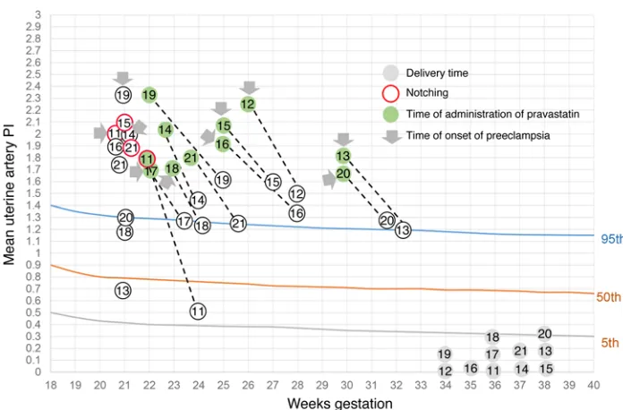
Twenty-one women with persistent aPL antibodies participated in this study. Most women met APS criteria before the current pregnancy, and a few of them met criteria during the current preg-nancy (Tables 1 and 2). All women had a poor obstetric history, as can be observed in Tables 1 and 2. Regardless of the fact that some women did not fulfill the criteria for APS at the beginning of the current pregnancy, all women were treated with antithrombotic therapy from the time of the first positive pregnancy test (35). The live birth rate prior to this study in the control group was 3.5% and 8% in the group that received pravastatin. All women were treated with LMWH (enoxaparin or tinzaparin, 40 mg subcutaneously, once daily) plus LDA (80 mg orally, once daily). Despite treatment, all of the patients developed PE and/or IUGR (Tables 1 and 3). Most of the patients were diagnosed with early PE (21–23 weeks) and received methyldopa (MDP) to treat hypertension. None of the 21 patients had underlying chronic hypertension, and all pre-sented normal blood pressure (BP) values prior to the onset of PE. Increased impedance to blood flow in the uterine arteries as shown by a pulsatility index (PI) above the 95th percentile — frequently associated with pregnancy complications associated with defec-tive trophoblast invasion — (36, 37) was observed in all women during Doppler assessment of uteroplacental perfusion (Tables 1 and 2, and Figure 1). Presence of diastolic notching (a character-istic waveform indicating decreased early diastolic flow) in the uterine artery reflects low vessel elasticity (37). Bilateral diastolic notching in uterine arteries was observed in 7 patients.
Patients 1 through 10 continued to be treated with standard LMWH+LDA after PE and/or IUGR diagnosis. Doppler measure-ments in the uterine arteries remained abnormal with standard LMWH+LDA treatment. A slight decrease in BP was observed after the administration of MDP. After PE and/or IUGR was diag-nosed, pregnancies continued for approximately 4 weeks (medi-an, 4.5 weeks; interquartile range [IQR], 2–6). Three pregnancies ended in stillbirth at 25 to 26 weeks. Preterm cesarean delivery (C-section) was performed in 7 women due to fetal distress and or maternal health concerns (median C-section time, 26.3 weeks; IQR, 26–32). One neonate died 3 hours after birth. Because of pre-maturity (birth weight: median, 900 g; IQR, 580–1,100) all neo-nates were admitted to the neonatal intensive care unit (NICU). Of the 7 neonates admitted to the NICU, 2 neonates died — one at 3 days and the other at 2 months of age — due to infection. The remaining 5 neonates were discharged from the NICU, and 3 of them currently present neurological and gastrointestinal develop-mental abnormalities.
In patients 11 to 21, pravastatin (20 mg) was added to the stan-dard of care treatment as soon as signs of PE and/or IUGR were observed until delivery. This group also received MDP to treat hypertension. Following the addition of pravastatin to conven-tional therapy, uteroplacental blood flow improved (Figure 1) and proteinuria and BP diminished significantly (Table 3). To evaluate improvement of maternal signs of PE after pravastatin treatment, a threshold of BP of 130/90 mmHg or less and proteinuria of 300 mg/dl or less was set. The BP/proteinuria values that reached these thresholds after pravastatin treatment are shown in Table 3. After reaching the threshold values, BP and proteinuria remained within those values or improved until the end of pregnancy. Time directed specifically to the underlying causes. Although delivery
is always beneficial for the mother, it may not be optimal for the fetus, as it might be extremely premature.
Efforts to reduce the risks of placenta insufficiency in APS remain critically important, and developing effective pharma-cological strategies will be of significant clinical benefit for mothers and fetuses.
Clinical studies suggest that the main cause of placental insuf-ficiency is a disturbance in uteroplacental circulation. Although placenta malperfusion is unique to pregnancy, there are many bio-logical and pathobio-logical similarities as well as risk factors in com-mon with cardiovascular disease (CVD). Normal development of maternal hyperlipidemia of pregnancy is exaggerated in PE (18). In addition, acute atherosis affects uteroplacental spiral arteries in 20% to 40% of cases of PE and resembles the early stages of atherosclerosis characterized by lipid deposition and inflammatory cells in the walls of maternal uterine arteries (19, 20). Thus, both conditions, PE and atherosclerotic CVD, are characterized by arte-rial foam cell deposition, endothelial dysfunction, oxidative stress, and inflammation. Moreover, patients with aPL antibodies have a high rate of atherosclerosis (21) and probably also high rates of ear-ly gestation development of uteroplacental acute atherosis (22, 23). Statins block the biosynthesis of cholesterol, and their favor-able effects have been well established for the prevention of ath-erosclerotic CVD. However, the benefit achieved with statin treat-ment in patients with hypercholesterolemia cannot be attributed to their cholesterol-lowering effect alone. Indeed, lipid-indepen-dent pleiotropic effects, including endothelial protection and reg-ulation of immune, inflammatory, and procoagulant responses, have been described (24).
Studies in animal models support the hypothesis that statins may be an effective means of preventing pregnancy complications related to APS and PE (25–29). Pravastatin has been shown to pre-vent adverse pregnancy outcomes in mouse models of APS and PE by diminishing inflammation, increasing placental blood flow, and reversing angiogenic and redox imbalances (26, 28, 29). Therefore, animal models and the similarities between PE and CVD provide a strong basis for the use of statins in the prevention of placental insufficiency in APS in humans. We have previously reported the clinical improvement in a preeclamptic APS patient treated with pravastatin (30). Similarly, a recent study has reported the success-ful use of pravastatin in preventing intrauterine fetal death (IUFD) in a case of massive perivillous fibrin deposition in the placenta (31). Another recent study has shown that pravastatin stabilized clinical and biochemical features of PE in 4 women (32). Statins have been shown not to be teratogenic as demonstrated by several studies (33, 34). A study by Costantine et al. reported no identifiable safety risks associated with pravastatin use in a pilot randomized trial designed to determine pravastatin safety and pharmacokinetic parameters in pregnant women at high risk of PE (33). Finally, a recent landmark study did not find congenital malformations and organ-specific malformations in the offspring of over 800,000 women exposed to statins during the first trimester of pregnancy (34).
tatin, rEDV was observed in patients 1 and 8. Emergency C-sec-tions were performed in both patients 1 week after the detection of abnormal Dopplers because of fetal distress. One of the neonates died 3 days after birth, and the other spent 4 months in the NICU and currently shows neurological abnormalities.
In the pravastatin-treated group, patients 12 and 19 also showed rEDV and IUGR (Table 2). However, while fetal survival in this situation is very unlikely, fetal weight gain and normal car-diotocography (CTG) were observed with pravastatin treatment, and pregnancies continued for 8 and 12 weeks, respectively. Redis-(days) of threshold achievement was defined as the shortest time
at which BP and proteinuria thresholds were reached (Table 3). BP diminished in patients who developed PE and also in patients 13, 16, and 19, who showed borderline hypertension and severe IUGR. The initial response to treatment was observed at as early as 10 days (median, 14 days; IQR, 10–15) (Figure 1 and Table 3).
Abnormal umbilical artery Doppler (absent end diastolic flow [aEDV] or reverse end diastolic flow [rEDV]) in the presence of IUGR is an ominous finding that requires immediate delivery to prevent IUFD. In the control group that did not receive
pravas-Table 1. Antibody profile, obstetric and medical history, risk factors, characteristics of current pregnancy, and pregnancy/fetal outcomes in 10 APS patients (1–10) who were treated with standard LMW+LDA
Patient no.,
age Past medical history/ risk factors Current pregnancy LMWH +LDA delivery Time of (from PE/severe Latency time IUGR diagnosis)
Neonatal outcome
1, 32 yr APS (aCL IgM positive,
3 miscarriages) 12 wk: ↑ Ut art PI, 21 wk: bilateral notching, ↑ Ut art PI, 24 wk: PE and severe IUGR < 5%,
25 wk: rEDV in umbilical artery
Yes 26 wk emergency
C-section 2 wk Time NICU: 4 mo BW: 520 g Current status of infant:
abnormal neurodev 2, 27 yr APS (LA positive,
3 miscarriages) 21 wk: ↑ Ut art PI, severe IUGR < 5% Yes 26 wk: stillbirth FW: 400 g 5 wk – 3, 28 yr APS ( LA positive, aβ2GPI positive,
stillbirth) 21 wk: 32 wk: severe IUGR < 5%↑ Ut art PI, IUGR < 10%, Yes 34 wk: emergency C-section 2 wk Time NICU: 3 mo BW: 980 g, Current status of infant: abnormal
neurodev and GI abnormalities 4, 38 yr APS (aCL IgG, 3 miscarriages,
5 IVFs, age > 35 yr) 21 wk: 31 wk: severe IUGR < 5%↑ Ut art PI, 30 wk: severe PE, Yes 32 wk: emergency C-section 2 wk Time NICU: 3.5 mo, BW: 1100 g,
Pseudomonas aeruginosa infection Current status of infant:
normal development 5, 37 yr APS (LA positive,
1 miscarriage < 10 wk, 1 miscarriage > 10 wk, SLE treated with HCQ,
age > 35 yr)
21 wk: ↑ Ut art PI, bilateral notching,
severe IUGR < 5%, 23 wk: severe PE Yes 25 wk: stillbirth FW: 450 g 4 wk –
6, 31 yr APS (LA positive, HELLP syndrome
28 wk, postpartum DVT) 25 wk: fetal weight stagnation for 3 wk21 wk: severe IUGR < 5%, Yes 26 wk: stillbirth FW: 430 g 5 wk – 7, 32 yr APS (triple positivity, DVT,
severe early PE 24 wk, stillbirth 26 wk)
21 wk: ↑ Ut art PI, bilateral notching,
27 wk: severeIUGR< 5%,PE Yes 28 wk: emergency C-section 7 wk Admitted to NICU 2 mo later, BW: 700 g died of infection 8, 43 yr APS (LA positive, ACA IgM positive,
3 miscarriages > 10 wk, age > 35 yr, IVF twin pregnancy)
21 wk: bilateral notching, severeIUGR (boy < 1%, girl < 5%) mild hypertension,
26 wk: PE, severe IUGR, rEDV in umbilical artery (boy)
Yes 27 wk: emergency
C-section 6 wk BW: 580 g (boy), 900 g (girl) Born alive Admitted to NICU Boy died at 3 days Girl spent 4 mo in NICU 9, 31 yr APS (LA positive), HELLP syndrome,
stillbirth (29 wk) 26 wk: severe IUGR < 5%, 21 wk: ↑ Ut art PI, 32 wk: weigh stagnation
Yes 33 wk: emergency
C-section 7 wk 1 mo at NICU FW: 1100 g Developmental problems 10, 37 yr APS (ACA IgM, aβ2GPI IgM,
miscarriage > 10 wk) 21 wk: 22 wk: severe PE and IUGR, ↑ Ut art PI, hypertension, IUGR, 24 wk: weight stagnation
Yes 25 wk: emergency
C-section 4 wk Baby died 3 hours after birth
Median 26.5 wk 4.5 wk BW: 900 g
IQR (26–32) (2–6) (580–1,100)
[image:4.585.42.550.91.544.2]pravastatin delivered at 36 weeks or later, with excellent neonatal outcomes (Figure 1 and Table 3), compared with the control group in which 3 stillbirths occurred and 100% of the remaining deliv-eries occurred preterm. All of the patients cotreated with pravas-tatin delivered after 34 weeks, thus diminishing substantially the chances for any prematurity-associated adverse neonatal out-comes. All women received antenatal steroids to improve respi-ratory neonatal outcome. Three patients (patients 12, 16, and 19) (27%) delivered at 34 to 35 weeks. Patient 12 had a twin pregnancy, and patient 19 showed early signs of severe IUGR. Neonates from patients 12 (twin pregnancy) and 19 (singleton) were admitted to the NICU; the length of stay varied from 10 days to 90 days and was related to gestation and birth weight (Table 3), but the neo-nates are now healthy and show normal development for their age. Only one patient in the control group delivered at 34 weeks (patient 3). The neonate showed a significant growth restriction (980 g), spent 3 months in the NICU, and currently shows neuro-logical and gastrointestinal abnormalities (Table 1).
There were no congenital abnormalities or late fetal deaths and no evidence of maternal morbidity because of the use of pravastatin, adding to the growing evidence that statins are safe in pregnancy.
Discussion
The currently accepted first-line treatment for obstetric APS is throm-boprophylactic treatment with LDA and heparinoids. However, in tribution of fetal cardiac output in response to abnormal placental
perfusion to maintain delivery of oxygen and nutrients to the brain — brain sparing — was observed in all growth-restricted fetuses, but remained stable until delivery in patients treated with pravastatin.
[image:5.585.37.550.93.392.2]Preterm delivery, spontaneous or iatrogenic, is a complica-tion frequently associated with PE. Infants born preterm are vulnerable to many complications, including respiratory distress syndrome, chronic lung disease, injury to the intestines, com-promised immune system, cardiovascular disorders, hearing and vision problems, and neurological insult. Infants born at the lower limit of viability have the highest mortality rates and the highest rates of all complications. All patients in the control group that received standard therapy delivered preterm by emergency C-sections (median, 26.5 weeks; IQR, 26–32) because of fetal and/or maternal health concerns. Pravastatin prolonged pregnan-cies significantly after the onset of PE (median, 13 weeks; IQR, 10–15) compared with those women who did not receive pravas-tatin (median, 4.5 weeks; IQR, 2–6; P < 0.01) (Tables 1 and 3). With pravastatin treatment, women delivered close to term (median, 36 weeks; IQR, 35–36), increasing the chances of fetal matura-tion and survival. In the group that did not receive pravastatin, all premature neonates were admitted to the NICU and 3 perinatal deaths were observed. The obstetric end point that was set for the cohort of patients treated with pravastatin was to deliver them after 37 weeks. Interestingly, 8 (73%) of the patients who received
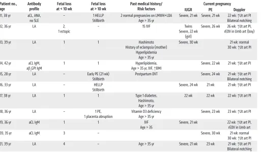
Table 2. Antibody profile, obstetric and medical history, risk factors, and characteristics of current pregnancy of the 11 APS patients (11–21) who were treated with standard LMW+LDA treatment supplemented with pravastatin
Patient no.,
age Antibody profile at < 10 wkFetal loss at > 10 wkFetal loss Past medical history/ Risk factors IUGR Current pregnancyPE Doppler 11, 38 yr aCL, ANA,
no SLE – Stillbirth1 HELLP 2 normal pregnancies on LMWH+LDA Age > 35 yr Severe, 21 wk Severe, 21 wk Bilateral notching22 wk: ↑Ut art PI 12, 36 yr LA 2,
1 ectopic – 15 IVF Severe, 22 wk Twins (girl)
Severe, 26 wk 26 wk: ↑Ut art PI, rEDV in Umb art (boy) 13, 39 yr LA 1 1 Hashimoto
History of eclampsia (mother) Hyperlipidemia
Age > 35 yr
Severe, 30 wk 21 wk: normal 30 wk: ↑Ut art PI
14, 42 yr aCL IgM, aβ2GPI IgM
1 1 Hyperlipidemia,
Age > 35 yr, IVF, ↑BMI Severe, 22 wk 21 wk: ↑Ut art PI 15, 28 yr LA – Early PE (21 wk)
Stillbirth Postpartum DVT Severe, 24 wk Bilateral notching21 wk: ↑Ut art PI 16, 33 yr LA – HELLP
Stillbirth Severe, 24 wk 21 wk 21 wk: ↑Ut art PI 17, 38 yr LA 1 1 Type 1 diabetes,
Hashimoto, Age > 35 yr
22 wk 22 wk 22 wk: ↑Ut art PI
18, 36 yr LA – 1 PE,
1 placenta abruption Vitamin D3 deficiency Age > 35 yr Severe, 23 wk 23 wk: ↑Ut art PI 19, 36 yr aCL IgM 1 1 IVF
Age > 35 Severe, 21 wk 22 wk: rEDV in Umb art↑Ut art PI, 20, 35 yr aCL IgM 3 – Severe, 30 wk 21 wk: normal
30 wk: ↑Ut art PI 21, 39 yr LA 4 – Age > 35 yr Severe, 21 wk 23 wk 21 wk: ↑Ut art PI
Bilateral notching
Limitations of the present study include the small number studied.
MDP remains one of the most widely used drugs for the treatment of PE. The use of antihypertensive therapy might also contribute to the improvement of maternal signs of PE in this cohort of women. However, treatment of preeclamptic women remote from term with antihypertensive drugs showed limited benefits in prolonging gestation and improving peri-natal outcomes (40). In fact, in 5 out of the 10 women in the control group who did not receive pravastatin, MDP did not decrease BP significantly and pregnancies were prolonged for a short time (median, 4 weeks; IQR, 2–5). Some women in this study were put on bed rest. While some studies suggest this might help control BP, there are no data to support the benefit of bed rest in prolonging gestation (41).
The rapid response to pravastatin observed in uterine artery hemodynamic parameters suggests that pravastatin might be tar-geting placental vasculopathy in APS patients who develop PE. It is tempting to speculate that pravastatin might increase placen-tal blood flow by its antiatherogenic effects and by stimulating the release of vasoactive substances from the endothelium, such as nitric oxide or carbon monoxide (42, 43). Statins have also been shown to downregulate tissue factor, a crucial molecule in the crosstalk between inflammation and thrombosis in mice and humans with APS (29, 44). These protective effects of pravastatin on the endothelium together with its effect restoring angiogenic balance (24) might also explain the amelioration of placental and maternal preeclamptic signs.
approximately 20% of obstetric APS cases, good pregnancy outcomes cannot be achieved with this conventional treatment (38). In fact, women with APS that received LDA+LMWH treatment throughout pregnancy had higher rates of PE than control women without APS (7). That the 21 patients described in this study developed PE and/or IUGR despite the use of antithrombotic therapy suggests that avert-ing thrombosis is not always effective in preventavert-ing placental insuf-ficiency–associated pregnancy complications in APS (5, 6).
The risk of neonatal death in a preeclamptic pregnancy is considerable, especially the risk associated with prematurity (15–17). Early onset PE is associated with the highest neona-tal morneona-tality. Moreover, the risk of prematurity and perinaneona-tal death is exacerbated in APS pregnancies. IUGR (26.3% of the total live births) and prematurity (48.2%) are the most frequent fetal morbidities in APS (39). Following pravastatin addition to conventional standard of care therapy, placental blood flow and maternal signs of PE improved significantly in this cohort, lead-ing to live births in 100% of the patients. The beneficial effects of pravastatin were observed within a short time period and allowed pregnancies to survive. On the other hand, only 45% of pregnancies resulted in live births in the patients who received conventional antithrombotic therapy, and 40% of the surviving neonates showed developmental abnormalities.
[image:6.585.43.553.95.307.2]Considering the adverse outcomes in previous pregnancies and in patients who received LMWH+LDA without pravastatin supplementation, the present study suggests that women with refractory obstetric APS may have improved pregnancy outcomes with pravastatin taken at the time of onset of PE or severe IUGR.
Table 3. Gestational age at diagnosis of PE/IUGR, BP and proteinuria values before and after pravastatin treatment, gestational age at delivery and fetal outcomes
Patient
no. Gestational age at diagnosis PE/IUGR (wk)
Before pravastatin Pravastatin treatment started at
(wk)
Values < threshold after pravastatin Time of threshold achievement
(d)
Pregnancy survival after
pravastatin (wk)
Gestational age at delivery
(wk)
Birth weight
(g) BP (mmHg) Proteinuria
(mg/dl) BP (mmHg) Proteinuria (mg/dl) Systolic Diastolic Systolic Diastolic
11 21 165 80 680 22 130 85 205 14 14 36 2,230 12 26 160 100 520 26 130 90 290 14 8 34 1,400 girlA,
1,900 boyB
13 30 130 92 350 30 125 85 204 15 8 38 2710 14 22 180 115 400 23 142 90 250 15 14 37 3,250 15 24 160 94 432 25 130 90 280 14 13 38 2,830 16 24 130 90 300 25 120 80 210 20 10 35 2,390 17 22 150 110 568 22 130 87 290 10 14 36 3,100 18 23 190 110 1014 23 130 92 429 15 13 36 2,310 19 21 138 90 320 23 120 85 250 20 12 34 1,800C
20 30 180 115 390 30 130 89 230 10 8 38 2,700 21 23 160 98 380 24 130 89 260 10 13 36 2,390 Median 23 160 98 400 24 130D 89E 250F 14 13 36 2390
IQR 22–26 138–180 90–110 350–568 23–26 125–130 85–90 210–290 10–15 8–14 35–38 2,065–2,770
Pravastatin ameliorates preeclamptic features in all APS patients. Threshold values (BP ≤ 130/90 mmHg, proteinuria ≤ 300 mg/dl) were set to evaluate treatment efficacy. Values after pravastatin correspond to the values less than or equal to the threshold that remained constant or improved until delivery. Median time of threshold achievement was 14 days (IQR, 10–15). Live births were observed in all of the patients. A10 days in NICU. B90 days
in NICU. C30 days in NICU. DDifference from before pravastatin, P = 0.0005. EDifference from before pravastatin, P = 0.002. FDifference from before
Overall, the results obtained in this study in which preg-nant women with APS who did not respond to conventional antithrombotic therapy responded satisfactorily to pravas-tatin appear encouraging in a patient population with frequent adverse pregnancy outcomes and warrant further investiga-tion. Randomized clinical trials should be organized to confirm these observations.
Methods
Between 2013 and 2015, at the Hippokration General Hospital of Thessaloniki, twenty-one women with APS had been treated with LMWH (enoxaparin or tinzaparin, 40 mg subcutaneously, once dai-ly) plus LDA (80 mg orally, once daidai-ly) from the first positive preg-nancy test. APS was defined by the presence of clinical criteria and laboratory criteria as follows. Clinical criteria included 1 or more clinical episodes of thrombosis and pregnancy morbidity. Pregnan-cy morbidity was defined as 1 or more unexplained fetal deaths at or beyond the 10th week of gestation or 1 or more premature births before the 34th week of gestation because of placental insufficiency such as PE or 3 or more unexplained consecutive spontaneous abor-tions before the 10th week of gestation. Lupus anticoagulant (LA) and/or anticardiolipin (aCL) and/or β2 glycoprotein-I IgG or IgM antibody present in plasma or serum on 2 or more occasions, at least 12 weeks apart, are included in the laboratory criteria for APS (45). While some of the women met the criteria for obstetric APS before this study and others had persistent aPL antibodies and met crite-ria during the current pregnancy, all women received conventional LDA+LMWH treatment (35).
Despite the aforementioned antithrombotic treatment, all of the patients developed placental insufficiency–related complica-tions such as PE and IUGR. Most of the patients were diagnosed with early PE (21–23 weeks).
PE was defined according to the report of the American College of Obstetricians and Gynecologists Task Force on Hypertension in Preg-nancy (46). PE was defined as BP elevation after 20 weeks of gestation with proteinuria or any of the following features: thrombocytopenia,
impaired liver function, new development of renal insufficiency, pul-monary edema, or new onset of cerebral or visual disturbances. IUGR was defined as fetal weight below the 10th percentile, and severe IUGR was defined as fetal weight below the 5th percentile for gestational age and abdominal circumference below the 2.5th percentile (47).
The patients’ past obstetric and medical history, risk factors, and characteristics of current pregnancy, including Doppler studies, are presented in Table 1 (patients who were treated with LMWH+LDA) and Table 2 (patients who were supplemented with pravastatin). All women had a poor obstetric history with a low rate of live births. Elev-en patiElev-ents consElev-ented and were started on pravastatin (20 mg/d) from the time they were diagnosed with PE or IUGR, and 10 received only standard LMWH+LDA treatment.
[image:7.585.35.383.53.282.2]Median age for the patients who were supplemented with pravas-tatin was 38 years (IQR, 35–39), and 32 years (IQR, 31–37) was the median age for the group that received only standard therapy. Women with PE were given MDP (1 g/d in 2 divided doses) to treat hyperten-sion. All patients were admitted to the hospital, most of them until delivery. Because these were inpatients in the high-risk pregnancy unit, close maternal and fetal monitoring was undertaken. Maternal monitoring included measurement of BP 3 times a day, measurement of proteinuria, and blood tests to assess liver and kidney function and number of platelets twice weekly. Ultrasound assessment of fetal growth was performed every week, amniotic fluid volume was assessed twice weekly, and umbilical, middle cerebral artery, ductus venosus, and uterine artery Doppler velocimetry were performed twice weekly to monitor fetal status. Daily monitoring of the fetus with CTG was also performed. Maternal and fetal progress were reviewed regularly. Patients with PE were discharged to the community when BP was less than 130/90 mmHg and blood results were stable or improving and, in the case of IUGR fetuses, when acceleration of fetal weight gain was observed along with stable or improved fetal and maternal Doppler measurements. After discharge, women were advised to check their BP twice daily and to visit the antenatal day assessment unit 3 times a week in order to have a CTG and to review their measurements. Blood tests were also performed during these visits, and the quantity
Figure 1. Mean uterine artery PI versus gestational age in the general population (estimated 5th, 50th, and 95th percentiles are shown) and in the 11 patients with APS (11–21) who were treated with LMWH+LDA
supplemented with pravastatin. PI values
of urinary protein was checked once a week. Doppler assessment after discharge was performed twice weekly. Despite improvement after pravastatin treatment, some patients chose to remain as inpatients in the high-risk pregnancy unit until delivery for close monitoring, main-ly because of maternal anxiety.
Statistics. Statistical analysis to compare BP and proteinuria
val-ues before and after pravastatin treatment and time from diagnosis to delivery between pravastatin-treated women and control was con-ducted using a paired t test. P < 0.05 was considered statistically sig-nificant. Medians and IQRs are reported for all variables measured. All analysis was conducted with GraphPad Prism statistical software (GraphPad Software Inc.).
Study approval. This study was approved by the Ethical Review
Committee at Hippokration General Hospital of Thessaloniki, and informed consent was obtained from all pregnant patients.
Author contributions
GG and EL conceived and designed the study. GG and EL ana-lyzed and interpreted data. GG created graphs and wrote the man-uscript. TD, CV, and AM collected data. DR was responsible for the overall supervision of the patients.
Acknowledgments
This work was funded by local sources from the Hippokration General Hospital of Thessaloniki.
Address correspondence to: Guillermina Girardi, Division of Women’s Health, The Rayne Institute - 4th Floor Lambeth Wing, St. Thomas’ Hospital, King’s College London, London SE1 7EH, United Kingdom. Phone: 44.0.207.188.1101; E-mail: guillermina. girardi@kcl.ac.uk.
1. Danza A, Ruiz-Irastorza G, Khamashta M. Antiphospohlipid syndrome in obstetrics. Best Pract Res Clin Obstet Gynaecol. 2012;26(1):65–76. 2. Stone S, et al. The placental bed in pregnancies
complicated by primary antiphospholipid syn-drome. Placenta. 2006;27(4–5):457–467. 3. Girardi G, et al. Complement C5a receptors
and neutrophils mediate fetal injury in the antiphospholipid syndrome. J Clin Invest. 2003;112(11):1644–1654.
4. Meroni PL, Gerosa M, Raschi E, Scurati S, Grossi C, Borghi MO. Updating on the pathogenic mechanisms 5 of the antiphospholipid antibod-ies-associated pregnancy loss. Clin Rev Allergy Immunol. 2008;34(3):332–337.
5. Laskin CA, et al. Low molecular weight heparin and aspirin for recurrent pregnancy loss: results from the randomized, controlled HepASA Trial. J Rheumatol. 2009;36(2):279–287.
6. Branch DW, Silver RM, Blackwell JL, Reading JC, Scott JR. Outcome of treated pregnancies in women with antiphospholipid syndrome: an update of the Utah experience. Obstet Gynecol. 1992;80(4):614–620.
7. Bouvier S, et al. Comparative incidence of preg-nancy outcomes in treated obstetric antiphos-pholipid syndrome: the NOH-APS observational study. Blood. 2014;123(3):404–413.
8. Girardi G, Redecha P, Salmon JE. Heparin pre-vents antiphospholipid antibody-induced fetal loss by inhibiting complement activation. Nat Med. 2004;10(11):1222–1226.
9. Lefkou E, Hunt B. Pre-eclampsia. In: Pavord S, Hunt B, eds. The Obstetric Hematology Manual. Cambridge, United Kingdom: Cambridge Uni-versity Press; 2010:203–217.
10. Duckitt K, Harrington D. Risk factors for pre-eclampsia at antenatal booking:systematic review of controlled studies. BMJ. 2005;330(7491):565.
11. Nodler J, Moolamalla SR, Ledger EM, Nuwayhid BS, Mulla ZD. Elevated antiphospholipid anti-body titers and adverse pregnancy outcomes: analysis of a population-based hospital dataset. BMC Pregnancy Childbirth. 2009;9:11. 12. Heilmann L, Schorsch M, Hahn T, Fareed J.
Antiphospholipid syndrome and pre-eclampsia. Semin Thromb Hemost. 2011;37(2):141–145.
13. Adams T Yeh C, Bennett-Kunzier N, Kinzler WL. Long-term maternal morbidity and mortal-ity associated with ischemic placental disease. Semin Perinatol. 2014;38(3):146–150. 14. Dekker GA. Management of preeclampsia.
Preg-nancy Hypertens. 2014;4(3):246–247.
15. Maternal mortality. World Health Organization. http://www.who.int/mediacentre/factsheets/ fs348/en/. Updated November 2015. Accessed June 27, 2016.
16. Cousens S, et al. National, regional, and world-wide estimates of stillbirth rates in 2009 with trends since 1995: a systematic analysis. Lancet. 2011;377(9774):1319–1330.
17. Vogel JP, et al. Maternal complications and perina-tal morperina-tality: findings of the World Health Orga-nization Multicountry Survey on Maternal and Newborn Health. BJOG. 2014;121(suppl 1):76–88. 18. Wetzka B, Winkler K, Kinner M, Friedrich I, März
W, Zahradnik HP. Altered lipid metabolism in preeclampsia and HELLP syndrome: links to enhanced platelet reactivity and fetal growth. Semin Thromb Hemost. 1999;25(5):455–462. 19. Staff AC, Dechend R, Pijnenborg R. Learning
from the placenta: acute atherosis and vascular remodelling in preeclampsia-novel aspects for atherosclerosis and future cardiovascular health. Hypertension. 2010;56(6):1026–1034.
20. Staff AC, Johnsen GM, Dechend R, Redman CW. Preeclampsia and uteroplacental acute athero-sis: immune and inflammatory factors. J Reprod Immunol. 2014;101–102:120–126.
21. Ames PR, Margarita A, Alves JD. Antipohospho-lipid antibodies and atherosclerosis: insights from systemic lupus erythematosus and primary antiphospholipid syndrome. Clin Rev Allergy Immunol. 2009;37(1):29–35.
22. Levy RA, Avvad E, Oliveira J, Porto LC. Placental pathology in antiphospholipid syndrome. Lupus. 1998;7(suppl 2):S81–S85.
23. Nayar R, Lage JM. Placental changes in a first trimester missed abortion in maternal systemic lupus erythematosus with antiphospholipid syn-drome; a case report and review of the literature. Hum Pathol. 1996;27(2):201–206.
24. Girardi G. Can statins prevent pregnancy compli-cations? J Reprod Immunol. 2014;101–102:161–167. 25. Ahmed A, Singh J, Khan Y, Seshan SV, Girardi G.
A new mouse model to explore therapies for pre-eclampsia. PLoS One. 2010;5(10):e13663. 26. Costantine MM, et al. Using pravastatin to
improve the vascular reactivity in a mouse model of soluble fms-like tyrosine kinase-1- induced preeclampsia. Obstet Gynecol. 2010;116(1):114–120.
27. Kumasawa K, et al. Pravastatin induces placental growth factor (PGF) and ameliorates preeclamp-sia in a mouse model. Proc Natl Acad Sci U S A. 2011;108(4):1451–1455.
28. Singh J, Ahmed A, Girardi G. Role of complement component C1q in the onset of preeclampsia in mice. Hypertension. 2011;58(4):716–724. 29. Redecha P, Franzke CW, Ruf W, Mackman N,
Girardi G. Neutrophil activation by the tissue fac-tor/Factor VIIa/PAR2 axis mediates fetal death in a mouse model of antiphospholipid syndrome. J Clin Invest. 2008;118(10):3453–3461. 30. Lefkou E, et al. Clinical improvement and
suc-cessful pregnancy in a preeclamptic patient with antiphospholipid syndrome treated with pravas-tatin. Hypertension. 2014;63(5):e118–e119. 31. Chaiworapongsa T, et al. Pravastatin to prevent
recurrent fetal death in massive perivillous fibrin deposition of the placenta (MPFD). J Matern Fetal Neonatal Med. 2016;29(6):855–862.
32. Brownfoot FC, et al. Effects of Pravastatin on human placenta, endothelium, and women with severe preeclampsia. Hypertension. 2015;66(3):687–697.
33. Costantine MM, et al.Safety and pharmacokinet-ics of pravastatin used for the prevention of pre-eclampsia in high-risk pregnant women: a pilot randomized controlled trial. Am J Obstet Gynecol. 2015;214(6):720.e1–720.e17.
34. Bateman BT, et al. Statins and congenital malfor-mations: cohort study. BMJ. 2015;350:h1035. 35. Arachchillage DR, Machin SJ, Mackie IJ, Cohen
H. Diagnosis and management of non-criteria obstetric antiphospholipid syndrome. Thromb Haemost. 2015;113(1):13–19.
36. Campbell S, et al. New doppler technique for assessing uteroplacental blood flow. Lancet. 1983;1(8326 pt 1):675–677.
restriction. Prenat Diagn. 2010;30(4):293–308. 38. Lopez-Pedrera C, et al. Immunotherapy in
antiphospholipid syndrome. Int Immunopharma-col. 2015;27(2):200–208.
39. Cervera R, et al. Morbidity and mortality in the antiphospholipid syndrome during a 10-year period: a multicentre prospective study of 1000 patients. Ann Rheum Dis. 2015;74(6):1011–1018. 40. Mutch LM, Moar VA, Ounsted MK, Redman
CW. Hypertension during pregnancy, with and without specific hypotensive treatment. Perinatal factors and neonatal morbidity. Early Human Develop. 1977;1(1):45–57.
41. Bigelow C, Stone J. Bed rest in pregnancy. Mt
Sinai J Med. 2011;78(2):291–302. 42. Kaesemeyer WH, Caldwell RB, Huang J,
Caldwell RW. Pravastatin sodium activates endo-thelial nitric oxide synthase independent of its cholesterol-lowering actions. J Am Coll Cardiol. 1999;33(1):234–241.
43. Ramma W, Ahmed A. Therapeutic potential of statins and the induction of heme oxygenase-1 in preeclampsia. J Reprod Immunol. 2014; 101–102:153–160.
44. Erkan D, et al. A prospective open-label pilot study of fluvastatin on proinflammatory and prothrombotic biomarkers in antiphospholipid antibody positive patients. Ann Rheum Dis.
2014;73(6):1176–1180.
45. Miyakis S, et al. International consensus state-ment on an update of the classification criteria for definite antiphospholipid syndrome (APS). J Thromb Haemost. 2006;4(2):295–306. 46. American College of Obstetricians and
Gynecolo-gists, Task Force on Hypertension in Pregnancy. Hypertension in pregnancy. Report of the Ameri-can College of Obstetricians and Gynecologists’ Task Force on Hypertension in Pregnancy. Obstet Gynecol. 2013;122(5):1122–1131.