Hypertension is a leading contributor to cardiovascular mortality worldwide. Despite this, its
underlying mechanism(s) and the role of excess salt in cardiorenal dysfunction are unclear.
Previously, we have identified cross-talk between mineralocorticoid receptor (MR), a
nuclear transcription factor regulated by the steroid aldosterone, and the small GTPase
Rac1, which is implicated in proteinuric kidney disease. We here show that high-salt
loading activates Rac1 in the kidneys in rodent models of salt-sensitive hypertension,
leading to blood pressure elevation and renal injury via an MR-dependent pathway. We
found that a high-salt diet caused renal Rac1 upregulation in salt-sensitive Dahl (Dahl-S)
rats and downregulation in salt-insensitive Dahl (Dahl-R) rats. Despite a reduction of serum
aldosterone levels, salt-loaded Dahl-S rats showed increased MR signaling in the kidneys,
and Rac1 inhibition prevented hypertension and renal damage with MR repression. We
further demonstrated in aldosterone-infused rats as well as adrenalectomized Dahl-S rats
with aldosterone supplementation that salt-induced Rac1 and aldosterone acted
interdependently to cause MR overactivity and hypertension. Finally, we confirmed the key
role of Rac1 in modulating salt susceptibility in mice lacking Rho GDP–dissociation
inhibitor
a
. Therefore, our data identify Rac1 as a determinant of salt sensitivity and provide
insights into the mechanism of salt-induced hypertension and kidney injury.
Research Article
Nephrology
Find the latest version:
Research article
Rac1 GTPase in rodent kidneys
is essential for salt-sensitive
hypertension via a mineralocorticoid
receptor–dependent pathway
Shigeru Shibata,1,2 ShengYu Mu,1 Hiroo Kawarazaki,1 Kazuhiko Muraoka,1
Ken-ichi Ishizawa,1 Shigetaka Yoshida,1 Wakako Kawarazaki,1 Maki Takeuchi,1
Nobuhiro Ayuzawa,1 Jun Miyoshi,3 Yoshimi Takai,4 Akira Ishikawa,2
Tatsuo Shimosawa,1 Katsuyuki Ando,1 Miki Nagase,1 and Toshiro Fujita1
1Department of Nephrology and Endocrinology and 2Division of Total Renal Care Medicine, University of Tokyo Graduate School of Medicine,
Tokyo, Japan. 3Department of Molecular Biology, Osaka Medical Center for Cancer and Cardiovascular Diseases, Osaka, Japan. 4Department of Molecular and Cellular Biology, Kobe University Graduate School of Medicine, Kobe, Japan.
Hypertension is a leading contributor to cardiovascular mortality worldwide. Despite this, its underlying
mechanism(s) and the role of excess salt in cardiorenal dysfunction are unclear. Previously, we have identified
cross-talk between mineralocorticoid receptor (MR), a nuclear transcription factor regulated by the steroid
aldosterone, and the small GTPase Rac1, which is implicated in proteinuric kidney disease. We here show
that high-salt loading activates Rac1 in the kidneys in rodent models of salt-sensitive hypertension, leading to
blood pressure elevation and renal injury via an MR-dependent pathway. We found that a high-salt diet caused
renal Rac1 upregulation in salt-sensitive Dahl S) rats and downregulation in salt-insensitive Dahl
(Dahl-R) rats. Despite a reduction of serum aldosterone levels, salt-loaded Dahl-S rats showed increased MR
signal-ing in the kidneys, and Rac1 inhibition prevented hypertension and renal damage with MR repression. We
further demonstrated in aldosterone-infused rats as well as adrenalectomized Dahl-S rats with aldosterone
supplementation that salt-induced Rac1 and aldosterone acted interdependently to cause MR overactivity and
hypertension. Finally, we confirmed the key role of Rac1 in modulating salt susceptibility in mice lacking Rho
GDP–dissociation inhibitor
α
. Therefore, our data identify Rac1 as a determinant of salt sensitivity and
pro-vide insights into the mechanism of salt-induced hypertension and kidney injury.
Introduction
Hypertension continues to be a substantial public health prob-lem worldwide (1), contributing to death from stroke, myocardial infarction, and end-stage renal disease. Although its causes remain unclear, owing to the trait’s complexity, increased salt sensitivity of blood pressure is a major contributing factor in a subgroup of hypertensive subjects (2, 3). Notably, the findings of single gene dis-orders that cause blood pressure variation have established the key role of renal salt handling in the pathogenesis of hypertension (4, 5). However, genetic defects that directly alter renal sodium transport are observed only in a small percentage of hypertensive subjects, and the mechanism of essential hypertension as well as that of interindi-vidual variation in salt sensitivity remain poorly understood.
The partnership between aldosterone and mineralocorticoid receptor (MR) in the kidneys tightly maintains sodium and fluid status by changing serum aldosterone levels counterbalanced by dietary salt. Nonetheless, the increased sodium intake in industri-alized societies has caused an excess of salt in the body, leading to hypertension (6), cardiovascular damage (7, 8), and kidney disease progression (9) despite suppression of serum aldosterone. Several lines of evidence indicate that an impaired feedback regulation
between sodium and aldosterone/MR appears to impact the salt-induced hypertension and cardiorenal damage (10–12). Indeed, in subjects with primary aldosteronism, high-sodium intake aggra-vates hypertension and end-organ damage that are associated with the inappropriate suppression of aldosterone, and the salt-medi-ated organ dysfunction is prevented by MR blockade, suggesting that aldosterone and high-salt levels synergistically potentiate MR-mediated signaling (13–15). In addition, excessive MR signaling is considered to be the key mechanism of end-organ damage even with low or normal serum aldosterone status, especially in the context of a high-sodium intake (16–18). Although these observations sup-port the existence of a definite pathway to augment MR signaling in a high-salt status, its molecular basis remains largely speculative.
Recently, we have identified signaling cross-talk between MR and the small GTPase Rac1 as a novel pathway that modulates MR func- tion (19). In this study, we show that a high-salt status acts synergis- tically with aldosterone to activate renal Rac1 in salt-sensitive hyper-tension, leading to high blood pressure and renal damage through potentiating MR signaling. We also show that the different response of renal Rac1 to high-sodium loading is a key mechanism that mod-ulates the salt sensitivity of blood pressure and kidney injury.
Results
Rac1 GTPase is involved in the heterogeneity of salt sensitivity through MR modulation. We first used Dahl salt-insensitive (Dahl-R) and Dahl
salt-sensitive (Dahl-S) rats, a model with an inherited predisposi-Authorship note: Shigeru Shibata, ShengYu Mu, and Hiroo Kawarazaki contributed equally to this work.
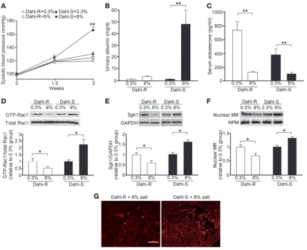
tion to salt-dependent hypertension. As compared with the control group fed a normal-salt (0.3%) diet, Dahl-R rats fed an 8%-salt diet did not show blood pressure elevation during 3 weeks of treatment period (Figure 1A). In contrast, salt-loaded Dahl-S rats developed significant hypertension at 3 weeks, which was accompanied by pathological albuminuria (Figure 1, A and B). In the normal-salt diet–fed groups, serum aldosterone concentrations were higher in Dahl-R rats than Dahl-S rats (Figure 1C); the results were identical to those in the previous studies (20). Salt loading suppressed serum aldosterone in both strains to similar levels (Figure 1C). In order to address the possibility that Rac1 is involved in the pathogenesis of salt-sensitive hypertension in this model, we assessed Rac1 activity in the kidneys. GST pull-down assay revealed that GTP-bound, active Rac1 expression was significantly elevated in the kidneys of Dahl-S rats by high-salt loading (Figure 1D). In sharp contrast, Rac1 activity was significantly reduced in Dahl-R rats in response to high-salt diet. The serine/threonine kinase Sgk1 is a key downstream effector of
[image:3.585.72.516.80.442.2]MR signaling in the kidneys, orchestrating the early and late respons- es through the regulation of epithelial sodium channel activity, traf-ficking, and transcription (21–23). The Sgk1 expression level also serves as a surrogate marker for MR signaling activity in vivo (15, 24). High-salt diet upregulated Sgk1 in the kidneys of Dahl-S rats, where-as its expression was significantly reduced in salt-loaded Dahl-R rats (Figure 1E). These results are in line with the previous observations indicating the role of Sgk1 in the increased salt sensitivity of Dahl-S rats (25, 26). When activated, MR translocates into the nucleus to regulate the transcription of targeted genes. Consistent with the Sgk1 expression, salt-loaded Dahl-S rats showed increased MR expression in the nuclear fraction, whereas it was decreased in salt- loaded Dahl-R rats (Figure 1F), indicating that MR signaling is actu-ally enhanced in the salt-loaded Dahl-S model. We also performed an immunofluorescence study for MR (Figure 1G). In salt-loaded Dahl-S rats, MR was strongly stained with a nuclear pattern in the distal nephron. In accordance with the previous studies, MR staining was also noted in glomerular cells (27, 28). No signals were detected in proximal tubules. Nuclear staining of MR in the distal nephron
Figure 1
research article
was less clear in salt-loaded Dahl-R rats. Taken together, salt-sensitive hypertension in the Dahl-S model is associated with the increased MR activity in the aldosterone-sensitive distal nephron. In further support of this possibility, serum potassium concentrations were significantly decreased in salt-loaded Dahl-S rats (4.34 ± 0.12 mEq/l in high-salt diet–fed Dahl-S rats versus 4.83 ± 0.13 mEq/l in normal-salt diet–fed Dahl-S group; P
< 0.05). This is consistent with the pre-vious observation by another group that noted a modest decrease in serum potassium levels in a similar model (29).
[image:4.585.84.511.82.565.2]To prove more moderate levels of salt intake would cause renal Rac1 elevation in Dahl-S rats, we examined the effects of a 4%-salt diet. After 4 weeks of treatment period, 4%-salt diet induced hyper-tension and albuminuria in Dahl-S rats (Supplemental Figure 1, A and B; supplemental material available online with this article;
Figure 2
doi:10.1172/JCI43124DS1). As expected, serum aldosterone levels were significantly suppressed in 4%-salt diet–fed rats (Supplemen-tal Figure 1C). Identical to that in the previous experiments, renal Rac1 activity was significantly elevated by 4%-salt diet (Supplemen-tal Figure 1D). Moreover, both nuclear MR and Sgk1 expression levels were significantly elevated by the salt diet (Supplemental Figure 1, E and F), and these observations were accompanied by the significant decrease in serum potassium levels (4.46 ± 0.08 mEq/l in 4%-salt diet–fed Dahl-S group versus 4.96 ± 0.13 mEq/l in nor-mal-salt diet–fed Dahl-S group; P < 0.01).
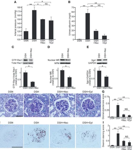
The above observations led us to the speculation that enhanced MR signaling and salt-dependent hypertension in Dahl-S rats can be attributable to Rac1 activation in the kidneys and that the dif- ferent response of renal Rac1 to salt intake is an underlying mecha-nism for the variation in salt sensitivity in Dahl model. To provide evidence that renal Rac1 activity affects salt sensitivity, Dahl-S rats fed an 8%-salt diet were treated with Nsc23766, a small molecule inhibitor that specifically reduces Rac activation (30, 31). We evalu-ated mean arterial pressure by direct measurement, using catheters inserted into lower aorta. As shown in Figure 2A, Nsc23766 par-tially but significantly reduced the arterial pressure in salt-loaded Dahl-S rats, confirming the causal role of Rac1 activation in salt-dependent elevation of blood pressure. In Dahl-S rats treated with the selective MR antagonist eplerenone, mean arterial pressure was reduced to the same extent as after treatment with Nsc23766. Both treatments successfully reduced the albuminuria in this model (Figure 2B). We next investigated the effects of Rac1 inhibition on MR signaling. Notably, the increased nuclear MR expression in salt-loaded Dahl-S rats was markedly prevented by Nsc23766, along with the repression of Rac1 activity (Figure 2, C and D). Sgk1 upregulation was also prevented by the treatment (Figure 2E). Although we did not detect significant change in serum potassium levels of Nsc23766-treated rats (4.70 ± 0.15 mEq/l), urinary potassi-um excretion was significantly decreased as compared with that in salt-loaded Dahl-S rats at 3 weeks of treatment (3.83 ± 0.12 mEq/d in Nsc23766-treated rats versus 4.41 ± 0.13 mEq/d in 8%-salt– loaded Dahl-S rats; P < 0.01). Consistent with the reduction in albuminuria, Nsc23766 and eplerenone ameliorated the glomeru-losclerotic changes seen in this model (Figure 2, F and G), and the
protective effects were confirmed by desmin staining, a marker for glomerular podocyte damage (Figure 2, H and I). We additionally evaluated the role of Rho-mediated signaling in the development of salt-dependent hypertension. In accordance with the findings of a previous study (32), the Rho/Rho-kinase inhibitor fasudil had no depressor effect in this model, and an antiproteinuric effect was not observed at this stage (Supplemental Figure 2). These results indicate that Rac1 activation induced by high-salt diet is a causal mechanism for the development of salt-dependent hypertension through potentiating MR activity in Dahl-S rats.
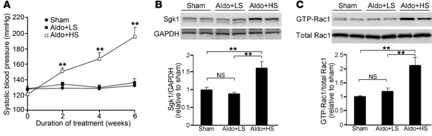
[image:5.585.80.501.84.217.2]Role of Rac1 in the synergistic action of salt and aldosterone. Several lines of evidence indicate that the injurious effect of aldosterone is aug-mented by high-sodium intake and that salt and aldosterone act synergistically to increase MR activity by an unidentified mecha-nism (14, 15, 33). We next set out to examine a role of Rac1 GTPase in the unsolved question, using Sprague-Dawley (SD) rats that received a fixed dose of aldosterone. An 8%-salt diet did not elevate blood pressure in SD rats in the absence of aldosterone infusion (data not shown). In the normal SD rats, serum aldosterone was suppressed by high-sodium diet (Supplemental Figure 3A). In addi-tion, Rac1 activity and Sgk1 were significantly reduced by high-salt diet (Supplemental Figure 3, B and C), similar to those observed in Dahl-R rats. Next, in order to evaluate the influence of salt status on Sgk1 and Rac1 activity under a fixed aldosterone condition, SD rats were continuously infused with aldosterone (0.75 μg/h s.c.) (10) and received a 0.05%-salt (Aldo+LS) or an 8%-salt (Aldo+HS) diet. As shown in Figure 3A, blood pressure remained constant in the Aldo+LS group, and there was no significant difference in blood pressure between sham-operated control rats and Aldo+LS rats after 6 weeks of treatment. In contrast, Aldo+HS rats showed progressive blood pressure elevation (Figure 3A); the results were consistent with those in previous studies in similar models (10, 11). Compared with controls, Sgk1 was not altered in the kidneys of Aldo+LS rats (Figure 3B). Interestingly, however, Sgk1 expression was significantly increased in the Aldo+HS group (Figure 3B). We then analyzed the profiles of renal Rac1 activity in this model. Active Rac1 expression remained unchanged between control and Aldo+LS rats (Figure 3C). In contrast, Aldo+HS rats showed signifi-cant elevation of Rac1 activity in the kidneys (Figure 3C).
Figure 3
Coadministration of salt and aldosterone causes blood pressure elevation with Rac1 and Sgk1 induction in the kidneys. (A) Systolic blood pres-sure was meapres-sured at 2, 4, and 6 weeks in the indicated animals. Sham, sham-operated control rats that received a normal-salt (0.3%) diet. (B) Sgk1 expression in the kidneys. Samples from Sham kidneys are indicated in left 2 lanes, samples from Aldo+LS kidneys are indicated in middle 2 lanes, and samples from Aldo+HS kidneys are indicated in right 2 lanes. The bar graph shows the results of densitometric analysis. (C) GTP-bound active Rac1 expression in the kidneys. The bar graph shows the results of densitometric analysis. Data are expressed as mean ± SEM;
research article
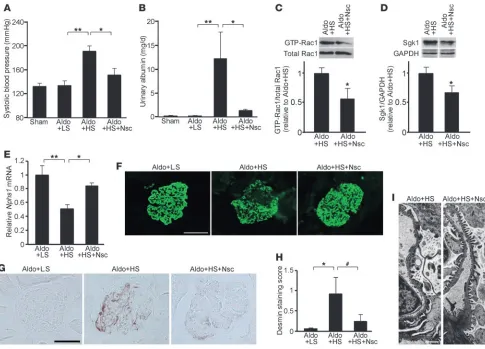
The pathogenetic role of Rac1 elevation in this model was con- firmed by the effects of Nsc23766, because the treatment signifi-cantly ameliorated hypertension and albuminuria in this model (Figure 4, A and B). In addition, Nsc23766 significantly reduced Rac1 activity and Sgk1 (Figure 4, C and D). We also evaluated the effects on glomerular podocyte damage in this model. The gene expression of nephrin (Nphs1), a principle component of the slit diaphragms, was considerably reduced in the glomerular fraction of Aldo+HS rats, and Nsc23766 treatment significantly prevented its downregulation (Figure 4E). An immunofluorescence study showed that the decrease in nephrin protein was also prevented by Rac1 inhibition (Figure 4F). Desmin was upregulated along the capillary loop in Aldo+HS rats but to a lesser degree in the Nsc23766-treated rats (Figure 4, G and H). Consistent with these results, a structural analysis using transmission electron micros- copy (TEM) revealed that the retraction of the podocyte foot pro-cesses in Aldo+HS rats was ameliorated by Nsc23766 (Figure 4I).
These results indicate that the Rac1-mediated pathway is involved in the unfavorable synergism between salt and aldosterone.
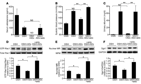
[image:6.585.50.535.81.430.2]In SD rats, we observed that the fixed aldosterone condition was necessary for salt-induced Rac1 activation. These results were some- what different from the data of Dahl-S rats, in which salt loading acti-vated Rac1 despite the suppression of aldosterone. To gain further insights into this difference, we next investigated the contribution of aldosterone in salt-loaded Dahl-S rats. For this purpose, Dahl-S rats were adrenalectomized, followed by high-sodium loading. Serum aldosterone levels were undetectable at 3 weeks after adrenalectomy (Figure 5A). As shown in Figure 5, B and C, hypertension and albu-minuria in salt-loaded Dahl-S rats were almost completely blocked by adrenalectomy. Notably, renal Rac1 activity was significantly sup-pressed in the adrenalectomized group (Figure 5D), indicating that the presence of aldosterone is essential for the salt-induced Rac1 acti-vation in this model. Nuclear MR accumulation and Sgk1 expression were also significantly reduced in adrenalectomized group (Figure 5,
Figure 4
E and F). In order to validate that the effects of adrenalectomy are actually attributable to the depletion of aldosterone, we supple-mented adrenalectomized Dahl-S rats with aldosterone (8 μg/kg/d, s.c.). In these rats, serum aldosterone levels were nearly identical to those of salt-loaded Dahl-S rats (Figure 5A). Aldosterone supplement reversed the depressor effect of adrenalectomy and its effects on reducing albuminuria (Figure 5, B and C). Importantly, the reduced Rac1 activity in adrenalectomized rats was also reversed by aldoste-rone infusion (Figure 5D). In addition, nuclear MR and Sgk1 were significantly increased in aldosterone-supplemented group (Figure 5, E and F). These data illustrate the interdependent nature of aldoste-rone and the Rac1-mediated pathway in causing excess MR signaling and salt-sensitive hypertension in vivo.
Arhgdia–/– mice exhibit salt-sensitive elevation of blood pressure and renal
injury . We have shown that Rac1 GTPase in the kidneys is an impor- tant determinant of salt sensitivity. Rac1 activity is known to be regu-lated by 3 groups of molecules: GDP/GTP exchange factor (GEF), GTPase-activating protein (GAP), and GDP-dissociation inhibitor (GDI) (34). Therefore, it is possible that the genetic variation in these regulatory molecules affects blood pressure through modulating salt susceptibility. In support of this possibility, the gene encoding human GDIα (ARHGDIA) is located in chromosome 17q25.3, in which several genome-wide linkage analyses in human hyperten-sion detected evidence of a quantitative trait loci influencing blood pressure among people of African descendant and North American
Indians (35, 36). GDIα binds to the GDP-bound, inactive form of Rac1 and inhibits both basal and GEF-stimulated dissociation of GDP from the inactive Rac1 (34). Thus, deletion of this molecule can affect blood pressure response to salt through the dysregulation of Rac1. To test this, we next evaluated the effects of high-salt loading on blood pressure in mice lacking GDIα (Arhgdia–/–
mice). As expect-ed, blood pressure remained constant in wild-type mice regardless of the salt status (Figure 6A). By contrast, blood pressure was sig-nificantly altered between high- and low-salt diet groups in Arhgdia–/–
mice (Figure 6A), indicating the increased salt sensitivity in this model. In a radiotelemetry analysis, we consistently found the salt-dependent elevation of blood pressure in Arhgdia–/– mice (Figure 6B),
confirming the role of GDIα in salt susceptibility. We addition-ally evaluated the histology of the kidney. As shown in Figure 6C, the renal phenotype, including glomerulosclerosis and associated tubulointerstitial damage, was substantially augmented by high-sodium loading. Furthermore, renal Rac1 activity in Arhgdia–/– mice
was significantly influenced by salt status (Figure 6D). GDIα not only inhibits basal Rac1 activity but also prevents Rac1 activation through inhibiting GEF-stimulated GDP dissociation (34). There-fore, the increased Rac1 activity in salt-loaded Arhgdia–/– mice is likely
[image:7.585.55.533.77.360.2]to result from GEF-stimulated GDP dissociation. We thus explored the activity of several Rac-specific GEFs known to be present in the kidneys using a G15ARac1 pull-down assay (37). We found that T cell lymphoma invasion and metastasis 1 (Tiam1), but not βpix or Sos1,
Figure 5
research article
was activated in response to salt loading in this model (Figure 6E). Tiam1 is known to activate several nuclear transcription factors through Rac1 induction (38). It is also of note that Nsc23766 blocks the interaction of Rac1 and several GEFs, including Tiam1, thereby suppressing Rac1 activation (30). The role of Tiam1 in salt-medi- ated Rac1 activation was further validated in an aldosterone infu-sion model (Supplemental Figure 4). Although serum aldosterone levels were suppressed in salt-loaded Arhgdia–/– mice (Figure 6F), Sgk1
expression was significantly upregulated (Figure 6G). In order to evaluate the contribution of Rac1 and MR activation in this model, we administered Nsc23766 or eplerenone. The blood pressure eleva-tion seen in salt-loaded Arhgdia–/– mice was significantly reduced by
both treatments (103.8 ± 4.2 mmHg in salt-loaded Arhgdia–/– mice,
90.3 ± 1.1 mmHg in Nsc23766-treated group, P < 0.05 versus salt-loaded Arhgdia–/– mice; 91.8 ± 2.7 mmHg in eplerenone-treated group,
[image:8.585.51.538.78.499.2]P < 0.05 versus salt-loaded Arhgdia–/– mice). Notably, albuminuria in Figure 6
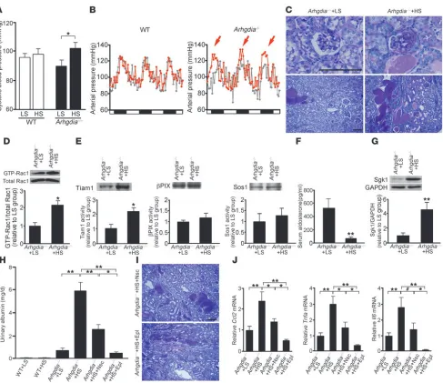
Blood pressure and renal injury in Arhgdia–/– mice are salt sensitive. (A) Systolic blood pressure in wild-type and Arhgdia–/– mice that received a
0.05%-salt (LS) or 8%-salt (HS) diet for 4 weeks (n = 8, except n = 6 for Arhgdia–/– mice that received LS diet [Arhgdia–/–+LS mice]). (B) Hourly
averages of 3-day recordings of mean arterial pressure by radiotelemetry before (gray) and after (red) salt loading. White and black boxes below the graphs represent subjective day and night, respectively. Mean arterial pressure was elevated by high salt in Arhgdia–/– mice (red arrows).
(C) Representative PAS-stained kidney sections. Scale bar: 100 μm. (D) GTP-bound and total Rac1 in kidneys. The bar graph shows the results of densitometric analysis (n = 4). (E) Activity of Rac-GEFs evaluated by G15ARac1 pull-down assay (n = 4). Equal protein amounts of kidney homogenates were used as samples for each analysis. (F) Serum aldosterone concentration in Arhgdia–/–+LS mice (n = 6) and Arhgdia–/– mice
that received HS diet (Arhgdia–/–+HS mice) (n = 8). (G) Sgk1 expression in the kidneys (n = 4). (H) Effects of Nsc23766 and eplerenone on
albuminuria (wild-type mice that received LS diet [WT+LS mice], n = 5; wild-type mice that received HS diet [WT+HS mice], n = 4; Arhgdia–/–+LS
mice, n = 7; Arhgdia–/–+HS mice, n = 8; Arhgdia–/–+HS mice that received Nsc23766, n = 8; Arhgdia–/–+HS mice that received eplerenone, n = 4).
(I) Representative renal histology in the Nsc23766- or eplerenone-treated group. Scale bar: 100 μm. (J) Quantitative analysis of Ccl2, Tnfa, and
Arhgdia–/–
mice was markedly aggravated by high-salt loading (Fig-ure 6H), consistent with the histological observations of the kidneys. Nsc23766 treatment could significantly reduce albuminuria in this model (Figure 6H). The renoprotective effect of eplerenone was even more marked (P < 0.01 versus salt-loaded Arhgdia–/– mice; P < 0.05
versus Nsc23766-treated group). The results were different from those in the Dahl-S model, because there was no significant differ-ence in the renoprotective effect between Nsc23766 and eplerenone in salt-loaded Dahl-S rats. These 2 models were also different in that we did not observe renal injury in Dahl-S rats without a high-salt diet (Figure 1B), whereas pathological albuminuria was present in
Arhgdia–/– mice even with a low-salt diet. These differences between
Dahl-S rats and Arhgdia–/– mice might result from the artificial
nature of Arhgdia–/– mice, or alternatively, the observations can be
attributed to the species difference. It is also possible that an addi-tional mechanism may coexist in causing renal injury in this model. The renoprotective effects of the pharmacological interventions in salt-loaded Arhgdia–/– mice were confirmed by histology (Figure 6I)
and by the reduction of inflammatory cytokines (Figure 6J). These results establish an important role of the Rac1-mediated pathway in the pathogenesis of salt-sensitive hypertension and renal injury.
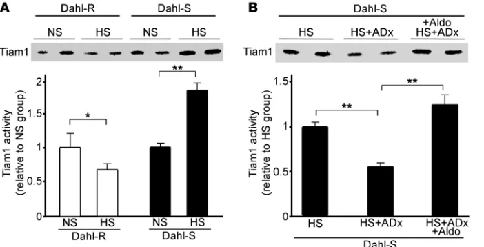
As described above, high-salt loading augmented Tiam1 activity in
Arhgdia–/– mice and in aldosterone-infused rats. In the Dahl-S model,
however, we observed that adrenalectomy abolished Rac1 activation and that aldosterone supplementation reversed the effects, indicat-ing that aldosterone can also be an important regulator of Rac1. To gain further insights into the mechanisms for the regulation of Rac1 activity, we finally evaluated Tiam1 activity in the Dahl model. Tiam1 levels changed differently with high-salt diet in Dahl-S and Dahl-R rats, with upregulation in the former and downregulation in the lat-ter (Figure 7A). Importantly, Tiam1 activation in salt-loaded Dahl-S rats was prevented by adrenalectomy, and this effect was abolished in the aldosterone-supplemented adrenalectomized group (Figure 7B). These data provide additional support for the hypothesis that salt and aldosterone interdependently regulate Rac1 activity. Moreover, the decreased Tiam1 activity in salt-loaded Dahl-R rats underscores the role of Tiam1 in the regulation of Rac1-MR signaling.
Discussion
The current study demonstrates that high-salt loading triggers Rac1 activation in the kidneys in salt-sensitive hypertension, which is the key event in the pathogenesis of blood pressure elevation and
kidney injury via an MR-dependent pathway. We first showed that renal Rac1 activation caused salt-dependent hypertension and renal injury in Dahl-S rats via potentiating MR signaling. Conversely, Rac1 activity was reduced in salt-loaded Dahl-R rats, supporting the close relationship between Rac1 and salt susceptibility. Using an aldosterone infusion model and adrenalectomized Dahl-S rats, we next showed that Rac1 was involved in the unfavorable interac- tion between salt and aldosterone; salt-induced Rac1 and aldoste- rone synergistically caused blood pressure elevation and renal dam-age. Finally, the key role of Rac1 and its regulatory molecules in determining salt susceptibility was confirmed in Arhgdia–/– mice.
The aldosterone/MR signaling is closely related to dietary salt intake in both health and disease. Although several investigators have provided evidence that supports the paradoxical response of MR activity to sodium loading in models of salt-sensitive hyperten-sion (15, 25, 26), the mechanism that augments MR activity in a high-salt status has long been a mystery. In this study, we showed that salt loading increased not only Sgk1 but also nuclear MR accumulation in Dahl-S rats and that the enhanced MR signaling was prevented by Rac1 inhibition. In contrast, high-salt loading decreased Rac1 and MR signaling in salt-resistant models, including Dahl-R and SD rats, indicating that high-salt loading potentiates Rac1-MR signaling only in salt-sensitive models, not in salt-resistant subjects. The negative action of high salt on Rac1 in salt-resistant models can be attributed to the suppression of serum aldosterone, because high-salt loading activated Rac1 in SD rats that received a fixed dose of aldosterone. In Dahl-S rats, however, serum aldoste-rone, even at low levels, is an important positive regulator of Rac1, considering the data that adrenalectomy decreased Rac1 activity during the high-salt diet and that aldosterone supplementation reversed the effect. Our data that aldosterone removal in Dahl-S rats influenced the activity of Rac-specific GEF Tiam1 further support an essential role of aldosterone in regulating Rac1 activity. Taking these observations together, we consider that aldosterone and high-salt diet interdependently regulate Rac1 activity, and, presumably due to the augmented response of Rac1 to these factors, Dahl-S rats show salt-dependent hypertension (see our cartoon; Figure 8). From our data, it is not entirely clear what level of aldosterone allows Rac1 to respond to salt loading in the Dahl-S model. Nevertheless, our study sheds light on the long-standing mystery and establishes what we believe to be a novel role of Rac1 as a key factor for the paradoxi-cal increase of MR activity in the context of a high-salt intake.
[image:9.585.47.398.83.262.2]research article
A key area of interest is the molecular basis for high-salt loading changes renal Rac1 activity. We showed that Rac1 activity was altered in opposite directions by high-salt diet in Dahl-S and Dahl-R rats. Because environmental conditions were identical, the differences in response to the same treatment can be attributed to the genomic heterogeneity in these models. One possibility is that genetic altera-tion in molecules that regulate Rac1 might be involved. Although the causative genes for hypertension in Dahl model remain unknown, previous studies have identified several loci cosegregating with blood pressure. As an example, congenic strain analysis revealed that chro-mosome 10 contains a quantitative trait locus (QTL) affecting blood pressure in Dahl-S model (39, 40). The gene encoding CAMGAP1, a GAP toward Rac1 that is abundantly expressed in the kidneys (41), is located within the QTL. In addition, another Rho family regulator,
Arhgap1 gene (encoding RhoGAP1), is located within the blood pres-sure QTLs in chromosome 3 (42). Although we showed that Tiam1 activity is altered in the Dahl model, involvement of other regula-tors is not excluded, considering that a multitude of GEFs and GAPs coordinately regulate small GTPases. The other possibility is that the humoral factors that affect Rac1 activity can be altered between the 2 strains. Local angiotensin II within the kidneys modulates renal function independently of systemic angiotensin II (43) and causes sodium and water retention that is at least partly attributable to the enhanced reabsorption in distal nephron (44–46). Of note, Kobori et al. revealed the paradoxical enhancement of intrarenal angioten- sinogen levels in salt-loaded Dahl-S rats (47). Interestingly, angio-tensin II can activate MR via mechanisms other than intracellular aldosterone synthesis (48). Tiam1 is activated by Src kinase, a key player in angiotensin II–mediated cellular response (49). Moreover, angiotensin II is shown to elevate Rac1 activity in vitro, leading to
nuclear transcription factor induction (50). Thus, it is also possible that humoral factors such as angiotensin II are involved in the differ-ent responses between 2 strains. Although it remains elusive how salt loading upregulates and downregulates Rac1 in Dahl-S and Dahl-R rats, respectively, future research on key alterations in genes that con-fer the different responses would be promising.
A potential limitation of our study is that we have not fully excluded other mechanisms that might be involved in pathological MR acti- vation. One hypothesis that has been postulated to explain the dis- crepancy between circulating aldosterone levels and tissue MR signal-ing activity is that the extraadrenal production of aldosterone might induce organ-specific MR activation. Indeed, tissue aldosterone levels in the heart are reported to be increased in cardiac hypertrophy, with the upregulation of aldosterone synthase (51). However, adrenalec-tomy almost completely prevented hypertension and kidney damage in this study, supporting the importance of systemic aldosterone, at least in our model. It is also of note that aldosterone supplementation to the same level as that of salt-loaded Dahl-S rats fully reversed the effects of adrenalectomy. Another hypothesis explaining the discrep-ancy is that glucocorticoid might act as a ligand for MR. Endogenous glucocorticoids, cortisol in humans and corticosterone in rodents, manifest a similar affinity for MR as aldosterone. The agonistic activ-ity of glucocorticoids is considered to be negligible in physiological milieu; however, they can act as agonists in some pathological con-ditions (18, 52, 53). Although we acknowledge that glucocorticoids can activate MR, especially in tissue damage in heart failure (52, 54), corticosterone may not potentiate the development of salt-sensitive hypertension in our study, considering that aldosterone supplemen-tation fully nullified the protective effects of adrenalectomy. As an alternative mechanism that modulates MR activity, we have previously demonstrated the interaction between Rac1 and MR (19). In the cur-rent study, we show that Rac1 activity was increased in response to high-sodium loading and that Rac1 inhibition reduced blood pressure and renal injury along with MR repression, underscoring the role of Rac1 as a regulator of MR activity and salt susceptibility. Nonetheless, several other possibilities have also been postulated to modulate MR-dependent responses (55, 56). In our study, the renoprotective effects of Nsc23766 were moderate as compared with those of eplerenone in salt-loaded Arhgdia–/–
mice, although there were no significant differ-ences between Nsc23766 and eplerenone in blood pressure–lowering and renoprotective effects in salt-loaded Dahl-S rats. One possible explanation for this observation is that alternative factors in addition to Rac1 are involved in the regulation of MR overactivity. There are sev- eral factors modulating nuclear receptors, including estrogen recep-tor, glucocorticoid receptor, and MR; MR is also activated by protein kinase A and Ubc9 (56, 57). Therefore, it is possible that additional pathways are involved in the regulation of MR-dependent responses in this model, and Rac1-mediated signaling represents one of the key mechanisms modulating MR activity in a high-salt status in salt-sensi-tive hypertensive animals. Another potential limitation is that, in some experiments, we evaluated blood pressure only by the indirect method. Although we acknowledge this limitation, the use of tail-cuff method can be validated, considering the substantial differences among groups in these studies (58). In addition, the albumin excretion data in our study certainly are consistent with the blood pressure data.
[image:10.585.47.285.78.295.2]In summary, we demonstrate that renal Rac1 GTPase regulates salt susceptibility of blood pressure and kidney injury via an MR-dependent mechanism. We also show that salt-induced Rac1 and aldosterone act in concert to induce pathological MR overactiv-ity, providing a theoretical basis for the unfavorable interaction
Figure 8
by the Animal Care Committee of the University of Tokyo.
In the Dahl model, 4-week-old male Dahl-S or Dahl-R rats (SLC) were fed an 8%- or 0.3%-NaCl diet for 3 weeks. In some experiments, Dahl-S rats were fed a 4%-salt diet for 4 weeks. In the drug treatment studies, Dahl-S rats were randomly assigned to one of the following treatment groups 3 days before salt loading: eplerenone (Pfizer; 1.25 g/kg chow); Nsc23766 (Tocris Biosciences; at a dose of 8 mg/kg/d, s.c.); and fasudil (Asahi Kasei Pharma; 100 mg/kg/d in drinking water). The doses of eplerenone used in this study correspond to the estimated intake of 100 mg/kg/d, which effectively inhib- its aldosterone/salt-induced hypertension and provided end-organ protec-tive effects in the kidneys in previous studies (10). The dosages of Nsc23766 used were determined according to the inhibitory effects of renal Rac1 activ- ity in vivo without apparent toxicity. The dosage of fasudil was in accor-dance with that in the previous studies, which has been shown to prevent Rho-kinase activation (59). Bilateral adrenalectomy was performed through a dorsal incision under ether anesthesia. Adrenalectomized rats received dexamethasone (Wako) at a dose of 60 μg/kg/d s.c., 3 days per week.
In aldosterone infusion experiments, SD rats (Tokyo Laboratory Animals Science), weighing 250–270 g, received either sham operation or implanta-tion of osmotic minipump (Alzet) containing aldosterone (0.75 μg/h s.c.). Aldosterone-infused rats were randomly assigned to 8%- or 0.05%-salt diet group for 6 weeks. Sham-operated rats received normal chow.
Arhgdia–/–
mice were generated as described previously (60). Male off-spring derived from Arhgdia+/– intercrosses were analyzed in this study.
The mice were divided into 8%- and 0.05%-NaCl diet groups at 4 weeks of age. In some experiments, Nsc23766 or eplerenone was given at a dose of 20 mg/kg/d s.c. or 1.67 g/kg, respectively.
Urine was collected for 24 hours using an individual metabolic cage (Nat- sume). Urine collecting tubes were lubricated with mineral oil to avoid evap-oration. We determined urine albumin using a rat or mouse albumin ELISA according to the manufacturer’s instructions (Shibayagi). Serum aldosterone was measured at SRL. Glomeruli were isolated by the sieving method (61).
Blood pressure measurements. Systolic blood pressure of conscious animals was measured using the tail-cuff method (27). A dark cover was placed over animals to reduce stress. We also performed direct blood pressure measure-ment. Under light anesthesia, a polyethylene catheter was implanted through the left femoral artery into the lower aorta. The distal end of catheter was threaded through the s.c. space and exteriorized from the subcapsular region. The catheter was filled with heparinized saline to avoid clotting. Mean arte-rial pressure was monitored in a conscious and unrestrained condition. In some experiments, arterial pressure was monitored by radiotelemetry. Mice were anesthetized with ether, and the left carotid artery was isolated. The tip of the catheter of the PhysioTel Transmitters (model PA-C10, Data Sci-ences International) was inserted in the carotid artery and advanced in the aortic arch, with the telemeter body positioned in a s.c. pocket on the left
was assessed by the pull-down assay (19).
Quantitative RT-PCR . Gene expression was quantitatively analyzed by real-time RT-PCR as described previously (61).
Immunohistochemistry . Immunostaining was performed as described previ- ously (61). Antibody to nephrin was provided by H. Kawachi (Niigata Uni- versity, Niigata, Japan). Desmin staining in the glomeruli was semiquantita-tively scored in accordance with the previously described method (62).
Histomorphometric analysis and TEM. After dissection, kidneys were rinsed in phosphate-buffered saline, fixed in 4% paraformaldehyde solution, and embedded in paraffin. The degree of glomerulosclerosis was semiquanti-tatively scored according to an established scoring system (19, 27). In TEM analysis, small pieces of cortex were fixed, dehydrated, and embedded as described previously (61).
Rac-GEF activation assay. Activity of GEFs was determined by the pull-down assay using nucleotide-free Rac1 mutant (G15ARac1), which forms a high- affinity binary complex with active GEFs (37), in accordance with the man-ufacturer’s instruction (Cell Biolabs Inc.). Kidneys were homogenized in a buffer consisting of 1% NP-40, 20 mM HEPES, pH 7.5, 150 mM NaCl, 5 mM MgCl2
, protease inhibitors, and phosphatase inhibitors. Lysates were centri-fuged at 14,000 g for 5 minutes, and equal amounts of protein (3,000–4,000
μg) were incubated at 4°C for 60 minutes with 20 μl G15ARac1 agarose beads. Samples were immunoblotted using antibodies against Tiam1 (Cell Biolabs Inc.), Sos1 (BD Biosciences), and βPIX (Millipore).
Statistics. The data are summarized as mean ± SEM. Unpaired t test was used for comparisons between 2 groups. For multiple comparisons, statis- tical analysis was performed by ANOVA with Tukey post-hoc tests. Histo- logical data were analyzed using nonparametric analysis with Kruskal-Wal-lis test, followed by Mann-Whitney U test. P values of less than 0.05 were considered to be significant.
Acknowledgments
We thank Satoshi Fukuda (University of Tokyo) for the excellent technical assistance in electron microscopic analysis. Antibody against nephrin was provided by Hiroshi Kawachi (Niigata Univer- sity). This work was supported in part by Grant-in-Aid for Scientif-ic Research (S) and by Takeda Science Foundation (to T. Fujita).
Received for publication February 23, 2011, and accepted in revised form June 1, 2011.
Address correspondence to: Toshiro Fujita, Department of Nephrol-ogy and Endocrinology, University of Tokyo Graduate School of Medicine, 7-3-1 Hongo, Bunkyo-ku, Tokyo 113-8655, Japan. Phone: 81.3.5800.9735; Fax: 81.3.5800.9736; E-mail: fujita-dis@ h.u-tokyo.ac.jp.
1. Kearney PM, Whelton M, Reynolds K, Muntner P, Whelton PK, He J. Global burden of hyper-tension: analysis of worldwide data. Lancet.
2005;365(9455):217–223.
2. Fujita T, Henry WL, Bartter FC, Lake CR, Delea
CS. Factors influencing blood pressure in salt-sensitive patients with hypertension. Am J Med. 1980;69(3):334–344.
research article
excretion and blood pressure. Results for 24 hour urinary sodium and potassium excretion. Intersalt Cooperative Research Group. BMJ. 1988;297(6644):319–328.
4. Lifton RP, Gharavi AG, Geller DS. Molecular mechanisms of human hypertension. Cell. 2001; 104(4):545–556.
5. Guyton AC. Blood pressure control--special role of the kidneys and body fluids. Science. 1991; 252(5014):1813–1816.
6. He FJ, MacGregor GA. Effect of longer-term modest salt reduction on blood pressure. Cochrane Database Syst Rev. 2004;(3):CD004937.
7. Tuomilehto J, et al. Urinary sodium excretion and cardiovascular mortality in Finland: a prospective study. Lancet. 2001;357(9259):848–851.
8. Bibbins-Domingo K, et al. Projected effect of dietary salt reductions on future cardiovascular disease. N Engl J Med. 2010;362(7):590–599. 9. Cianciaruso B, et al. Salt intake and renal outcome
in patients with progressive renal disease. Miner Electrolyte Metab. 1998;24(4):296–301.
10. Blasi ER, Rocha R, Rudolph AE, Blomme EA, Polly ML, McMahon EG. Aldosterone/salt induces renal inflammation and fibrosis in hypertensive rats.
Kidney Int. 2003;63(5):1791–1800.
11. Nishiyama A, et al. Possible contributions of reactive oxygen species and mitogen-activated protein kinase to renal injury in aldosterone/salt-induced hyper-tensive rats. Hypertension. 2004;43(4):841–848. 12. Fujita T. Mineralocorticoid receptors, salt-sensitive
hypertension, and metabolic syndrome. Hypertension. 2010;55(4):813–818.
13. Rossi GP, Sechi LA, Giacchetti G, Ronconi V, Straz- zullo P, Funder JW. Primary aldosteronism: cardio-vascular, renal and metabolic implications. Trends Endocrinol Metab. 2008;19(3):88–90.
14. Wang Q, et al. Chronic hyperaldosteronism in a transgenic mouse model fails to induce cardiac remodeling and fibrosis under a normal-salt diet. Am J Physiol Renal Physiol. 2004;286(6):F1178–F1184. 15. Nagase M, Matsui H, Shibata S, Gotoda T, Fujita T. Salt-induced nephropathy in obese spontaneously hypertensive rats via paradoxical activation of the mineralocorticoid receptor: role of oxidative stress. Hypertension. 2007;50(5):877–883. 16. Nagata K, et al. Mineralocorticoid receptor antago-nism attenuates cardiac hypertrophy and failure in low-aldosterone hypertensive rats. Hypertension. 2006;47(4):656–664.
17. Williams GH, et al. Efficacy of eplerenone versus enalapril as monotherapy in systemic hyperten-sion. Am J Cardiol. 2004;93(8):990–996.
18. Funder JW. Is aldosterone bad for the heart? Trends Endocrinol Metab. 2004;15(4):139–142.
19. Shibata S, et al. Modification of mineralocorti-coid receptor function by Rac1 GTPase: impli-cation in proteinuric kidney disease. Nat Med. 2008;14(12):1370–1376.
20. Rapp JP, Dahl LK. Suppression of aldosterone in salt susceptible and salt resistant rats. Endocrinology. 1973;92(4):1286–1289.
21. Wulff P, et al. Impaired renal Na(+) reten-tion in the sgk1-knockout mouse. J Clin Invest. 2002;110(9):1263–1268.
22. Pearce D, Kleyman TR. Salt, sodium channels, and SGK1. J Clin Invest. 2007;117(3):592–595. 23. Zhang W, et al. Aldosterone-induced Sgk1 relieves
Dot1a-Af9-mediated transcriptional repres-sion of epithelial Na+ channel alpha. J Clin Invest. 2007;117(3):773–783.
24. Quinkler M, et al. Increased expression of miner- alocorticoid effector mechanisms in kidney biop-sies of patients with heavy proteinuria. Circulation. 2005;112(10):1435–1443.
25. Farjah M, Roxas BP, Geenen DL, Danziger RS. Dietary salt regulates renal SGK1 abundance: rele-vance to salt sensitivity in the Dahl rat. Hypertension.
2003;41(4):874–878.
26. Aoi W, et al. Abnormal expression of ENaC and SGK1 mRNA induced by dietary sodium in Dahl salt-sensitively hypertensive rats. Cell Biol Int. 2007; 31(10):1288–1291.
27. Shibata S, Nagase M, Yoshida S, Kawachi H, Fujita T. Podocyte as the target for aldosterone: roles of oxidative stress and Sgk1. Hypertension. 2007;49(2):355–364.
28. Terada Y, et al. Aldosterone stimulates proliferation of mesangial cells by activating mitogen-activated protein kinase 1/2, cyclin D1, and cyclin A. J Am Soc Nephrol. 2005;16(8):2296–2305.
29. Raij L, Luscher TF, Vanhoutte PM. High potassium diet augments endothelium-dependent relaxations in the Dahl rat. Hypertension. 1988;12(6):562–567. 30. Gao Y, Dickerson JB, Guo F, Zheng J, Zheng Y.
Rational design and characterization of a Rac GTPase-specific small molecule inhibitor. Proc Natl Acad Sci U S A. 2004;101(20):7618–7623. 31. Cancelas JA, Lee AW, Prabhakar R, Stringer KF,
Zheng Y, Williams DA. Rac GTPases differentially integrate signals regulating hematopoietic stem cell localization. Nat Med. 2005;11(8):886–891. 32. Nishikimi T, et al. Fasudil, a Rho-kinase inhibitor,
attenuates glomerulosclerosis in Dahl salt-sensitive rats. J Hypertens. 2004;22(9):1787–1796.
33. Brilla CG, Weber KT. Mineralocorticoid excess, dietary sodium, and myocardial fibrosis. J Lab Clin Med. 1992;120(6):893–901.
34. Takai Y, Sasaki T, Matozaki T. Small GTP-binding proteins. Physiol Rev. 2001;81(1):153–208.
35. Franceschini N, et al. A quantitative trait loci-spe- cific gene-by-sex interaction on systolic blood pres-sure among American Indians: the Strong Heart Family Study. Hypertension. 2006;48(2):266–270. 36. Wilk JB, et al. Genome-wide linkage analyses for
age at diagnosis of hypertension and early-onset hypertension in the HyperGEN study. Am J Hyper-tens. 2004;17(9):839–844.
37. Garcia-Mata R, Wennerberg K, Arthur WT, Noren NK, Ellerbroek SM, Burridge K. Analysis of activat-ed GAPs and GEFs in cell lysates. Methods Enzymol. 2006;406:425–437.
38. Simon AR, Vikis HG, Stewart S, Fanburg BL, Cochran BH, Guan KL. Regulation of STAT3 by direct binding to the Rac1 GTPase. Science. 2000; 290(5489):144–147.
39. Deng AY, Dutil J, Sivo Z. Utilization of marker-assisted congenics to map two blood pressure quantitative trait loci in Dahl rats. Mamm Genome. 2001;12(8):612–616.
40. Rapp JP, Garrett MR, Deng AY. Construction of a double congenic strain to prove an epistatic inter-action on blood pressure between rat chromosomes 2 and 10. J Clin Invest. 1998;101(8):1591–1595. 41. Sakakibara T, Nemoto Y, Nukiwa T, Takeshima
H. Identification and characterization of a novel Rho GTPase activating protein implicated in receptor–mediated endocytosis. FEBS Lett. 2004; 566(1–3):294–300. 42. Palijan A, Dutil J, Deng AY. Quantitative trait loci with opposing blood pressure effects demonstrat-ing epistasis on Dahl rat chromosome 3. Physiol Genomics. 2003;15(1):1–8. 43. Seikaly MG, Arant BS Jr, Seney FD Jr. Endogenous angiotensin concentrations in specific intrarenal fluid compartments of the rat. J Clin Invest. 1990; 86(4):1352–1357.
44. Crowley SD, et al. Distinct roles for the kidney and systemic tissues in blood pressure regulation by the renin-angiotensin system. J Clin Invest. 2005; 115(4):1092–1099.
45. Hall JE, Guyton AC, Trippodo NC, Lohmeier TE, McCaa RE, Cowley AW Jr. Intrarenal control of electrolyte excretion by angiotensin II. Am J Physiol. 1977;232(6):F538–F544.
46. Zhao D, Seth DM, Navar LG. Enhanced distal neph- ron sodium reabsorption in chronic angiotensin II-infused mice. Hypertension. 2009;54(1):120–126.
47. Kobori H, Nishiyama A, Abe Y, Navar LG. Enhance- ment of intrarenal angiotensinogen in Dahl salt-sensitive rats on high salt diet. Hypertension. 2003; 41(3):592–597.
48. Jaffe IZ, Mendelsohn ME. Angiotensin II and aldosterone regulate gene transcription via func-tional mineralocortocoid receptors in human coronary artery smooth muscle cells. Circ Res. 2005; 96(6):643–650.
49. Servitja JM, Marinissen MJ, Sodhi A, Bustelo XR, Gutkind JS. Rac1 function is required for Src-induced transformation. Evidence of a role for Tiam1 and Vav2 in Rac activation by Src. J Biol Chem. 2003;278(36):34339–34346.
50. Tsai CT, et al. Angiotensin II activates signal trans-ducer and activators of transcription 3 via Rac1 in atrial myocytes and fibroblasts: implication for the therapeutic effect of statin in atrial structural remodeling. Circulation. 2008;117(3):344–355. 51. Takeda Y, Yoneda T, Demura M, Furukawa K,
Miyamori I, Mabuchi H. Effects of high sodium intake on cardiovascular aldosterone synthesis in stroke-prone spontaneously hypertensive rats.
J Hypertens. 2001;19(3 pt 2):635–639.
52. Rossier MF, Lenglet S, Vetterli L, Python M, Mat-urana A. Corticosteroids and redox potential modulate spontaneous contractions in isolated rat ventricular cardiomyocytes. Hypertension. 2008;52(4):721–728.
53. Mihailidou AS, Loan Le TY, Mardini M, Funder JW. Glucocorticoids activate cardiac mineralocorticoid receptors during experimental myocardial infarc-tion. Hypertension. 2009;54(6):1306–1312. 54. Funder JW. Minireview: Aldosterone and miner-alocorticoid receptors: past, present, and future. Endocrinology. 2010;151(11):5098–5102. 55. Krug AW, et al. Elevated mineralocorticoid recep-tor activity in aged rat vascular smooth muscle cells promotes a proinflammatory phenotype via extracellular signal-regulated kinase 1/2 mitogen-activated protein kinase and epidermal growth factor receptor-dependent pathways. Hypertension. 2010;55(6):1476–1483.
56. Yokota K, et al. Coactivation of the N-terminal transactivation of mineralocorticoid receptor by Ubc9. J Biol Chem. 2007;282(3):1998–2010. 57. Massaad C, Houard N, Lombes M, Barouki R.
Modulation of human mineralocorticoid recep-tor function by protein kinase A. Mol Endocrinol. 1999;13(1):57–65. 58. Kurtz TW, Griffin KA, Bidani AK, Davisson RL, Hall JE. Recommendations for blood pressure mea-surement in humans and experimental animals. Part 2: Blood pressure measurement in experimen-tal animals: a statement for professionals from the subcommittee of professional and public educa-tion of the American Heart Association council on high blood pressure research. Hypertension. 2005;45(2):299–310.
59. Higashi M, et al. Long-term inhibition of Rho-kinase suppresses angiotensin II-induced car-diovascular hypertrophy in rats in vivo: effect on endothelial NAD(P)H oxidase system. Circ Res. 2003;93(8):767–775.
60. Togawa A, et al. Progressive impairment of kidneys and reproductive organs in mice lacking Rho GDI-alpha. Oncogene. 1999;18(39):5373–5380.
61. Shibata S, Nagase M, Fujita T. Fluvastatin amelio- rates podocyte injury in proteinuric rats via modu-lation of excessive Rho signaling. J Am Soc Nephrol. 2006;17(3):754–764.