Copyright © 1999, American Society for Microbiology. All Rights Reserved.
Genetic Diversity and Population Structure of
Vibrio cholerae
PILAR BELTRA´ N,1GABRIELA DELGADO,1ARMANDO NAVARRO,1FRANCISCA TRUJILLO,1
ROBERT K. SELANDER,2*ANDALEJANDRO CRAVIOTO1
Departamento de Salud Pu´blica de la Facultad de Medicina, Universidad Nacional Auto´noma de Me´xico, Me´xico, D.F., Me´xico,1and Institute of Molecular Evolutionary Genetics, Pennsylvania State University,
University Park, Pennsylvania 168022
Received 9 September 1998/Returned for modification 17 November 1998/Accepted 8 December 1998
Multilocus enzyme electrophoresis (MLEE) of 397Vibrio choleraeisolates, including 143 serogroup reference
strains and 244 strains from Mexico and Guatemala, identified 279 electrophoretic types (ETs) distributed in two major divisions (I and II). Linkage disequilibrium was demonstrated in both divisions and in subdivision Ic of division I but not in subdivision Ia, which includes 76% of the ETs. Despite this evidence of relatively frequent recombination, clonal lineages may persist for periods of time measured in at least decades. In addition to the pandemic clones of serogroups O1 and O139, which form a tight cluster of four ETs in subdivision Ia, MLEE analysis identified numerous apparent clonal lineages of non-O1 strains with intercon-tinental distributions. A clone of serogroup O37 that demonstrated epidemic potential in the 1960s is closely related to the pandemic O1/O139 clones, but the nontoxigenic O1 Inaba El Tor reference strain is not. A strain
of serogroup O22, which has been identified as the most likely donor of exogenousrfbregion DNA to the O1
progenitor of the O139 clone, is distantly related to the O1/O139 clones. The close evolutionary relationships of the O1, O139, and O37 epidemic clones indicates that new cholera clones are likely to arise by the modification of a lineage that is already epidemic or is closely related to such a clone.
For Vibrio cholerae, the causative agent of cholera and a natural inhabitant of aquatic environments, the conventional method of identifying and classifying strains is a serotyping scheme in which nearly 200 serogroups (or serovars) have been distinguished on the basis of epitopic variation in the cell surface lipopolysaccharide (LPS) (52). From an epidemiolog-ical standpoint, the species has been divided into serogroup O1 and serogroup non-O1 strains, which were long believed to differ in ability to cause epidemic cholera (4, 10). Historically, O1 strains have been responsible for all major epidemics, in-cluding seven pandemics (19), but in 1992 an epidemic clone of serogroup O139 (Bengal) appeared in southern India (30). It rapidly spread throughout much of Southeast Asia (1, 9) and reached western Africa in 1994 (37).
The emergence of the O139 clone with pandemic potential stimulated increased interest in the molecular basis of patho-genesis inV. choleraeand the degree to which genes determin-ing serotype and virulence properties are subject to horizontal transfer and recombination among strains (17). Molecular ge-netic studies have shown that the origin of the O139 clone involved a complex rearrangement of therfbregion in a strain of O1 El Tor, which included deletion of genes responsible for the biosynthesis and assembly of the side chains of the O1 cell surface LPS and insertion of exogenous DNA mediating syn-thesis of the O139 LPS core (2, 3, 40–42) and a capsule (12, 45). The observations that strains with identical nucleotide sequences of the aspartate semialdehyde dehydrogenase gene (asd) may express different O antigens and that O1 isolates are heterogeneous in sequence provided further evidence of the horizontal transfer of genes mediating O-antigen synthesis (19). Most surprisingly, it was discovered that the CTX ele-ment, which includes the structural genes (ctxAand ctxB) for
the subunits of cholera toxin, is the integrated genome of a filamentous bacteriophage, CTXø, and is transmissible (31, 46). Moreover, the bacterial receptor for CTXø, the toxin-coregulated pilus, is encoded by an operon (tcp) that is part of a transmissible pathogenicity island (20, 21). These findings raise the possibility that all strains of V. cholerae have the potential to become agents of epidemic cholera.
Previous research on the evolutionary genetics ofV. cholerae
has been primarily concerned with the identification and epi-demiology of O1 and O139 strains that are responsible for cholera epidemics and pandemics. This work includes the ap-plication of multilocus enzyme electrophoresis (MLEE) to as-sess genotypic diversity in a collection of 181 O1 and 79 non-O1 strains (33) and the extensive use of this technique, in conjunction with ribotyping and restriction fragment length polymorphism analysis of the ctxAgene, to study various as-pects of the molecular epidemiology of cholera in Latin Amer-ica and elsewhere (8, 15, 43, 44). Additionally, ribotyping and comparative sequence analysis of theasdgene have been em-ployed to reconstruct the evolutionary history of O1 clones involved in the sixth and seventh pandemics (18, 19).
We report here the results of an analysis of 397 isolates ofV. cholerae by MLEE undertaken to determine the extent of genetic diversity in the species as a whole, the relationships of the epidemic O1 and O139 clones to strains of other sero-groups, and the genetic population structure of the non-O1 segment of the species.
MATERIALS AND METHODS
Bacterial strains.This study was based on 397 strains received asV. cholerae.
The sample included 143 strains in the serogroup reference collection main-tained at the National Institute of Infectious Diseases in Japan (52). These strains were isolated from worldwide sources in the period from 1932 to 1993; 117 of them were recovered from humans, 13 from animals, 6 from river water, 3 from seawater, and 3 from unknown sources. The reference strains for sero-groups O1 through O83 were provided by T. Cheasty, and those for serogroup O155 and serogroups O84 through O140, together with strain CA-385 (rough), which is used in serotyping, were obtained from T. Shimada.
A collection of 191 strains from Sonora, Tabasco, and 14 other states in Mexico was provided by the Instituto Nacional de Diagno´stico y Referencia
* Corresponding author. Mailing address: Institute of Molecular Evolutionary Genetics, Mueller Laboratory 516, Pennsylvania State University, University Park, PA 16802. Phone: (814) 234-8997. Fax: (814) 863-4706. E-mail: rks3@psu.edu.
581
on May 15, 2020 by guest
http://jcm.asm.org/
Downloaded from
on May 15, 2020 by guest
http://jcm.asm.org/
Downloaded from
on May 15, 2020 by guest
http://jcm.asm.org/
Epidemiolo´gicos, the Laboratorio Estatal de Salud Pu´blica (Sonora), and the Laboratorio Regional de Salud Pu´blica (Tabasco). A sample of 53 strains recov-ered from humans in Guatemala was obtained from the Instituto de Nutricio´n de Centroame´rica y Panama´. Of the total of 244 strains from Mexico and Guate-mala, 172 were recovered from humans, 41 from well and sewage water, 15 from fish, 7 from other environmental sources, 2 from food, and 7 from unspecified sources.
Five serogroup O139 strains from Thailand were furnished by P. Echeverria. From the Centers for Disease Control and Prevention (CDC), we received single isolates of O1 El Tor from Australia, Romania, Peru, and Louisiana and an O139 isolate from an imported human case of cholera in the United States in 1993.
The strains from Mexico and Guatemala, almost all of which were isolated in the period from 1991 to 1995, were serotyped in our laboratory by the standard method of Sakazaki and Donovan (32). Eight of these strains were of a serotype that was not then represented in the reference collection; for purposes of this paper, we have designated this serotype OA.
All of the strains used in this study have been deposited in the collection of the Facultad de Medicina, Universidad Nacional Auto´noma de Me´xico (FMU).
MLEE.MLEE was performed by the methods described by Selander et al.
(34). Seventeen enzyme loci were assayed for allelic variation: 6PG (6-phospho-gluconate dehydrogenase), G6P (glucose 6-phosphate dehydrogenase), IDH (isocitrate dehydrogenase), NSP (nucleoside phosphorylase), ALD (alanine de-hydrogenase), SHK (shikimate dede-hydrogenase), CAT (catalase), LAP (leucine aminopeptidase), GOT (glutamic-oxalacetic transaminase), ME (malic enzyme), MDH (malate dehydrogenase), PLP (phenylalanyl-leucine peptidase), PGI (phosphoglucose isomerase), HEX (hexokinase), PGM (phosphoglucomutase), IPO (indophenol oxidase), and THD (threonine dehydrogenase).
Electromorphs (mobility variants) were equated with alleles, an absence of enzyme activity was scored as a null allele, and distinctive allele profiles for the 17 loci were designated electrophoretic types (ETs).
Statistical analysis.From the allele profiles of the ETs, mean genetic diversity
per locus (H) and pairwise genetic distance were calculated as described by Selander et al. (34), and dendrograms were constructed by the unweighted pair-group method with arithmetic mean (UPGMA). As a measure of multilocus population structure (linkage disequilibrium), we calculated an index of associ-ation of alleles (IA) by using the equation 12VO/VE, whereVOis the variance
of the observed distribution of number of mismatched alleles between ET pairs andVEis the mismatch variance expected when allele associations are random
(linkage equilibrium) (24, 49). Calculations were made with computer programs written by T. S. Whittam (Pennsylvania State University, University Park).
Detection of thectxAgene.Tests for the presence of thectxAgene, which
encodes a subunit protein of cholera toxin, were performed by colony blot assay with a digoxigenin-labeled oligonucleotide probe, according to the procedure of Maniatis et al. (23), and by PCR amplification. The nucleotide sequences of the probe and the primers were those specified by Shirai et al. (39).
RESULTS
Serogroups.Of the 244 strains from Mexico and Guatemala,
230 were of 59 serogroups and 14 were serologically nontype-able (NT). The reference collection currently includes nearly 200 serogroups (52), and thus about 30% of the described serotypic diversity of the species is exhibited by the Mexico-Guatemala strains.
In this paper, individual reference strains are referred to by their serogroup designations, marked with an asterisk (e.g., O155*).
Multilocus enzyme genotypes.All 17 loci assayed were
poly-morphic, with an average of 9.5 alleles per locus and a range of 4 (IPO) to 15 (CAT and LAP). Among the total sample of 397 isolates, 279 ETs, each representing a distinctive allele profile, were distinguished. The 142 serogroup reference strains were of 133 ETs, and the 244 Mexico-Guatemala strains were as-signed to 154 ETs.
For each of the 17 enzymes assayed, an absence of activity was rare. Among the 279 ETs, only 26 null alleles, distributed over 12 of the loci, were recorded. Thus, only 0.5% of a total of 4,743 (173279) alleles scored were nulls, and 10 of them were represented in the profiles of the two strains (ETs 276 and 277) that form evolutionary subdivision x (see below).
The mean genetic diversities per locus were 0.436 for all ETs and 0.430 for the ETs of the reference strains (Table 1).
Estimates of genetic distance among the 279 ETs are sum-marized in the dendrograms presented in Fig. 1 and 2. All but four of the ETs are members of two major divisions
(designat-ed I and II) that diverge from one another at a distance of about 0.7, which roughly corresponds to an 11-locus difference (Fig. 1). In addition, there are two lineages (labeled x and y) that branch from the major divisions and from one other at distances greater than 0.9. Each of these lineages includes two distantly related ETs.
Division II consists of 12 ETs, each of which is represented by a single serogroup reference strain. Seven of these strains are from India and the Far East, as follows: India, two strains, both from humans; China, one human isolate; the Philippines, one human strain; and Japan, two strains, from a crab and from river water. Three strains (O114*, O115*, and O116*) were cultured from river water (1979), a human (1980), and seawa-ter (1978) in the United States; one isolate (O71*) was recov-ered from a bird in Denmark (1978); and one strain (O29*) was isolated from a human at an unspecified locality (1968).
The vast majority of strains are of ETs in division I, in which for purposes of reference we have designated six subdivisions (Ia to If), each consisting of a single ET or a group of related ETs.
Most of the 51 strains of the 40 ETs that form subdivision Ic are from Mexico and Guatemala, but 8 strains (representing 8 ETs) are from the serogroup reference collection, as follows: O50*, O75*, O78*, O82*, and O126* from India; O155* from Thailand; and O107* and O92* from Japan.
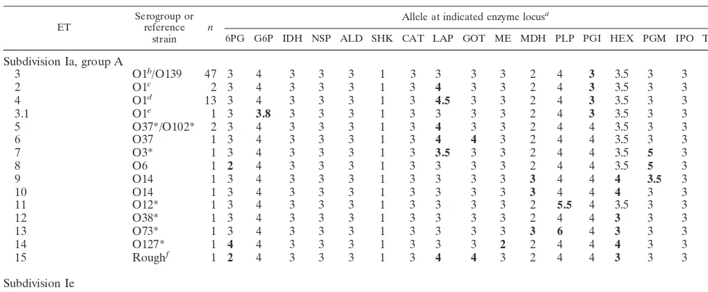
Subdivision Ia includes 82% of the reference collection strains and 78% of the Mexico-Guatemala strains. All but 10 of the 214 ETs in this subdivision form three branches (designat-ed A, B, and C in Fig. 1) that diverge from one another at a genetic distance of about 0.4. With the singular exception of the O1 Inaba El Tor reference strain (ET 259, in subdivision Ie), all O1 strains in our sample, together with all O139 iso-lates, are of 4 ETs that form a tight cluster in group A of subdivision Ia, which consists of a total of 37 ETs (Fig. 2).
Multilocus population structure. Estimates of multilocus
[image:2.612.311.551.90.311.2]association of alleles for all 279 ETs and for the ETs in several segments of the dendrograms (Fig. 1 and 2) are shown in Table
TABLE 1. Measures of mean genetic diversity per locus and multilocus linkage equilibrium, based on ETs
Sample No. of: Meanno. of alleles H
a I
Ab
Isolates ETs
All strains
Total 397 279 9.5 0.436 1.24860.082*
Divisions I and II 392 275 7.5 0.421 0.72860.083* Division I 380 263 6.7 0.397 0.23760.085*
Subdivision Ia
Group A 103 37 3.4 0.297 0.24860.228 Group B 148 123 4.8 0.309 20.04860.125 Group C 55 44 3.3 0.274 0.05360.210 Subdivision Ic 47 40 3.2 0.384 0.93160.217* Division II 12 12 2.8 0.375 1.20460.399* Reference strainsc
Total 142 133 7.5 0.441 1.75960.120*
aH, mean genetic diversity per locus. bI
A, index of association of alleles. *,P#0.05. cStrain CA-385 (rough) not included.
on May 15, 2020 by guest
http://jcm.asm.org/
1. For the total sample of 279 ETs, the 133 ETs of the refer-ence strains, and the ETs of each of the divisions I and II and subdivision Ic, there is evidence of significant nonrandom as-sociations of alleles (linkage disequilibrium), but allele associ-ations are not demonstrably nonrandom for the ETs of groups A, B, and C in subdivision Ia.
Relationships of O1 and O139 epidemic strains.With
al-lowance for a difference in the panels of enzymes employed in MLEE analysis, our findings for the epidemic O1 and O139 strains are fully consistent with those reported by Evins et al. (15) and in earlier studies by Wachsmuth’s group at the CDC (43, 44). These workers assayed variation in 16 enzymes, only 9 of which were included in our panel of 17 enzymes. In the interest of consistency, we have numbered the ETs of our O1
and O139 isolates to correspond to the ET designations of the CDC group (Table 2; Fig. 2).
ET 4 is represented by 13 isolates of O1 Inaba El Tor. Eleven of these strains are from Mexico (Quintana Roo, Campeche, Tabasco, Veracruz, Puebla, and Hidalgo), one is from Guatemala, and one is from Peru. ET 4 marks the orig-inal, or first-wave, Latin American epidemic clone (15).
[image:3.612.61.295.71.514.2]ET 3 includes 2 strains of O1 Inaba El Tor, 38 strains of O1 Ogawa El Tor, and 7 strains of O139. The two Inaba El Tor strains (FMU strains 90501 and 90500) were recovered from humans in Tabasco in 1991 and 1993. The sample of O1 Ogawa El Tor isolates includes the reference strain, 35 isolates from Mexico (Tabasco, Morelos, and the state of Me´xico), a strain from Australia, and an isolate from Romania. ET 3 is the seventh-pandemic type, a clone that in Latin America was first identified in Mexico in 1991 and is now widely distributed in
[image:3.612.307.545.258.691.2]FIG. 1. Dendrogram showing genetic relationships of the ETs ofV. cholerae, based on MLEE analysis (17 loci). The dendrogram was constructed from a matrix of pairwise genetic distances by the UPGMA method. The lineages of subdivision Ic and of groups A, B, and C of subdivision Ia are truncated. The relationships of the 37 ETs in group A are shown in the dendrogram in Fig. 2.
FIG. 2. Dendrogram showing genetic relationships of the 37 ETs ofV. chol-eraein group A of subdivision Ia of division I, based on MLEE analysis (17 loci). The dendrogram was constructed from a matrix of pairwise genetic distances by the UPGMA method.
on May 15, 2020 by guest
http://jcm.asm.org/
Mexico and Central America (15). The Australian isolate (CDC 2463-88) was distinguished, as ET 1, from isolates of ET 3 by Evins et al. (15) on basis of its carrying a different allele for diaphosrase 1. ET 1 marks a distinctive Australian toxigenic clone (13, 43).
Included in ET 2 are an O1 Inaba El Tor isolate (CDC 2164-78) collected in Louisiana in 1978 and an O1 Inaba clas-sical strain (FMU 87295/0) recovered from a tourist returning to the United States from Cancu´n, Quintana Roo, in 1983. ET 2 marks a toxigenic clone that is endemic to the Gulf Coast of Mexico and the United States (15).
With our panel of 17 enzymes, the sole basis for distinguish-ing ETs 2, 3, and 4 is allelic variation at the LAP locus (Table 2). One O1 Ogawa El Tor isolate (from Tabasco) is ET 3.1, which differs from ET 3 in having a distinctive G6P allele. This variant genotype was not detected in previous studies.
Relationship of serogroup O37 strains. The reference
strains O37* (India, 1969) and O102* (China, 1988), both of which are of ET 5 (Fig. 2), differ from strains of the O1/O139 cluster (ETs 2 to 4) at a single locus, that for PGI, and share the LAP 4 allele with strains of ET 2 (Table 2). A second O37 strain (from Guatemala) is of ET 6, which differs from ET 5 in having a 4 allele rather than a 3 allele at the GOT locus. Two other O37 isolates in the collection, both of which were cul-tured from well water in Campeche, represent ETs 75 and 149, which are in group B of subdivision Ia.
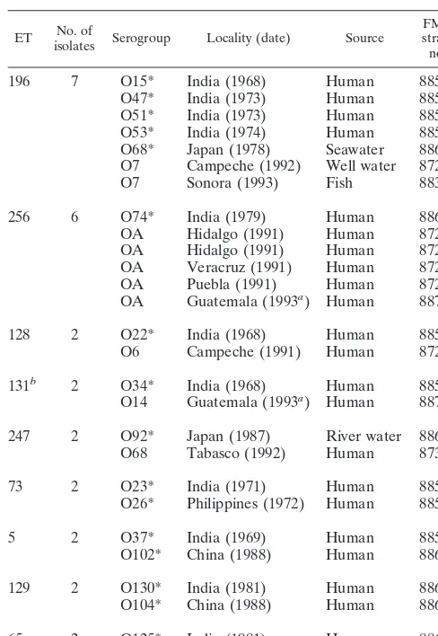
Serotypic diversity among non-O1 strains of the same ET.
In addition to ETs 2 to 4 of the epidemic O1 and O139 clones, MLEE identified nine ETs that are each represented by non-O1 isolates from different continents or other major land masses (Table 3). In all cases, the strains from different con-tinents are of different serogroups. For example, ET 196 (a member of group C of subdivision Ia) is represented by a total of seven isolates, including four (O15*, O47*, O51*, and O53*) recovered from humans in India between 1968 and
1974, a strain (O68*) obtained from seawater in Japan in 1978, and two isolates (both O7) cultured from well water in Campeche and a fish in Sonora in 1992 and 1993. The infer-ence is that ET 196 marks a widely distributed clone that had persisted for at least 24 years, with several modifications in serotype through mutation or recombination of genes of therfb
region.
ETs 128 and 131 are each represented by a strain collected in India in 1968 and an isolate recovered in Campeche or Guatemala in the early 1990s. It is noteworthy that ET 131 differs at only a single locus (that for LAP) from ET 132 of the O14 reference strain, which was recovered in India in 1964 (see Table 5). Thus, ETs 131 and 132 are members of a clonal lineage that was extant for at least 28 years.
Nine ETs are represented by pairs or multiple strains from different states in Mexico (Table 4). Five of these ETs are represented by strains of the same serogroup, and four of them are represented by strains of different serogroups. For exam-ple, ET 16 was represented by seven O5 isolates from Hidalgo, Tabasco, Yucata´n, and an unspecified locality in Mexico. Sig-nificantly, ET 16 differs by only one locus (that for PLP) from ET 17 of the O5 reference strain, which was collected in India in 1964 (see Table 5).
Serotypic diversity among strains of closely related ETs.We
[image:4.612.53.562.83.293.2]identified 19 cases in which pairs of ETs that differ at a single enzyme locus are represented by strains collected on different continents or major land masses (Table 5). In four of these cases, the strains are of the same serotype; these are O5* from India (1964) and seven O5 isolates from Hidalgo and other states in Mexico (1991 and 1992), O14* from India (1964) and O14 from Guatemala (1993), O37* from India (1969) and O37 from Guatemala (1993), and O44* from India (1973) and O44 from Chiapas (1991). In all other cases, the strains are of different serotypes or one of them was NT.
TABLE 2. Allele profiles of 16 ETs in division I
ET Serogroup orreference strain n
Allele at indicated enzyme locusa
6PG G6P IDH NSP ALD SHK CAT LAP GOT ME MDH PLP PGI HEX PGM IPO THD
Subdivision Ia, group A
3 O1b/O139 47 3 4 3 3 3 1 3 3 3 3 2 4 3 3.5 3 3 1
2 O1c 2 3 4 3 3 3 1 3 4 3 3 2 4 3 3.5 3 3 1
4 O1d 13 3 4 3 3 3 1 3 4.5 3 3 2 4 3 3.5 3 3 1
3.1 O1e 1 3 3.8 3 3 3 1 3 3 3 3 2 4 3 3.5 3 3 1
5 O37*/O102* 2 3 4 3 3 3 1 3 4 3 3 2 4 4 3.5 3 3 1
6 O37 1 3 4 3 3 3 1 3 4 4 3 2 4 4 3.5 3 3 1
7 O3* 1 3 4 3 3 3 1 3 3.5 3 3 2 4 4 3.5 5 3 1
8 O6 1 2 4 3 3 3 1 3 3 3 3 2 4 4 3.5 5 3 1
9 O14 1 3 4 3 3 3 1 3 3 3 3 3 4 4 4 3.5 3 1
10 O14 1 3 4 3 3 3 1 3 3 3 3 3 4 4 4 3 3 1
11 O12* 1 3 4 3 3 3 1 3 3 3 3 2 5.5 4 3.5 3 3 1
12 O38* 1 3 4 3 3 3 1 3 3 3 3 2 4 4 3 3 3 1
13 O73* 1 3 4 3 3 3 1 3 3 3 3 3 6 4 3 3 3 1
14 O127* 1 4 4 3 3 3 1 3 3 3 2 2 4 4 4 3 3 1
15 Roughf 1 2 4 3 3 3 1 3 4 4 3 2 4 4 3 3 3 1
Subdivision Ie
259 O1*g 1 3.5 2 3 3 0 1 1 4.5 3 3 2 5 3 1 3 3 1
aMinority alleles are indicated in boldface type.
bThirty-nine isolates of O1 Ogawa El Tor and 2 isolates of O1 Inaba El Tor. cOne isolate of O1 Inaba classical and 1 isolate of O1 Inaba El Tor. dThirteen isolates of O1 Inaba El Tor from Mexico, Guatemala, and Peru. eOne isolate of O1 Ogawa El Tor from Tabasco.
fStrain CA-385.
gO1 Inaba El Tor reference strain.
on May 15, 2020 by guest
http://jcm.asm.org/
Genetic diversity within serogroups. MLEE analysis dem-onstrated that strains of the same serogroup may belong to two or more widely divergent ET lineages. Thus, for example, strains of serogroup O29 were assigned to four ETs that occur in division I (ET 50 and ET 52 in group B and ET 173 in group C) and division II (ET 274), and O53 strains are found in group C of division I (ET 196) and also in lineage y (ET 278). Some estimated levels of genetic diversity between or among the ETs of strains of the same serogroup are shown in Table 6. In several cases, the estimated diversity is at least equivalent to that obtained for the 279 ETs identified among all 397 isolates.
Distribution of the ctxA gene. When tested with the ctxA
probe, 13 of the 143 reference strains were positive, as shown in Table 7. With PCR amplification of the gene, the same strains were positive, with the exception of O1 Inaba El Tor*. Among 104 of 254 nonreference strains that were randomly selected for testing, 2 isolates of O1 Ogawa El Tor, 3 isolates of O1 Inaba El Tor, an isolate of O1 Inaba classical, and an isolate of O6 were positive forctxAby both colony blot assay and PCR amplification.
All but 3 of the total of 21ctxA-positive strains are members of subdivision Ia of division I (Fig. 1 and 2); the exceptions are
the reference strains O1 Inaba El Tor*, in subdivision Ie, and O135* and O138*, in division II.
DISCUSSION
Species limits.Strains of ETs 276 to 279 in the deep lineages
x and y (Fig. 1) are sufficiently differentiated from all other strains as to raise the question of whether they should be included in the speciesV. cholerae. It is likely that assessment of genomic relatedness by DNA-DNA hybridization would show relative degrees of annealing with other strains somewhat below the 70% standard adopted for species inclusion by the CDC (6).
Genetic diversity. The estimate of 0.436 for the mean
ge-netic diversity per locus among the 279 ETs ofV. cholerae
represented in the present study is larger than the comparable value of 0.343 reported for theEscherichia colireference col-lection (35) but smaller than the corresponding value of 0.627 obtained for Salmonella enterica (36). In a previous MLEE study of allelic variation at 13 enzyme loci among 260 isolates ofV. cholerae(most of which were serogroup O1), Salles and Momen (33) detected an average of 4.3 alleles per locus, iden-tified 73 ETs, and estimated the mean genetic diversity per locus as 0.326.
Genetic structure of populations.Comparisons of the
ob-served and expected variances of the mismatch distributions for ETs at several hierarchical levels of dendrogram structure yielded only limited evidence of linkage disequilibrium (Table 1). The cases in which the observed variance exceeded the upper 95% confidence limit of the variance expected under random association of alleles were those involving all 279 ETs, the 275 ETs of divisions I and II combined, the 263 ETs of division I, the 12 ETs of division II, and the 40 ETs of subdi-vision Ic of disubdi-vision I. Within each of the groups A, B, and C of subdivision Ia, which include 204 ETs (Fig. 1), significant levels of nonrandom association were not demonstrable. The inference is that, at least among the strains of ETs in subdivi-sion Ia, the rate of horizontal transfer and recombination of housekeeping enzyme genes is sufficiently high to prevent the development and long-term maintenance of distinctive allele complexes. On the whole, it seems likely that the frequency of recombination, both intragenic and assortative (50), of house-keeping genes inV. choleraeis somewhat higher than in either
E. coli(48) orS. enterica(36), a conclusion also reached by Karaolis et al. (19) from a comparative sequence analysis of theasd gene. But even within subdivision Ia, clonal lineages may persist for periods of time measured in at least decades. The most obvious examples are the epidemic and pandemic strains of serogroups O1 and O139 (ETs 2 to 4), but our analysis identified numerous clones and clonal lineages of non-O1 strains with widespread, if not global, distributions (Tables 3 to 5).
A factor that has not been evaluated in studies of the genetic structure of bacterial populations on the basis of MLEE data is the convergent evolution of electromorphs, which cannot be equated with isoalleles. Similarity in electrophoretic mobility resulting from convergence in the net electrostatic charge of an enzyme will lessen the likelihood that linkage disequilibrium is detected from the analysis of MLEE data. Studies of sequence variation in several housekeeping enzymes among multiple strains ofE. coliandS. enterica(5, 26–28, 47) have shown that individual electromorphs may exhibit substantial heterogeneity in amino acid sequence, much of which clearly stems from convergence rather than mutational divergence from a com-mon ancestral sequence.
[image:5.612.51.295.91.445.2]There was already evidence for recombination of genes of
TABLE 3. ETs represented by non-O1 isolates from different continents
ET isolatesNo. of Serogroup Locality (date) Source FMUstrain no.
196 7 O15* India (1968) Human 88554
O47* India (1973) Human 88586
O51* India (1973) Human 88590
O53* India (1974) Human 88592
O68* Japan (1978) Seawater 88607 O7 Campeche (1992) Well water 87242
O7 Sonora (1993) Fish 88354
256 6 O74* India (1979) Human 88613
OA Hidalgo (1991) Human 87250
OA Hidalgo (1991) Human 87256
OA Veracruz (1991) Human 87264
OA Puebla (1991) Human 87246
OA Guatemala (1993a) Human 88778
128 2 O22* India (1968) Human 88561
O6 Campeche (1991) Human 87268
131b 2 O34* India (1968) Human 88573
O14 Guatemala (1993a) Human 88729
247 2 O92* Japan (1987) River water 88631 O68 Tabasco (1992) Human 87311
73 2 O23* India (1971) Human 88562
O26* Philippines (1972) Human 88565
5 2 O37* India (1969) Human 88576
O102* China (1988) Human 88641
129 2 O130* India (1981) Human 88669
O104* China (1988) Human 88643
65 2 O125* India (1981) Human 88664
O132* Thailand (1981) Human 88671
aDate of receipt at the Departamento de Salud Pu´blica de la Facultad de
Medicina.
bET 131 differs from ET 132 of strain O14* (India, 1964) at a single locus (that
for LAP) (Table 5).
on May 15, 2020 by guest
http://jcm.asm.org/
therfbregion ofV. cholerae, based on studies of relatively small numbers of strains and serogroups. Our observation that strains of the same serogroup frequently are found in diver-gent, even distantly related, lineages supports earlier evidence (2, 19) that therfbgenes are subject to horizontal transfer and further suggests that this process occurs with relatively high frequency. Convergence in serotype is, of course, an alterna-tive explanation, but reasoning by analogy from the lack of evidence for convergence in epitope structure in the serologi-cally diverse flagellins of S. enterica (22), we favor the first hypothesis. The issue can be settled by comparative sequencing of the epitope-encoding segments of therfbregion.
The fact that strains of the same ET may express different O antigens can be explained by recombination or by spontaneous mutation of the genes encoding O somatic properties.
Epidemic non-O1 clones. There are two examples of
epi-demicV. choleraeexpressing a non-O1 antigen. The first is the serogroup O139 clone, which emerged in India and Bang-ladesh through modification of the El Tor O1 pandemic strain by acquisition of genes mediating the synthesis of the O139 LPS and a polysaccharide capsule. The second is the O37 strain that reportedly was responsible for a large outbreak of cholera in the Sudan in 1968 (54). By IS1004fingerprinting, Bik et al. (3) determined that an O37 isolate from the Sudan is closely related to classical O1 strains. The O37 reference strain (ET 5), which was recovered from a patient in India in 1969,
presumably represents the same clone as the O37 Sudan strain. As determined by MLEE, it is closely related to O1 El Tor and other epidemic O1 strains (Fig. 2), thus confirming the result obtained by IS1004fingerprinting. In fact, ET 5 is distinguished from ETs 2 to 4 of the O1/O139 cluster solely by possession of a 4 allele (versus a 3 allele) at the PGI locus. It carries thectxA
gene and expresses cholera toxin. Yamamoto et al. (53) re-ported that the amino acid sequence of the cholera toxin pro-duced by O37 strain S7 differs from that of most O1 strains in having single substitutions in both the CtxA and CtxB seg-ments (29), which are presumed to cause the formation of an unusually large subunit B oligomer. Recently, Karaolis et al. (20) reported that, almost uniquely among non-O1 strains, a Sudan 1968 outbreak strain carries a chromosomal pathoge-nicity island that is characteristic of epidemic and pandemic strains.
Honma et al. (16) studied an O37 isolate that produces a hemolysin (O37-Hly) that is antigenically similar to O1 El Tor hemolysin (El Tor-Hly) but different in molecular size, hemo-lytic activity, and glucose-binding capacity. The gene encoding O37-Hly differs from that encoding O1 El Tor-Hly by the presence of a 4-bp insertion that generates a premature stop codon in the downstream region. Thus, the O37-Hly is a trun-cated derivative of O1 El Tor-Hly, sharing 90% of the N-terminal region.
[image:6.612.54.550.82.438.2]In the Mexico-Guatemala collection, there are three O37
TABLE 4. ETs represented by isolates from two or more states in Mexico
ET No. of isolates Serogroup Locality (date) Source FMU strain no.
Of same serogroup
16a 7 O5 Hidalgo (1991) Human 87243
O5 Hidalgo (1991) Human 87259
O5 Tabasco (1991) Human 87139
O5 Tabasco (1991) Human 87672
O5 Mexico (1991) Human 87288
O5 Tabasco (1992) Human 87306
O5 Yucata´n (1992) Human 87291
228 2 O149 Guanajuato (1991) Human 87297
O149 Tabasco (1991) Human 87673
219 2 O149 Yucata´n (1991) Human 87262
O149 Zacatecas (1991) 87299
107 2 O64 Veracruz (1991) Human 87257
O64 Hidalgo (1991) Human 87289
124 2 O24 Veracruz (1991) Human 87282
O24 Tabasco (1992) Human 87434
Of different serogroups
181 4 O35 Sonora (1993) Sewage water 88374
O35 Sonora (1993) Sewage water 88375
O42 Sonora (1993) Fish 88351
NT Guanajuato (1991) Human 87287
52 3 O79 Zacatecas (1991) Human 87295
O43 Tabasco (1992) Human 87307
O29 Sonora (1993) Sewage water 88371
234 2 O5 Campeche (1992) Well water 87240
O62 Tabasco (1992) Human 87304
171 2 O41 Sonora (1993) Septic tank 88366
NT Guerrero (1991) 87271
aET 16 differs from ET 17 of strain O5* (India, 1964) at a single locus (that for PLP) (Table 5).
on May 15, 2020 by guest
http://jcm.asm.org/
TABLE 5. Serogroups and geographic sources of non-O1 strains representing pairs of ETs that differ at a single locus
Strains from different continents Strains from same continent
ET Serogroup Locality (date) strain no.FMU Locus ET Serogroup Locality (date) strain no.FMU Locus
17 O5* India (1964) 88544 PLP 194 O4* India (1932) 88543 LAP
16 O5 Hidalgo (1991)a 87243 PLP 193 O96* India (1976) 88635 LAP
132 O14* India (1964) 88553 LAP 166 O6* India (1962) 88545 PGM
131 O14 Guatemala (1993b) 88729 LAP 165 O79* India (1976) 88618 PGM
5 O37* India (1969)c 88576 GOT 132 O14* India (1964) 88553 LAP
6 O37 Guatemala (1993b) 88777 GOT 131 O34* India (1968) 88573 LAP
109 O44* India (1973) 88583 PLP 196 O15* India (1968) 88554 MDH
108 O44 Chiapas (1991) 87245 PLP 195 O35* India (1969) 88574 MDH
102 O18* India (1964) 88557 PGI 115 O54* India (1974) 88593 LAP
101 O14 Campeche (1992) 87440 PGI 114 O94* India (1976) 88633 LAP
152 O16* India (1971) 88555 PGI 168 O95* India (1976) 88634 CAT
151 O48 Tabasco (1991) 87675 PGI 167 O129* India (1981) 88668 CAT
169 O134* India (1991) 88673 CAT
122 O49* India (1974) 88588 6PG
121 NT Michoacan (1992) 87293 6PG 155 O112* Japan (1989)h 88651 IDH
123 NT Sonora (1993) 88352 6PG 154 O113* Japan (1989)h 88652 IDH
192 O60* India (1975) 88599 IDH 31 O12 Tabasco (1992) 87432 G6P
191 O68 Sonora (1993) 88392 IDH 32 O12 Guatemala (1993b) 88750 G6P
134 O55* India (1975) 88594 LAP 107 O64 Veracruz (1991) 87257 LAP
133 NT Hidalgo (1991) 87263 LAP 106 O64 Chiapas (1991) 87272 LAP
186 O81* India (1978) 88620 6PG 229 O149 Guanajuato (1991) 87290 CAT
185 O80 Campeche (1992) 87437 6PG 228 O149 Guanajuato (1991) 87297 CAT
230 O155 Campeche (1992) 90594 CAT
73 O23* India (1971)d 88562 G6P
72 O93 Guatemala (1993b) 88773 G6P 22 O14 Campeche (1992) 87661 PGM
21 O14 Campeche (1992) 87778 PGM
256 O74*c India (1979) 88613 IPO
257 OA Guatemala (1993b) 88733 IPO 59 O44 Sonora (1993) 88380 PGM
58 O5 Sonora (1993) 88382 PGM
247 O92* Japan (1987)f 88631 CAT
246 O31 Tabasco (1992) 87303 CAT 157 O141 Hidalgo (1991) 87274 LAP
156 O82 Sonora (1993) 88389 LAP
136 O120* Japan (1991) 88659 PGM
135 O52 Sonora (1993) 88356 PGM 233 NT Hidalgo (1991) 87298 CAT
234 O5 Campeche (1992) 87240 CAT
53 O108* Japan (1989) 88637 PGM
52 O79 Zacatecas (1991) 87295 PGM 225 O41 Tabasco (1992) 87309 LAP
54 O105 Sonora (1993) 88387 PGM 226 NT Tabasco (1992) 87305 LAP
98 O122* Romania (1980) 88661 CAT 221 O35 Veracruz (1991) 87279 THD
97 O97 Veracruz (1991) 87283 CAT 220 O151 Sonora (1993) 88367 THD
265 O20* India (1962) 88559 IDH 51 O43 Guatemala (1993b) 88727 CAT
264 O101* China (1988) 88640 IDH 50 O40 Guatemala (1993b) 88744 CAT
128 O22* India (1968)g 88561 PGM
127 O83* India (1978) 88622 PGM
129 O104* China (1988) 88643 PGM
200 O86* Philippines (1981) 88625 GOT
199 O133* India (1991) 88672 GOT
aThere are six additional O5 isolates of ET 16 from Hidalgo, Yucata´n, Tabasco, and “Mexico” (Table 4). bDate of receipt at the Departamento de Salud Pu´blica de la Facultad de Medicina, UNAM.
cStrain O37* (India, 1969) is of the same ET as O102* from China (1988) (Table 3).
dStrain O23* (India, 1971) is of the same ET as strain O16* from the Philippines (1972) (Table 3).
eFive additional OA isolates from Mexico and Guatemala (1991 to 1993) are of the same ET as strain O74* from India (1979) (Table 3). fStrain O92* (Japan, 1987) is of the same ET as a strain of serotype O68 from Tabasco (1992) (Table 3).
gStrain O22* (India, 1968) is of the same ET as an O6 strain from Campeche (1991) (Table 3). hSource: rat.
on May 15, 2020 by guest
http://jcm.asm.org/
isolates, one of which (ET 6) is almost identical in MLEE genotype (it carries a GOT 4 rather than a GOT 3 allele) to the O37 reference strain (ET 5) from India but apparently lacks thectxAgene. It was isolated from a patient in Guatemala. The two other O37 isolates, both of which were cultured from well water in Campeche, are distantly related (six- and seven-locus differences) to both the reference O37 and Guatemala O37 strains, as well as to one another (four-locus difference), and neither one carries thectxAgene.
It is noteworthy that strain O102*, which was recovered from a patient with diarrhea in China in 1988, is identical in MLEE genotype (ET 5) to strain O37* but apparently does not carry thectxAgene.
In sum, there is a clone of serogroup O37 that has epidemic potential and was present in Africa and India in 1968 and 1969. Because it is closely related to O1 El Tor and the other O1 pandemic clones, it apparently represents a case similar to that
of the O139 clone, in which an already-established pathogenic lineage of serogroup O1 acquired a new serotype by horizontal DNA transfer and rearrangement of therfbregion genes. The O37 strain from Guatemala may be an offshoot of this clone in which the CTX genetic element has been deleted. The two O37 strains from Campeche presumably have independent acquisi-tions of the O37 polysaccharide gene region.
O1 Inaba El Tor reference strain. The O1 Inaba El Tor
reference strain (ET 259), which does not produce cholera toxin although it carries at least part of thectxAgene, is not closely related to the O1/O139 cluster of pandemic strains or to the toxigenic O37 clone (ET 5). According to T. Shimada (38a), the reference strain is the NIH 35-a-3 isolate listed by Burrows et al. (7) as one of the strains used for vaccine prep-aration by the U.S. Army in the early 1940s. It was received from the Central Research Institute in Kasauli, India, in 1942, without indication of collection date or source of isolation. Perhaps it is related to the O1 strain that caused a cholera-like disease in Hong Kong in the 1950s (43).
Relationships of serogroup O1 strains.Colwell et al. (11)
hypothesized that non-O1 cells may convert to the O1 serotype and vice versa under suitable conditions, a possible strategy for survival in the environment. As noted earlier, O37* and O102* (both of ET 5) may represent cases in which O1 clones have acquired new serotypes. In our collection, the only apparent case of conversion of a non-O1 strain to the O1 serotype (apart from O139) is the O1 Inaba El Tor reference strain (ET 259), which occurs in subdivision Ie (Fig. 1) and is distantly related to the O1 epidemic strains (ETs 2 to 4) in group A of subdi-vision Ia (Fig. 2).
Source ofrfbregion DNA in the emergence of the epidemic
O139 clone.The putative source of the exogenousrfb region
[image:8.612.53.293.91.237.2]DNA that was involved in the transformation of an O1 El Tor strain to the epidemic O139 clone has been identified as a strain of serogroup O22, O141, or O155 on the basis of sero-typic cross-reactions with O139 (2, 38, 42). Molecular analysis
TABLE 6. Mean genetic diversity per locus among multiple ETs of the same serogroup
Serogroup No. of: diversity per locusMean genetic Isolates ETs
O12 4 4 0.206
O37 4 4 0.314
O6 12 10 0.316
O8 14 4 0.333
O5 14 8 0.345
O38 4 2 0.412
O155 9 7 0.423
O42 3 3 0.490
O29 4 4 0.559
O39 5 4 0.618
O30 2 2 0.824
O53 3 2 1.000
TABLE 7. Strains testing positive forctxAa
Serogroup FMU strain no. ET subdivision (group)Division or Source
Reference
O1 Ogawa El Tor* 3 Ia (A) India (1941), human
O1 Inaba El Tor* 259 Ie India (1942), human
O19* 41 Ia (B)
O33* 204 Ia (C) India (1968), human
O37* 5 Ia (A) India (1969), human
O43* 43 Ia (B) India (1973), human
O44* 109 Ia (B) India (1972), human
O54* 115 Ia (B) India (1974), human
O105* 116 Ia (B) India (1988), human
O106* 64 Ia (B) China (1988), human
O135* 268 II India (1992), human
O138* 270 II Japan (1992), crab
O139* 3 Ia (A) India (1993), human
Nonreference
O1 Ogawa El Tor 87668 3 Ia (A) Morelos, human
O1 Ogawa El Tor 90334 3 Ia (A) Mexico, human
O1 Inaba El Tor 87269 4 Ia (A) Campeche, human
O1 Inaba El Tor 88696 2 Ia (A) Louisiana, human
O1 Inaba El Tor 90500 3 Ia (A) Tabasco, human
O1 Inaba classical 87395/0 2 Ia (A) Quintana Roo, human
O6 88751 27 Ia (A) Guatemala, human
O139 88230 3 Ia (A) United States, imported case
aAll strains were positive when tested with thectxAprobe and by PCR, with the exception of O1 Inaba El Tor*, which was probe positive but PCR negative.
on May 15, 2020 by guest
http://jcm.asm.org/
[image:8.612.53.547.473.717.2]showed that, in common with O139, they have two open read-ing frames in therfaDregion that are lacking in O1 strains.
Through study of the gene content and organization of the
rfbregion adjacent to IS1358in strains of O139 and 13 other serogroups, including O22 and O155, Dumontier and Berche (14) recently identified the clone represented by strain O22* (Shimada strain 169-68, from India) as the most likely donor, although the possibility of a multistep rearrangement in the recipient O1 strain cannot be excluded. As determined by MLEE analysis, O22* (ET 128) falls in group B of subdivision Ia and differs in ET from the epidemic O1 and O139 strains at four or five loci.
In our sample of strains, there were nine serogroup O155 isolates belonging to seven ETs. ET 224 of the O155 reference strain (Thailand, 1993) and five other ETs (represented by isolates from Tabasco and Campeche) are in subdivision Ic, where, however, they do not form a tight cluster. The remain-ing ET, which is represented by two isolates from Sonora, is in group B of subdivision Ia. Thus, strains of serogroup O155 belong to a moderately diverse group of ETs, none of which is closely related to the ETs of the epidemic O1 and O139 clones. This suggests that genes mediating expression of the O155 LPS antigen are transferred with relatively high frequency.
Genesis of epidemic clones. Because genes for the major
virulence factors can be transferred horizontally and antigenic conversion can be achieved by the acquisition and loss ofrfb
genes, there is the formal possibility that anyV. choleraecell could be transformed into a virulent strain, even an epidemic one (19, 25). However, the close evolutionary relationships of the O1, O139, and O37 epidemic clones indicate that new epidemic or other strongly virulent clones are likely to arise by the modification of a lineage that is already epidemic or is closely related to such a clone. Thus, O139 evolved from an El Tor O1 clone by acquisition of a transposon carrying genes for the O139 LPS and a polysaccharide capsule and deletion of most of the genes mediating synthesis of the serotype O1 LPS (see the review by Rubin et al. [31]). Also, the toxigenic O37 clone that caused outbreaks in the Sudan and India in the late 1960s is closely related to the cluster of O1/O139 epidemic clones. Analogously, E. coli O157:H7, which emerged as an agent of hemorrhagic colitis by acquisition of the O157 anti-gen, a Shiga-like toxin, and the enterohemorrhagic E. coli
plasmid, is an evolutionary derivative of an O55:H7 clone that is associated with infantile diarrhea (51).
ACKNOWLEDGMENTS
We thank Thomas Cheasty, Peter Echeverria, Alma Rosa Gonza´lez, Sergio Leo´n, Claudio Lezana, Jose´ Luis Navarro-Heinze, and Toshio Shimada for supplying strains and Delia Licona and Jose´ Luis Me´ndez for technical assistance in the laboratory.
This research was supported by grants from the Consejo Nacional de Ciencia y Tecnologı´a (project 2397PB); the Direccio´n General de Apoyo al Personal Acade´mico, UNAM (project IN211496); and the National Institutes of Health (AI-22144).
REFERENCES
1.Albert, M. J., A. K. Siddique, M. S. Islam, A. S. Faruque, M. Ansaruzzaman,
S. M. Faruque, and R. B. Sack.1993. Large outbreak of clinical cholera due
toVibrio choleraenon-O1 in Bangladesh. Lancet341:704.
2.Bik, E. M., A. E. Bunschoten, R. D. Gouw, and F. R. Mooi.1995. Genesis of
the novel epidemic Vibrio choleraeO139 strain: evidence for horizontal transfer of genes involved in polysaccharide synthesis. EMBO J.14:209–216.
3.Bik, E. M., R. D. Gouw, and F. R. Mooi.1996. DNA fingerprinting ofVibrio
choleraestrains with a novel insertion sequence element: a tool to identify epidemic strains. J. Clin. Microbiol.34:1453–1461.
4.Blake, P. A., R. E. Weaver, and D. G. Hollis.1980. Diseases of humans (other
than cholera) caused by vibrios. Annu. Rev. Microbiol.34:341–367.
5.Boyd, E. F., K. Nelson, F.-S. Wang, T. S. Whittam, and R. K. Selander.1994.
Molecular genetic basis of allelic polymorphism in malate dehydrogenase
(mdh) in natural populations ofEscherichia coliandSalmonella enterica. Proc. Natl. Acad. Sci. USA91:1280–1284.
6.Brenner, D. J., A. G. Steigerwalt, P. Epple, W. F. Bibb, R. M. McKinney,
R. W. Starnes, J. M. Colville, R. K. Selander, P. H. Edelstein, and C. W.
Moss.1988.Legionella pneumophilaserogroup Lansing 3 isolated from a
patient with fatal pneumonia, and descriptions ofL. pneumophilasubsp.
pneumophilasubsp. nov.,L. pneumophilasubsp.fraserisubsp. nov., andL. pneumophilasubsp.pasculleisubsp. nov. J. Clin. Microbiol.26:1695–1703.
7.Burrows, W., A. N. Mather, M. E. Elliott, and S. M. Wagner.1946. Studies
on immunity to Asiatic cholera. J. Infect. Dis.79:159–167.
8.Chen, F., G. M. Evins, W. L. Cook, R. Almeida, N. Hargrett-Bean, and K.
Wachsmuth. 1991. Genetic diversity among toxigenic and nontoxigenic
Vibrio choleraeO1 isolated from the Western Hemisphere. Epidemiol. In-fect.107:225–233.
9.Chongsa-nguan, M., W. Chaicumpa, P. Moolasart, P. Kandhasingha, T.
Shimada, H. Kurazono, and Y. Takeda.1993.Vibrio choleraeO139 Bengal in
Bangkok. Lancet342:430–431.
10. Colwell, R. R. 1996. Global climate and infectious disease: the cholera
paradigm. Science274:2025–2031.
11. Colwell, R. R., A. Huq, M. A. R. Chowdhury, P. R. Brayton, and B. Xu.1995.
Serogroup conversion ofVibrio cholerae. Can. J. Microbiol.41:946–950.
12. Comstock, L. E., D. Maneval, Jr., P. Panigrahi, A. Joseph, M. M. Levine,
J. B. Kaper, J. G. Morris, Jr., and J. A. Johnson.1995. The capsule and O
antigen inVibrio choleraeO139 Bengal are associated with a genetic region not present inVibrio choleraeO1. Infect. Immun.63:317–323.
13. Desmarchelier, P. M., H. Momen, and C. A. Salles.1988. A zymovar analysis
ofVibrio choleraeisolated in Australia. Trans. R. Soc. Trop. Med. Hyg.
82:914–917.
14. Dumontier, S., and P. Berche.1998.Vibrio choleraeO22 might be a putative
source of exogenous DNA resulting in the emergence of the new strain of
Vibrio choleraeO139. FEMS Microbiol. Lett.164:91–98.
15. Evins, G. M., D. N. Cameron, J. G. Wells, K. D. Greene, T. Popovic, S.
Giono-Cerezo, I. K. Wachsmuth, and R. V. Tauxe. 1995. The emerging
diversity of the electrophoretic types ofVibrio choleraein the Western Hemi-sphere. J. Infect. Dis.172:173–179.
16. Honma, Y., K. Yamamoto, and M. Iwanaga.1995. Aberrant gene for El Tor
hemolysin fromVibrio choleraenon-O1, N037. FEMS Microbiol. Lett.133:
151–154.
17. Kaper, J. B., J. G. Morris, Jr., and M. M. Levine.1995. Cholera. Clin.
Microbiol. Rev.8:48–86.
18. Karaolis, D. K. R., R. Lan, and P. R. Reeves.1994. Molecular evolution of
the seventh-pandemic clone ofVibrio choleraeand its relationship to other pandemic and epidemicV. choleraeisolates. J. Bacteriol.176:6199–6206.
19. Karaolis, D. K. R., R. Lan, and P. R. Reeves.1995. The sixth and seventh
cholera pandemics are due to independent clones separately derived from environmental, nontoxigenic, non-O1Vibrio cholerae. J. Bacteriol.177:3191– 3198.
20. Karaolis, D. K. R., J. A. Johnson, C. C. Bailey, E. C. Boedeker, J. B. Kaper,
and P. R. Reeves.1998. AVibrio choleraepathogenicity island associated with
epidemic and pandemic strains. Proc. Natl. Acad. Sci. USA95:3134–3139.
21. Kovach, M. E., M. D. Shaffer, and K. M. Peterson.1996. A putative integrase
gene defines the distal end of a large cluster of ToxR-regulated colonization genes inVibrio cholerae. Microbiology142:2165–2174.
22. Li, J., K. Nelson, A. C. McWhorter-Murlin, T. S. Whittam, and R. K.
Selander.1994. Recombinational basis of serovar diversity inSalmonella
enterica. Proc. Natl. Acad. Sci. USA91:2552–2556.
23. Maniatis, T., E. F. Fritsch, and J. Sambrook.1982. Molecular cloning: a
laboratory manual. Cold Spring Harbor Laboratory, Cold Spring Harbor, N.Y.
24. Maynard Smith, J., N. H. Smith, M. O’Rourke, and B. G. Spratt.1993. How
clonal are bacteria? Proc. Natl. Acad. Sci. USA90:4384–4388.
25. Mekalanos, J. J., E. J. Rubin, and M. K. Waldor.1997. Cholera: molecular
basis for emergence and pathogenesis. FEMS Immunol. Med. Microbiol.
18:241–248.
26. Nelson, K., T. S. Whittam, and R. K. Selander.1991. Nucleotide
polymor-phism and evolution in the glyceraldehyde-3-phosphate dehydrogenase gene (gapA) in natural populations ofSalmonellaandEscherichia coli. Proc. Natl. Acad. Sci. USA88:6667–6671.
27. Nelson, K., and R. K. Selander.1992. Evolutionary genetics of the proline
permease gene (putP) and the control region of the proline utilization operon in populations ofSalmonellaandEscherichia coli. J. Bacteriol.174:
6886–6895.
28. Nelson, K., and R. K. Selander.1994. Intergeneric transfer and
recombina-tion of the 6-phosphogluconate dehydrogenase gene (gnd) in enteric bacte-ria. Proc. Natl. Acad. Sci. USA91:10227–10231.
29. Olsvik, Ø., J. Wahlberg, B. Petterson, M. Uhlen, T. Popovic, I. K.
Wachs-muth, and P. I. Fields.1993. Use of automated sequencing of polymerase
chain reaction-generated amplicons to identify three types of cholera toxin subunit B inVibrio choleraeO1 strains. J. Clin. Microbiol.31:22–25.
30. Ramamurthy, T., S. Garb, R. Sharma, S. K. Bhattacharya, G. B. Nair, T.
Shimada, T. Takeda, T. Karasawa, H. Kurazano, A. Pal, and Y. Takeda.
1993. Emergence of novel strain ofVibrio choleraewith epidemic potential in
on May 15, 2020 by guest
http://jcm.asm.org/
southern and eastern India. Lancet341:703–704.
31.Rubin, E. J., M. K. Waldor, and J. J. Mekalanos.1998. Mobile genetic
elements and the evolution of new epidemic strains ofVibrio cholerae, p. 147–161.InR. M. Krause (ed.), Emerging infections. Academic Press, San Diego, Calif.
32.Sakazaki, R., and T. J. Donovan.1984. Serology and epidemiology ofVibrio
choleraeandVibrio mimicus. Methods Microbiol.16:271–289.
33.Salles, C. A., and H. Momen.1991. Identification of Vibrio choleraeby
enzyme electrophoresis. Trans. R. Soc. Trop. Med. Hyg.85:544–547.
34.Selander, R. K., D. A. Caugant, H. Ochman, J. M. Musser, M. N. Gilmour,
and T. S. Whittam.1986. Methods of multilocus enzyme electrophoresis for
bacterial population genetics and systematics. Appl. Environ. Microbiol.
51:873–884.
35.Selander, R. K., D. A. Caugant, and T. S. Whittam.1987. Genetic structure
and variation in natural populations ofEscherichia coli, p. 1625–1648.In
F. C. Neidhardt, J. L. Ingraham, K. B. Low, B. Magasanik, M. Schaechter, and H. E. Umbarger (ed.),Escherichia coliandSalmonella typhimurium: cellular and molecular biology, vol. 2. American Society for Microbiology, Washington, D.C.
36. Selander, R. K., J. Li, and K. Nelson.1996. Evolutionary genetics of
Salmo-nella enterica, p. 2691–2707. InF. C. Neidhardt, R. Curtiss III, J. L. In-graham, E. C. C. Lin, K. B. Low, B. Magasanik, W. S. Reznikoff, M. Riley, M. Schaechter, and H. E. Umbarger (ed.),Escherichia coliandSalmonella: cellular and molecular biology, 2nd ed., vol. 2. ASM Press, Washington, D.C.
37. Sharma, C., A. Ghosh, A. Dalsgaard, A. Forslund, R. K. Ghosh, S. K.
Bhattacharya, and G. B. Nair.1998. Molecular evidence that a distinctVibrio
choleraeO1 biotype El Tor strain in Calcutta may have spread to the African continent. J. Clin. Microbiol.36:843–844.
38. Shimada, T., E. Arakawa, K. Itoh, T. Nakazato, T. Okitsu, S. Yamai, M.
Kusum, G. B. Nair, and Y. Takeda.1994. Two strains ofVibrio cholerae
non-O1 possessing somatic (O) antigen factors in common withV. cholerae
serogroup O139 synonym “Bengal.” Curr. Microbiol.29:331–333.
38a.Shimada, T.Personal communication.
39. Shirai, H., M. Nishibuchi, T. Ramamurthy, S. K. Bhattacharya, S. C. Pal,
and Y. Takeda.1991. Polymerase chain reaction for detection of the cholera
enterotoxin operon ofVibrio cholerae. J. Clin. Microbiol.29:2517–2521.
40. Stroeher, U. H., K. E. Jedani, B. K. Dredge, R. Morona, M. H. Brown, L. E.
Karageorgos, M. J. Albert, and P. A. Manning.1995. Genetic
rearrange-ments in therfbregions ofVibrio choleraeO1 and O139. Proc. Natl. Acad. Sci. USA92:10374–10378.
41. Stroeher, U. H., G. Parasivam, B. K. Dredge, and P. A. Manning.1997.
NovelVibrio choleraeO139 genes involved in lipopolysaccharide biosynthe-sis. J. Bacteriol.179:2740–2747.
42. Vimont, S., S. Dumontier, V. Escuyer, and P. Berche.1997. TherfaDlocus:
a region of rearrangement inVibrio choleraeO139. Gene185:43–47.
43. Wachsmuth, I. K., G. M. Evins, P. I. Fields, Ø. Olsvik, T. Popovic, C. A.
Bopp, J. G. Wells, C. Carrillo, and P. A. Blake.1993. The molecular
epide-miology of cholera in Latin America. J. Infect. Dis.167:621–626.
44. Wachsmuth, K., Ø. Olsvik, G. M. Evins, and T. Popovic.1994. Molecular
epidemiology of cholera, p. 357–370.InI. K. Wachsmuth, P. A. Blake, and Ø. Olsvik (ed.),Vibrio choleraeand cholera: molecular to global perspectives. ASM Press, Washington, D.C.
45. Waldor, M. K., R. Colwell, and J. J. Mekalanos.1994. TheVibrio cholerae
O139 serogroup antigen includes an O-antigen capsule and lipopolysaccha-ride virulence determinants. Proc. Natl. Acad. Sci. USA91:11388–11392.
46. Waldor, M. K., and J. J. Mekalanos.1996. Lysogenic conversion by a
fila-mentous phage encoding cholera toxin. Science272:1910–1914.
47. Wang, F.-S., T. S. Whittam, and R. K. Selander.1997. Evolutionary genetics
of the isocitrate dehydrogenase gene (icd) inEscherichia coliandSalmonella enterica. J. Bacteriol.179:6551–6559.
48. Whittam, T. S.1996. Genetic variation and evolutionary processes in natural
populations ofEscherichia coli, p. 2708–2720.InF. C. Neidhardt, R. Curtiss III, J. L. Ingraham, E. C. C. Lin, K. B. Low, B. Magasanik, W. S. Reznikoff, M. Riley, M. Schaechter, and H. E. Umbarger (ed.),Escherichia coliand
Salmonella: cellular and molecular biology, 2nd ed., vol. 2. ASM Press, Washington, D.C.
49. Whittam, T. S., H. Ochman, and R. K. Selander.1983. Multilocus genetic
structure in natural populations ofEscherichia coli. Proc. Natl. Acad. Sci. USA80:1751–1755.
50. Whittam, T. S., and S. E. Ake.1993. Genetic polymorphisms and
recombi-nation in natural populations ofEscherichia coli, p. 223–245.InN. Takahata and A. G. Clark (ed.), Mechanisms of molecular evolution. Sinauer Associ-ates, Inc., Sunderland, Mass.
51. Whittam, T. S., E. A. McGraw, and S. D. Reid.1998. PathogenicEscherichia
coliO157:H7: a model for emerging infectious diseases, p. 163–183.InR. M. Krause (ed.), Emerging infections. Academic Press, San Diego, Calif.
52. Yamai, S., T. Okitsu, T. Shimada, and Y. Katsube.1997. Distribution of
serogroups ofVibrio choleraenon-O1 non-O139 with specific reference to their ability to produce cholera toxin, and addition of novel serogroups. Kansenshogaku Zasshi71:1037–1045. (In Japanese.)
53. Yamamoto, K., G. R. F. Do Valle, M. Xu, T. Miwatani, and T. Honda.1995.
Amino acids of the cholera toxin fromVibrio choleraeO37 strain S7 which differ from those of strain O1. Gene163:155–156.
54. Zinnaka, Y., and C. C. Carpenter, Jr.1972. An enterotoxin produced by
noncholera vibrios. Johns Hopkins Med. J.131:403–411.
on May 15, 2020 by guest
http://jcm.asm.org/
AUTHOR’S CORRECTION
Genetic Diversity and Population Structure of
Vibrio cholerae
PILAR BELTRA´ N, GABRIELA DELGADO, ARMANDO NAVARRO, FRANCISCA TRUJILLO, ROBERT K. SELANDER,ANDALEJANDRO CRAVIOTO
Departamento de Salud Pu´blica de la Facultad de Medicina, Universidad Nacional Auto´noma de Me´xico, Me´xico, D.F., Me´xico, and Institute of Molecular Evolutionary Genetics, Pennsylvania State University,
University Park, Pennsylvania 16802
Volume 37, no. 3, p. 581–590, 1999. In our article, we suggested that non-O1 strains of identical or closely similar electrophoretic type (ET) collected on different continents represent clonal lineages (Tables 3 and 5 in the original article). Reconsideration of our data, however, indicates that for many of these sets of strains an equally plausible if not more likely explanation of multilocus genotypic similarity is the independent recombinational assembly of common alleles. This is the case for ET 196 and ET 128 (Table 3), each of which has a genotype consisting of alleles that occur in high or moderate frequency in populations, as well as for many of the ETs listed in Table 5, which pertains to sets of strains differing at single loci.
However, for a number of ETs of non-O1 strains, a clonal relationship is probable because their genotypes include unique or rare alleles. Thus, for example, the genotype of ET 256, which was represented by a strain collected in India in 1979 and five strains recovered from patients in Mexico and Guatemala in the early 1990s, includes a unique allele of leucine aminopeptidase, an extremely rare indophenol oxidase allele, and uncommon alleles at four additional loci. Similarly, a clonal relationship is indicated for a pair of serotype O44 strains from India (1973) and Mexico (1991) that share a unique allele of nucleoside phosphorylase and a rare allele of phosphoglucomutase.
Multilocus enzyme electrophoresis is of limited use in identifying clonal lineages because, as we have noted, electromorphs cannot be equated with isoalleles and convergence in electrophoretic mobility of an enzyme is not infrequent. To determine clonal relationships among strains with a high degree of confidence, sequence data for multiple housekeeping genes will be required. This reinterpretation of the likely status of certain groups of non-O1 strains does not affect any other aspect of the work reported in our study.