Copyright © 2004, American Society for Microbiology. All Rights Reserved.
Development and Clinical Evaluation of a Highly Sensitive DNA
Microarray for Detection and Genotyping of
Human Papillomaviruses
TaeJeong Oh,
1† ChangJin Kim,
2† SukKyung Woo,
1TaeSeung Kim,
3DongJun Jeong,
2MyungSoon Kim,
1Sunwoo Lee,
1HyunSill Cho,
1and Sungwhan An
1*
Research and Development, GenomicTree, Inc., Taejon,
1Department of Pathology, College of Medicine,
Soonchunhyang University, Chonan,
2and Department of Pathology, College of Medicine,
Yonsei University, Seoul,
3South Korea
Received 22 September 2003/Returned for modification 14 December 2003/Accepted 15 March 2004
Human papillomavirus (HPV) has been found in cervical cancer, tonsillar cancer, and certain types of head
and neck cancers. We report on a DNA microarray-based method for the simultaneous detection and typing of
HPVs. The genotype spectrum discriminated by this HPV DNA microarray includes 15 high-risk HPV
geno-types and 12 low-risk HPV genogeno-types. The HPV DNA microarray showed high degrees of specificity and
reproducibility. We evaluated the performance of the HPV DNA microarray by application to three
HPV-positive cell lines (HeLa, Caski, and SiHa cells) and two HPV-negative cell lines (C33A and A549 cells). The
HPV DNA microarray successfully identified the known types of HPV present in the cell lines. The detection
limit of the HPV DNA microarray was at least 100-fold higher than that of PCR. To assess the clinical
applicability of the HPV DNA microarray, we performed the HPV genotyping assay with 73 nonmalignant and
malignant samples from 39 tonsillar cancer patients. Twenty-five of the 39 (64.1%) malignant samples were
positive for HPV, whereas 3 of 34 (8.8%) nonmalignant samples were positive for HPV. This result shows a
preferential association of HPV with tonsillar carcinomas. The correlations of the presence of HPV with the
grade of differentiation and risk factors were not significant. Our data show that the HPV DNA microarray may
be useful for the diagnosis and typing of HPV in large-scale epidemiological studies.
Epidemiological and molecular studies have demonstrated
that high-risk types of human papillomavirus (HPV) not only
are etiologically related to the development of most cases of
uterine cervical carcinoma (2, 8, 14, 16) but also are associated
with certain types of carcinomas in the head and neck (7, 11).
Until now, more than 100 different HPV genotypes have been
identified on the basis of the DNA sequence of the L1, E6, and
upstream regulatory regions (3, 17, 29). The mucosal HPV
genotypes are generally classified into low-risk and high-risk
groups on the basis of their association with malignant lesions
and phylogenetic relationships (13, 15, 29). Furthermore, it has
been demonstrated in tonsillar carcinoma that HPV types 16
and 33 express the E6 and E7 oncogenes and that transcription
is localized in the cancer cells and does not occur in the
sur-rounding stroma (24, 28). Because HPV genotyping
informa-tion is clinically useful for prognosis and therapy based on the
risk type, it is important that the HPV genotype be identified
by as sensitive and as specific a method as possible.
At present, eight main strategies are used to detect and type
various HPVs. All of these strategies have advantages and
disadvantages, depending on their application (5, 10, 26).
Sev-eral consensus PCR systems have been conveniently used in
several large-scale epidemiological studies (9, 10, 19).
How-ever, consensus PCR products do not provide practical
infor-mation for genotyping (26). Meanwhile, because it is difficult to
design compatible multiple primer sets for genotype-specific
PCR, the maximum number of HPVs detectable in a single
assay is relatively limited (17). Although recent work has
re-ported on HPV DNA microarray systems capable of typing
mul-tiple HPV genotypes (1, 4, 12, 16, 18), they still have technical
limitations.
To overcome the existing limitations of the HPV detection
and genotyping methodologies available, we report on an
im-proved PCR-based HPV DNA microarray. The detection
lim-it, reproducibility, and specificity of the HPV DNA microarray
were estimated. To assess the applicability of the HPV DNA
microarray in clinical practice, we performed DNA microarray
hybridizations with samples from 39 Korean patients with
ton-sillar squamous carcinoma.
MATERIALS AND METHODS
Clinical samples and cell lines.Five-micrometer sections of
paraffin-embed-ded tonsillar carcinoma tissues from 39 patients diagnosed with tonsillar carci-noma were prepared. The genomic DNAs were isolated from microdissected nonmalignant and malignant tonsillar tissues of each patient in parallel. The cervical cell lines SiHa (HPV type 16 [HPV-16] positive), Caski (HPV-16 posi-tive), HeLa (HPV-18 posiposi-tive), and C33A and the lung cancer cell line A549 were kindly provided by the Cancer Metastasis Center of Yonsei University (Seoul, South Korea). Cells were cultured in Dulbecco’s modified Eagle’s me-dium supplemented with 10% fetal bovine serum, 1% penicillin, and 1% strep-tomycin at 37°C with 5% CO2. The genomic DNA was prepared by using a
Wizard Genomic DNA Purification kit (Promega Biosciences Inc., Madison, Wis.), according to the instructions of the manufacturer.
Construction of the HPV type-specific probes.Type-specific 30-bp sequences
of probes specific for HPV types 6, 11, 16, 18, 31, 33, 34, 35, 39, 40, 42, 43, 44, 45, 51, 52, 54, 56, and 58 were selected as reported previously (13). The DNA sequences of probes specific for HPV types 59, 62, 66, 67, 68, 69, 70, and 72 were obtained from a public HPV sequence database (http://hpv-web.lanl.gov/stdgen
* Corresponding author. Mailing address: Research and
Develop-ment, GenomicTree, Inc., 461-6 Jonmin-dong Yusong, Taejon
305-811, South Korea. Phone: 82-42-861-4550. Fax: 82-42-861-4552.
E-mail: [email protected].
† T.O. and C.K. contributed equally to this work.
3272
on May 15, 2020 by guest
http://jcm.asm.org/
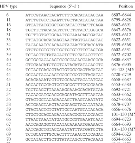
/virus/hpv), and their probe sequences were designed by multiple-sequence align-ment analysis with the CLUSTAL X (version 1.81) program. The 30-bp type-specific probe sequences are listed in Table 1.
The protocol for HPV-specific probe synthesis is outlined in Fig. 1. To facil-itate amplification and binding to the solid support of the HPV probes, we cloned the 30-bp DNAs into plasmid vectors. Equal amounts (5g) of each comple-mentary oligonucleotide probe were mixed and completely denatured by heating at 100°C for 3 min. The denatured oligonucleotides were left at room temper-ature for 1 h to make duplex oligonucleotides. To clone the HPV-specific probes, 2g of each of the duplex oligonucleotides was subjected to an extension pro-cedure in order to add an adenine base to both ends by PCR for 2 h in the presence of dATP only. The adenine-tailed HPV probes were cloned into the pGEM T Easy vector (Promega Biosciences Inc.). The sequences of the 27 HPV type-specific probes were confirmed by DNA sequencing with an automatic DNA sequencer (ABI 3700; Applied Biosystems, Foster City, Calif.). The⬃180-bp positive con-trol probe, the sequence of which corresponds to the L1 region that is highly conserved among most known types of genital HPVs, was amplified with primers MY11 and GP6⫹from the genomic DNA of the Caski (HPV-16 positive) and HeLa (HPV-18 positive) cell lines and cloned into the pGEM T Easy vector.
The PCR control,⬃550 bp of human glyceraldehyde-3-phosphate dehydro-genase (GAPDH) cDNA, was amplified from the genomic DNA of Caski cells by reverse transcription-PCR with GAPDH-specific primers GAPDH-LEFT and GAPDH-RIGHT. To use GAPDH cDNA as an indicator of the adequacy of PCR for the amplification of HPV DNA by MYH-PCR, we added sequences specific for the MY09 and MY11 primers to both ends of the GAPDH cDNA. The GAPDH cDNA clone carrying the MY09 and MY11 primer sequences was cloned into the pGEM T Easy vector.
A negative control probe (180 bp) carrying a partialEscherichia coli lacZgene of the pGEM T Easy vector was amplified from the multiple-cloning site of the pGEM T Easy vector. The standard PCR for preparation of the HPV DNA microarray was simply performed with the pSP6 and pT7 primer sets. The PCR mixture (50l) contained 2.5 mM deoxynucleoside triphosphates, primers pSP6 and pT7 (25 pmol each), 1⫻PCR buffer, and 5 U ofTaqpolymerase (Solgent Co., Taejon, South Korea). The amplification steps were as follows: 95°C for 10 min and 30 cycles of 95°C for 1 min, 55°C for 1 min, and 72°C for 1 min. This was followed by a final extension for 10 min. PCR was performed in a Primus-HP thermal cycler (MWG Biotechnology, Ebersberg, Germany). All the primer sequences used for the PCR are listed in Table 2.
Target probe labeling by PCR.The MYH-PCR (9, 19) was used for
[image:2.603.45.283.80.332.2]amplifi-cation of HPV DNA from the cell lines and tonsillar tissue. A standard PCR was carried out with a 50-l mixture containing the primer set MY09, MY11, and HMB01 (25 pmol each of primers MY09 and MY11 and 5 pmol of HMB01); 10 pg of a PCR control DNA (GAPDH); 5l of tonsillar tissue DNA or 1 ng of cell line genomic DNA; a mixture of deoxynucleoside triphosphates (dATP, dCTP, dGTP, and dUTP, 10 mM each) containing 1l of cyanine 5 (Cy5)-dUTP (25 nM; Pharmacia); and 5 U ofTaqpolymerase (Solgent Co.) in the presence of 1⫻ PCR buffer. To avoid contamination of the template with DNA from a previous PCR, we pretreated the PCR mixture with uracilN⬘-glycosylase (0.5 U; Perkin-Elmer) at 50°C for 5 min before PCR (14). The amplification cycles were carried out as follows: 95°C for 10 min and 35 cycles of 95°C for 1 min, 55°C for 1 min, and 72°C for 1 min. This was followed by a final extension for 10 min, and the PCR products labeled with Cy5 were purified by using a PCR purification kit (Qiagen), as recommended by the manufacturer. The purified amplicons were stored at⫺20°C under darkness until use. The amount of DNA added to a PCR mixture represents 1 to 5% of the DNA obtained from the tonsillar tissues. Negative and positive controls were included in all PCR experiments.
TABLE 1. The 30-bp sequences of the HPV type-specific probes
HPV type Sequence (5⬘–3⬘) Position
6 ATCCGTAACTACATCTTCCACATACACCAA 6807–6844
11 ATCTGTGTCTAAATCTGCTACATACACTAA 6799–6828
16 GTCATTATGTGCTGCCATATCTACTTCAGA 6662–6691
18 TGCTTCTACACAGTCTCCTGTACCTGGGCA 6647–6676
31 TGTTTGTGCTGCAATTGCAAACAGTGATAC 6583–6612
33 TTTATGCACACAAGTAACTAGTGACAGTAC 6622–6651
34 TACACAATCCACAAGTACAACTGCACCATA 6539–6568
35 GTCTGTGTGTTCTGCTGTGTCTTCTAGTGA 6602–6531
39 TCTACCTCTATAGAGTCTTCCATACCTTCT 6672–6702
40 GCTGCCACACAGTCCCCCACACCAACCCCA 6808–6837
42 CTGCAACATCTGGTGATACATATACAGCTG 6876–6905
43 TCTACTGACCCTACTGTGCCCAGTACATAT 94–123 (MY)a
44 GCCACTACACAGTCCCCTCCGTCTACATAT 6720–6749
45 ACACAAAATCCTGTGCCAAGTACATATGAC 6658–6687
51 AGCACTGCCACTGCTGCGGTTTCCCCAACA 6553–6583
52 TGCTGAGGTTAAAAAGGAAAGCACATATAA 6692–6721
54 TACAGCATCCACGCAGGATAGCTTTAATAA 6633–6662
56 GTACTGCTACAGAACAGTTAAGTAAATATG 6627–6656
58 ACTGAAGTAACTAAGGAAGGTACATATAAA 6678–6707
59 CTACTACTCTCTATTCCTAATGTATACACA 6656–6676
62 CTGCTGCAGCAGAATACACGGCTACCAACT 101–130 (MY)a
66 TTAACTAAATATGATGCCCGTGAAATCAAT 6694–6723
67 AATCAGAGGCTACATACAAAAATGAAAACT 6664–6693
68 AATCAGCTGTACCAAATATTTATGATCCTA 101–130 (MY)a
69 TCTGCATCTGCCACTTTTAAACCATCAGAT 6594–6623
70 GCCATACCTGCTGTATATAGCCCTACAAAG 6634–6663
[image:2.603.302.538.260.657.2]72 TCTGTATCAGAATATACAGCTTCTAATTTT 6843–6872 aThe nucleotide sequences indicate the positions in⬃450 bp of DNA derived from MYH-PCR.
FIG. 1. Protocol for HPV-specific probe synthesis by
oligonucleo-tide shuffling (25). Oligonucleooligonucleo-tides were synthesized by the standard
phosphoramidite method. Equal amounts (5
g each) of two
comple-mentary oligonucleotides corresponding to 27 HPV probes were
com-bined and subsequently heated at 100°C for 3 min. The denatured
oligonucleotides were left at room temperature for 1 h to allow
an-nealing. Details are described in Materials and Methods.
on May 15, 2020 by guest
http://jcm.asm.org/
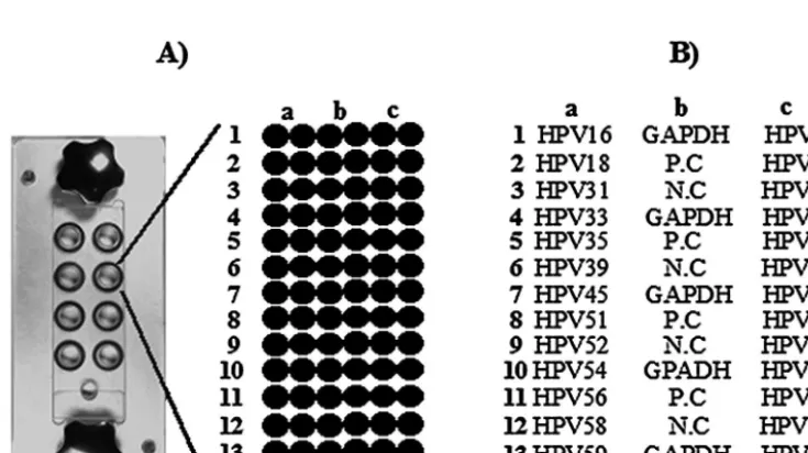
Design of HPV DNA microarray.Twenty-seven HPV-specific probes, GAPDH cDNA-specific probes, HPV DNA-positive control probes, and negative control probes were printed on DNA microarray slides with a robotic microspotting microarrayer (OmniGrid II; GeneMachine, Ann Arbor, Mich.). All amplified probes were dissolved in 10l of 50% dimethyl sulfoxide (AMRESCO) at final concentrations of 200 to 240 ng/l. As shown in Fig. 2, the average size of the spots was 250m. Amine-coated GAPS II slides (Corning Co., Corning, N.Y.) were used for the microarray.
Microarray hybridization and HPV genotyping.HPV DNA microarray
hy-bridization was performed as described previously (27). HPV DNA microarray hybridization was carried out at 55°C for 2 h in an eight-well platform hybrid-ization chamber (GenomicTree, Inc.). The hybridhybrid-ization mixture (100l) con-tained half (25l) of the amplified target probe, 3.5⫻SSC (1⫻SSC is 0.15 M NaCl plus 0.015 M sodium citrate), 5g of salmon sperm DNA (Gibco BRL, Paisley, United Kingdom), and 0.2% sodium dodecyl sulfate. It was heated for 2 min at 95°C and immediately applied onto the HPV DNA microarray.
To determine the HPV genotype, the HPV DNA microarray was imaged with an Axon 4000B scanner (Axon Instruments, Union City, Calif.) and image analysis was performed. The signal intensities were measured and analyzed by using GenePix Pro (version 4.0) software. The HPV genotype was determined by showing that a probe specific for a given HPV type displayed a reliable signal. We
considered 55% pixels in a spot showing a signal 1.5-fold higher than that of the local background as a reliable signal.
Test of HPV DNA microarray specificity.To test the specificities of the HPV
DNA-specific probes, we performed HPV DNA microarray hybridizations with the type-specific probe clones labeled with fluorescent Cy5-dUTP. Twenty-seven HPV type-specific probes were cloned into a TA site of a different cloning vector, TOPO pCR2.1 (Invitrogen, Breda, The Netherlands). One nanogram (⬃2.33⫻ 108copies) of each cloned HPV probe was amplified with primers TOPO-LEFT
and TOPO-RIGHT, together with 10 pg of the plasmid containing the GAPDH cDNA-specific probe, in the presence of Cy5-dUTP. Each product was indepen-dently hybridized onto an array of the 27 immobilized HPV-specific probes, and the hybridization results were evaluated by scanning the microarrays.
Test of HPV DNA microarray reproducibility.To examine the consistency of
PCR-mediated microarray hybridization, we performed a standard MYH-PCR in replicates in the presence of Cy5-dUTP using 1 ng of genomic DNA from a Caski cell (HPV-16 positive). Each 20% volume of the amplified products was hybridized on the HPV DNA microarray. Data are represented as the mean intensities of positive signals, with standard deviations, from three independent experiments.
Detection limit of HPV DNA microarray.To assess the detection limit of the
[image:3.603.45.542.81.210.2]HPV DNA microarray, we performed a serial dilution test with plasmid DNA
TABLE 2. PCR primers used in this study
Primer name and HPV type Sequence (5⬘–3⬘)a Position
MY09
CGT CCM ARR GGA WAC TGA TC
7033–7014 in HPV-16
MY11
GCM CAG GGW CAT AAY AAT GG
6582–6601 in HPV-16
GP6
⫹
GAA AAA TAA ACT GTA AAT CA
6765–6746 in HPV-16
HMB01
bGCG ACC CAA TGC AAA TTG GT
GAPDH-LEFT
TCA ACG GAT TTG GTC GTA TT
77–96
GAPDH-RIGHT
TAG AGG CAG GGA TGA TGT TC
664–645
pSP6
ATT TAG GTG ACA CTA TAG AA
139–158 in pGEM T Easy
pT7
TAA TAC GAC TCA CTA TAG GG
2999–3 in pGEM T Easy
TOPO-LEFT
AGC TTG GTA CCG AGC TCG GAT
255–235 in pCR2.1-TOPO
TOPO-RIGHT
CGA ATT GGG CCC TCT AGA TGC
363–343 in pCR2.1-TOPO
MY09GAP
CGT CCM ARR GGA WAC TGA TC TCA ACG GAT TTG GTC GTA TT
MY11GAP
GCM CAG GGW CAT AAY AAT GG TAG AGG CAG GGA TGA TGT TC
aNucleotide abbreviations are according to the International Union of Pure and Applied Chemistry system of nomenclature: M, A⫹C; R, A⫹G; W, A⫹T; Y, C⫹T.
bHMB01 is directed to the minus strand of HPV-51.
FIG. 2. Microarray design for HPV genotyping. (A) Eight-well platform hybridization reaction chamber. Each well contains 84 probes
corresponding to the PCR control, the HPV-positive control, the HPV-negative control, and type-specific probes. (B) Schematic diagram of the
HPV DNA microarray probe positions. GAPDH cDNA, HPV-positive controls (P.C), and HPV-negative controls (N.C) were spotted on the
center of the slide. Each HPV type-specific probe was printed on the both sides, as shown, and all probes were printed in duplicate.
on May 15, 2020 by guest
http://jcm.asm.org/
[image:3.603.109.477.479.685.2]containing cloned HPV-16. The plasmid copy numbers were calculated from optical density measurements, and dilution series containing 1011to 100copies of
HPV DNA were made. The standard PCR mixture contained 10 pg of the plasmid with the GAPDH cDNA-specific probe, 2 ng of HPV-negative C33A genomic DNA, Cy5-dUTP, and a serial diluent of the plasmid with the probe for HPV-16 in order to mimic the complex nucleic acid environment present during amplification of genomic DNA. After amplification, each 20% volume of PCR product was analyzed by agarose gel electrophoresis and hybridization by the HPV DNA microarray.
Statistical analysis.Pearson’s2test was used to compare the correlation of
the presence of HPV with the grade of differentiation, risk factors, and malig-nancy by the Excel program (Microsoft, Redmond, Wash.). Differences with Pvalues of⬍0.01 were regarded as significant.
RESULTS
Design and specificity of HPV DNA microarray.
Our HPV
DNA microarray contained 27 HPV type-specific probes and 3
kinds of probes as controls, including PCR-positive,
HPV-positive, and negative controls, as shown in Fig. 2. The
PCR-positive control determines the adequacy of the PCR for the
target specimen. Since we selected the 180-bp sequence as an
HPV-positive control probe corresponding to the highly
con-served region of the L1 gene, which is common among most
known types of HPV, the positive control is capable of
detect-ing any type of HPV, if any is present.
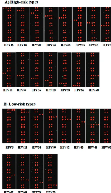
To evaluate the specificity of the HPV type-specific probes,
we performed HPV DNA microarray hybridization with PCR
amplicons from plasmids containing probes for the 27 HPV
types. As shown in Fig. 3, all HPV-specific probes hybridized
specifically to the corresponding targets of each of the HPV
genotypes, and no cross-hybridization with other HPV types
was observed. The hybridization signal intensities varied due to
slight differences in probe sequences and amounts of PCR
amplicons. Nonetheless, assignment to a genotype was not
significantly affected.
Detection limit and reproducibility of the HPV DNA
mi-croarray.
To assess the detection limit of the HPV DNA
mi-croarray, the serially diluted plasmid templates and a fixed
amount of PCR control template were used for the end point
dilution test, as described in Materials and Methods. After
amplification, each 20% volume of the PCR products was
analyzed by agarose gel electrophoresis and hybridization to
the HPV DNA microarray (Fig. 4B and C). The HPV DNA
microarray showed a highly significant linear relationship
be-tween the log of the input HPV copy number and the log of the
signal intensities. The HPV DNA microarray efficiently
de-tected 100 copies of the HPV starting template, while
MYH-PCR detected 10
4copies of the plasmid with the
HPV-16-specific probe by agarose gel analysis. This result indicates that
the detection limit of the HPV DNA microarray is at least
100-fold higher than that of the conventional MYH-PCR
method under our analysis conditions. The signals for the
HPV-positive control and HPV-16 varied with the HPV copy
number, while the signals for the PCR control were steadily
maintained (Fig. 4C).
In addition, to test the consistency of HPV DNA microarray
hybridization, we performed three independent microarray
ex-periments with the same amount of DNA derived from the
Caski cell line carrying HPV-16. The relative standard
devia-tions of positive signals from the PCR control, the HPV
con-trol, and the HPV-16-specific probes were within 10% of the
mean intensities (Fig. 4D). The results revealed that HPV DNA
microarray hybridization shows a high degree of reproducibility.
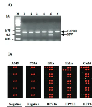
HPV DNA microarray analysis with cell lines.
To address
the applicability of the HPV DNA microarray, we carried out
HPV DNA microarray hybridization with HPV-positive and
-negative cell lines. The most representative HPV-positive cell
lines, SiHa (HPV-16 positive; 1 to 10 copies per cell), HeLa
(HPV-18 positive; 10 to 50 copies/cell), and Caski (HPV-16
positive; 60 to 600 copies/cell) cells, were examined. The
re-sults of the PCR and hybridization images with these cell lines
are shown in Fig. 5. The MYH-PCR was performed in the
presence of 1 ng of each template DNA, and the 20% volumes
of the PCR products were separated on an agarose gel (Fig.
5A). The HPV-negative A549 and C33A cell lines showed no
HPV DNA on the gel. The amounts of the PCR products for
the HeLa and Caski cell lines are dependent on the viral copy
number, while we did not observe any significant PCR product
from SiHa cells carrying HPV at low copy numbers. The same
amounts of PCR products were hybridized with the HPV DNA
microarray (Fig. 5B). Positive signals appeared for the PCR
control, the positive control, and the type-specific probes, as
expected. We found that the PCR products of the SiHa and
Caski cell genomic DNAs specifically hybridized to the
HPV-16-specific probes and the PCR product of the HeLa cell
genomic DNA hybridized to the HPV-18-specific probes.
Hy-bridization signals were recorded only for the PCR controls for
HPV-negative cell lines. The signal intensities of the HPV
type-specific probes were related to the HPV copy number
present in the cell lines and closely matched the PCR results.
Prevalence of HPV in tonsillar squamous carcinoma.
To
examine the clinical performance of the HPV DNA
microar-ray, we performed HPV DNA microarray experiments with 73
clinical tonsillar tissue samples from 39 patients with tonsillar
squamous carcinoma. Of the 39 patients (mean age, 58.2 years;
age range, 32 to 77 years), 35 were men (mean age, 58.9 years; age
range, 32 to 77 years) and 4 were women (mean age, 52 years; age
range, 40 to 62 years). Thirty-nine of the 73 specimens were
ma-lignant tonsillar squamous epithelia and 34 were from an
ad-jacent nonmalignant part. As shown in Table 3, the tonsillar
carcinoma tissues of 25 (64.1%) of the 39 patients were found to
be HPV positive, while nonmalignant tonsillar squamous
epi-thelia from 3 (8.8%) of 34 patients were HPV positive.
HPV-16 was the most frequent HPV type found in the HPV-positive
carcinomas (23 of 25 samples) and nonmalignant epithelia (3
of 3 samples). One patient was found to be infected with
HPV-33, and another patient was found to be coinfected with
HPV-6 and HPV-58. The results showed significant
hybridiza-tion signals and a high degree of specificity to probes specific
for the corresponding genotype. We carried out the
2test (
P
⬍
0.01) to determine the correlation of HPV infection with
ma-lignancy, the grade of differentiation, and two risk factors
(Ta-ble 3). The results revealed that the presence of HPV showed no
significant correlation with histology (
P
⫽
0.603), smoking (
P
⫽
0.43), or drinking of alcohol (
P
⫽
0.04), while the presence of
HPV was significantly correlated with malignancy (
P
⫽
0.001).
DISCUSSION
It is important that HPV genotypes be determined by as
elaborate a method as possible, because the HPV genotype
on May 15, 2020 by guest
http://jcm.asm.org/
FIG. 3. Specificities of the type-specific probes for the 27 HPV types amplified from plasmids. (A) Hybridization results with high-risk
type-specific probes; (B) hybridization results with low-risk type-specific probes. Each HPV type is indicated at the bottom.
on May 15, 2020 by guest
http://jcm.asm.org/
FIG. 4. Detection limit and reproducibility of the HPV DNA microarray. (A) Agarose gel electrophoresis of the MYH-PCR products with a
serial dilution of the plasmid with the HPV-16-specific probe. Ten microliters of each 50-
l PCR product was separated on a 1.0% agarose gel.
The input copy numbers of the plasmid with the HPV-16-specific probe are as follows in the indicated lanes: M, 1-kb ladder; 1, 10
10copies; 2, 10
9copies; 3, 10
8copies; 4, 10
7copies; 5, 10
6copies; 6, 10
5copies; 7, 10
4copies; 8, 10
3copies; 9, 10
2copies; 10, 10 copies; 11, 1 copy. (B) HPV DNA
microarray hybridization results with the amplicon obtained by MYH-PCR of the plasmid with the HPV-16-specific probe. Ten microliters of the
50-
l PCR products derived from 10
5to 10
0copies were hybridized with HPV DNA on the microarray for 2 h at 55°C. The starting plasmid copy
number is shown at the bottom. (C) Standard curves of signal intensities for HPV-16 with serial dilutions of 10
6to 10
0copies. Linear regression
was based on the titration series for the plasmid with the HPV-16-specific probe, and each curve is based on the average of three replicates.
(D) Reproducibility of the HPV DNA microarray. Three independent experiments were performed with the genomic DNA of Caski cells. The
mean
⫾
standard deviation was calculated from the signal intensity of each probe at 635 nm. Each probe is indicated at the bottom: GAPDH, PCR
control; Positive, HPV-positive control; HPV-16, HPV-16-specific probe.
3277
on May 15, 2020 by guest
provides information useful for the prognosis of malignant
degeneration. Although PCR is known to be the most sensitive
method for the detection of HPV infection in clinical samples
(17, 23), it has some drawbacks, in that false-positive results
because of amplification of related organisms can occur and
false-negative amplifications may result from variations in the
sequences of the primer binding sites for the target region of
the virus. Furthermore, in order to type the consensus
ampli-con, additional labor-intensive procedures, such as sequencing
or type-specific PCR, are required (27). Recently, several
stud-ies involving genotyping of HPV with type-specific
oligonucle-otides and by DNA microarray analysis have been reported (1,
4, 12, 16). However, those reports did not address technical
details, including type specificity and reproducibility in studies
with type-specific oligonucleotides, nor did they present
evi-dence of the clinical performance of DNA microarray analysis.
[image:7.603.142.441.62.407.2]FIG. 5. HPV genotyping results with the cell lines. (A) Agarose gel electrophoresis of the amplified products from the cell lines. Ten microliters
of each 50-
l PCR product was separated on a 1.0% agarose gel. Lanes: M, 1-kb ladder; 1, A549 cells; 2, C33A cells; 3, SiHa cells; 4, HeLa cells;
5, Caski cells; 6, PCR control. (B) Ten microliters of each 50-
l PCR product was hybridized with the HPV DNA microarray at 55°C for 2 h.
HPV-negative cell lines A549 and C33A showed hybridization signals with the PCR controls only. HPV-positive hybridization signals were shown
with the SiHa (HPV-16 positive), HeLa (HPV-18 positive), and Caski (HPV-16 positive) cell lines. The cell lines are indicated at the top, and the
HPV types are indicated at the bottom.
TABLE 3. Correlation of HPV prevalence with clinical data in tonsillar carcinomas
aMalignancyb Histologyc Smokingd Drinkinge
Type Frequencyf Type Frequency Amt Frequency Amt Frequency
T
25/39 (64.1)
WD
2/3 (66.67)
⫺
3/5 (60.0)
⫺
1/5 (20.0)
N
3/34 (8.8)
MD
15/25 (60.0)
⫹
3/6 (50.0)
⫹
7/8 (87.5)
PD
8/11 (72.7)
⫹⫹
21/28 (75.0)
⫹⫹
18/26 (69.2)
aPvalues were calculated by the2test and were considered statistically significant if thePvalue was⬍0.01. ThePvalues were 0.001, 0.603, 0.43, and 0.04 for the
malignancy, histology, smoking, and drinking groups, respectively. bN, nontumor; T, tumor.
cCell tumor differentiation grade according to histopathology: MD, moderate differentiation; PD, poor differentiation; WD, good differentiation. d⫺, nonsmoker;⫹, smoker for less than 20 years;⫹⫹, smoker for more than 20 years.
e⫺, nondrinker;⫹, less than one alcoholic drink per week;⫹⫹, more than one alcoholic drink per week. fFrequency data are presented as number of samples HPV DNA positive/total number of samples tested (percent).
on May 15, 2020 by guest
http://jcm.asm.org/
In this work, we report on a more useful MYH-PCR-mediated
HPV DNA microarray system capable of genotyping 15
high-risk HPV types and 12 low-high-risk HPV types.
We chose the hypervariable region within the L1 genes of
the 27 HPV types for use as type-specific probes for HPV
genotyping and synthesized 30-bp oligonucleotides whose
se-quences were specific for the 27 types. All oligonucleotides
selected contained more than six mismatches with the
corre-sponding regions of the other HPVs. Although a high copy
number, equal to 10
8copies of the HPV-specific probe on the
plasmid used as the template, was used for the PCR, a signal
derived from cross-hybridization was not observed. This result
indicates that even high yields of HPV DNA from clinical
samples will not induce cross-hybridization. The chance of
cross-hybridization in assays with cervical swab specimens and
other types of clinical samples can thus be ignored. We also
measured the detection limit of the HPV DNA microarray.
HPV DNA microarray hybridization with the same amount of
starting template showed a higher detection limit than
MYH-PCR alone. In addition, we tested the reproducibilities of the
HPV DNA microarray experiments. The relative standard
de-viations of positive signals for each probe were found to be
within 10% of the mean values. This result indicates that the
hybridization results showed a high degree of reproducibility.
The preclinical performance of the HPV DNA microarray
was evaluated by testing the cell lines A549 (HPV negative),
C33A (HPV negative), SiHa (HPV-16), HeLa (HPV-18
posi-tive), and Caski (HPV-16 positive). In the microarray
hybrid-ization experiments with HPV-positive cell lines, hybridhybrid-ization
signals appeared for each known type of HPV probe and
HPV-positive controls, while HPV-negative cell lines did not show
any positive hybridization signal with the type-specific probes.
The signal intensities of the type-specific probes are related to
the HPV copy number present in the cells. This result indicates
that the HPV DNA microarray is able to determine the
rela-tive viral loads by the differences in hybridization signals.
On the basis of the results obtained with the HPV DNA
microarray and the different cell lines, we examined the HPV
DNA microarray in clinical practice. We performed HPV
genotyping with 34 samples of nonmalignant tissues and 39
samples of malignant tissues from 39 Korean patients with
tonsillar squamous carcinoma. It is known that the frequency
of HPV identified in patients with head and neck cancer varies
widely (2 to 76%), depending on clinical preparation of the
patients, the materials and methods used for analysis, as well as
the number of cases included (6, 20, 21, 24). In this work, HPV
DNA was found in 25 (64.1%) of the 39 carcinoma tissue
samples. Recently, an increasing number of reports have
shown that among all head and neck carcinomas the high-risk
HPV type HPV-16 is the most frequent HPV type detected in
tonsillar carcinomas (45 to 70%) (20–22). Our data also
showed that the most frequent HPV type in tonsillar
carcino-mas is HPV-16. In addition, we have analyzed the correlation
of the HPV infection status with malignancy, the grade of
differentiation, and two risk factors, including smoking and
drinking. The results showed that the presence of HPV is not
correlated with the grade of differentiation or risk factors,
while it was closely correlated with malignancy, as shown in
Table 3. Our results support the hypothesis that the presence
of HPV is closely associated with tonsillar carcinomas. In the
HPV DNA microarray hybridization experiments with clinical
samples, we did not observe any false-positive or -negative
signals compared with the results of detection by PCR under
our experimental conditions. Because the use of an extremely
sensitive and reliable method is required for the diagnosis of
HPV infection in clinical practice, the use of the HPV DNA
microarray technology has distinct advantages.
In conclusion, we developed a microarray-based system for
HPV genotyping. Our data demonstrate that HPV DNA
mi-croarray analysis coupled with MYH-PCR can be applied to
HPV detection and genotyping. This diagnostic tool will
un-doubtedly be useful for the clinical diagnosis of HPV infection
and large-scale epidemiological studies.
ACKNOWLEDGMENTS
We thank Chiwang Yoon and Daekyung Yoon (GenomicTree, Inc.)
for fabrication of the HPV DNA microarrays.
REFERENCES
1. An, H. J., N. H. Cho, S. Y. Lee, I. H. Kim, C. Lee, S. J. Kim, M. S. Mun, S. H.
Kim, and J. K. Jeong.2003. Correlation of cervical carcinoma and
precan-cerous lesions with human papillomavirus (HPV) genotypes detected with the HPV DNA chip microarray method. Cancer97:1672–1680.
2. Bernard, H. U., S. Y. Chan, M. M. Manos, C. K. Ong, L. L. Villa, H. Delius,
C. L. Peyton, H. M. Bauer, and C. M. Wheeler.1994. Identification and
assessment of known and novel human papillomaviruses by polymerase chain reaction amplification, restriction fragment length polymorphisms, nu-cleotide sequence, and phylogenetic algorithms. J. Infect. Dis.170:1077– 1085.
3. Chan, S. Y., H. Delius, A. L. Halpern, and H. U. Bernard.1995. Analysis of
genomic sequences of 95 papillomavirus types: uniting typing, phylogeny, and taxonomy. J. Virol.69:3074–3083.
4. Cho, N. H., H. J. An, J. K. Jeong, S. Kang, J. W. Kim, Y. T. Kim, and T. K.
Park.2003. Genotyping of 22 human papillomavirus types by DNA chip in
Korean women: comparison with cytologic diagnosis. Am. J. Obstet. Gy-necol.188:56–62.
5. Cox, J. T., A. T. Lorincz, M. H. Schiffman, M. E. Sherman, A. Cullen, and
R. J. Kurman.1995. Human papillomavirus testing by hybrid capture
ap-pears to be useful in triaging women with a cytologic diagnosis of atypical squamous cells of undetermined significance. Am. J. Obstet. Gynecol.172:
946–954.
6. Friesland, S., H. Mellin, E. Munck-Wikland, A. Nilsson, J. Lindholm, T.
Dalianis, and R. Lewensohn.2001. Human papilloma virus (HPV) and p53
immunostaining in advanced tonsillar carcinoma—relation to radiotherapy response and survival. Anticancer Res.21:529–534.
7. Frisch, M., and R. Biggar.1999. Aetiological parallel between tonsillar and
anogenital squamous-cell carcinomas. Lancet354:1442–1443.
8. Gaarenstroom, K. N., P. Merkert, J. M. M. Walboomers, A. J. C. van den
Brule, P. J. F. van Bommel, C. J. L. M. Meijer, F. J. Voorhorst, P. Kenemans,
and T. J. M. Helmerhorst.1994. Human papillomavirus DNA and
geno-types: prognostic factors for progression of cervical intraepithelial neoplasia. Int. J. Gynecol. Cancer4:73–78.
9. Graviit, P. E., C. L. Peyton, T. Q. Alessi, C. M. Wheeler, F. Coutle´e, A.
Hildesheim, M. H. Schiffman, D. R. Scott, and R. J. Apple.2000. Improved
amplification of genital human papillomavirus. J. Clin. Microbiol.38:357– 361.
10. Hart, K. W., O. M. Williams, N. Thelwell, A. N. Fiander, T. Brown, L. K.
Borysiewicz, and C. M. Gelder.2001. Novel method for detection, typing,
and quantification of human papillomavirus in clinical samples. J. Clin. Microbiol.39:3204–3212.
11. Hemminski, K., C. Dong, and M. Frisch.2000. Tonsillar and other upper
aerodigestive tract cancers among cervical cancer patients and their hus-bands. Eur. J. Cancer Prev.9:433–437.
12. Hwang, T. S., J. K. Jeong, M. Park, H. S. Han, H. K. Choi, and T. S. Park.
2003. Detection and typing of HPV genotypes in various cervical lesions by HPV oligonucleotide microarray. Gynecol. Oncol.90:51–56.
13. Jacobs, M. V., A. M. de R. Husman, A. J. C. van den Brule, P. J. F. Snijders,
C. J. L. Miejer, and J. M. M. Walboomers.1995. Group-specific
differenti-ation between high- and low-risk human papillomavirus genotypes by gen-eral primer-mediated PCR and two cocktails of oligonucleotide probes. J. Clin. Microbiol.33:901–905.
14. Josefsson, A., K. Livak, and U. Gyllensten.1999. Detection and quantitation
of human papillomavirus by using the fluorescent 5⬘ exonuclease assay. J. Clin. Microbiol.37:490–496.
15. Kataja, V., S. Syrjanen, R. Mantyhjarvi, M. Yliskoski, S. Saarikoski, and K.
on May 15, 2020 by guest
http://jcm.asm.org/
Syrjanen.1992. Prognositic factors for cervical human papillomavirus infec-tions. Sex. Transm. Dis.19:154–160.
16. Kim, C. J., J. K. Jeong, M. Park, T. S. Park, T. C. Park, S. E. Namkoong, and
J. S. Park.2003. HPV oligonucleotide microarray-based detection of HPV
genotypes in cervical neoplastic lesions. Gynecol. Oncol.89:210–217.
17. Kleter, B., L. van Doorn, L. Schrauwen, A. Molijn, S. Sastrowijoto, J. ter
Schegget, J. Lindeman, B. ter Harmsel, M. Burger, and W. Quint.1999.
Development and clinical evaluation of a highly sensitive PCR-reverse hy-bridization line probe assay for detection and identification of anogenital human papillomavirus. J. Clin. Microbiol.37:2508–2517.
18. Liu, C. H., W. L. Ma, R. Shi, Y. Q. Ou, B. Zhang, and W. L. Zheng.2003.
Possibility of using DNA chip technology for diagnosis of human papilloma-virus. J. Biochem. Mol. Biol.36:349–353.
19. Manos, M. M., Y. Ting, D. K. Wright, A. J. Lewis, T. R. Broker, and S. M.
Wolinsky.1989. The use of polymerase chain reaction amplification for the
detection of genital human papillomavirus. Cancer Cells7:209–214.
20. Mckaig, R. G., R. S. Baric, and A. F. Olshan.1997. Human papillomavirus
and head and neck cancer: epidemiology and molecular biology. Head Neck
20:250–265.
21. Mellin, H., S. Friesland, R. Lewensohn, T. Dallianis, and E.
Munck-Wik-land.2000. Human papillomavirus (HPV) DNA in tonsillar cancer: clinical
correlates, risk of relapse, and survival. Int. J. Cancer89:300–304.
22. Mellin, H., L. Dahlgren, E. Munck-Wikland, J. Lindholm, H. Rabbani, M.
Kalantari, and E. Dalianis.2002. Human papillomavirus type 16 is episomal
and a high viral load may be correlated to better prognosis in tonsillar cancer. Int. J. Cancer102:152–158.
23. Qu, W., G. Jiang. Y. Cruz, C. J. Chang, G. Y. F. Ho, R. S. Klein, and R. D.
Burk.1997. PCR detection of human papillomavirus: comparison between
MY09/MY11 and GP5⫹/GP6⫹primer systems. J. Clin. Microbiol.35:1304– 1310.
24. Snijders, P. J. F., F. V. Cromme, A. J. C. van den Brule, H. F. J.
Schrijne-makers, G. B. Snow, C. J. L. M. Meijer, and J. M. M. Walboomers.1992.
Prevalence and expression of human papillomavirus in tonsillar carcinomas, indicating a possible viral etiology. Int. J. Cancer51:845–850.
25. Stemmer, W. P. C., A. Crameri, D. H. Kim, T. M. Brennan, and H. L.
Heyneker.1995. Single-step assembly of a gene and entire plasmid from
large numbers of oligodeoxyribonucleotides. Gene164:49–53.
26. Vernon, S. D., E. R. Unger, and D. Williams.2000. Comparison of human
papillomavirus detection and typing by cycle sequencing, line blotting, and hybrid capture. J. Clin. Microbiol.38:651–655.
27. Wang, D., L. Coscoy, M. Zylberberg, P. C. Avila, H. A. Boushey, D. Ganem,
and J. L. DeRisi.2002. Microarray-based detection and genotyping of viral
pathogens. Proc. Natl. Acad. Sci. USA26:15687–15692.
28. Wilczynski, S. P., B. T. Y. Lin, X. Xie Yuan, and I. B. Paz.1998. Detection
of human papillomavirus DNA and oncoprotein overexpression are associ-ated with distinct morphological patterns of tonsillar squamous cell carcino-mas. Am. J. Pathol.152:145–156.
29. zur Hausen, H.1996. Papillomavirus infections—a major cause of human
cancers. Biochim. Biophys. Acta1288:F55–F78.