Copyright © 2002, American Society for Microbiology. All Rights Reserved.
Molecular Epidemiology of Astrovirus Infection in
Barcelona, Spain
Susana Guix,
1Santiago Caballero,
1Cristina Villena,
1Rosa Bartolome´,
2Cristina Latorre,
3Nuria Rabella,
4Maria Simo´,
5Albert Bosch,
1* and Rosa M. Pinto´
1Enteric Virus Laboratory, Department of Microbiology, University of Barcelona,1Laboratory of Microbiology, Hospital
de la Vall d’Hebron,2Laboratory of Microbiology, Hospital de Sant Joan de Deu,3Laboratory of Microbiology,
Hospital de la Santa Creu i Sant Pau,4and Laboratory of Microbiology,
Consorci Sanitari de Terrassa,5Barcelona, Spain
Received 26 February 2001/Returned for modification 18 August 2001/Accepted 3 November 2001
A 3-year study involving 2,347 gastroenteritis samples was conducted to determine the prevalence, time distribution, and medical significance of human astrovirus infection in Barcelona, Spain. The overall incidence of astrovirus was found to be 4.9%. Mixed infections with other enteric agents were detected in 17.2% of all astrovirus-positive samples. During the 3-year period, the highest astrovirus incidence was reported in the winter months, although infections also occurred in summer. The peak detection rate was observed in children between 2 and 4 years of age. Overall, HAstV-1 was the most prevalent type, followed by HAstV-4, HAstV-3, HAstV-8, and HAstV-2. HAstV-5, HAstV-6, and HAstV-7 were not detected during these 3 years. From our serotype data for each age group, we observed that HAstV-1, HAstV-2, and HAstV-3 affected mostly children younger than 3 years of age, while HAstV-4 and HAstV-8 had a greater impact in older children. Genetic variability was analyzed between astroviruses isolated in Barcelona and strains isolated in other parts of the world. A fourth lineage was described for HAstV-1, most likely due to the large number of assayed samples, which may also explain the high level of genetic variability observed in the astrovirus isolates.
Astroviruses are nonenveloped single-stranded RNA viruses that were first detected in 1975 by electron microscopy in stool specimens from children with acute gastroenteritis (14). The astrovirus genome contains three open reading frames (ORFs): ORF1a and ORF1b, which encode the viral protease and polymerase, respectively, and ORF2, which encodes the capsid precursor. A subgenomic RNA that contains ORF2 may be detected in the cytoplasm of astrovirus-infected cells.
Human astroviruses have been increasingly identified as im-portant agents of diarrheal disease in children and the elderly (17). Outbreaks of diarrhea due to astrovirus have frequently been reported (1, 20, 24, 26, 32), and astroviruses have also been associated with nosocomial infections in hospitals (29, 33). They have also been detected in immunocompromised (24) and AIDS-infected patients (13). Astrovirus infections occur worldwide, and their incidence in children with gastro-enteritis in both developing and developed countries ranges from 2 to 9% (2, 5, 6, 22, 31, 34), although some studies report prevalences up to 26% (15).
Although astrovirus epidemiological studies have been com-monly based on electron microscopy and enzyme immunoassay techniques, during the past few years the number of surveys using molecular techniques, mainly reverse transcription-PCR (RT-PCR), has substantially increased. There is a widespread belief that astrovirus incidence may have been underestimated, since enzyme immunoassay is far less sensitive than RT-PCR (6, 20). Furthermore, seroprevalence studies indicate that most
children acquire astrovirus antibodies during the first years of life (10, 11). Consequently, a new appreciation for the role of astrovirus in diarrheal disease has evolved, and in many cases, astroviruses are regarded as the second most common cause of viral gastroenteritis in children after rotavirus (7, 9).
Presently, astroviruses are classified into seven (HAstV-1 to HAstV-7) serotypes according to the reactivity of the capsid proteins with type-specific antibodies. These seven antigenic groups (serotypes) correlate perfectly with seven genotypes that can be determined according to the nucleotide sequence of a 348-bp region of ORF2 (25). The existence of an eighth type (HAstV-8) has been suggested based on three complete capsid protein gene sequences deposited in GenBank. Most studies in different countries around the world indicate that HAstV-1 is the most common serotype, while HAstV-6 and HAstV-7 have rarely been isolated (6, 12, 22, 24, 28, 33).
The aim of the present study was to determine the preva-lence, time distribution, serotype frequencies, and medical sig-nificance of astrovirus infections from children with gastroen-teritis in Barcelona, Spain, during a 3-year period. In addition, we analyzed the genetic diversity of astrovirus isolates in order to further investigate the molecular epidemiology of this gas-troenteritis agent.
MATERIALS AND METHODS
Stool samples.Between May 1997 and April 2000, 2,291 fecal samples were collected from infants and children with gastroenteritis at four hospitals in the Barcelona area (Hospital de la Vall Hebron, Hospital de Terrassa, Hospital de Sant Joan de Deu, and Hospital de la Santa Creu i Sant Pau). Fifty-six adult samples were also analyzed. Routine diagnostic tests for common bacterial pathogens, rotavirus, adenovirus, and several parasites were carried out for a substantial number of samples following the responsible physician’s advice (Ta-ble 1). For astrovirus detection, stools were suspended (10%, wt/vol) in phos-phate-buffered saline containing 2 M NaNO3, 1% bovine serum albumin;
frac-* Corresponding author. Mailing address: Department of Microbi-ology, University of Barcelona, Avda Diagonal 645, 08028 Barcelona, Spain. Phone: (34) 934034620. Fax: (34) 934034629. E-mail: albert @porthos.bio.ub.es.
133
on May 15, 2020 by guest
http://jcm.asm.org/
tion V), and 0.1% Triton X-100 (pH 7.2) and pelleted at 1,000⫻gfor 5 min, and the resulting supernatant was stored at⫺70°C for later analysis.
Astrovirus detection.Astrovirus was detected by RT-PCR after extraction of its RNA and subsequently confirmed by Southern blot hybridization with an internal probe. RNA was purified from 50l of fecal supernatant by guanidine thiocyanate extraction, as previously described (3). RT-PCR was carried out with primers A1(5⬘-CCTGCCCCGAGAACAACCAAGC-3⬘) and A2(5⬘-GTAAGA TTCCCAGATTGGTGC-3⬘), which amplify a fragment of ORF1a (35). Five microliters of the extracted RNA was heated to 99°C for 5 min and immediately placed on ice. First-strand cDNA was synthesized at 42°C for 60 min by adding 1M primer A2 and 3 U of reverse transcriptase (Expand; Roche) in 10l (final volume) containing 50 mM Tris-HCl (pH 8.3), 40 mM KCl, 5 mM MgCl2, 10 mM
dithiothreitol, 0.5 mM Tween 20, and 0.2 mM concentrations of each de-oxynucleoside triphosphate. Five microliters of the RT product was amplified by using 0.5 U of the Expand high-fidelity PCR system enzyme mix (Roche) and 0.5 M (each) primers A1 and A2 in a total volume of 50l containing 5l of Expand high-fidelity buffer (Roche), 2 mM MgCl2, and each deoxynucleoside
triphosphate at 0.2 mM. After a denaturation step of 3 min at 95°C, 40 cycles of amplification (94°C, 30 s; 55°C, 30 s; 72°C, 30 s) were performed followed by a final extension of 7 min at 72°C. Ten microliters of the PCR product was analyzed on a 1.5% agarose gel and detected by ethidium bromide staining. PCR products were confirmed by Southern blot hybridization with an internal digoxi-genin-labeled probe (5⬘-AAGAAAGAGAAACAACCAG-3⬘) under stringent conditions.
Astrovirus typing.All specimens positive for astrovirus were typed by sequenc-ing the RT-PCR product as previously described by Noel et al. (25), after amplification of a 413-bp region of ORF2. Conditions for RT-PCR reactions with primers Mon244 and Mon245 were identical to those with primers A1 and A2, but primer and MgCl2concentrations in the PCR were 1 and 1.5M,
respectively. The PCR program consisted of 3 min at 95°C followed by 40 cycles of amplification (94°C, 1 min; 55°C, 30 s; 72°C, 1 min) and a final extension of 7 min at 72°C. Forty microliters of the RT-PCR product was run on a 1% agarose gel, and the DNA was purified with a High Pure PCR product purification kit (Roche), according to the manufacturer’s instructions. The nucleotide sequence of a 348-bp fragment of the PCR product was determined from 2 to 7l of the purified DNA by using the Thermo Sequenase II dye terminator cycle sequenc-ing premix kit (Amersham Pharmacia Biotech) and the primers Mon244 and Mon245. Each nucleotide sequence was compared to those of reference strains by using the BLAST program (National Center for Biotechnology Information) in order to assign a serotype.
Phylogenetic analysis. Both nucleotide and amino acid multiple-sequence alignments were carried out with the CLUSTALW program. Phylogenetic trees were constructed, on the basis of the 348-bp DNA sequences from ORF2 de-scribed above, by the neighbor-joining method. Nucleotide distance matrices were calculated by Kimura’s two-parameter method (PHYLIP package version 3.5 c; distributed by J. Felsenstein, Department of Genetics, University of Wash-ington, Seattle). Phylogenetic relationships were analyzed with and without a bootstrap of 100 replicates. The consensus tree was visualized by using TreeView software, and it was rerooted by using feline astrovirus sequence as an outgroup (AF056197). Sequence information for HAstV-1 to HAstV-7 was obtained from Oxford reference strains and also from astrovirus type 1 Newcastle (36). For HAstV-8, three sequences available at GenBank were used (Z66541, AF175261, and AF260508), as well as some sequences from astroviruses isolated elsewhere (18, 22). ThePiparameter was used as a measure of nucleotide diversity (nu-cleotide changes per site), and it was calculated with DnaSP 3.0 software (26).
Statistical analysis.The chi-square test was used to compare prevalence rates among 1-year periods of the study and also to evaluate the differences between astrovirus incidence among age groups. Association between astrovirus serotype and age group was studied by analysis of variance (ANOVA).
Nucleotide sequence accession numbers. The nucleotide sequences deter-mined in this study have been deposited in GenBank and have been assigned accession numbers AF348753 to AF348801.
RESULTS
Epidemiology of astrovirus infections.Over a 3-year period,
from May 1997 to April 2000, a total of 2,347 gastroenteritis fecal samples were analyzed for the presence of astrovirus. The overall incidence of astrovirus was found to be 4.9% (116 out of 2,347 samples).
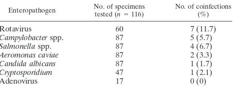
A significant number of samples were also subjected to other microbiological analyses at the different hospitals (Table 1). A total of 325 samples were positive for some of the following agents: rotavirus, adenovirus,Salmonellaspp.,Campylobacter
spp.,Aeromonasspp.,Yersiniaspp.,Enterococcusspp.,
Staph-ylococcusspp.,Candidaspp.,Giardia,Entamoeba, and
Crypto-sporidium. Additionally, mixed infections were detected in 20
of the 116 astrovirus-positive samples, which represent 17.2% of all positive samples (Table 2).
Clinical features of astrovirus infections were documented for a group of 52 pediatric patients from the Hospital Vall d’Hebron whose samples were positive. Of these children, 51.9% did not require hospitalization, while 34.6% were hos-pitalized because of gastroenteritis (the mean number of days in the hospital was 10.7; range, 1 to 57), and 13.5% of the infections were considered nosocomial infections in nongas-troenteritis patients (mainly suffering from oncologic diseases). Although the disease induced by astrovirus in our community was mild, the role of astrovirus as a cause of nosocomial in-fections should not be neglected. Clinical symptoms were di-arrhea (86.3%), temperature above 37.8°C (54.9%), and vom-iting (49.0%). Resolution was good in all patients, although one child suffered from two episodes of astroviral gastroenter-itis within the same year (see below).
[image:2.587.302.543.85.175.2]For the pediatric population (up to 15 years), the average age was 2.8 years (33.5 months), and the median age was 1.6 years (19 months). For astrovirus-positive samples, the average and median ages were 2.5 years (29.6 months) and 1.9 years (23 months). Although astrovirus infections occurred in most age groups (Fig. 1), the number of cases was higher for children between 2 and 3 years of age, and 80% of astrovirus infections occurred in children under 3 years of age (Fig. 1A). Three astrovirus cases were also detected in newborns. Detection rates for each age group are shown in Fig. 1B. The highest detection rate was observed among children of 13 to 14 years. TABLE 1. Incidence of enteropathogens detected in stool
specimens
Enteropathogena No. (%) of specimens
Tested/total Positive Negative
Bacteria and yeasts 1,896/2,347 171 (9) 1,725 (91)
Parasites 888/2,347 10 (1) 878 (99)
Rotavirus 1,265/2,347 151 (12) 1,114 (88)
Adenovirus 404/2,347 8 (2) 396 (98)
Astrovirus 2,347/2,347 116 (5) 2,229 (95)
aBacteria and yeasts were detected by standard culture techniques. Parasites
were detected by optical microscopy and immunological procedures. Rotaviruses and adenoviruses were detected by latex agglutination tests or enzyme-linked immunosorbent assay procedures.
TABLE 2. Mixed infections in astrovirus-positive cases
Enteropathogen No. of specimenstested (n⫽116) No. of coinfections(%)
Rotavirus 60 7 (11.7)
Campylobacterspp. 87 5 (5.7)
Salmonellaspp. 87 4 (6.7)
Aeromonas caviae 87 2 (3.3)
Candida albicans 87 1 (1.7)
Cryptosporidium 47 1 (2.1)
Adenovirus 17 0 (0)
on May 15, 2020 by guest
http://jcm.asm.org/
[image:2.587.44.284.94.175.2]However, since the number of analyzed samples is remarkably higher in the age groups of less than 5 years and the number of cases is significantly higher in infants between 2 and 4 years of age than in those of 13 to 14 years, it can be concluded that the maximum detection rate occurs in patients between 2 and 4 years of age.
A group of adult patients with ages between 15 and 82 years
was also analyzed. Astrovirus RNA was found in 4 of 56 sam-ples (ages: 27, 28, 59, and 69 years), which represents 7% of the adult patients, who were immunocompromised patients (bone marrow transplantation or multiple myeloma) or suffered from severe syndromes (brain stroke or ulcerous colitis).
The monthly distribution of astrovirus incidence is shown in Fig. 2. During the 3-year period, detection rates were higher in the winter months, although astrovirus infections also occurred in summer. In our study, the incidence of astrovirus between May 1998 and April 1999 was higher than those observed in the 1-year periods 1997–1998 and 1999–2000, being 8, 2.6, and 2.5%, respectively. When analyzed by a2test, astrovirus
in-cidence in 1998–1999 was significantly higher than inin-cidences in 1997–1998 and 1999–2000 (2⫽37.43,P⬍0.01).
The annual incidence of each serotype is shown in Fig. 3. In 1997–1998, HAstV-1 was the only serotype detected, while in 1998–1999, five different serotypes affected the studied popu-lation, with HAstV-3 being the most prevalent, followed by HAstV-4. In the following 1-year period, HAstV-1 reappeared as the most common type accounting for 67% of the cases, while HAstV-4 and HAstV-8 showed a decreasing incidence and HAstV-2 and HAstV-3 were not detected. The analysis of the genetic variability of isolates allowed us to describe the emergence of a different HastV-1 strain during 1999–2000 (see below). During the study period, one child suffered from an episode of HAstV-3 diarrhea in November 1998 and had a subsequent episode of HAstV-1 infection 9 months later, sug-gesting a lack of heterotypic immunity between the different antigenic types, which could be responsible for the changes in serotype distribution observed in consecutive years.
Overall, HAstV-1 was the most prevalent type (38%) fol-lowed by HAstV-4 (26%), HAstV-3 (19%), HAstV-8 (11%), and HAstV-2 (6%). HAstV-5, HAstV-6, and HAstV-7 were not detected during these 3 years.
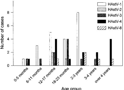
[image:3.587.47.280.68.362.2]From our serotype data for each age group, we observed that HAstV-1, HAstV-2, and HAstV-3 affected mostly children younger than 3 years of age, while HAstV-4 and HAstV-8 had a greater impact in older children (Fig. 4). Average ages for each serotype were 18.8 months for HAstV-1, 17.3 months for FIG. 1. Age distribution of patients with astrovirus gastroenteritis
[image:3.587.135.448.536.714.2]from April 1997 to May 2000. (A) Number of astrovirus-positive sam-ples in every age group; (B) astrovirus detection rate in every age group. In, number of samples analyzed. Age information was available for 2,309 samples.
FIG. 2. Monthly distribution of astrovirus detected in stool specimens from May 1997 to April 2000.
on May 15, 2020 by guest
http://jcm.asm.org/
HAstV-2, 19.1 months for HAstV-3, 43.1 months for HAstV-4, and 57 months for HAstV-8. When we subjected these data to analysis of variance, we determined that the average ages for HAstV-8 and HAstV-4 were significantly different from those for HAstV-1 and HAstV-3 (P⬍0.05). The lack of significance with HAstV-2 average age is due to the low number of positive samples for this serotype.
Genetic variation. Forty-nine Spanish samples were
ana-lyzed at the nucleotide and amino acid levels based on the 348-bp fragment (116 amino acids) of the capsid region deter-mining the serotype (25). Genetic variability was analyzed be-tween astroviruses isolated in Barcelona and other astroviruses isolated in Australia, Colombia, Venezuela, Mexico, and the United Kingdom (18, 19, 22, 36). The degree of genetic vari-ability, measured as the number of nucleotide changes by nu-cleotide site (Pi), was calculated in all antigenic groups. The group with higher nucleotide diversity was shown to be sero-type 2 (Pi⫽0.08118), followed by serotype 1 (Pi⫽0.05622), serotype 8 (Pi ⫽ 0.02083), serotype 4 (Pi ⫽ 0.01547), and finally serotype 3 (Pi⫽0.01228).
Nucleotide variation was not detected among serotype 2 and serotype 8 Spanish isolates. Nucleotide homologies were 90 to 100% among HAstV-1 isolates, 97 to 100% among HAstV-3
isolates, and 95 to 100% among HAstV-4 isolates. Comparing Spanish isolates with prototypic strains, nucleotide identities were high in all cases (90 to 97%) except for HAstV-2, for which it was only 85%.
At the amino acid level, conservation was high within all antigenic types, even serotype 2. All samples were 100% iden-tical to reference strains, except one HAstV-3, all HastV-4, and all HastV-8 isolates. One conserved amino acid change, not previously reported, was detected in one serotype 3 isolate (Bcn3.10). It contained an Arg3His change at amino acid 84 of the capsid protein. This position is quite variable, being Arg in HAstV-1, HAstV-2, HAstV-3, HAstV-5, and HAstV-7, Lys in HAstV-6, and Thr in HAstV-4 and HAstV-8.
Regarding HAstV-4, all serotype 4 Spanish isolates had a Pro3Ala change with regard to the prototype Oxford strain at the amino acid 87. Ala is also present in all HAstV-4 sequences available at the databases from other studies, and it happens also to be a highly conserved amino acid in all serotypes. In addition, a Val3Ile substitution was observed at residue 192 of the capsid region in the Bcn4.9 isolate. This position appears to also be variable in HAstV-8 isolates. Four Spanish HAstV-8 isolates had a Val and two isolates displayed an lle residue. This substitution is also present in the Mexico isolate Yuc-8, in the prototype sequence of HAstV-6, and in the South Amer-ican serotype 1 isolates (Col418, Col509, and Col526). Further-more, it should be noted that at the amino acid level, serotype 8 isolates were 100% similar to most serotype 4 isolates, show-ing a close relationship in this capsid region.
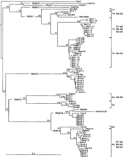
A phylogenetic tree was constructed to study the genetic relationship between the astroviruses isolated in this study and other published sequences (Fig. 5). In the present study, type 1 isolates clustered into 4 major branches with a high confi-dence level. Within serotype 2 isolates, only two major branches were observed. Serotype 4 isolates could also be distributed into 2 major lineages, showing at least 7% sequence diversity. Regarding serotypes 3 and 8, no major lineages could be discriminated.
DISCUSSION
[image:4.587.132.452.73.215.2]This is the first long-term study of astrovirus infection in Spain, and it reveals an overall incidence of 4.9%. This result is consistent with other studies, which report rates of 2 to 9% FIG. 3. Serotype distribution of astrovirus identified in every 1-year period of the study.
FIG. 4. Serotype distribution of astrovirus infections according to age group.
on May 15, 2020 by guest
http://jcm.asm.org/
[image:4.587.45.283.531.704.2]in developed countries (2, 6, 22). Coinfections with other pathogens were detected in 17.2% of the astrovirus-positive samples, although this percentage ranges from 33 to 65% in the literature (4, 8, 33). Most of these studies were carried out
in developing countries where poor hygienic conditions may contribute to multiple enteric infections.
[image:5.587.92.494.76.580.2]The maximum detection rate was observed in children be-tween 2 and 4 years of age. Reports from other countries like FIG. 5. Phylogenetic tree of the 348-bp region of ORF2 of 49 astrovirus isolates from Barcelona (Bcn), the 7 Oxford reference strains (HAstV-1 to HAstV-7), 2 serotype 8 strains from Mexico (Yuc-8) and the United Kingdom (HAstV-8 UK), 9 strains from Australia (Melb), 9 strains from Colombia (Col), 3 strains from the United Kingdom (Newcastle), and 1 strain from Venezuela (Ven). Bootstrap values are given at the branch points. The scale bar indicates an evolutionary distance of 0.10 nucleotide per position in the sequence. Sequences were obtained from the GenBank database with the following accession numbers: Oxford strains; L23513, L13745, L38505, L38506, U15136, L38507, and L38508; serotype 1 Newcastle strain, Z25771; serotype 8 strains, Z66541, AF260508, and AF175261; other Australian strains, AF175253, AF175254, AF175255, AF175256, AF175257, AF175258, AF175259, and AF175260; Colombian strains, AF211957, AF211958, AF211959, AF211960, AF211961, AF211962, AF211963, AF211964, and AF211965; Venezuelan strain, AF211952, AF211953, and AF211956.
on May 15, 2020 by guest
http://jcm.asm.org/
Mexico, Thailand, Guatemala, France, Australia, Colombia, and Venezuela have shown higher incidences in younger pop-ulations (2, 4, 8, 9, 18, 22). However, in Guatemala and France (2, 4), the detection rate at the age of 2 was also high.
Detection rates were higher in winter, although astrovirus infections also occurred in summer months. This seasonal pat-tern is consistent with other epidemiological studies from tem-perate regions (17). However, reports exist which describe high astrovirus incidences during spring and summer (9, 24). Nev-ertheless, some other long-term studies (22) describe a slightly different pattern without a distinct winter peak in each year. Our study, although limited to 3 years, seems to support stud-ies in the United Kingdom, South Africa, and Australia (12, 22, 30) that suggest a greater impact on the pediatric population in alternate years. In the United Kingdom, higher frequency of serotype 1 in alternate years was observed during a 5-year period. This observation was also reported by Mustafa et al. (22) in Australia, together with the lack of an evident winter peak in the years with a lower incidence of serotype 1. How-ever, in our study, serotype 1 showed an increasing incidence every year (see below) and a winter peak was also detected in each 1-year period. Consequently, different reasons should ac-count for this apparent biennial pattern.
Fifty-four of 116 astrovirus-positive samples could be sero-typed using the method described by Noel et al. (25). This apparent lack of typing sensitivity has been reported previously (23). Overall, the most prevalent type was HAstV-1, followed by HAstV-4, HAstV-3, HAstV-8, and HAstV-2, while sero-types 5, 6, and 7 were not detected during these 3 years. Serotype 1 has been considered the most frequent and domi-nant type in most parts of the world, with a few exceptions (8). The second most common serotype differs depending on the country. In the present study, it appears to be HAstV-4, as in Australia and Bangladesh (22, 33).
More than 50% of serotype 1 and 3 samples were from children under 2 years of age, while the majority of serotype 4 and 8 samples were from children older than 3 years. This statistically significant different age pattern suggests that the immune protection against the most prevalent serotypes ac-quired earlier in life fails to confer any protection against new serotypes or variants appearing thereafter.
The genetic variability of the 348-bp fragment determining the serotype (25), measured as the number of mutations per nucleotide site, was highest in the serotype 2 sequences, fol-lowed by serotypes 1, 8, 4, and 3. In fact, a high degree of variation among serotype 2 isolates has been previously de-scribed (22). Spanish HAstV-2 isolates were much closer to Australian strains than to South American isolates, which clus-tered with prototype Oxford strains.
The genetic relationship among the present astrovirus iso-lates and the previously published isoiso-lates allowed us to dis-tinguish several lineages or clusters into each serotype. In the case of serotype 1, together with the three previously described lineages (18) a new cluster (lineage 1d) is reported, including at the moment only sequences from Spain. Other Spanish isolates clustered in lineages 1a and 1b, meaning that Spanish type 1 isolates may represent variants of three different astro-virus strains. Additionally, strains belonging to lineage 1d were mostly isolated in 1997–1998 and 1998–1999 and occasionally in 1999–2000, while strains 1a and 1b were found only in
1999–2000. This suggests that strains 1a and 1b may have replaced strain 1d over time. This observation could also ex-plain the remarkable increase in the prevalence of serotype 1 in 1999–2000, assuming that infection with 1d strains does not confer complete protection against strains 1a and 1b. However, this hypothesis could be accepted only provided that the epitope structure includes amino acids not only from the present fragment of the capsid region but also from other parts of ORF2, or even residues from other ORFs, such as ORF1a, that seem to be involved in the antigenic structure (16), since the region studied here is 100% identical among the different serotype 1 lineages at the amino acid level.
Within serotype 2 and within serotype 4, two lineages are reported. Although all Spanish serotype 4 isolates clustered in lineage 4a, two subgroups of samples (with 4% sequence di-versity) belonging to different 1-year periods could be distin-guished. This may also indicate that these two strains would not have cocirculated in the community.
Finally, no lineages could be described for serotypes 3 and 8. We emphasize the description of the fourth lineage for serotype 1, as it may be a consequence of the large number of samples analyzed but may on the other hand also explain the high level of genetic variability observed in the astrovirus isolates.
ACKNOWLEDGMENTS
We acknowledge the skillful assistance of A` ngels Rabasso´ and the technical expertise of the Serveis Cientific-Te`cnics of the University of Barcelona.
This work was supported in part by grant 1997SGR 00224 from the Generalitat de Catalunya.
REFERENCES
1.Belliot, G., H. Laveran, and S. S. Monroe.1997. Outbreak of gastroenteritis in military recruits associated with serotype 3 astrovirus infection. J. Med. Virol.51:101–106.
2.Bon, F., P. Fascia, M. Dauvergne, D. Tenenbaum, H. Planson, A. M. Petion, P. Pothier, and E. Kohli.1999. Prevalence of group A rotavirus, human calicivirus, astrovirus, and adenovirus type 40 and 41 infections among chil-dren with acute gastroenteritis in Dijon, France. J. Clin. Microbiol.37:3055– 3058.
3.Boom, R., C. J. A. Sol, M. M. M. Salimans, C. L. Jansen, P. M. E. Wertheim-van Dillen, and J. Wertheim-van der Noordaa.1990. Rapid and simple method for purification of nucleic acids. J. Clin. Microbiol.28:495–503.
4.Cruz, J. R., A. V. Bartlett, J. E. Herrmann, P. Caceres, N. R. Blacklow, and F. Cano.1992. Astrovirus-associated diarrhea among Guatemalan ambula-tory rural children. J. Clin. Microbiol.30:1140–1144.
5.Foley, B., J. O’Mahony, S. M. Morgan, C. Hill, and J. G. Morgan.2000. Detection of sporadic cases of Norwalk-like virus (NLV) and astrovirus infection in a single Irish hospital from 1996 to 1998. J. Clin. Virol.17:109– 117.
6.Gaggero, A., M. O’Ryan, J. S. Noel, R. I. Glass, S. S. Monroe, N. Mamani, V. Prado, and L. F. Avendan˜o. 1998. Prevalence of astrovirus infection among Chilean children with acute gastroenteritis. J. Clin. Microbiol.36:
3691–3693.
7.Glass, R. I., J. Noel, D. Mitchell, J. E. Herrmann, N. R. Blacklow, L. K. Pickering, P. Dennehy, G. Ruiz-Palacios, M. L. de Guerrero, and S. S. Monroe.1996. The changing epidemiology of astrovirus-associated gastro-enteritis: a review. Arch. Virol. Suppl.12:287–300.
8.Guerrero, M. L., J. S. Noel, D. K. Mitchell, J. J. Calva, A. L. Morrow, J. Martı´nez, G. Rosales, F. R. Vela´zquez, S. S. Monroe, R. I. Glass, L. K. Pickering, and G. M. Ruiz-Palacios.1998. A prospective study of astrovirus diarrhea of infancy in Mexico City. Pediatr. J. Infect. Dis.17:723–727. 9.Herrmann, J. E., D. N. Taylor, P. Echeverria, and N. R. Blacklow.1991.
Astroviruses as a cause of gastroenteritis in children. N. Engl. J. Med.
324:1757–1760.
10.Koopmans, M. P. G., M. H. L. Bijen, S. S. Monroe, and J. Vinje´.1998. Age-stratified seroprevalence of neutralizing antibodies to astrovirus types 1 to 7 in humans in The Netherlands. Clin. Diagn. Lab. Immunol.5:33–37. 11.Kriston, S., M. M. Willcocks, M. J. Carter, and W. D. Cubitt.1996.
Sero-prevalence of astrovirus types 1 to 6 in London, determined using
on May 15, 2020 by guest
http://jcm.asm.org/
nant virus antigen. Epidemiol. Infect.117:159–164.
12.Lee, T. W., and J. B. Kurtz.1994. Prevalence of human astrovirus serotypes in the Oxford region 1976–92, with evidence for two new serotypes. Epide-miol. Infect.112:187–193.
13.Liste, M. B., I. Natera, J. A. Suarez, F. H. Pujol, F. Liprandi, and J. E. Ludert.2000. Enteric virus infections and diarrhea in healthy and human immunodeficiency virus-infected children. J. Clin. Microbiol.38:2873–2877. 14.Madeley, C. R., and B. P. Cosgrove. 1975. 28 nm particles in faeces in
infantile gastroenteritis. Lancetii:451–452.
15.Maldonado, Y., M. Cantwell, M. Old, D. Hill, M. L. Sanchez, L. Logan, F. Millan-Velasco, J. L. Valdespino, J. Sepulveda, and S. Matsui.1998. Popu-lation-based prevalence of symptomatic and asymptomatic astrovirus infec-tion in rural Mayan infants. J. Infect. Dis.178:334–339.
16.Matsui, S. M., J. P. Kim, H. B. Greenberg, L. M. Young, L. S. Smith, T. L. Lewis, J. E. Herrmann, N. R. Blacklow, K. Dupuis, and G. R. Reyes.1993. Cloning and characterization of human astrovirus immunoreactive epitopes. J. Virol.67:1712–1715.
17.Matsui, S. M., and H. B. Greenberg.1996. Astroviruses, p. 811–824.InB. N. Fields, D. M. Knipe, P. M. Howley, R. M. Chanock, J. L. Melnick, T. P. Monath, B. Roizman, and S. E. Straus (ed.), Fields virology, 3rd ed. Lippin-cott-Raven, Philadelphia, Pa.
18.Medina, S. M., M. F. Gutierrez, F. Liprandi, and J. E. Ludert.2000. Iden-tification and type distribution of astroviruses among children with gastro-enteritis in Colombia and Venezuela. J. Clin. Microbiol.38:3481–3483. 19.Me´ndez-Toss, M., P. Romero-Guido, M. E. Munguı´a, E. Me´ndez, and C. F.
Arias.2000. Molecular analysis of a serotype 8 human astrovirus genome. J. Gen. Virol.81:2891–2897.
20.Mitchell, D. K., S. S. Monroe, X. Jiang, D. O. Matson, R. I. Glass, and L. K. Pickering.1995. Virologic features of an astrovirus diarrhea outbreak in a day care center revealed by reverse transcriptase-polymerase chain reaction. J. Infect. Dis.172:1437–1444.
21.Mitchell, D. L., D. O. Matson, X. Jiang, T. Berke, S. S. Monroe, M. J. Carter, M. M. Willcocks, and L. K. Pickering.1999. Molecular epidemiology of childhood astrovirus infection in child care centers. J. Infect. Dis.180:514– 517.
22.Mustafa, H., E. A. Palombo, and R. F. Bishop.2000. Epidemiology of astrovirus infection in young children hospitalized with acute gastroenteritis in Melbourne, Australia, over a period of four consecutive years, 1995 to 1998. J. Clin. Microbiol.38:1058–1062.
23.Naficy, A. B., M. R. Rao, J. L. Holmes, R. Abu-Elyazeed, S. J. Savarino, T. F. Wierzba, R. W. Frenck, S. S. Monroe, R. I. Glass, and J. D. Clemens.2000. Astrovirus diarrhea in Egyptian children. J. Infect. Dis.182:685–690. 24.Noel, D., and D. Cubitt.1994. Identification of astrovirus serotypes from
children treated at the Hospitals for Sick Children, London 1981-93. Epide-miol. Infect.113:153–159.
25.Noel, J. S., T. W. Lee, J. B. Kurtz, R. I. Glass, and S. S. Monroe.1995. Typing of human astroviruses from clinical isolates by enzyme immunoassay and nucleotide sequencing. J. Clin. Microbiol.33:797–801.
26.Oishi, I., K. Yamazaki, T. Kimoto, Y. Minekawa, E. Utagawa, S. Yamazaki, S. Inouye, G. S. Grohmann, S. S. Monroe, S. E. Stine, C. Carcamo, T. Ando, and R. I. Glass.1994. A large outbreak of acute gastroenteritis associated with astrovirus among students and teachers in Osaka, Japan. J. Infect. Dis.
170:439–443.
27.Rozas, J., and R. Rozas.1999. DnaSP version 3: an integrated program for molecular population genetics and molecular evolution analysis. Bioinfor-matics15:174–175.
28.Sakamoto, T., H. Negishi, Q. Wang, S. Akihara, B. Kim, S. Nishimura, K. Kaneshi, S. Nakaya, Y. Ueda, K. Sugita, T. Motohiro, T. Nishimura, and H. Ushijima.2000. Molecular epidemiology of astroviruses in Japan from 1995 to 1998 by reverse transcription-polymerase chain reaction with serotype-specific primers (1 to 8). J. Med. Virol.61:326–331.
29.Shastri, S., A. M. Doane, J. Gonzales, U. Upadhyayula, and D. M. Bass.
1998. Prevalence of astroviruses in a children’s hospital. J. Clin. Microbiol.
36:2571–2574.
30.Steele, A. D., H. R. Basetse, N. R. Blacklow, and J. E. Herrmann.1998. Astrovirus infection in South Africa: a pilot study. Ann. Trop. Paediatr.
18:315–319.
31.Svenungsson, B., A. Lagergren, E. Ekwall, B. Evengard, K. O. Hedlund, A. Ka¨rnell, S. Lo¨fdahl, L. Svensson, and A. Weintraub.2000. Enteropathogens in adult patients with diarrhea and healthy control subjects: a 1-year pro-spective study in a Swedish clinic for infectious diseases. Clin. Infect. Dis.
30:770–778.
32.Taylor, M. B., F. E. Marx, and W. O. K. Grabow.1997. Rotavirus, astrovirus and adenovirus associated with an outbreak of gastroenteritis in a South African child care centre. Epidemiol. Infect.119:227–230.
33.Unicomb, L. E., N. N. Banu, T. Azim, A. Islam, P. K. Bardhan, A. S. G. Faruque, A. Hall, C. L. Moe, J. S. Noel, S. S. Monroe, M. J. Albert, and R. I. Glass.1998. Astrovirus infection in association with acute, persistent and nosocomial diarrhea in Bangladesh. Pediatr. Infect. Dis. J.17:611–614. 34.Walter, J. E., and D. K. Mitchell.2000. Role of astroviruses in childhood
diarrhea. Curr. Opin. Pediatr.12:275–279.
35.Willcocks, M. M., N. Ashton, J. B. Kurtz, W. D. Cubitt, and M. J. Carter.
1994. Cell culture adaptation of astrovirus involves a deletion. J. Virol.
68:6057–6058.
36.Willcocks, M. M., T. D. Brown, C. R. Madeley, and M. J. Carter.1994. The complete sequence of a human astrovirus. J. Gen. Virol.75:1785–1788.