Copyright © 2001, American Society for Microbiology. All Rights Reserved.
Direct Identification of Bacteria from Positive Blood Cultures by
Amplification and Sequencing of the 16S rRNA Gene:
Evaluation of BACTEC 9240 Instrument
True-Positive and False-True-Positive Results
QINFANG QIAN,1† YI-WEI TANG,1‡ CHRISTOPHER P. KOLBERT,1CATHERINE A. TORGERSON,1 JOHN G. HUGHES,1EMILY A. VETTER,1W. SCOTT HARMSEN,2STACY O. MONTGOMERY,3
FRANKLIN R. COCKERILL III,1*ANDDAVID H. PERSING1§
Division of Clinical Microbiology, Department of Laboratory Medicine and Pathology,1and Department of Health Sciences Research,2Mayo Clinic, Rochester, Minnesota 55905, and PE Biosystems, Foster City, California 944043
Received 11 January 2001/Returned for modification 14 May 2001/Accepted 16 July 2001
In a previous study which evaluated the BACTEC 9240 automated blood culture system (Becton Dickinson Diagnostic Instrument Systems, Sparks, Md.), we noted a 1.3% “instrument false-positive” rate. That is, the BACTEC system signaled that a bottle (BACTEC Plus Aerobic/F bottle or BACTEC Anaerobic Lytic/10 bottle) culture was positive but a Gram stain was negative and there was no growth of bacteria or yeasts on subculture to chocolate agar. Furthermore, from the same sample of blood, cultures for fungi using the Isolator blood culture system (Wampole Laboratories, Cranbury, N.J.) were negative for growth. For the present study, we evaluated 76 instrument false-positive samples for the presence of 16S ribosomal DNA using the MicroSeq 500 kit (PE Biosystems, Foster City, Calif.). These samples also were negative for fungi by the Isolator method. This kit has a PCR module and sequencing module for the amplification and sequencing of the 16S RNA gene and provides a database for sequence alignment and identification of bacteria. To optimize the assay, we evaluated the effect of adding 0.5% bovine serum albumin to the sample from blood culture bottles and found that it decreased the effects of inhibitors on the PCR. Two control groups of blood culture specimens were also eval-uated. One group (nⴝ45) were “instrument true positives”; the instrument signaled positive, and subsequent Gram stains were positive and subcultures on chocolate agar grew bacteria. The other group (nⴝ20) were “instrument true negatives”; the instrument signaled negative, the Gram stain was negative, and subcultures on chocolate agar and from the Isolator tube on fungal media showed no growth. None of the 76 instrument false-positive samples had evidence for 16S rRNA gene sequences. All of the instrument true-positive samples and all of the instrument true-negative specimens were positive and negative, respectively, using the MicroSeq 500 kit. Total peripheral white blood cell counts were statistically significantly higher for patients who had instrument false-positive results than for patients who had instrument true-positive or true-negative results (Pⴝ0.001). We conclude that instrument false positives signaled by the BACTEC 9240 system are not due to bacteria in the blood culture samples.
Bacteremia has become a major nosocomial infection in most medical centers and represents a formidable source of morbidity and mortality (1, 14, 19, 26). Patients with blood-stream infections have a three-times-greater risk of dying than that expected from the underlying diseases alone (27). It has been estimated that there are 500,000 cases of bacteremia each year, with an associated crude mortality of 35% (28). The rapid and reliable detection and subsequent identification of micro-organisms from blood remain critical so that the appropriate antimicrobial therapy can be provided.
DNA sequencing of conserved regions within phylogeneti-cally informative genetic targets, such as the small-subunit
(16S) rRNA gene, is promising as a means for identifying bacteria (5, 9, 22, 27). Broad-range PCR primers can be used to amplify highly conserved regions of the 16S rRNA gene from a wide range of organisms; highly variable regions within the amplicon permit phylogenetic analysis and, in many cases, species-level identification (7, 29). Recently, a commercial sys-tem, MicroSeq 500 (PE Biosystems, Foster City, Calif.), for identifying bacterial species based on amplification and se-quence analysis of the first 527 bp of the 16S rRNA genes has become available (18, 23). In the present study, we evaluated the ability of the MicroSeq 500 system to detect and identify bacteria directly from blood cultures signaled as positive by the BACTEC 9240 blood culture system (Becton Dickinson Diag-nostic Instrument Systems, Sparks, Md.).
The BACTEC 9240 blood culture system uses infrared spec-trophotometry to monitor carbon dioxide produced by micro-organisms on a continuous basis. This system has been shown to be more sensitive than other systems for the detection of a variety of microorganisms in blood. A recent study by our group demonstrated that this system had a 1.3% “instrument false-positive” rate; a positive signal occurred, but both Gram stain and subculture of the specimen on chocolate agar media * Corresponding author. Mailing address: Department of
Labora-tory Medicine and Pathology, Hilton 470B, Mayo Clinic, 200 First St., S.W., Rochester, MN 55905. Phone: (507) 2901. Fax: (507) 284-4272. E-mail: cockerill.franklin@mayo.edu
† Present address: Department of Pathology, Yamins 309, Beth Is-rael Deaconess Medical Center, Boston, MA 02215.
‡ Present address: Departments of Medicine and Pathology, Van-derbilt University Medical Center, Nashville, TN 37232-2605.
§ Present address: Infectious Disease Research Institute/Corixa Corporation, Seattle, WA 98104.
3578
on May 15, 2020 by guest
http://jcm.asm.org/
showed no growth of bacteria or yeasts. Furthermore, culture of the same sample of blood for fungi using the Isolator blood culture system (Wampole Laboratories, Cranbury, N.J.) were negative for growth (2).
The objectives of the present study were to determine the utility of the MicroSeq 500 system to directly identify bacteria in blood culture bottles that were (i) BACTEC instrument true positives (the instrument signaled positive, and subsequent Gram stains were positive, and subcultures on chocolate agar grew bacteria) and (ii) BACTEC instrument false positives.
MATERIALS AND METHODS
Clinical specimens.Aliquots of broth from blood culture bottles (BACTEC Plus Aerobic/F bottle and BACTEC Anaerobic Lytic/10 bottle) that signaled positive by the BACTEC 9240 blood culture system were collected and saved at 4°C. All DNA extractions were done within 2 to 5 days after the positive ment signal occurred. A total of 76 instrument false-positive samples, 45 instru-ment true-positive samples (n⫽45), and 20 instrument true-negative samples (negative Gram stain and no growth of bacteria or yeast on subculture on chocolate agar and for the same sample no growth of fungi from the Isolator blood culture system) were collected. Inoculation, processing, and incubation procedures for the BACTEC 9240 blood culture system (used for the BACTEC Plus Aerobic/F bottle and the BACTEC Anaerobic Lytic/10 bottle) and the Isolator blood culture system were described previously (2).
Patient leukocyte count.All leukocyte counts were performed in the Mayo Clinic Hematology Laboratory using a model STKS counter (Coulter Corpora-tion, Miami, Fla.).
Acridine orange stain.Samples of all instrument false-positive blood cultures were stained with acridine orange (Becton Dickinson) in accordance with the manufacturer’s instructions. Smears were examined at 1,000⫻magnification with an oil immersion objective using a fluorescence microscope.
Phenotypic identification of bacteria.Conventional biochemical tests were used for the phenotypic identification of aerobic and anaerobic bacteria (22, 24). For some nonfermenting aerobic bacteria, the MicroLog system, release 4.0 (Biolog, Inc., Hayward, Calif.), was used.
DNA extraction and PCR amplification.Nucleic acids from 200 l of the contents of blood culture aerobic and anaerobic bottles were extracted by using the IsoQuick kit (ORCA Research, Inc., Bothell, Wash.) according to the man-ufacturer’s instructions. Extracted DNA was suspended in 100l of water and was stored at⫺70°C until use. DNA samples were further diluted with water at 1:10 and 1:100 prior to PCR. Amplification of the first 527 bp of the 16S rRNA gene of the bacteria was done using the MicroSeq 500 kit (PE Biosystems) in accordance with the manufacturer’s instructions with minor modifications: 20l of DNA samples and 5l of 5% bovine serum albumin (BSA; Sigma, St. Louis, Mo.) were used instead of 25l of DNA samples. Briefly, 20l of undiluted or diluted extracted DNA was mixed with 5l of 5% BSA (Sigma) and 25l of PCR master mixture, which included a broad-range primer set (0005F and 0531R). Amplifications were performed on a PE Biosystems GeneAmp 9600 thermal cycler. A total of 25 cycles were performed, and each cycle consisted of 30 s of melting at 95°C, 30 s of annealing at 60°C, and 45 s of extension at 72°C. Prior to the first cycle, the samples were heated to 95°C for 10 min, and the last cycle was followed by a final extension at 72°C for 10 min. Each 10l of PCR product was electrophoresed on an agarose gel (1.5% Nusieve [FMC, Rockland, Maine] and 1.5% electrophoresis grade agarose [Bethesda Research Laborato-ries, Gaithersburg, Md.]; a total of 3% agarose gel) to determine the presence of the amplicon. A single band was visualized with UV light by staining the gel with ethidium bromide (10g/ml) if the PCR was positive (15).
Genotypic identification of bacteria by sequence analysis of the 16S rRNA gene.The remaining PCR products were purified prior to sequencing. Each 6l of PCR product was mixed with 1l of 1-U/ml shrimp alkaline phosphatase (Amersham Life Science, Inc., Piscataway, N.J.) and 1l of 10-U/ml exonuclease I (Amersham Life Science, Inc.), incubated at 37°C for 30 min, and then incu-bated at 80°C for 15 min. The PCR products (527 bp) were then sequenced with the fluorescent dye terminator cycle sequencing kit, which included 16S sequenc-ing primers 0005F and 0531R; sequencsequenc-ing was performed on a GeneAmp PCR 9600 thermal cycler. Sequences were determined by electrophoresis with the ABI PRISM 377 automated DNA sequencer. Using the MicroSeq 500 microbial identification and analysis software, fragmentary sequence information was aligned and assembled, and the final consensus sequence was compared with over 1,200
validated 16S ribosomal DNA (rDNA) sequences in the database (18, 21). Identi-fication of a specific bacterium was determined by phylogenetic analysis.
RESULTS
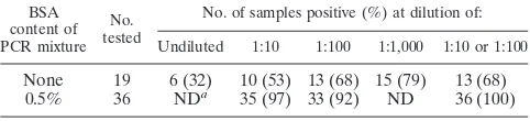
Effect of BSA on PCR.During the evaluation of instrument true-positive blood cultures, we found that samples from the BACTEC 9240 aerobic and anaerobic blood culture bottles demonstrated significant inhibition of PCR. Enough material was available from 36 of the 45 true-positive samples to study the role that BSA may play in overcoming the PCR-inhibitory effects. As shown in Table 1, if DNA samples were not diluted before amplification, the rate of positivity was only 32%. Upon dilution of samples to 1:1,000, the amplification rate improved to 79%. Pretreatment of specimens by red blood cell lysis and/or centrifugation before extraction of DNA did not appre-ciably affect PCR inhibition (data not shown). However, by addition of 0.5% BSA to the PCR mixture, inhibition of PCR was essentially eliminated (Table 1). The PCR-positive rate increased from 53 to 97% (35 out of 36 samples) with BSA at a DNA sample dilution of 1:10. There was a reduction in sensitivity (92%) of the PCR when samples were diluted to 1:100. However, when running samples at both 1:10 and 1:100 dilutions, we could achieve 100% sensitivity. All the 20 instru-ment true negatives were PCR negative.
Comparison of bacteria identification by sequencing the 16S rDNA with conventional phenotypic identification methods. The results for genotypic versus phenotypic identification for the 45 instrument true-positive samples are shown in Table 2. Initial samples were chosen on a random basis without an awareness of phenotypic identification. Additional samples were selected after phenotypic identification to assure a more diverse group of organisms. The samples included 13 species of gram-positive and 17 species of gram-negative aerobic bacteria and 3 species of gram-positive and 4 species of gram-negative anaerobic bacteria. There was 100% agreement for phenotypic and genotypic identification methods for all 37 aerobic isolates to the species or genus level. Results for six of eight (75%) anaerobic isolates by both phenotypic and genotypic methods agreed to the species or genus level. One isolate was identified asPorphyromonas gingivalisby the phenotypic method but as Porphyromonas macacae(salivosa) by the sequencing method. Another isolate was identified asPeptostreptococcus prevotiiby the phenotypic method but asRuminococcus( Peptostreptococ-cus)productus(83.2% sequence homology) or as Peptostrepto-coccus prevotii(80% sequence homology) by the sequencing method.
[image:2.587.301.542.95.150.2]Evaluation of the instrumental false-positive samples by acridine orange stain and 16S rDNA amplification.None of the 76 instrument false-positive samples showed evidence of any microorganisms by acridine orange stain. Furthermore, no TABLE 1. Effect of 0.5% BSA on PCR amplification of 16S rDNA
from instrument true-positive blood culture samples
BSA content of PCR mixture
No. tested
No. of samples positive (%) at dilution of:
Undiluted 1:10 1:100 1:1,000 1:10 or 1:100
None 19 6 (32) 10 (53) 13 (68) 15 (79) 13 (68)
0.5% 36 NDa 35 (97) 33 (92) ND 36 (100)
aND, not done.
on May 15, 2020 by guest
http://jcm.asm.org/
PCR amplicon was produced for any of these samples using broad-range primers for the bacterial 16S rRNA gene.
Blood leukocyte counts and instrument detection time of blood culture samples.To investigate if high blood leukocyte counts contribute to the false signaling of the BACTEC 9240 blood culture system, we obtained peripheral leukocyte counts for patients for the following numbers of samples: 73 of 76 instrument false-positive specimens, 43 of 45 instrument true-positive specimens, and 20 of 20 instrument true-negative spec-imens (Table 3). Of the 73 patients with instrument false pos-itives, 51 (70%) had higher-than-normal leukocyte counts (normal range, 3.5⫻109to 10.5⫻109leukocytes/liter). Of 43
positive-control patients and 20 negative-control patients, 18 (42%) and 11 (55%), respectively, had higher-than-normal leu-kocyte counts. Elevated leuleu-kocyte counts were statistically sig-nificantly more frequently associated with the instrument false-positive samples than with the two other groups of samples (P⫽0.001).
We also observed that more than one-half (54%, 38 of 70) of the instrument false-positive samples and none (0 of 45) of the instrument true-positive samples were signaled within 0 to 24 h of incubation, 41% (29 of 70) of instrument false-positive sam-ples and 67% (30 of 45) of instrument true-positive samsam-ples were signaled within 24 to 48 h of incubation, and 4% (3 of 70) of instrument false-positive and 33% (15 of 45) of instrument true-positive samples were signaled within 48 to 120 h of in-cubation (Table 4). Instrument false-positive samples were sig-naled significantly earlier than instrument true-positive sam-ples (P⬍0.0001).
DISCUSSION
[image:3.587.301.540.94.173.2]The development of continuously monitored blood culture instruments has led to a decrease in the detection time of bloodstream infections. However, specific identification of bac-teria still requires conventional phenotypic methods. DNA probe assays have been developed for a limited number of pathogens that are frequently isolated from blood cultures (3, 13). For fastidious bacteria, subculture of the bacteria from blood culture bottles to solid media may require several days to weeks before phenotypic assays can provide an identification. These organisms are infrequently a cause of infection, and so immunologic or DNA probe assays have not been developed. Recently, Turenne et al. (23) reported a rapid identification method for bacteria from blood cultures by using multiplex PCR amplification of the 16S rRNA gene and analysis of the amplified fragments using nondenaturing electrophoresis. TABLE 2. Agreement of genotypic (MicroSeq 500) identification
with phenotypic identification of bacteria grown in blood culture bottles
Sequencing result % of sequencehomology Conventionalidentification
Streptococcus pneumoniae(3 isolates) 96.5–100 S. pneumoniae Streptococcus vestibularis 100 Streptococcusspp.,
viridans group
Streptococcus pyogenes 100 S. pyogenes Staphylococcus epidermidis 100 Staphylococcusspp.,
coagulase negative
Staphylococcus hominis 99.4 Staphylococcusspp., coagulase negative
Staphylococcus aureus(3 isolates) 97.9–99.8 S. aureus
Corynebacterium jeikeium 100 Corynebacteriumsp.
Micrococcus luteus 99.3 Micrococcussp.
Enterococcus faecalis 99.8 E. faecalis Enterococcus faecium 99.8 E. faecium Enterococcus gallinarum 99.8 E. gallinarum Enterococcus flavescens 99.8 E. casseliflavusor
E. flavescensa Brevibacterium casei 95.4 Brevibacteriumsp.
Escherichia coli(3 isolates) 99.4–100 E. coli Haemophilus influenzae 100 H. influenzae Enterobacter cloacae 99.9 E. cloacae Enterobacter aerogenes 99.9 E. aerogenes Pseudomonas aeruginosa(2 isolates) 99.8–100 P. aeruginosa Klebsiella pneumoniae 99.0 K. pneumoniae Proteus mirabilis 99.9 P. mirabilis
Cardiobacterium hominis 100 Cardiobacteriumsp.
Serratia marcescens 99.4 S. marcescens Stenotrophomonas maltophilia 98.9 S. maltophilia Capnocytophaga ochracea 92.2 C. sputigenaor
C. ochraceaa Citrobacter youngae 99.6 C. freundiicomplex
Citrobacter freundii 98.0 C. freundii Acinetobacter junii 100 Acinetobactersp.
Ralstonia(Alcaligenes)eutropha 96.3 Ralstoniasp.
Sphingomonas paucimobilis 100 S. paucimobilis Morganella morganii 99.8 M. morganii Propionibacterium acnes 100 Propionibacteriumsp.
Fusobacterium nucleatum 97.6 F. nucleatum Fusobacterium necrophorum 99.5 F. necrophorum Bacteroides fragilis 99.6 B. fragilis Bacteroides fragilis 99.8 B. necrophorum Lactobacillus fermentum 97.2 Lactobacillussp.
Porphyromonas salivosa 87.8 P. gingivalisb Ruminococcus(Peptostreptococcus)
productus 83.2 Peptostreptococcusprevotiib aThe biochemical pattern could not differentiate between the two species. bThe genotypic identification disagreed with the phenotypic identification.
TABLE 3. Peripheral total leukocyte counts related to false signaling of BACTEC 9240 blood culture system
Leukocyte levelb
No. of blood culture samples with indicated reading/no. sampled (%) Instrument
false positive true positiveInstrument true negativeInstrument
Low 2/73 (3) 8/43 (19) 1/20 (5)
Normal 20/73 (27) 17/43 (40) 8/20 (40)
High 51/73 (70)a 18/43 (42)a 11/20 (55)a
aStatistically significant (P⫽0.001).
bLow,⬍3.5⫻109/liter; normal, 3.5⫻109to 10.5⫻109/liter; high,⬎10.5⫻
[image:3.587.43.282.100.555.2]109/liter.
TABLE 4. Time of signaling of blood culture samples by BACTEC 9240 blood culture systema
Incubation time (h)
No. of blood culture samples with indicated reading/no. sampled (%)
Instrument false positive Instrument true positive
0–24 38/70 (54) 0/45 (0)
24–48 29/70 (41) 30/45 (67)
48–120 3/70 (4) 15/45 (33)
aInstrument false-positive samples signaled statistically significantly sooner
than instrument true-positive samples (P⬍0.0001).
on May 15, 2020 by guest
http://jcm.asm.org/
[image:3.587.301.540.641.712.2]Their method could not differentiate two important pathogens, Staphylococcus aureus and Streptococcus pneumoniae, from other bacteria of their respective genera presumably because of the limited resolution of macromolecular-gel-based separa-tion techniques.
Previously, our group used the MicroSeq kit to amplify the first 418, 527, or 1,189 bp of the 16S rRNA gene for identifi-cation of 72 unusual aerobic gram-negative bacilli isolated from clinical specimens on solid culture media (21). We found that sequencing the first 527 bp of the 16S rRNA gene pro-vided rapid, unambiguous identification of all 72 isolates to genus level and 97.9% of nonfermenting gram-negative bacte-ria and 84.0% of fermenting gram-negative bactebacte-ria to species level.
In the present study, the MicroSeq 500 method was applied directly to samples from blood culture bottles, which were signaled positive by the BACTEC 9240 blood culture system. The first 527 bp of the16S rRNA gene were analyzed. Results were obtained with the MicroSeq 500 method. Results for all 37 (100%) aerobic gram-positive and gram-negative isolates agreed with results obtained by conventional phenotypic meth-ods to species or genus level (Table 2). The time required to complete the MicroSeq 500 analysis (DNA extraction, ampli-fication, sequencing, and analysis of sequence data) was ap-proximately 2 days.
Recently, it was demonstrated that the MicroSeq 500 kit was an accurate and rapid method for the identification of Myco-bacteriumspecies from isolated colonies (18). However, there has been no reported study on the analysis of anaerobic bac-teria by these methods. Although the number of samples was small in the present study, the agreement between MicroSeq results and conventional phenotypic analyses for anaerobic bacteria was lower than for aerobic bacteria. This may be due to the need for more anaerobic representatives in the Micro-Seq database, shortcomings in conventional identification techniques, or a combination of both factors. Few taxonomic or phylogenetic studies have addressed broad-range rDNA PCR as a method for anaerobic bacterial identification. Al-though the 23S rRNA gene is larger (approximately 2.5 kb, compared to 1.5 kb for the 16S rRNA gene) and may allow for more-complete identification of anaerobic bacteria (21), the 16S rRNA gene may well contain enough phylogenetically informational sites to perform well as a diagnostic target.
We, like other investigators, discovered that samples of blood culture specimens taken from the BACTEC Plus Aero-bic/F bottle or BACTEC Anaerobic Lytic/10 bottle contained substantial PCR-inhibitory activity (11, 12). If DNA samples were not diluted before performing the PCR, an amplicon was obtained only 32% of the time. When the DNA samples were diluted to 1:1,000, an amplicon was obtained 79% of the time. Hemin in blood has been shown to inhibit PCR (8, 10, 17). Fredricks and Relman (6) demonstrated that sodium polyanet-holesulfonate, a common additive to blood culture media was a potent PCR inhibitor.
Investigators have tried to use different methods to over-come the inhibitory effect of clinical specimens for PCR test-ing. Kulski and Pryce (12) determined that DNA extracted by alkali wash and heat lysis contained less inhibitory activity than that extracted by a sodium iodide-isopropanol method. Benzyl
alcohol-guanidine hydrochloride organic DNA extraction can also overcome the PCR-inhibitory effect (6).
Several studies have shown that BSA can lessen PCR inhi-bition. Forbes and Hicks (4) reported that 0.05% BSA de-creased the effects of PCR inhibition in 95% of their sputum samples. Kreader (10) found that inhibition from hemin and unknown inhibitors from feces and natural water samples was decreased 10- to 1,000-fold by BSA but that BSA could not relieve the interference from minimum inhibitory levels of a hemin degradation product, such as bilirubin. In the present study, blood culture samples taken from the BACTEC bottles contained not only hemin but also sodium polyanetholesulfon-ate and/or other unknown inhibitors. By supplementing the PCR mixture with 0.5% BSA and testing at both 1:10 and 1:100 dilutions of samples, the inhibitory effects on PCR were sig-nificantly reduced but not completely eliminated (Table 1).
The BACTEC 9240 blood culture system permits continu-ously monitoring specimens for growth of bacteria and fungi. We and others (2, 16, 20) have demonstrated that this system has a small but still significant instrument false-positive rate; a positive signal occurs, but both Gram stain and subculture of the specimen to solid (chocolate) media showed no organisms, and fungal cultures using the Isolator blood culture method from the same sample of blood showed no growth.
It has been suggested by the manufacturer that elevated peripheral leukocyte counts may contribute to the false signal-ing of the BACTEC 9240 blood culture system. In the present study, elevated leukocyte counts were statistically significantly associated with instrument false-positive samples compared to instrument true-positive and instrument true-negative samples (Table 3). We also found that instrument false-positive sam-ples were signaled by the BACTEC system statistically signif-icantly earlier than positive controls (Table 4).
Although elevated total peripheral leukocyte counts were more frequently associated with the instrument false-positive samples, 31% of these samples had normal or lower-than-normal peripheral leukocyte counts. Therefore, elevated leu-kocyte counts were not the only cause for the instrument false-positive signals for this group of samples. Theoretically, there might be unusual and/or fastidious bacteria including myco-bacteria (although mycomyco-bacteria do not commonly grow in the BACTEC bottles studied) present in these instrument false-positive samples; however, in none of these 76 samples was a 16S rDNA amplicon generated. These samples also had neg-ative acridine orange stains. Acridine orange stains nucleic acids of bacteria and thus is a more sensitive method than Gram stain. It is theoretically possible that lysis of some or-ganisms (e.g.,Streptococcus pneumoniae) may occur in blood culture systems to the extent that no bacteria are subsequently cultured. In such a scenario, the instrument may signal posi-tive. Further studies are required to verify this possibility and might include frequent sampling of blood cultures for subcul-ture, stains, antigen detection, and nucleic acid analysis.
In conclusion, the MicroSeq 500 PCR and sequencing kit is a reliable rapid method for detecting aerobic bacteria directly from blood culture bottles of the BACTEC 9240 blood culture system. Adding 0.5% BSA to the PCR mixture greatly dimin-ished PCR inhibitors in these specimens. The BACTEC 9240 instrument false-positive samples appeared negative for bacte-rial 16S rDNA. Elevated peripheral leukocyte counts were
on May 15, 2020 by guest
http://jcm.asm.org/
statistically significantly more frequently associated with in-strument false-positive samples.
ACKNOWLEDGMENTS
We thank Roberta Kondert for her efforts in preparing the manu-script, Jeffrey J. Germer and Mark J. Espy for technical advice and support, and the technologists in the bacteriology and mycology labo-ratories for their contributions to this evaluation.
REFERENCES
1.Adal, K. A., and B. M. Farr.1996. Central venous catheter-related infections: a review. Nutrition12:208–213.
2.Cockerill, F. R., III, G. S. Reed, J. G. Hughes, C. A. Torgerson, E. A. Vetter, W. S. Harmsen, J. C. Dale, G. D. Roberts, D. M. Ilstrup, and N. K. Henry. 1997. Clinical comparison of BACTEC 9240 plus aerobic/F resin bottles and the isolator aerobic culture system for detection of bloodstream infections. J. Clin. Microbiol.35:1469–1472.
3.Davis, T. E., and D. D. Fuller.1991. Direct identification of bacterial isolates in blood cultures by using a DNA probe. J. Clin. Microbiol.29:2193–2196. 4.Forbes, B. A., and K. E. Hicks.1996. Substances interfering with direct detection ofMycobacterium tuberculosisin clinical specimens by PCR: effects of bovine serum albumin. J. Clin. Microbiol.34:2125–2128.
5.Fredricks, D. N., and D. A. Relman.1996. Sequence-based identification of microbial pathogens: a reconsideration of Koch’s postulates. Clin. Microbiol. Rev.9:18–33.
6.Fredricks, D. N., and D. A. Relman.1998. Improved amplification of micro-bial DNA from blood cultures by removal of the PCR inhibitor sodium polyanetholesulfonate. J. Clin. Microbiol.36:2810–2816.
7.Greisen, K., M. Loeffelholz, A. Purogit, and D. Leong.1994. PCR primers and probes for the 16S rRNA gene of most species of pathogenic bacteria, including bacteria found in cerebrospinal fluid. J. Clin. Microbiol.32:335– 351.
8.Higuchi, R. 1989. Simple and rapid preparation of samples for PCR, p. 31–38.InH. A. Erlich (ed.), PCR technology: principles and applications for DNA amplification. Stockton Press, New York, N.Y.
9.Kolbert, C. P., and D. H. Persing.1999. Ribosomal DNA sequencing as a tool for identification of bacterial pathogens. Curr. Opin. Microbiol.2:299– 305.
10.Kreader, C. A.1996. Relief of amplification inhibition in PCR with bovine serum albumin or T4 gene 32 protein. Appl. Environ. Microbiol.62:1102– 1106.
11.Kulski, J. K., C. Khinsoe, T. Pryce, and K. Christiansen.1995. Use of a multiplex PCR to detect and identifyMycobacterium aviumandM. intracel-lularein blood culture fluids of AIDS patients. J. Clin. Microbiol.33:668– 674.
12.Kulski, J. K., and T. Pryce.1996. Preparation of mycobacterial DNA from blood culture fluids by simple alkali wash and heat lysis method for PCR detection. J. Clin. Microbiol.34:1985–1991.
13.Kuritza, A. P., C. E. Getty, P. Shaughnessy, R. Hesse, and A. A. Salyers. 1986. DNA probe for identification of clinically important bacteroides spe-cies. J. Clin. Microbiol.23:343–349.
14.Make, D. G. 1981. Nosocomial bacteremia: an epidemiologic overview. Am. J. Med.70:719–732.
15.Mendez, J. C., M. J. Espy, T. F. Smith, J. A. Wilson, and C. V. Paya.1998. Evaluation of PCR primers for early diagnosis of cytomegalovirus infection following liver transplantation. J. Clin. Microbiol.36:526–530.
16.Nolte, F. S., J. M. Williams, R. C. Jerris, J. A. Morello, C. D. Leitch, S. Matushek, L. D. Schwabe, F. Dorigan, and F. E. Kocka.1993. Multicenter clinical evaluation of a continuous monitoring blood culture system using fluorescent-sensor technology (BACTEC 9240). J. Clin. Microbiol.31:552– 557.
17.Panaccio, M., and A. Lew.1991. PCR in the presence of 8% (v/v) blood. Nucleic Acids Res.19:1151–1156.
18.Patel, J. B., D. G. B. Leonard, X. Pan, J. M. Musser, R. E. Berman, and I. Nachamkin.2000. Sequence-based identification ofMycobacteriumspecies using MicroSeq 500 16S rDNA bacterial identification system. J. Clin. Mi-crobiol.38:246–251.
19.Salzman, M. B., and L. G. Rubin.1995. Intravenous catheter-related infec-tions. Adv. Pediatr. Infect. Dis.10:337–368.
20.Shigei, J. T., J. A. Shimabukuro, M. T. L. Pezzlo. M. de la Maza, and E. M. Peterson.1995. Value of terminal subcultures for blood cultures monitored by BACTEC 9240. J. Clin. Microbiol.33:1385–1388.
21.Tang, Y., N. M. Ellis, M. K. Hopkins, D. H. Smith. D. E. Dodge, and D. H. Persing.1998. Comparison of phenotypic and genotypic techniques for iden-tification of unusual aerobic pathogenic Gram-negative bacilli. J. Clin. Mi-crobiol.36:3674–3679.
22.Temesgen, Z., D. R. Toal, and F. R. Cockerill III.1997. Leclercia adecar-boxylata infections: case report and review. Clin. Infect. Dis.25:79–81. 23.Turenne, C. Y., E. Witwicki, D. J. Hoban, J. A. Karlowsky, and A. M. Kabani.
2000. Rapid identification of bacteria from positive blood cultures by fluo-rescence-based PCR-single-strand conformation polymorphism analysis of the 16S rRNA gene. J. Clin. Microbiol.38:513–520.
24.Washington, J. A.1985. Laboratory procedures in clinical microbiology, 2nd ed. Springer-Verlag, New York, N.Y.
25.Weisburg, W. G., S. M. Barns, D. A. Pelletier, and D. J. Lane.1991. 16S ribosomal DNA amplification for phylogenetic study. J. Bacteriol.173:697– 703.
26.Wenzel, R. P., M. R. Pinsky, R. J. Ulevitch, and L. Young.1996. Current understanding of sepsis. Clin. Infect. Dis.22:407–413.
27.Wenzel, R. P.1992. Anti-endotoxin monoclonal antibodies—a second look. N. Engl. J. Med.326:1151–1153.
28.Woese, C. R.1987. Bacterial evolution. Microbiol. Rev.51:221–271.