University of East London Institutional Repository: http://roar.uel.ac.uk
This paper is made available online in accordance with publisher policies. Please scroll down to view the document itself. Please refer to the repository record for this item and our policy information available from the repository home page for further information.
To see the final version of this paper please visit the publisher’s website. Access to the published version may require purchase or a subscription. Author(s): Jansari, Ashok S; Davis, Kavus; McGibbon, Terence; Firminger, Stephanie; Kapur, Narinder.
Title:When "long-term memory" no longer means "forever": analysis of accelerated long-term forgetting in a patient with temporal lobe epilepsy.
Year of publication: 2010
Citation: Jansari, AS; Davis, K; McGibbon, T; Firminger, S; Kapur N. (2010) ’ When "long-term memory" no longer means "forever": analysis of accelerated long-term forgetting in a patient with temporal lobe epilepsy.’ Neuropsychologia 48 (6) 1707-1715
Link to published version:
When “long-term memory” no longer means “forever”: Analysis of accelerated
long-term forgetting in a patient with temporal lobe epilepsy
Ashok S Jansari*, Kavus Davis*, Terence McGibbon*, Stephanie Firminger* & Narinder Kapur+
* School of Psychology, University of East London
+
Addenbrooke’s Hospital, Cambridge
Short Title: Accelerated Long-term Forgetting and Epilepsy Running Head: Long-Term Amnesia
Address for correspondence: Dr Ashok Jansari
School of Psychology University of East London Romford Rd
London E15 4LZ UK
Abstract
Classical amnesia involves a difficulty in transferring information to long-term memory and can be detected with standard clinical tests. However, there are some patients who pass these tests but nonetheless show longer-term memory impairments. A case study is presented of a patient, RY, with temporal lobe epilepsy, who
exhibited such a profile of “accelerated long-term forgetting”. To investigate the effect of recalling information on later retention, recall and recognition for pairs of novel stories were tested at five intervals ranging from 30 minutes to 4 weeks; we also manipulated whether or not recall and recognition were repeatedly tested for stories. Two studies are reported, one before RY commenced treatment with anticonvulsant medication, and one following 6 months of treatment. Very similar memory profiles were observed in both settings. Against a background of above average cognitive function, results showed that RY’s free recall, although initially average or above, was significantly impaired at extended delays (within 24 hours) for non-repeatedly recalled episodic information. However, this contrasted with normal performance for information that had been repeatedly recalled. An unresolved issue in the field is the impact of anticonvulsant medication on alleviating long-term forgetting, and the current study shows that anticonvulsant medication can have negligible beneficial effects in improving the rate of long-term forgetting in this type of patient. In addition, our study highlights the possible protective effect of active review of recent episodic memories.
1. Introduction
How does memory for what happened two minutes ago differ from memory for what happened twenty years ago and is there a process that the former undergoes to become the latter? Early functional models of memory suggested a transfer of information from a temporary short-term store to a more long-lasting and possibly permanent long-term one (Atkinson & Schiffrin, 1968). A neurobiological process, consolidation, was postulated to occur at a synaptic level to aid this process (Hebb, 1949). The evidence from studies on patients with selective memory problems (e.g. Scoville & Milner, 1957) supported such a distinction between the two stores or forms of memory, namely short-term memory (STM) and long-term memory (LTM). The major memory impairments of importance, and associated clinical tests, have revolved around intact or impaired STM and intact or impaired LTM.
Recently, however, a number of individual case and group studies have reported patients who pass the standard clinical tests of memory but nonetheless complain of profound long-term memory problems (e.g. Kapur et al., 1996, 1997; O’Connor, Sieggreen, Ahern, Schomer & Mesulam, 1997; Blake, Wroe, Breen & McCarthy, 2000; Mayes et al., 2003; Mameniskiene, Jatuzis, Kaubrys & Budrys, 2006; Butler et al., 2007; Butler et al., 2009). Following presentation of information, such studies have typically tested at 30 minutes (the delay used in most clinical tests), and found no impairment, then tested again at a single long delay of between 24 hours
sensitive to detect mild deficits in early processing. Although the exact timing of the onset and the progression of the patients’ accelerated forgetting is still unclear, this phenomenon of “long-term amnesia” (LTA; Kapur et al., 1997), or “accelerated long-term forgetting” (ALF; Butler & Zeman, 2008a), poses a challenge to the standard clinical measures as well as to the underlying theoretical assumptions. It should be noted that in a review of neuroimaging studies, Gilboa (2004) has
suggested that the brain mechanisms for recalling artificial stimuli in the laboratory (and by extrapolation in the clinic) are different from those involved in recollecting personal autobiographical memory. This difference may go some way to explaining the difficulty in capturing levels of real-world accelerated forgetting within the clinic.
Traditionally, information that has been retained for even a few minutes was assumed to have made the transition from short-term or working memory to a long-term store (Parkin, 1993). This view has often made the assumption that this transition involves a single-stage process of consolidation (Weingartner & Parker, 1984). However, evidence from studies of LTA suggests that after the initial ‘fixation’ of memory within the first 30 minutes, subsequent preservation may require further stages of consolidation before information that is initially encoded and learned is set down in a more permanent store(cf. Frankland and Bontempi, 2005). A failure of these secondary consolidation processes could explain the pattern of accelerated forgetting distinctive to ALF.
gradients in such cases, (which can extend for months or even years (e.g. Zola-Morgan, Squire & Amaral, 1986), with older memories intact while memory for newer pre-morbid material is degraded, suggest that at least some memories are at first reliant on the MTL, but through secondary consolidation become less reliant on this structure over time.
In ALF cases involving epilepsy, a further possible confound is the impact of epilepsy medication. In most or all reported cases patients were under medication at the time of test (e.g. Mayes et al., 2003; Kapur et al., 1996; Mameniskiene, Jatuzis, Kaubrys & Budrys, 2006). While O’Connor et al. (1997) found that anticonvulsant medication improved memory indirectly, through minimising seizures, and
Midorikawa and Kawamura (2007) report a case with improvement in ALF under successful anticonvulsant medication, high serum levels of epilepsy drugs have also been shown to impair retention (but not acquisition), of new information (Jokeit, Kramer & Ebner, 2005). Butler and Zeman (2008a) argue that the accelerated forgetting seen in cases of TEA is unlikely to be a direct result of anticonvulsant treatment as patients subjectively report symptoms prior to treatment, and often report improvements after treatment. Theorising about the cause(s) of ALF would be aided by objective studies of ALF in epilepsy cases prior to administration of
medication. If combined with comparative testing after treatment commences, such studies would also assist in clarifying the impact of anticonvulsant medication on memory deficits in such cases.
Repetition and rehearsal have been extensively studied within the general cognitive field of memory research and also in memory rehabilitation, and both have been found beneficial in consolidating memories (e.g. Ebbinghaus, 1885; Wilson,
Baddeley, Evans & Shiel 1994). However, while these approaches may be useful for learning lists of words or other information which can be re-presented, they are not easily applied to memory for events and episodes in general life, unless these have been recorded in some way (e.g. use of a portable, automatic camera, “SenseCam”, Berry et. al., 2007).
Although such repeated recall has not been specifically tested in cases of ALF, there is some general evidence for its benefits. Successful retrieval of an item has been shown to have a positive effect on later ability to recall the same item (Whitten, 1978), sometimes referred to as the retrieval practice effect (Baddeley, 1997), and active recall of material can enhance memory more than a second, passively
received, representation of the information (McDaniel & Masson, 1985; Roediger & Karpicke, 2006). Repeated recall at expanding delays, a method referred to as spaced retrieval, has proven successful with dementia patients, amnesics and normal healthy participants (e.g. Landauer & Bjork, 1978; Brush & Camp, 1998; Cull, Shaugnessy & Zechmesiter, 1996). However, it should be noted that spaced retrieval procedures typically include re-presentation after retrieval failure, to maintain performance at ceiling.
whole new trace is laid down conjointly with a trace that is reactivated (see also the ‘multi-trace theory’ of Nadel & Moscovitch, 1997).
The current study investigated the impact of repeated recall without re-presentation of novel information on the memory performance of a patient, RY, who exhibited ALF. The time course of the patient’s accelerated forgetting was also studied through testing at multiple delays. Two experiments were run, separated by 9 months. As a result of Experiment 1 the patient was referred for neurological investigations and was subsequently diagnosed with temporal lobe epilepsy. Since the Experiment 1 results were obtained prior to this diagnosis and the commencement of
anticonvulsant drug treatment, they are free from any drug-related confounds. Experiment 2 was run after 6 months of drug treatment, and we therefore had an ideal opportunity to investigate the impact of anticonvulsant medication on accelerated long-term forgetting.
2. Case History
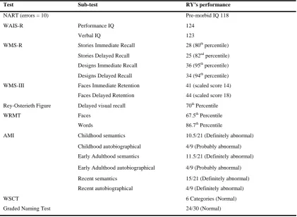
Table 1: Neuropsychological assessment of RY
Table 1
Test Sub-test RY’s performance
NART (errors = 10) Pre-morbid IQ 118
WAIS-R Performance IQ 124
Verbal IQ 123
WMS-R Stories Immediate Recall 28 (80th percentile) Stories Delayed Recall 25 (82nd percentile) Designs Immediate Recall 36 (95th percentile) Designs Delayed Recall 34 (94th percentile) WMS-III Faces Immediate Retention 41 (scaled score 14)
Faces Delayed Retention 44 (scaled score 18) Rey-Osterieth Figure Delayed visual recall 70th Percentile
WRMT Faces 67.5th Percentile
Words 86.7th Percentile
AMI Childhood semantics 10.5/21 (Definitely abnormal) Childhood autobiographical 4/9 (Probably abnormal)
Early Adulthood semantics 11.5/21 (Definitely abnormal) Early Adulthood autobiographical 4/9 (Probably abnormal) Recent semantics 15/21 (Definitely abnormal) Recent autobiographical 4/9 (Definitely abnormal)
WSCT 6 Categories (Normal)
Graded Naming Test 24/30 (Normal)
WAIS-R= Wechsler Adult Intelligence Scale Revised; WMS-R= Wechsler Memory Scale Revised; WMS-III=
Wechsler Memory Scale III; WRMT= Warrington Recognition Memory Test; AMI= Autobiographical Memory
Interview; WSCT= Wisconsin Card Sorting Test
Other than cardiac surgery in 2005, RY has an unremarkable medical history. (It is interesting to note that Zeman, Boniface & Hodges (1998) found that a history of cardiac disease was common in their series of patients with TEA.) RY also reported that since childhood he experienced what he referred to as ‘turns’ where his
awareness changes and he feels a sense of déjà vu for about 20 seconds. This feeling of déjà vu is followed by a ‘dreamlike’ episode which may involve forgotten
fade rapidly. Although he had experienced episodes as a child, they had become frequent and noticeable just before 2000 and at the time of presentation, were occurring in clusters of four or five episodes about twice a month and usually occurred in the morning after a lack of sleep. These episodes were not associated with any olfactory, gustatory or epigastric sensations. Clinical investigations conducted when RY first complained of memory problems did not find evidence of epilepsy. However, subsequent to the testing in Experiment 1 of the current study - which revealed significant memory problems both on laboratory tasks and
autobiographical memory measures - in conjunction with RY’s description of his turns and feelings of déjà vu the possibility of subclinical epilepsy was investigated. A sleep-deprived EEG subsequently showed right temporal spike activity (with a greater number of epileptiform discharges occurring while asleep than while awake), and he was given a diagnosis of temporal lobe epilepsy by a consultant neurologist. He has since been prescribed anticonvulsant medication (Lamotrigine, 50mg, twice daily). Neuropsychiatric evaluation performed at diagnosis identified no
Fig. 1. T2 weighted 3D coronal images of patient RY showing normal
hippocampi bilaterally
3. Experiment 1
3.1 Method
3.1.1 Normal Controls
RY’s performance was compared to 8 age- and reading-score derived IQ-matched healthy control subjects who were free of neurological or psychiatric disorders. RY: age at time of testing = 63, NART IQ=118. Control group: N=8; 3 males, 5 females, mean age 66.3, SD 4.9 years, mean NART IQ 117.88, SD 6.29. All participants gave informed written consent to take part in the study, which was approved by the local ethical committee.
3.1.2 Stimuli & Procedure
Participants were tested on recall and recognition of structured prose material using ten stories made up of between 200 and 250 words (Jansari, & Tranel, 1999).Each story (identified by a one-word title, e.g. concert) had been created to include twenty idea units of information to allow free recall to be assessed systematically.
story to test recognition ability. During the presentation phase, each story was read out loud by the experimenter with the participant following it on a written copy; following this they were given one additional minute for silent reading. Stories were presented in pairs with each pair being separated by unrelated material or general conversation to avoid confusion. The time delay between successive pairs of stories was usually about ten minutes. It should be noted that the stories were only presented at this one unique time point and never again.
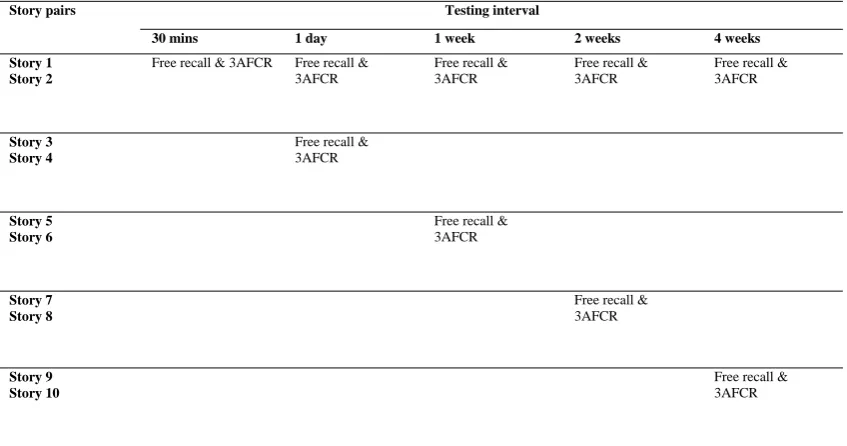
Table 2: Experiment 1 presentation and testing regime (3AFCR = 3 Alternative
[image:15.595.94.518.156.379.2]Forced Choice Recognition)
Table 2
Testing interval Story pairs
30 mins 1 day 1 week 2 weeks 4 weeks
Story 1 Story 2
Free recall & 3AFCR Free recall & 3AFCR
Free recall & 3AFCR
Free recall & 3AFCR
Free recall & 3AFCR
Story 3 Story 4
Free recall & 3AFCR
Story 5 Story 6
Free recall & 3AFCR
Story 7 Story 8
Free recall & 3AFCR
Story 9 Story 10
Free recall & 3AFCR
3.2 Results
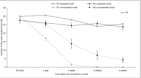
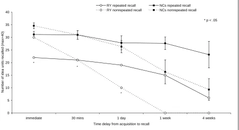
For free recall of the repeatedly recalled stories (1 & 2), RY performed similarly to controls (Fig. 2). However, a very different picture emerged when assessing retention of material that was only ever recalled once. Using Crawford & Garthwaite’s (2002) method for comparing a single case with a group of control subjects, the free recall scores showed this differing performance most dramatically - RY’s recall is
disparity in RY’s pattern of retention between the two types of information and clearly demonstrates a reinforcement effect for repeatedly recalled material1.
Fig. 2. Free-recall of stories in Experiment 1 (error bars represent one standard
error) 0 5 10 15 20 25 30 35
30 mins 1 day 1 week 2 weeks 4 weeks
Time delay from acquisition to recall
N
um
be
r of i
de a un it s r eca lled ( m ax=4 0)
RY repeated recall NCs repeated recall
RY nonrepeated recall NCs nonrepeated recall
* p < .05
*
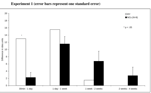
Given that RY displays accelerated forgetting for the non-repeatedly recalled stories, the timeframe of this forgetting was analysed by exploring the loss of information between consecutive recall sessions. For each participant, the difference in free recall scores between adjacent timepoints was computed (Fig. 3). Analysis of this
forgetting data showed that RY lost significantly more information than controls between 30 minutes and 1 day (t(7)=2.63, p<.05), confirming that his accelerated forgetting starts within the first 24 hours.
1
Fig. 3. Reduction in free-recall performance between test intervals in
Experiment 1 (error bars represent one standard error)
0 2 4 6 8 10 12 14 16 18 20
30min - 1 day 1 day - 1 week 1 week - 2 weeks 2 weeks - 4 weeks
D if fe ren ce i n i dea un it s RY NCs (N=8) *
* p < .05
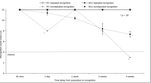
Recognition for information from the repeatedly recalled stories was at ceiling (a maximum score of 12) for both RY and all controls at all time points (Fig. 4). In contrast, RY’s performance for recognition of the non-repeatedly recalled
information was variable. He was impaired for the 1 day stories (t(7)=3.01, p<.05), and for the 4 week stories (t(7)=2.58, p<.05) for which he performed below chance.
Fig. 4. Recognition of stories in Experiment 1 (error bars represent one standard error) 0 2 4 6 8 10 12
30 mins 1 day 1 week 2 weeks 4 weeks
Time delay from acquisition to recognition
R ec ogni ti on s c or e (m ax =12)
RY repeated recognition NCs repeated recognition
RY nonrepeated recognition NCs nonrepeated recognition
chance
* p < .05
*
*
3.3 Discussion of Experiment 1
reflects his and his wife’s anecdotal report of the time-frame of his memory loss. Thus RY’s memory profile for non-repeatedly recalled information was found to reflect that of other ALF patients (Ahern et al., 1994; De Renzi & Lucchelli, 1993; Kapur et al., 1996; Lucchelli & Spinnler, 1998; O’Connor et al., 1997; Mayes et al., 2003; Butler et al., 2008; Butler et al., 2009), where memory for episodic
information is initially preserved, but then found to be impaired at longer time intervals.
4. Experiment 2
Experiment 2 was a partial replication of Experiment 1, performed after RY had been taking medication for 6 months (see Case History), and was intended to identify any changes in RY’s ALF as a result of the medication. At this time RY reported that he had been seizure-free since commencing drug treatment. If RY’s ALF is directly due to seizures, and if the medication did successfully eliminate or greatly reduce seizure occurrence, then an improvement in his memory would be predicted.
4.1 Method
4.1.1 Normal Controls
RY’s performance was compared to a new group of 6 age, sex and IQ-matched control subjects who were free of neurological or psychiatric disorders (mean age, 61.83, SD 5.41 years, mean NART IQ 122, SD 5.79).All participants gave informed written consent to take part in the study, which was approved by the local ethical committee.
4.1.2 Stimuli & Procedure
Eight new stories made up of between 200 and 250 words (based on the format used by Jansari, & Tranel, 1999) were prepared. Presentation and test procedures were identical to Study 1 with two exceptions.
material had been successfully encoded, and therefore any subsequent forgetting was of already-encoded material. Secondly, given RY’s equally poor performance at two and four weeks in Experiment 1, it was felt that testing at both intervals was
unnecessary, and it was therefore decided to omit the two week test point.
4.2 Results
Fig. 5. Free-recall of stories in Experiment 2 (error bars represent one standard
error). The immediate recall for the non-repeatedly recalled stories is based on
the average of all three pairs of stories which were then only recalled at one
unique timepoint. 0 5 10 15 20 25 30 35 40
immediate 30 mins 1 day 1 week 4 weeks
Time delay from acquisition to recall
N u mber of i d e a u n it s r e c a lle d ( m a x =40)
RY repeated recall NCs repeated recall RY nonrepeated recall NCs nonrepeated recall
* p < .05
* *
*
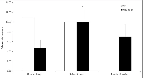
Comparing the curves for recall of ‘repeatedly recalled’ and non-repeatedly recalled’ information in Figure 5 shows the disparity in RY’s pattern of retention of the two types of information and again demonstrates a reinforcement effect for repeatedly recalled material. To further explore the rate of memory deterioration, the
significance levels (t(5)=1.47, p=0.10). This adds support to the suggestion that RY's forgetting begins within the first 24 hours.
Fig. 6. Reduction in free-recall performance between test intervals in
Experiment 2 (error bars represent one standard error)
0.00 2.00 4.00 6.00 8.00 10.00 12.00 14.00
30 mins - 1 day 1 day - 1 week 1 week - 4 weeks
D
iffer
en
c
e i
n
id
ea
un
it
s
RY
NCs (N=6)
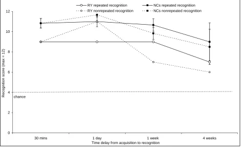
Fig. 7. Recognition of stories in Experiment 2 (error bars represent one standard error) 0 2 4 6 8 10 12
30 mins 1 day 1 week 4 weeks Time delay from acquisition to recognition
R e cogn it ion sc or e ( m a x = 1 2 )
RY repeated recognition NCs repeated recognition RY nonrepeated recognition NCs nonrepeated recognition
chance
4.3 Discussion of Experiment 2
1 day and rapidly falls to the floor thereafter. As stated in the Discussion of
Experiment 1, the turn that RY experienced at the 1 day testing meant that it was not possible to state unequivocally that his accelerated forgetting started within the first 24hrs since it could have contributed to both his poor recall and recognition of the non-repeatedly recalled material. However, in the absence of any turns being reported during Experiment 2, the rapid forgetting that occurred for equivalent material strongly suggests that indeed, the rapid forgetting is beginning this early2. In contrast, and echoing the findings of Experiment 1, repeated recall has a beneficial impact. RY’s initial (immediate) recall of the first two stories was significantly worse than that of the controls and this probably contributed to his poorer performance when recalling these stories 30 minutes later. However, despite this ‘poor start’, repeated recall then brings his memory within normal limits and although his memory is never the same as that of controls, there is no catastrophic forgetting to floor levels within a week as there is for the non-repeatedly recalled stories.
In Experiment 1 recognition of non-repeatedly recalled information matched controls at 2 weeks, but then fell to chance by 4 weeks, reflecting an accelerated forgetting. In Experiment 2, however, recognition for this material type was maintained within normal levels at all intervals. This appears to indicate an improvement in recognition memory.
2
Relative to Experiment 1, there thus appears to have been no improvement in RY’s free recall, and some improvement in recognition memory; this is coupled with impaired initial encoding of material which was not evaluated in Experiment 1. This is a complex picture and addressed in the General Discussion.
5. General Discussion
This study has identified a further case of accelerated long-term forgetting (ALF), thus confirming again the existence of patients who can pass all standard clinical memory tests, yet still suffer from significant long-term memory deficits. In this case the patient, RY, displays normal memory performance at 30 minutes, but
significantly degraded free recall of non-repeatedly recalled episodic information after 1 day. Using a new paradigm comparing the impact of repeatedly recalling the same information against recalling information only at one unique timepoint, it was possible to show that the former can have a protective effect on memory traces. If this does not happen, the patient's memory for novel information falls to floor levels within two weeks. Finally, because the patient had not been given a diagnosis of epilepsy before the research began and only received one after the completion of Experiment 1, it was possible to evaluate the impact of anti-epileptic medication by testing him before and after six months of drug treatment. While recognition memory seemed better, no overall improvement in free recall was found following
medication.
ALF has been found to have a close association with temporal lobe epilepsy
generally (TLE; e.g. Blake et al., 2000; Martin et al., 1991) and with a specific type of TLE known as transient epileptic amnesia (TEA; e.g. Butler et al., 2007). Both TLE and TEA patients often report persistent memory problems (e.g. Baxendale et al., 1998; Mameniskiene, Jatuzis, Kaubrys & Budrys, 2006; Butler et al., 2007). Accelerated forgetting has also been identified for material learnt shortly after
As RY has reported experiencing ‘turns’ since childhood and has only reported memory problems in the last decade, any direct connection between the frequency of epileptiform activity and his memory loss remains unclear. However, there is little evidence as yet that points to other sources of pathology that could account for his memory impairment. A lack of any obvious pathology has been reported in other ALF cases (e.g. Lucchelli & Spinnler, 1998), and although Butler et al. (2009) identified a small reduction in hippocampal volume in TEA cases they found no correlation between this atrophy and accelerated forgetting. It therefore might have been expected that RY’s memory would improve with successful medication. However his free recall performance after drug treatment is, in fact, little changed (and if anything his initial encoding of material is somewhat impaired). This suggests either that his ALF is not related to overt seizure frequency, or that the drug may have an amnestic effect which counteracts any benefits gained from reduced seizure frequency. Importantly for clinical practice, this suggests that even where medication eliminates overt seizures this will not necessarily resolve memory problems. This picture is slightly complicated by the fact that RY’s recognition memory
In addition, although RY has reported that his overt turns have ceased, this does not mean that all epileptiform activity has been eliminated. It is possible that subclinical seizures are still present, but are occurring within medial structures where they cannot be detected by standard scalp EEG. In an analysis of intracranial EEG reports Zangaladze et al. (2008) found that the majority of temporal lobe subclinical seizures originated from medial structures (amygdala or hippocampus) and remained
localized within that region. A further possibility is that seizures are occurring during sleep. As reported in the Case History, the telemetry data used to diagnose TLE highlighted that RY experienced a greater number of partial seizures while asleep than while awake. No EEG monitoring during sleep has been performed since commencing treatment and it is therefore not possible to make an unequivocal statement on this issue. A possible link to sleep may be significant considering the mounting evidence for the importance of sleep to consolidation of declarative memories (e.g. Ellenbogen, Hulbert, Stickgold, Dinges & Thompson-Schill, 2006; Drosopoulos, Schulze, Fischer & Born, 2007). In addition, a clear association between waking and amnesic attacks in TEA has led Butler and Zeman (2008) to suggest the possibility that nocturnal subclinical epileptiform activity may be a causal factor in the memory deficits seen in this condition. EEG monitoring of inter-ictal brain activity over an extended period would be beneficial in clarifying the role of any remaining low-level epileptiform activity while awake or during sleep.
epileptiform activity might be expected to disrupt the function of the medial temporal lobes upon which this short term process is considered dependent (Mayes et al., 2003). Indeed, there are many cases of TLE patients who show just such impairments after short delays, (e.g. Giovagnoli & Avanzini, 1996), but yet a key characteristic of ALF is that memory function is normal for at least 30 minutes. One possible
explanation is that the epileptiform activity in ALF cases may occur with low frequency, such that the amount encountered in a 30 minute period is insufficient to disrupt memory, while the amount encountered over 24 hours or several days is sufficient. Alternatively there may be low level disruption within 30 minutes which existing tests are insufficiently sensitive to detect. Indeed, in a preliminary study with RY, McGibbon, Jansari and Gaskell (2008) found evidence that ALF could be
detected at a one hour delay within the ‘clinical window’, using a specially developed word-pair association test.
Such cases suggest either a further functional deficit, or perhaps a slowly developing ALF which has been operating for many years or even decades.
In contrast, a post-consolidation dysfunction would be expected to also affect strongly learned memories, suggesting that continual rehearsal may be necessary to maintain even successfully consolidated memories indefinitely, and that the
immunity provided by repeated recall or rehearsal within the timeframe of this study may be temporary. Testing over extended time periods, controlling for level of rehearsal or recall, would be necessary to confirm this; to our knowledge no such testing has yet been published. Further work with RY to investigate the extent of any retrograde amnesia and the long term immunity effects of repeated recall would therefore be useful in distinguishing between causal deficits.
It should be noted, however, that ALF is not always accompanied by extensive retrograde amnesia (e.g. Kapur et al., 1997), and that therefore post-consolidation dysfunctions cannot be the sole cause of all ALF cases. A further possibility, therefore, is that both secondary consolidation and post-consolidation dysfunctions can cause ALF, either independently or in combination, and that patterns of
impairment displayed in each case will depend on the combination of dysfunctions present.
events, and 36% report navigational difficulties (Butler et al., 2007). In addition to the ALF discussed in the current study, RY also displays retrograde amnesia for autobiographical events (not reported here), and reports navigational difficulties. However, he has never experienced the transient amnestic episodes which are one of the diagnostic criteria for TEA (Zeman, Boniface & Hodges, 1998). RY does not, therefore, meet the criteria for TEA. However, similarities in persistent memory deficits suggest possible commonalities in underlying pathology or causal mechanisms. Further study will be required to clarify any such relationship.
In tests of free-recall for non-repeatedly recalled prose material, however, such reinforcement was not possible and here RY displayed a marked pattern of accelerated forgetting (reflecting patterns of other ALF patients e.g. Kapur et al., 1996; Mayes et al., 2003), reaching floor levels by two weeks. ALF cases such as RY and JL (Mayes et al., 2003), displaying strong evidence for some kind of initial ‘rehearsal immunity’, highlight the importance of further investigation into the immunity effects for strongly learned and frequently rehearsed information. In particular, ALF patients' accelerated forgetting of episodic memories, combined with their apparent responsiveness to repeated recall, highlights the benefits on long-term memory from repeated, spaced review of material. This technique has been found useful in improving autobiographical memory by repeated exposure to photographic images of earlier experiences that were generated by ‘SenseCam’, a portable, automatic camera (e.g. Berry et al., 2007). Such testing may also help distinguish between competing models of memory. If ALF cases show equal rehearsal-immunity for both semantic and episodic information then this would support models that postulate the relocation of both semantic and episodic memory indexing from the MTL to the neocortex (e.g. Alvarez & Squire, 1994). In contrast, if rehearsal-immunity differentially benefits semantic information then this would favour those models which maintain that while semantic indexing may relocate to the neocortex, episodic memory indexing remains reliant on the MTL indefinitely (Nadel & Moscovitch, 1997).
evidence of a class of patients who pass standard clinical tests of memory, yet display significant memory problems. Objective evidence for similar levels of accelerated forgetting both before, and after, medication has indicated that
anticonvulsant drugs alone cannot account for ALF, and that such medication does not necessarily ameliorate ALF even where overt seizures cease. This case highlights the importance of pursuing further investigations into the immunity effects of various forms of rehearsal (active recall, passive representation, etc.) on memory for
different types of material (verbal, visual, etc.) through detailed examinations of ALF patient memory profiles, and the possibility of using such techniques as the basis of protective strategies. Finally, the present study indicates the need for further
References
Ahern, G., O’Connor, M., Dalmau, J., Coleman, A., Posner, J. B., Schomer, D. L. et al. (1994). Paraneoplastic temporal lobe epilepsy with testicular neoplasm and atypical amnesia. Neurology, 44, 1270-1274.
Alvarez, P. & Squire, L. R. (1994). Memory consolidation and the medial temporal lobe: A simple network model. Proceedings of the National Academy of Sciences
USA, 91, 7041-7045.
Atkinson, R. C. & Schiffrin, R. M. (1968). Human memory: A proposed system and its control processes. In K.W. Spence (Ed.), The psychology of learning and
motivation: advances in research and theory (Vol 2, pp. 89-195). New York:
Academic Press.
Baddeley, A. (1997). Human Memory: Theory and Practice (rev. ed.). Hove, UK: Psychology Press Ltd.
Baxendale, S.A., van Paesschen, W., Thompson, P.J., Connelly, A., Duncan, J.S., Harkness, W.F. et al. (1998). The relationship between quantitative MRI and neurological functioning in temporal lobe epilepsy. Epilepsia, 39, 158–166.
Blake, R. V., Wroe, S. J., Breen, E. K & McCarthy, R. A. (2000). Accelerated forgetting in patients with epilepsy. Evidence for an impairment in memory consolidation. Brain, 123, 472-483.
Brush, J. A. & Camp, C. J. (1998). Using spaced retrieval as an intervention during speech-language therapy. Clinical Gerontologist, 19, 51-64.
Butler, C.R., Bhaduri, A., Acosta-Cabronero, J., Nestor, P.J., Kapur, N., Graham, K.S., Hodges, J.R. & Zeman, A.Z. (2009). Transient epileptic amnesia: regional brain atrophy and its relationship to memory deficits. Brain, 132, 357-368.
Butler, C.R., Graham, K.S., Hodges, Kapur, N., Wardlaw, J.M. & Zeman, A.Z. (2007). The Syndrome of Transient Epileptic Amnesia. Annals of Neurology, 61 ,
587-598.
Butler, C.R. & Zeman, A.Z. (2008a). Recent insights into the impairment of memory in epilepsy: transient epileptic amnesia, accelerated forgetting and remote
memory impairment. Brain, 131, 2243-2263.
Crawford, J.R. & Garthwaite, P.H. (2002). Investigation of the single case in neuropsychology: Confidence limits on the abnormality of test scores and test score differences. Neuropsychologia, 40, 1196-1208.
Cull, W.L., Shaughnessy, J.J. & Zechmeister, E.B. (1996). Expanding understanding of the expanding-pattern-of-retrieval mnemonic: Towards confidence in
applicability. Journal of Experimental Psychology: Applied, 2, 365-378.
Damasio, A.R. (1986). Time-locked multiregional retroactivation: A systems-level proposal for the neuronal substrates of recall and recognition. Cognition, 33, 25-62.
De Renzi, E & Lucchelli, F. (1993). Dense retrograde amnesia, intact learning capability and abnormal forgetting rate: A consolidation deficit. Cortex, 29, 449-466.
Drosopoulos, S., Schulze, C., Fischer, S. & Born, J. (2007). Sleep’s function in the spontaneous recovery and consolidation of memories. Journal of Experimental
Psychology: General, 136, 169-183.
Ebbinghaus, H. (1885). Memory: a Contribution to Experimental Psychology.
Ellenbogen, J. M., Hulbert, J. C., Stickgold, R., Dinges, D. F. & Thompson-Schill, S. L. (2006). Interfering with theories of sleep and memory: Sleep, declarative memory, and associative interference. Current Biology, 16, 1290-1294.
Frankland P, & Bontempi B. (2005). The organization of recent and remote memories. Nature Reviews Neuroscience, 6: 119-130.
Gilboa, A. (2004). Autobiographical and episodic memory - one and the same? Evidence from prefrontal activation in neuroimaging studies. Neuropsychologia, 42, 1336–1349.
Giovagnoli, A.R., & Avanzini, G. (1996). Forgetting rate and interference on a verbal memory distractor task in patients with temporal lobe epilepsy. Journal of
Clinical and ExperimentalNeuropsychology, 18, 259–264.
Hebb, D.O. (1949). The Organization of Behavior. New York, NY, USA: John Wiley & Sons.
Isaac, C. L. & Mayes, A. R. (1999a). Rate of forgetting in amnesia I: Recall and recognition of prose. Journal of Experimental Psychology: Learning, Memory
and Cognition, 25, 942-962.
Isaac, C. L. & Mayes, A. R. (1999b). Rate of forgetting in amnesia II: Recall and recognition of word lists at different levels of organization. Journal of
Jansari, A. & Tranel, D. (1999). Are confabulations mis-combined elements of veridical events? Journal of Cognitive Neuroscience, 18-18 Suppl.
Jokeit, H., Krämer, G. & Ebner, A. (2005). Do antiepileptic drugs accelerate forgetting? Epilepsy & Behavior, 6, 430-432.
Kapur, N., Millar, J., Colbourn, C., Abbott, P, Kennedy, P. & Docherty, T. (1997). Very long-term amnesia with temporal lobe epilepsy: Evidence for multiple-stage consolidation processes. Brain and Cognition, 35, 58-70.
Kapur, N., Scholey, K., Moore, E., Barker, S., Brice, J., Thompson, S. et al. (1996). Long-term retention deficits in two cases of disproportionate retrograde amnesia.
Journal of Cognitive Neuroscience, 8, 416-434.
Kopelman, M.D., Wilson, B.A. & Baddeley, A.D. (1990). The Autobiographical
Memory Interview. Bury St Edmunds: Thames Valley Test Company.
Landauer, T.K. & Bjork, R.A. (1978). Optimal rehearsal patterns and name learning. In K.M. Gruneberg, P.E. Morris & R.N. Sykes (Eds.), Practical Aspects of
Memory (pp. 625-632). New York: Academic Press.
Lewis, P. & Kopelman, M.D. (1998). Forgetting rates in neuropsychiatric disorders.
Lucchelli, F. & Spinnler, H. (1998). Ephemeral new traces and evaporated remote engrams: A form of neocortical temporal lobe amnesia? A preliminary case report. Neurocase, 4, 447–459.
Mameniskiene, R., Jatuzis, D., Kaubrys, G. & Budrys, V. (2006). The decay of memory between delayed and long-term recall in patients with temporal lobe epilepsy. Epilepsy & Behavior, 8, 278-288.
Martin, R. C., Loring, D. W., Meador, K. J., Lee, G. P., Thrash, N & Arena, J. G. (1991). Impaired long-term retention despite normal verbal learning in patients with temporal lobe dysfunction. Neuropsychology, 1, 3-12.
Mayes, A. R., Issac, C. L., Holdstock, J. S., Cariga, P., Gummer, A & Roberts, N. (2003). Long-term amnesia: A review and detailed illustrative case study. Cortex, 39, 567-603.
McDaniel, M.A. & Masson, M.E.J. (1985). Altering memory representation through retrieval. Journal of Experimental Psychology: Learning, Memory and
Cognition, 11, 371-385.
Meador, K.J., (2006). Cognitive and memory effects of the new anti-epileptic drugs.
Epilepsy Research, 68, 63-67
Midorikawa, A. & Kawamura, M. (2007). Recovery of long-term anterograde amnesia, but not retrograde amnesia, after initiation of an anti-epileptic drug in a case of transient epileptic amnesia. Neurocase, 13, 385-389.
Morrison, P.D., Alladyce, J. & McKane, J.P. (2002). Fear not: Neurobiological disruption of long-term memory. British Journal of Psychiatry, 180, 195–197.
Motamedi, G.K. & Meador, K.J. (2004). Antiepileptic drugs and memory. Epilepsy
& Behavior, 5, 435-439
Nadel, L. & Moscovitch, M. (1997). Memory consolidation, retrograde amnesia and the hippocampal complex. Current Opinion in Neurobiology, 7, 217-227.
O’Connor, M., Sieggreen, M. A., Ahern, G., Schomer, D & Mesulam, M. (1997). Accelerated forgetting in association with temporal lobe epilepsy and
paraneoplastic encephalitis. Brain and Cognition, 35, 71-84.
Paller, K.A. (1997). Consolidating dispersed neocortical memories: The missing link in amnesia. Memory, 5, 73–88.
Roediger, H.L. & Karpicke, J.D. (2006). Test-enhanced learning: Taking memory tests improves long-term retention. Psychological Science, 17, 249-255 Scoville, W.B. & Milner, B. (1957). Loss of recent memory after bilateral
hippocampal lesions. Journal of Neurology Neurosurgery and Psychiatry, 20, 11-21.
Squire, L.R. & Alvarez, P. (1995). Retrograde amnesia and memory consolidation: A neurological perspective. Current Opinion in Neurobiology, 5, 169–177.
Squire, L.R. (1981). Two forms of human amnesia: an analysis of forgetting. Journal
of Neuroscience, 1, 635-640.
Weingartner, H. & Parker, E. S. (1984). Memory Consolidation. Hillsdale: Erlbaum.
Whitten, W.B. (1978). Initial-retrieval “depth” and the negative recency effect.
Memory and Cognition, 6, 590-598.
Wilson, B.A., Baddeley, A.D., Evans, J.J. & Shiel, A. (1994). Errorless learning in the rehabilitation of memory impaired people. Neuropsychological
Rehabilitation, 4, 307-326.
Zeman, A.Z.J. Boniface, S.J. & Hodges, J. R. (1998). Transient epileptic amnesia: a description of the clinical and neuropsychological features in 10 cases and a review of the literature. Journal of Neurology, Neurosurgery & Psychiatry, 64,
435-43.