JOURNAL OFCLINICALMICROBIOLOGY, Feb. 2010, p. 581–585 Vol. 48, No. 2 0095-1137/10/$12.00 doi:10.1128/JCM.00543-09
Copyright © 2010, American Society for Microbiology. All Rights Reserved.
Multilocus Sequence Analysis for Typing
Leptospira interrogans
and
Leptospira kirschneri
䌤
†
Albertine Leon,
1Ste
´phane Pronost,
1* Guillaume Fortier,
1Genevie
`ve Andre-Fontaine,
2and Roland Leclercq
3Frank Duncombe Laboratory, 1 route de Rosel, 14053 Caen Cedex 4, France1; Leptospira Medical and Molecular Bacteriology Unit, Veterinary School of Nantes, Nantes, France2; and Microbiology Service and EA 2128, Relations Hoˆte et Microorganismes des
E´pithe´liums, University Hospital of Caen, Caen, France3
Received 17 March 2009/Returned for modification 18 June 2009/Accepted 22 November 2009
Fifty-three strains belonging to the pathogenic speciesLeptospira interrogansandLeptospira kirschneriwere analyzed by multilocus sequence analysis. The species formed two distinct branches. In theL. interrogans
branch, the phylogenetic tree clustered the strains into three subgroups. Genogroups and serogroups were superimposed but not strictly.
Leptospira spp. belong to the bacterial phylum “ Spiro-chaetes.” Members of the genus Leptospira are generally di-vided into a pathogenic species,Leptospira interrogans sensu lato, and a nonpathogenic species,Leptospira biflexa sensu lato
(3, 12). Pathogenic members are the causal agents of leptospi-rosis, a widespread zoonosis that is a major public health di-lemma. In the natural reservoirs of the bacteria, such as ro-dents, infection produces chronic and persistent asymptomatic shedding in the renal tubules, and bacteria are then excreted in urine.
A wide range of molecular methods to type leptospiral iso-lates, including PCR-restriction endonuclease analysis (4, 19), pulsed-field gel electrophoresis (7), and random amplification (6, 17), have been applied with more or less success. The genomic DNA-DNA hybridization method has been widely used for the determination of phylogenetic relationships be-tween closely related strains (3, 5, 14). Most procedures are time-consuming, and depending on the method, various limi-tations that include low degrees of reproducibility and high background levels have been pointed out.
The availability of an increasing number of sequenced ge-nomes has favored the application of sequence-based ap-proaches that can yield much deeper information than previ-ous methods about relationships between strains (13). Nucleotide sequence-based methods are more suitable than conventional procedures, as they facilitate direct, unambiguous comparison between isolates typed in different locations (15). Multilocus sequence typing (MLST) and multilocus se-quence analysis (MLSA) have been recently proposed as al-ternative ways for defining species or recognizing distinct strains of named species (8, 20). These techniques require identification of loci that evolve more rapidly than rRNA genes and analyses of multiple genes to provide a buffer against the
distorting effects of recombination at a single locus. The diver-sity and relationship of different isolates across related taxa are then assessed by using an appropriate phylogenetic or cladistic approach. This strategy has been used recently to obtain new perspectives onListeria monocytogenesevolution and to char-acterize the genomic diversity of Enterobacter cloacae (16),
Streptococcus agalactiae(10), andCampylobacter jejuni(11). In this study, we have applied an MLSA typing schema to patho-genic isolates ofLeptospira.
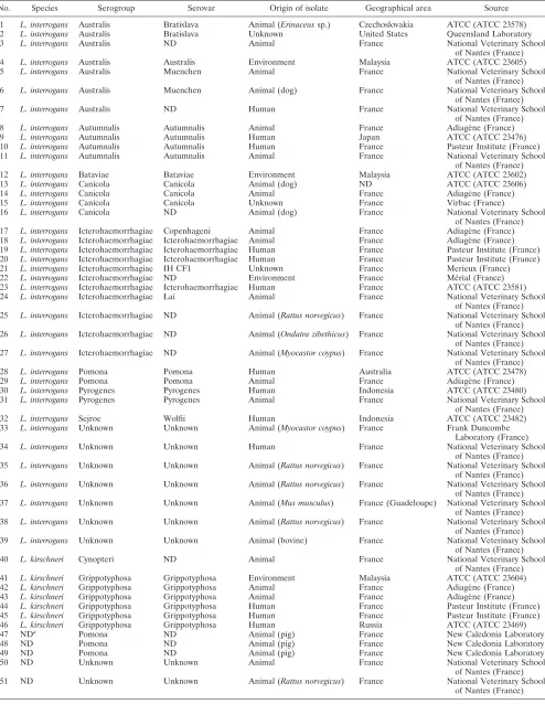
A total of 51 strains, including 11 reference strains, from different sources and geographical regions were analyzed in this study (Table 1). Species identification was performed by rRNA gene sequencing using LeptoA (5⬘-GGCGGCGCGTC TTAAACATG-3⬘) and LeptoB (5⬘-TTCCCCCCATTGAGCA AGATT-3⬘) nucleotide primers. In addition, all strains were checked for the presence of thehap1 fragment (262 bp) by amplification of pathogenicLeptospiraDNA with the specific primers described previously (2).
For MLSA, 14 housekeeping genes were selected in a prelim-inary study by comparison between twoLeptospiragenomic se-quences available in GenBank (http://www.ncbi.nlm.nih.gov): those ofL. interrogansserovar Copenhageni strain Fiocruz L1-130 chromosome I (accession number AE016823.1) andL. interrogans
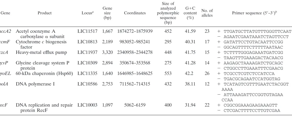
serovar Lai strain 56601 chromosome I (accession number AE010300.1). Since studies by Haake et al. revealed that outer membrane protein sequences have mosaic compositions consis-tent with horizontal transfer of DNA between related bacterial species (9), genes encoding surface proteins were excluded from the initial choice of gene targets. Primers were evaluated against the collection of reference strains. Based on the performance of primers and the number of alleles at a given locus, seven pairs of primers were selected. The nucleotide primers and the sizes of amplified DNA fragments from internal gene regions are shown in Table 2. PCRs were carried out using the following conditions: initial denaturation at 94°C for 5 min, followed by 45 cycles of 94°C for 30 s, 46°C (forrecF) or 50°C (for accA2,ccmF,czcA,
gcvP,groEL, andpolA) for 45 s, and 72°C for 60 s. The samples were maintained at 72°C for another 10 min. Both strands of each fragment were then sequenced.
Sequence data for the 51 study strains,L. interrogansserovar
* Corresponding author. Mailing address: Frank Duncombe Labo-ratory, Animal Health Department, 1 route de Rosel, 14053 Caen Cedex 4, France. Phone: 00 33 231 47 19 54. Fax: 00 33 231 47 19 00. E-mail: [email protected].
† Supplemental material for this article may be found at http://jcm .asm.org/.
䌤Published ahead of print on 2 December 2009.
581
on May 16, 2020 by guest
http://jcm.asm.org/
TABLE 1. Characteristics ofLeptospirastrains
No. Species Serogroup Serovar Origin of isolate Geographical area Source
1 L. interrogans Australis Bratislava Animal (Erinaceussp.) Czechoslovakia ATCC (ATCC 23578) 2 L. interrogans Australis Bratislava Unknown United States Queensland Laboratory 3 L. interrogans Australis ND Animal France National Veterinary School
of Nantes (France) 4 L. interrogans Australis Australis Environment Malaysia ATCC (ATCC 23605) 5 L. interrogans Australis Muenchen Animal France National Veterinary School
of Nantes (France) 6 L. interrogans Australis Muenchen Animal (dog) France National Veterinary School
of Nantes (France) 7 L. interrogans Australis ND Human France National Veterinary School
of Nantes (France) 8 L. interrogans Autumnalis Autumnalis Animal France Adiage`ne (France) 9 L. interrogans Autumnalis Autumnalis Human Japan ATCC (ATCC 23476) 10 L. interrogans Autumnalis Autumnalis Human France Pasteur Institute (France) 11 L. interrogans Autumnalis Autumnalis Animal France National Veterinary School
of Nantes (France) 12 L. interrogans Bataviae Bataviae Environment Malaysia ATCC (ATCC 23602) 13 L. interrogans Canicola Canicola Animal (dog) ND ATCC (ATCC 23606) 14 L. interrogans Canicola Canicola Animal France Adiage`ne (France) 15 L. interrogans Canicola Canicola Unknown France Virbac (France)
16 L. interrogans Canicola ND Animal (dog) France National Veterinary School of Nantes (France) 17 L. interrogans Icterohaemorrhagiae Copenhageni Animal France Adiage`ne (France) 18 L. interrogans Icterohaemorrhagiae Icterohaemorrhagiae Animal France Adiage`ne (France) 19 L. interrogans Icterohaemorrhagiae Icterohaemorrhagiae Human France Pasteur Institute (France) 20 L. interrogans Icterohaemorrhagiae Icterohaemorrhagiae Human France Pasteur Institute (France) 21 L. interrogans Icterohaemorrhagiae IH CF1 Unknown France Merieux (France) 22 L. interrogans Icterohaemorrhagiae ND Environment France Me´rial (France) 23 L. interrogans Icterohaemorrhagiae Icterohaemorrhagiae Human France ATCC (ATCC 23581) 24 L. interrogans Icterohaemorrhagiae Lai Animal France National Veterinary School
of Nantes (France) 25 L. interrogans Icterohaemorrhagiae ND Animal (Rattus norvegicus) France National Veterinary School
of Nantes (France) 26 L. interrogans Icterohaemorrhagiae ND Animal (Ondatra zibethicus) France National Veterinary School
of Nantes (France) 27 L. interrogans Icterohaemorrhagiae ND Animal (Myocastor coypus) France National Veterinary School
of Nantes (France) 28 L. interrogans Pomona Pomona Human Australia ATCC (ATCC 23478) 29 L. interrogans Pomona Pomona Animal France Adiage`ne (France) 30 L. interrogans Pyrogenes Pyrogenes Human Indonesia ATCC (ATCC 23480) 31 L. interrogans Pyrogenes Pyrogenes Animal France National Veterinary School
of Nantes (France) 32 L. interrogans Sejroe Wolfii Human Indonesia ATCC (ATCC 23482) 33 L. interrogans Unknown Unknown Animal (Myocastor coypus) France Frank Duncombe
Laboratory (France) 34 L. interrogans Unknown Unknown Human France National Veterinary School
of Nantes (France) 35 L. interrogans Unknown Unknown Animal (Rattus norvegicus) France National Veterinary School
of Nantes (France) 36 L. interrogans Unknown Unknown Animal (Rattus norvegicus) France National Veterinary School
of Nantes (France) 37 L. interrogans Unknown Unknown Animal (Mus musculus) France (Guadeloupe) National Veterinary School
of Nantes (France) 38 L. interrogans Unknown Unknown Animal (Rattus norvegicus) France National Veterinary School
of Nantes (France) 39 L. interrogans Unknown Unknown Animal (bovine) France National Veterinary School
of Nantes (France) 40 L. kirschneri Cynopteri ND Animal France National Veterinary School
of Nantes (France) 41 L. kirschneri Grippotyphosa Grippotyphosa Environment Malaysia ATCC (ATCC 23604) 42 L. kirschneri Grippotyphosa Grippotyphosa Animal France Adiage`ne (France) 43 L. kirschneri Grippotyphosa Grippotyphosa Animal France Adiage`ne (France) 44 L. kirschneri Grippotyphosa Grippotyphosa Human France Pasteur Institute (France) 45 L. kirschneri Grippotyphosa Grippotyphosa Human France Pasteur Institute (France) 46 L. kirschneri Grippotyphosa Grippotyphosa Human Russia ATCC (ATCC 23469)
47 NDa Pomona ND Animal (pig) France New Caledonia Laboratory
48 ND Pomona ND Animal (pig) France New Caledonia Laboratory
49 ND Pomona ND Animal (pig) France New Caledonia Laboratory
50 ND Unknown Unknown Animal France National Veterinary School
of Nantes (France) 51 ND Unknown Unknown Animal (Rattus norvegicus) France National Veterinary School
of Nantes (France)
a
ND, not determined.
on May 16, 2020 by guest
http://jcm.asm.org/
Copenhageni strain Fiocruz L1-130, andL. interrogansserovar Lai strain 56601 were used to construct a phylogenetic tree (Fig. 1). Sequenced DNA fragments were concatenated in the following order to generate a single 2,855-bp sequence:accA2,
ccmF,czcA,gcvP,groEL,polA, andrecF. Phylogenetic analysis was conducted with MEGA4 (21). The neighbor-joining method (18) was used for sequence comparison. The evolu-tionary distances were computed using the maximum compos-ite likelihood method (22).
16S RNA sequencing identified 39 strains of various sero-groups (Icterohaemorrhagiae, Australis, Autumnalis, Canic-ola, Pomona, Pyrogenes, and Bataviae) asL. interrogans geno-mospecies and 7 strains (serogroups Grippotyphosa and Cynopteri) as L. kirschneri genomospecies. For the five re-maining strains, the genomospecies could not be determined because of insufficient quantity of genomic material (Table 1). The seven loci selected were suitable for MLSA, as they could be amplified from all isolates and sequenced, irrespec-tive of the serogroup or species. Depending on the gene con-sidered, 12 to 26 alleles could be observed, with thegroELgene being the most discriminant.
Phylogenetic analysis clustered the sequences into two main clades. TheL. interrogansstrains grouped into one clade, and theL. kirschneristrains grouped into the other clade, except for one strain,L. kirschneriserogroup Grippotyphosa ATCC 23604, which was classified in theL. interrogansclade, in con-trast to the other reference strain, L. kirschneri serogroup Grippotyphosa ATCC 23469.
The major L. interrogans branch was further divided into several subgroups. A major subgroup comprised most Ictero-haemorrhagiae serogroup strains (n ⫽ 10), although it also included one Australis serogroup strain. Eight strains of un-known serogroups belonged to this subgroup. The three re-maining Icterohaemorrhagiae serogroup strains were placed into distant branches. Autumnalis (n⫽4), Canicola (n⫽4), and Pomona (n⫽5) serogroup strains were clustered into one group. The other subgroups were more heterogeneous,
com-prising Australis, Pyrogenes, and Bataviae serogroup strains (n⫽ 9). Individual gene analysis (see the figures in the sup-plemental material) showed that, in general, strains which clus-tered together in the multilocus analysis shared the same allele for each individual gene.
Noticeably, strain 17 (L. interrogansserogroup Icterohaem-orrhagiae serovar Copenhageni) was placed alone on a distinct branch. Individual gene analysis placed this strain in the Icterohaemorrhagiae clade, except for therecF allele, which was closely related to that of the Grippotyphosa strains (see the figures in the supplemental material). This finding may be explained by horizontal gene transfer events that are observed inLeptospiraand lead to mosaic compositions of sequences (9, 25). The positioning ofL. kirschneriserogroup Grippotyphosa ATCC 23604 in theL. interrogans clade was unexplained. In this case, clustering using individual gene analysis was similar to that using MLSA. To exclude the possibility of contamina-tion, we analyzed another sample of the isolate provided by the ATCC and found identical sequences. Possibly this strain was misidentified.
An MLSA technique forLeptospira species was developed previously by Ahmed et al. (1). Genotyping was based on DNA sequencing of six genes and allowed differentiation of the ma-jor species of Leptospira, L. interrogans, L. kirschneri, L. noguchii,L. santarosai, andL. borgpetersenii. The set of primers used permitted analysis of the genetic relationships among species and the respective evolution of the species. However, the clustering was limited to the species level. In contrast, the primers that we have used distinguished clusters at the sero-group level for the pathogenic species L. interrogansand L. kirschneri.
[image:3.585.45.541.82.280.2]Recently, an MLST schema based on the sequences of seven loci was used for typing ofLeptospiraisolates from humans and rodents in Thailand (23). Twelve sequence types (ST) were identified among the isolates. ST34 was predominant in human and rodent populations, representing 71 and 88% of isolates, respectively. In contrast, strains from a reference collection
TABLE 2. Details of gene loci and corresponding primer sequences used for MLSA
Gene Product Locusa
Gene size (bp)
Coordinates
Size of analyzed polymorphic
sequence (bp)
G⫹C content
(%) No. of
alleles Primer sequence (5⬘–3⬘) b
accA2 Acetyl coenzyme A LIC11517 1,667 1874272–1875939 452 41.59 23 ⫹ TTGATGCTTATGTTTGGGTTCAAT
carboxylase␣subunit ⫺ AGAATCGAATAAATCTAGTTCCT
ccmF Cytochromecbiogenesis LIC10813 2,189 983052–985241 295 40.31 17 ⫹ GATATTCCTGTACAATTCCGG
factor ⫺ GGCAGTTTTCTTTTTAATAAC
czcA Heavy-metal efflux pump LIC11937 3,320 2340958–2344278 448 41.75 15 ⫹ TCTTTTGGGAGAAATGATCGG ⫺ TAAGTTTGAAAGACTACAACG
gcvP Glycine cleavage system P LIC10309 2,894 350674–353568 275 41.28 14 ⫹ AAGAGCTAAAAGATCTGCAGC
protein ⫺ CTGGCCTTGAAATTTCGAACG
groEL 60-kDa chaperonin (Hsp60) LIC11335 1,640 1646985–1648625 553 42.2 26 ⫹ TCGCCTCGTCTCCATCCA ⫺ TGACGCAGAATCCATGGTAG
polA DNA polymerase I LIC10586 2,753 711562–714315 432 38.11 12 ⫹ TCATAGTCGTTTGAATCTACGGT AAAA
⫺ ATTAAAGATTCCGGTGTAGAA CCAA
recF DNA replication and repair LIC10003 1,097 5062–6159 400 31.94 22 ⫹ CGGCGGAAAGAAGAAAGTT
protein RecF ⫺ CTCGACTTTTCCTTGTCGAA
aL. interrogansserovar Copenhageni strain Fiocruz L1-130 has been used as a reference. b⫹, foward primer;⫺, reverse primer.
VOL. 48, 2010 NOTES 583
on May 16, 2020 by guest
http://jcm.asm.org/
had various STs. This study showed that ST34 may have a greater propensity than the other clones to cause human dis-eases in Thailand. Analysis of our sequences by the MLST method (15) showed a great diversity of STs and no clustering (data not shown). This result was not surprising, since our
lates.
In another study, Victoria et al. (24) showed that the S10-spc-␣ locus, which encodes ribosomal proteins and is highly conserved inL. interrogans, is a useful tool to elucidate evolu-tionary patterns. This analysis provided phylogenetic insights into the genusLeptospiraat the species level but only limited information on subspecies discrimination.
Development of various schemas for molecular classification of Leptospira will probably allow better characterization of subtypes ofLeptospirawith particular epidemicity or propen-sity to cause diseases in humans or animals. More strains in various ecological niches need to be tested to determine which loci among those proposed in this study and those by Ahmed et al. (1) and Thaipadungpanit et al. (23) are suitable for a definitive typing schema.
Nucleotide sequence accession numbers. Nucleotide se-quences determined in this study have been deposited in the GenBank data library under accession numbers GU113162 to GU113532.
This work received financial support from the Conseil Ge´ne´ral du Calvados, the Conseil Re´gional de Basse-Normandie, and the Associ-ation pour le De´veloppement de la Recherche Equine en France (ADREF).
We thank the team of Mathieu Picardeau for identification of strains by rrs gene sequencing, and Be´atrice Blanchard (Adiage`ne) and Denise Dessouter (New Caledonia Laboratory) for generous provision ofLeptospirastrains. This work was performed within the Competi-tiveness Cluster of the Horse Industry of Basse-Normandie.
REFERENCES
1.Ahmed, N., S. M. Devi, M. L. Valverde, P. Vijayachari, R. S. Machang’u, W. A. Ellis, and R. A. Hartskeerl.2006. Multilocus sequence typing method for identification and genotypic classification of pathogenicLeptospira spe-cies. Ann. Clin. Microbiol. Antimicrob.5:28.
2.Branger, C., B. Blanchard, C. Fillonneau, I. Suard, F. Aviat, B. Chevallier, and G. Andre-Fontaine.2005. Polymerase chain reaction assay specific for pathogenic Leptospira based on the genehap1encoding the hemolysis-associated protein-1. FEMS Microbiol. Lett.243:437–445.
3.Brenner, D. J., A. F. Kaufmann, K. R. Sulzer, A. G. Steigerwalt, F. C. Rogers, and R. S. Weyant.1999. Further determination of DNA relatedness between serogroups and serovars in the familyLeptospiraceaewith a proposal for
Leptospira alexanderisp. nov. and four newLeptospiragenomospecies. Int. J. Syst. Bacteriol.49:839–858.
4.Brown, P. D., and P. N. Levett.1997. Differentiation ofLeptospiraspecies and serovars by PCR-restriction endonuclease analysis, arbitrarily primed PCR and low-stringency PCR. J. Med. Microbiol.46:173–181.
5.Chang, H. W., Y. D. Nam, M. Y. Jung, K. H. Kim, S. W. Roh, M. S. Kim, C. O. Jeon, J. H. Yoon, and J. W. Bae. 2008. Statistical superiority of genome-probing microarrays as genomic DNA-DNA hybridization in reveal-ing the bacterial phylogenetic relationship compared to conventional meth-ods. J. Microbiol. Methods75:523–530.
6.Corney, B. G., J. Colley, and G. C. Graham.1997. Simplified analysis of pathogenic leptospiral serovars by random amplified polymorphic DNA fin-gerprinting. J. Med. Microbiol.46:927–932.
7.Galloway, R. L., and P. N. Levett.2008. Evaluation of a modified pulsed-field gel electrophoresis approach for the identification ofLeptospiraserovars. Am. J. Trop. Med. Hyg.78:628–632.
8.Gevers, D., F. M. Cohan, J. G. Lawrence, B. G. Spratt, T. Coenye, E. J. Feil, E. Stackebrandt, Y. Van de Peer, P. Vandamme, F. L. Thompson, and J. Swings.2005. Opinion: re-evaluating prokaryotic species. Nat. Rev. Micro-biol.3:733–739.
9.Haake, D. A., M. A. Suchard, M. M. Kelley, M. Dundoo, D. P. Alt, and R. L. Zuerner. 2004. Molecular evolution and mosaicism of leptospiral outer membrane proteins involves horizontal DNA transfer. J. Bacteriol.186:
2818–2828.
10.Honsa, E., T. Fricke, A. J. Stephens, D. Ko, F. Kong, G. L. Gilbert, F. Huygens, and P. M. Giffard.2008. Assignment ofStreptococcus agalactiae
isolates to clonal complexes using a small set of single nucleotide polymor-phisms. BMC Microbiol.8:140.
[image:4.585.43.286.45.562.2]11.Levesque, S., E. Frost, R. D. Arbeit, and S. Michaud.2008. Multilocus sequence typing ofCampylobacter jejuniisolates from humans, chickens, raw milk and environmental water in Quebec. J. Clin. Microbiol.46:3404–3411. 12.Levett, P. N.2001. Leptospirosis. Clin. Microbiol. Rev.14:296–326. FIG. 1. Phylogenetic tree constructed from concatenated
se-quences (2,855 bp) of seven housekeeping genes ofLeptospirastrains. The identification numbers of the strains, the serogroups, and the serovars (in parentheses) are indicated. Gri., Grippotyphosa; Ict., Ic-terohaemorrhagiae. The neighbor-joining method (18) was used for sequence comparison. Phylogenetic analysis was conducted with MEGA4 (21). The evolutionary distances were computed using the maximum composite likelihood method (22). Only bootstrap values greater than 70 are shown.
584
on May 16, 2020 by guest
http://jcm.asm.org/
13.Levett, P. N.2007. Sequence-based typing ofLeptospira: epidemiology in the genomic era. PLoS Negl. Trop. Dis.1:e120.
14.Levett, P. N., R. E. Morey, R. L. Galloway, and A. G. Steigerwalt.2006.
Leptospira broomiisp. nov., isolated from humans with leptospirosis. Int. J. Syst. Evol. Microbiol.56:671–673.
15.Maiden, M. C., J. A. Bygraves, E. Feil, G. Morelli, J. E. Russell, R. Urwin, Q. Zhang, J. Zhou, K. Zurth, D. A. Caugant, I. M. Feavers, M. Achtman, and B. G. Spratt.1998. Multilocus sequence typing: a portable approach to the identification of clones within populations of pathogenic microorganisms. Proc. Natl. Acad. Sci. U. S. A.95:3140–3145.
16.Paauw, A., M. P. Caspers, F. H. Schuren, M. A. Leverstein-van Hall, A. Deletoile, R. C. Montijn, J. Verhoef, and A. C. Fluit.2008. Genomic diversity within theEnterobacter cloacaecomplex. PLoS ONE3:e3018.
17.Ramadass, P., S. Meerarani, M. D. Venkatesha, A. Senthilkumar, and K. Nachimuthu. 1997. Characterization of leptospiral serovars by randomly amplified polymorphic DNA fingerprinting. Int. J. Syst. Bacteriol.47:575– 576.
18.Saitou, N., and M. Nei.1987. The neighbor-joining method: a new method for reconstructing phylogenetic trees. Mol. Biol. Evol.4:406–425. 19.Savio, M. L., C. Rossi, P. Fusi, S. Tagliabue, and M. L. Pacciarini.1994.
Detection and identification ofLeptospira interrogansserovars by PCR cou-pled with restriction endonuclease analysis of amplified DNA. J. Clin. Mi-crobiol.32:935–941.
20.Stackebrandt, E., W. Frederiksen, G. M. Garrity, P. A. Grimont, P. Ka-mpfer, M. C. Maiden, X. Nesme, R. Rossello-Mora, J. Swings, H. G. Truper, L. Vauterin, A. C. Ward, and W. B. Whitman.2002. Report of the ad hoc committee for the re-evaluation of the species definition in bacteriology. Int. J. Syst. Evol. Microbiol.52:1043–1047.
21.Tamura, K., J. Dudley, M. Nei, and S. Kumar.2007. MEGA4: Molecular Evolutionary Genetics Analysis (MEGA) software version 4.0. Mol. Biol. Evol.24:1596–1599.
22.Tamura, K., M. Nei, and S. Kumar.2004. Prospects for inferring very large phylogenies by using the neighbor-joining method. Proc. Natl. Acad. Sci. U. S. A.101:11030–11035.
23.Thaipadungpanit, J., V. Wuthiekanun, W. Chierakul, L. D. Smythe, W. Petkanchanapong, R. Limpaiboon, A. Apiwatanaporn, A. T. Slack, Y. Su-puttamongkol, N. J. White, E. J. Feil, N. P. Day, and S. J. Peacock.2007. A dominant clone ofLeptospira interrogansassociated with an outbreak of human leptospirosis in Thailand. PLoS. Negl. Trop. Dis.1:e56.
24.Victoria, B., A. Ahmed, R. L. Zuerner, N. Ahmed, D. M. Bulach, J. Quinteiro, and R. A. Hartskeerl.2008. Conservation of theS10-spc-alphalocus within otherwise highly plastic genomes provides phylogenetic insight into the ge-nusLeptospira. PloS ONE3:e2752.
25.Xue, F., J. Yan, and M. Picardeau.2009. Evolution and pathogenesis of
Leptospiraspp.: lessons learned from the genomes. Microbes Infect.11:328– 333.
VOL. 48, 2010 NOTES 585