0095-1137/10/$12.00 doi:10.1128/JCM.00289-10
Copyright © 2010, American Society for Microbiology. All Rights Reserved.
Systematic Internal Transcribed Spacer Sequence Analysis for
Identification of Clinical Mold Isolates in Diagnostic
Mycology: a 5-Year Study
䌤
†
Diana E. Ciardo,¶ Katja Lucke, Alex Imhof,‡ Guido V. Bloemberg, and Erik C. Bo
¨ttger*
Institut fu¨r Medizinische Mikrobiologie, Universita¨t Zu¨rich, Zu¨rich, Switzerland
Received 12 February 2010/Returned for modification 26 March 2010/Accepted 14 June 2010
The implementation of internal transcribed spacer (ITS) sequencing for routine identification of molds in
the diagnostic mycology laboratory was analyzed in a 5-year study. All mold isolates (nⴝ6,900) recovered in
our laboratory from 2005 to 2009 were included in this study. According to a defined work flow, which in addition to troublesome phenotypic identification takes clinical relevance into account, 233 isolates were subjected to ITS sequence analysis. Sequencing resulted in successful identification for 78.6% of the analyzed isolates (57.1% at species level, 21.5% at genus level). In comparison, extended in-depth phenotypic charac-terization of the isolates subjected to sequencing achieved taxonomic assignment for 47.6% of these, with a mere 13.3% at species level. Optimization of DNA extraction further improved the efficacy of molecular identification. This study is the first of its kind to testify to the systematic implementation of sequence-based identification procedures in the routine workup of mold isolates in the diagnostic mycology laboratory.
The diagnostic mycology laboratory is confronted with an increasing number of infections caused by an expanding spec-trum of fungal species (9, 18, 19, 23, 28). An important reason for this development is the progress in medicine that results in prolonged life expectancy for patients with a compromised immune system, many of whom are particularly vulnerable to fungal infections.
Conventional identification of fungi is mainly based on phe-notypic characteristics (11, 35). Micro- and macromorpholo-gies have to be carefully evaluated for correct species assign-ment, requiring a great deal of expertise because of excessive morphological variability. In addition, some isolates do not form their typical structural characteristics required for iden-tification under laboratory conditions (11). As a result, the diagnostic laboratory is often confronted with misidentifica-tions and/or lack of identification. Genetic methods for iden-tification and taxonomic classification of molds have been es-tablished in recent years (6, 38). In particular, the internal transcribed spacer (ITS) regions located between the 18S and 28S rRNA genes and including the ITS1 and ITS2 regions and the 5.8S rRNA gene have emerged as the most common target for molecularly based identification (4, 16, 37). The ITS re-gions occur in multiple copies in the fungal genome, and the number of ITS sequences available in public databases (such as GenBank, NCBI) is expanding continuously (31). Unfortu-nately, the quality of sequence entries is variable and
taxo-nomic assignments in public databases are in part unreliable, hampering their use for identification (5, 27).
For a cost- and time-effective application of molecular iden-tification procedures, we have previously defined selection cri-teria and assembled these in an algorithm. According to these criteria, mold isolates of clinical relevance that cannot readily be identified by standard conventional characteristics are sub-jected to ITS sequence-based identification (7). Analysis of the ITS regions is the most commonly used molecular method for identification of molds (3, 4, 29). Studies reporting on the use of molecularly based identification procedures in diagnostic mycology have mainly focused on method development (12, 25, 30, 40) and include anecdotal case reports (15, 17, 20). There are few studies addressing the use of systematic ITS sequencing implemented as a routine tool according to a de-fined work flow and its impact on diagnostic performance in a medical mycology laboratory (21). Here, we report on the results of a 5-year study on systematic implementation of ITS sequencing for identification of clinical mold isolates in the diagnostic laboratory.
MATERIALS AND METHODS
Conventional identification.Clinical specimens were cultured on general my-cology media (Sabouraud dextrose agar containing gentamicin and chloram-phenicol and brain heart infusion agar; Becton Dickinson AG, Allschwil, Swit-zerland) and on fungal selective media (Chromagar and Mycosel; Becton Dickinson AG) at 25°C for a maximum of 3 weeks and regularly examined for growth. Subcultures for identification were done as follows: (i)Aspergillusspp. on malt yeast agar (11) at 25°C, 35°C, and 42°C; (ii)Mucoraleson potato carrot agar (9) at 25°C, 37°C, 40°C, 45°C, 50°C, and 56°C; (iii) all other molds on Sabouraud dextrose agar containing gentamicin and chloramphenicol at 25°C and 35°C and on Mycosel and potato carrot agar, both at 25°C. Phenotypic identification was based on macro- and micromorphological criteria, as described in reference 11.
DNA extraction.Mold isolates were subjected to DNA extraction using the InstaGene matrix (Bio-Rad, Reinach BL, Switzerland), as described previously (7). For DNA extraction, a fungal mycelium with a surface area of 2 to 4 cm2was obtained from Mycosel or Sabouraud dextrose agar plates. In case of PCR failure with InstaGene extract as the template, a freshly collected fungal mycelium was digested with lyticase (until 27 September 2007) or proteinase K (following 27
* Corresponding author. Mailing address: Institut fu¨r Medizinische Mikrobiologie, Universita¨t Zu¨rich, Gloriastr. 30/32, 8006 Zu¨rich, Swit-zerland. Phone: 41 44 634 2703. Fax: 41 44 634 4906. E-mail: boettger @imm.uzh.ch.
† Supplemental material for this article may be found at http://jcm .asm.org/.
¶ Present address: Viollier AG, Basel, Switzerland.
‡ Present address: Medizinische Klinik, Spital Langenthal, Lan-genthal, Switzerland.
䌤Published ahead of print on 23 June 2010.
2809
on May 16, 2020 by guest
http://jcm.asm.org/
September 2007). For lyticase digestion, the mycelium was incubated at 37°C for 2 h with 30 U of lyticase (Sigma-Aldrich Chemie GmbH, Schnelldorf, Germany) in 200l digestion buffer (50 mM Tris-HCl, 1 mM EDTA, pH 8.0), followed by alkaline lysis with 10l 1 M NaOH and 10l 10% SDS, incubation for 10 min at 95°C, and neutralization with 10l 1 M HCl. For proteinase K digestion, the mycelium was vortexed for 3 min at the fastest setting with 80g proteinase K (recombinant PCR grade; Roche, Rotkreuz, Switzerland) and 50 mg glass beads (acid washed,⬍106m; Sigma-Aldrich Chemie GmbH, Buchs, Switzerland) in 200l digestion buffer (50 mM Tris-HCl, 1 mM EDTA [pH 8.0], 0.5% SDS), incubated at 56°C for 3 h, and vortexed again for 3 min at the fastest setting. DNA was extracted and purified using the QIAamp DNA blood minikit (Qiagen, Hombrechtikon, Switzerland) according to the instructions of the manufacturer and eluted in 100l elution buffer.
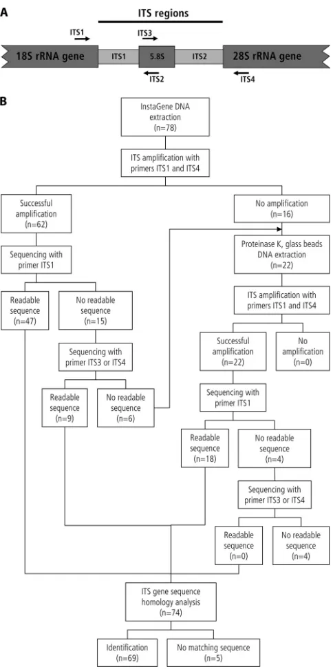
Amplification and sequencing.PCR of the ITS regions was performed as described before using the LightCycler FastStart DNA Master SYBR green I kit (LightCycler reagents; Roche, Rotkreuz, Switzerland) and a PerkinElmer GeneAmp PCR System 9600 or 9700 (Applied Biosystems, Rotkreuz, Switzer-land) (7). The amplified ITS regions were sequenced with primer ITS1 or, in case of failure, with primer ITS4, ITS3, or ITS2 (37) (Fig. 1) using the BigDye kit (Applied Biosystems, Rotkreuz, Switzerland) and an automated DNA sequencer (ABI Prism 3100-Avant genetic analyzer; Applied Biosystems). When the ITS1 and ITS2 regions were separately sequenced, a consensus sequence was created for homology analysis.
Sequence analysis.Sequences obtained (covering⬎90% of the ITS regions) were analyzed for homology using GenBank (NCBI) and the SmartGene ITS database (ITS validated database; SmartGene IDNS, Zug, Switzerland) in par-allel. The SmartGene ITS database is a curated database which is designed to include most of the clinically relevant fungi and which contains some ITS se-quences that are missing in GenBank. Sequence assignment to species and genus level was done according to guidelines published previously (7). A sequence was assigned to a species if the best matching reference sequence showedⱖ98% homology and the next best matching reference species showed at least 0.8% less sequence homology. A sequence was assigned to genus level on the basis of 95 to 98% homology to the best matching sequence or ofⱖ98% homology with sequence entries for several species from the same genus. “No identification” was defined as⬍95% homology with the best matching reference sequence or as sequence homology of⬎95% with various genera present (7).
Prospective study.Over the study period of 5 years (from January 2005 to October 2009), clinical mold isolates were subjected to phenotypic and sequence-based identification measures following a defined work flow (7). Isolates for which (i) phenotypic identification by conventional characteristics did not result in species assignment within 5 days following primary subcultivation and (ii) which were judged to be clinically relevant were analyzed by ITS sequencing. Clinical relevance was defined as follows: (i) isolates from primarily sterile specimen sites growing at 35°C, (ii) isolates from nonsterile specimen sites with direct microscopy positive for hyphae, (iii) isolates from nonsterile specimen sites growing at 35°C and clinical evidence of infection, (iv) isolates from der-matological samples positive for hyphae in direct microscopy and growing at 35°C (dermatophytes were excluded from the study). In addition to being se-quenced, the isolates included in this study were subjected to prolonged culti-vations to allow for forming morphological characteristics and to facilitate tax-onomic assignment based on phenotypic traits.
RESULTS
From January 2005 to October 2009, a total of 6,900 mold isolates were recovered in the diagnostic laboratory. These molds were found to represent 136 different species belonging to 81 different genera (see Table S1 in the supplemental ma-terial). Seven hundred twenty-six isolates could not be readily identified by standard phenotypic characteristics. Two hundred thirty-three (32.1%) of these isolates were categorized as po-tentially clinically relevant and subjected to ITS analysis. Se-quences determined were analyzed using two databases: the GenBank database and the SmartGene validated ITS data-base.
During the 5-year study period, the number of sequenced isolates decreased from 88 in 2005 to 20 in the first 10 months of 2009 (data not shown). Using the two databases combined,
[image:2.585.301.543.65.551.2]183 (78.6%) of the 233 isolates subjected to molecular identi-fication were successfully identified by ITS sequence analysis (Table 1). Most of these isolates (57.1%) were assigned to species level. Genus level assignment was achieved for 21.5%; inability to differentiate at species level for these isolates was mostly due to high interspecies homology of the genera in-volved. Use of the SmartGene validated ITS database resulted in a higher percentage of identification at species level than
FIG. 1. Flow chart for DNA extraction and ITS sequencing of molds as implemented from 27 September 2007 onwards. (A) Sche-matic representation of the fungal ITS regions, consisting of ITS1, the 5.8 rRNA gene, and ITS2. Positions and directions of primers ITS1, ITS2, ITS3, and ITS4 (37) are indicated with arrows. (B) Flow chart of DNA extraction and ITS sequencing. n, number of isolates.
on May 16, 2020 by guest
http://jcm.asm.org/
that obtained using the GenBank database (Table 1). Pro-longed cultivation and morphological characterization of the 233 isolates enabled taxonomic assignment in 47.6% of the cases, with 13.3% at species level and 34.3% at genus level (Table 1). Successful identification by ITS sequencing in-creased from 77.3% in 2005 to 95.0% in 2009. This was asso-ciated with an increase in species assignment from 50.0% in 2005 to 80.0% in 2009 (data not shown). The isolates subjected to sequence analysis were assigned to 87 species belonging to 55 different genera (see Table S1 in the supplemental mate-rial), covering more than one-half of the spectrum of molds encountered in our laboratory.
During the study period, 36 (15.4%) of the 233 isolates could not be identified by sequencing because matching ITS refer-ence sequrefer-ences for these were lacking in the databases (Table 1). By phenotypic methods, 5 of these 36 isolates could be
assigned toAcremoniumspp.,Alternariaspp.,Exophialaspp.,
Paecilomyces variotii, andScytalidiumspp. ITS sequence anal-ysis with no taxonomic assignment decreased from 18.2% in 2005 to 0.0% in 2009.
In the 5-year study period, 14 (6.0%) of the 233 isolates failed to yield an ITS amplicon (Table 1). On the basis of prolonged phenotypic identification procedures, 4 of these 14
isolates were identified as Aspergillus fumigatus, Microascus
cinereus, Onychocola canadensis, andPhialophora richardsiae. Genus level assignment was achieved for 3 of the 14 isolates as
Aspergillusspp.,Paecilomyces spp., andSyncephalastrum spp. Seven of the 14 isolates could not be assigned at any taxonomic level.
An effort was made to improve DNA extraction when the initial InstaGene extraction was not successful. For efficient
DNA release from fungal cells, digestion with lyticase was replaced by proteinase K digestion combined with mechanical rupture of the cells by glass beads. Following implementation of this modification (from 27 September 2007 onwards), 82% of the isolates for which the initial InstaGene extraction failed to yield an amplicon were successfully amplified and se-quenced (Fig. 1). The percentage of isolates for which ITS amplification failed was reduced from 12% in 2006 to 5% in 2008 to 2009.
DISCUSSION
From January 2005 to October 2009, a wide spectrum of fungal species were isolated in our diagnostic laboratory (see Table S1 in the supplemental material). This corroborates re-cent reports testifying to the growing number of potentially pathogenic fungal species (9, 10, 13, 14, 26, 36). Consequently, each laboratory isolate has to be evaluated carefully with re-gard to its pathogenic potential as well as its clinical relevance. The diagnostic laboratory initially has often limited or no ac-cess to clinical data on patients, which restricts available infor-mation mainly to the type of sample material, results of direct microscopy observation, and isolates’ growth conditions. Iso-lates recovered from nonsterile sites may readily be catego-rized as contaminants or as common colonizers if essential pathogenicity characteristics are lacking, such as growth at 37°C (11). Identification by phenotypic traits is often challeng-ing, since this requires growth on selected media at various temperatures and expertise in morphological characterization. For identification of clinically relevant fungi, molecular tech-niques based on sequencing of specific target regions are valu-able alternatives to traditional phenotypic identification pro-cedures, since sequence-based identification is independent of growth conditions and the formation of specific morphological structures (4, 6, 8).
Together with previously defined specific criteria along a diagnostic work flow that considers observations made in the laboratory as well as clinical information provided by the phy-sician on request, ITS sequencing proved a reliable tool to enhance the rate of identification of clinically relevant fungi. Within the study period, 726 isolates could not be identified by phenotypic characteristics, of which 233 (32.1%) were selected on the basis of clinical relevance for further molecular identi-fication (see Table S1 in the supplemental material). Sequence analysis of the ITS regions clearly improved identification in comparison with traditional morphology-based identification, assigning 78.6% of the analyzed 233 isolates to a defined taxon (Table 1). Identification at species level was possible for 57.1% of the sequenced isolates, and 21.5% were assigned to genus level. In comparison, further phenotypic characterization based on prolonged cultivations resulted in species assignment for 13.3% and in genus assignment for 34.3% of these isolates (Table 1). In comparison with GenBank a higher percentage of strains could be assigned to species level by using the Smart-Gene validated ITS database.
[image:3.585.41.284.105.317.2]The data collected in the 5-year study period revealed a substantial increase in species identification by ITS sequence analysis from 50.0% in 2005 to 80.0% in 2009. Genus level assignment remained constant at around 20%, and the isolates which could not be assigned to a given taxon decreased from
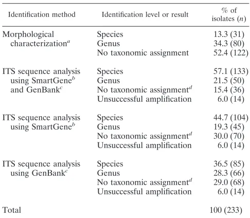
TABLE 1. Identification of fungal isolates selected for ITS sequence analysis based on defined criteria according
a diagnostic work flow from January 2005 to October 2009
Identification method Identification level or result % of isolates (n)
Morphological characterizationa
Species 13.3 (31)
Genus 34.3 (80)
No taxonomic assignment 52.4 (122)
ITS sequence analysis using SmartGeneb and GenBankc
Species 57.1 (133)
Genus 21.5 (50)
No taxonomic assignmentd 15.4 (36) Unsuccessful amplification 6.0 (14)
ITS sequence analysis using SmartGeneb
Species 44.7 (104)
Genus 19.3 (45)
No taxonomic assignmentd 30.0 (70) Unsuccessful amplification 6.0 (14)
ITS sequence analysis using GenBankc
Species 36.5 (85)
Genus 28.3 (66)
No taxonomic assignmentd 29.0 (68) Unsuccessful amplification 6.0 (14)
Total 100 (233)
a
Taxonomic assignment based on extended in-depth phenotypic identification measures.
b
SmartGene, homology analysis using the SmartGene ITS database.
c
GenBank, homology analysis using the NCBI database.
d
No identification due to the lack of homologous sequences.
on May 16, 2020 by guest
http://jcm.asm.org/
18.2% to 0.0% in 2009. While this may reflect in part the continuous updating and enlargement of databases (27), we also note that a strict adherence to clinical relevance as a selection criterion for ITS sequence analysis and increased diagnostic expertise gained during the 5-year study period re-duced the number of environmental isolates selected for se-quencing and thus the total number of isolates subjected to sequencing. In contrast to established pathogenic species, en-vironmental (and presumably nonpathogenic) isolates form a frequent source for ITS sequences which cannot be assigned to established taxa, since no corresponding sequence entries are available (7).
A systematic analysis comparing phenotypic identification and molecular identification revealed that some molds are particularly difficult to identify by phenotypic traits (see Table S1 in the supplemental material). Frequently isolated
patho-genic molds such asBeauveria spp.,Cladosporiumspp.,
Peni-cilliumspp. (at genus level),Paecilomyces lilacinus, Scedospo-rium apiospermum, Scopulariopsis brevicaulis, and Aspergillus
spp. (at species level) are readily identified on the basis of morphological characteristics. In contrast, reliable
identifica-tion of rarely encountered pathogens such as Aureobasidium
pullulans, Fusarium oxysporum, Fusarium solani, Penicillium marneffei, Phoma glomerata, Schizophyllum commune, and
Scytalidium dimidiatumwas possible only by sequence analysis (see Table S1 in the supplemental material).
Efficient extraction of high-quality DNA is crucial for PCR amplification and sequence analysis. The use of sufficient fun-gal starting material is essential, since the amount of DNA per mycelial mass may vary in different growth stages. In most
cases, the use of 2 to 4 cm2 of mycelium taken from solid
medium was found to be sufficient for successful DNA extrac-tion. A wide spectrum of fungal species, which may show vari-ations in cell wall composition and in genomic ITS copies, hampers the implementation of a universal DNA extraction method (2, 22, 33). In response, we applied a stepwise DNA extraction procedure using two different protocols (Fig. 1). Amplification of the ITS regions of some fungal isolates failed despite several attempts. This may reflect unsuccessful DNA extraction due to (i) low levels of DNA in the mycelial mass or (ii) inefficient disruption of the cell wall, possibly due to un-known modifications of cell wall composition. Alternatively, the PCR may have been inhibited by fungal components re-leased during cell disruption. Few of the isolates for which amplification of the ITS regions was not successful were iden-tified by further phenotypic investigations, indicating that these isolates belonged to various genera. Several isolates remained unidentified, since morphological characteristics did not form despite prolonged incubation. Interestingly, one out of the nine Aspergillus fumigatus isolates did not yield a successful amplification of the ITS regions (see Table S1 in the supple-mental material). It was noted that this isolate showed only sparse sporulation after repeated UV induction. Presumably, the amount of extracted DNA obtained from the mycelium was not sufficient for proper gene amplification.
A literature survey indicates that this study is the first and most exhaustive report on systematic implementation of ITS sequencing for the purpose of mold identification in a diag-nostic mycology laboratory. We defined a set of criteria and a diagnostic work flow for subjecting isolates to molecular
iden-tification. In comparison, available studies mainly reported on nonsystematic approaches analyzing smaller sets of isolates to address a more limited question (1, 34, 39). The initial pheno-typic part of our identification strategy including standard mor-phological characteristics can be readily adapted to the individual laboratory expertise or could be complemented by matrix-assisted laser desorption ionization–time of flight (MALDI-TOF) mass spectrometry in the near future (24, 32). ITS sequence analysis is a valuable diagnostic tool in the med-ical mycology laboratory, with a high percentage of species identification for molds which are difficult to identify by stan-dard phenotypic characteristics. The number of isolates sub-jected to sequence analysis can be reduced significantly (in this study from 726 to 233 isolates) based on defined selection criteria, resulting in cost reduction without loss of clinical di-agnostic quality. In conclusion, systematic and efficient combi-nation of phenotypic and molecular procedures substantially improves identification of mold isolates in the diagnostic lab-oratory.
ACKNOWLEDGMENTS
We thank the technicians in the mycology and molecular diagnostic laboratories for their invaluable help and Michael Hombach for crit-ical reading of the manuscript.
This study was supported in part by the University of Zu¨rich.
REFERENCES
1.Bagyalakshmi, R., K. L. Therese, S. Prasanna, and H. N. Madhavan.2008. Newer emerging pathogens of ocular non-sporulating molds (NSM) identi-fied by polymerase chain reaction (PCR)-based DNA sequencing technique targeting internal transcribed spacer (ITS) region. Curr. Eye Res.33:139– 147.
2.Bainbridge, B. W., C. L. Spreadbury, F. G. Scalise, and J. Cohen.1990. Improved methods for the preparation of high molecular weight DNA from large and small scale cultures of filamentous fungi. FEMS Microbiol. Lett.
54:113–117.
3.Balajee, S. A., L. Sigler, and M. E. Brandt.2007. DNA and the classical way: identification of medically important molds in the 21st century. Med. Mycol.
45:475–490.
4.Balajee, S. A., A. M. Borman, M. E. Brandt, J. Cano, M. Cuenca-Estrella, E. Dannaoui, J. Guarro, G. Haase, C. C. Kibbler, W. Meyer, K. O’Donnell, C. A. Petti, J. L. Rodriguez-Tudela, D. Sutton, A. Velegraki, and B. L. Wickes.2009. Sequence-based identification ofAspergillus,Fusarium, and
Mucoralesspecies in the clinical mycology laboratory: where are we and where should we go from here? J. Clin. Microbiol.47:877–884.
5.Bridge, P. D., P. J. Roberts, B. M. Spooner, and G. Panchal.2003. On the unreliability of published DNA sequences. New Phytol.160:43–48. 6.Chen, S. C., C. L. Halliday, and W. Meyer.2002. A review of nucleic
acid-based diagnostic tests for systemic mycoses with an emphasis on poly-merase chain reaction-based assays. Med. Mycol.40:333–357.
7.Ciardo, D. E., G. Scha¨r, M. Altwegg, E. C. Bo¨ttger, and P. P. Bosshard.2007. Identification of moulds in the diagnostic laboratory—an algorithm imple-menting molecular and phenotypic methods. Diagn. Microbiol. Infect. Dis.
59:49–60.
8.Ciardo, D. E., G. Scha¨r, E. C. Bo¨ttger, M. Altwegg, and P. P. Bosshard.2006. Internal transcribed spacer sequencing versus biochemical profiling for iden-tification of medically important yeasts. J. Clin. Microbiol.44:77–84. 9.Cornely, O.2008. Aspergillus to Zygomycetes: causes, risk factors,
preven-tion, and treatment of invasive fungal infections. Infection36:296–313. 10.Cristofaro, P., and M. D. Mileno.2006. Penicillium marneffei infection in
HIV-infected travelers. AIDS Alert21:140–142.
11.de Hoog, G. S., J. Guarro, J. Gene´, and M. J. Figueras.2000. Atlas of clinical fungi, 2nd ed. Centraalbureau voor Schimmelcultures, Utrecht, Netherlands, and Universitat Rovira i Virgili, Reus, Spain.
12.Etienne, K. A., R. Kano, and S. A. Balajee.2009. Development and valida-tion of a microsphere-based Luminex assay for rapid identificavalida-tion of clini-cally relevant aspergilli. J. Clin. Microbiol.47:1096–1100.
13.Fishman, J. A.2002. Overview: fungal infections in the transplant patient. Transpl. Infect. Dis.4(Suppl. 3):3–11.
14.Golan, Y.2005. Overview of transplant mycology. Am. J. Health Syst. Pharm.
62(Suppl. 1):17–21.
15.Goldani, L., V. Aquino, L. Lunardi, V. Cunha, and R. Santos.2009. Two specific strains ofHistoplasma capsulatumcausing mucocutaneous
on May 16, 2020 by guest
http://jcm.asm.org/
tations of Histoplasmosis: preliminary analysis of a frequent manifestation of Histoplasmosis in southern Brazil. Mycopathologia167:181–186. 16.Hinrikson, H. P., S. F. Hurst, T. J. Lott, D. W. Warnock, and C. J. Morrison.
2005. Assessment of ribosomal large-subunit D1-D2, internal transcribed spacer 1, and internal transcribed spacer 2 regions as targets for molecular identification of medically importantAspergillusspecies. J. Clin. Microbiol.
43:2092–2103.
17.Hipolito, E., E. Faria, A. Alves, G. de Hoog, J. Anjos, T. Gonc¸alves, P. Morais, and H. Esteva˜o.2009.Alternaria infectoriabrain abscess in a child with chronic granulomatous disease. Eur. J. Clin. Microbiol. Infect. Dis.
28:377–380.
18.Kauffman, C. A.2001. Fungal infections in older adults. Clin. Infect. Dis.
33:550–555.
19.Kaufman, D.2004. Fungal infection in the very low birthweight infant. Curr. Opin. Infect. Dis.17:253–259.
20.Li, D. M., D. R. Xiu, R. Y. Li, R. A. Samson, G. S. de Hoog, and D. L. Wang.
2008.Aspergillus flavusmyositis in a patient after liver transplantation. Clin. Transplant.22:508–511.
21.Linton, C. J., A. M. Borman, G. Cheung, A. D. Holmes, A. Szekely, M. D. Palmer, P. D. Bridge, C. K. Campbell, and E. M. Johnson.2007. Molecular identification of unusual pathogenic yeast isolates by large ribosomal subunit gene sequencing: 2 years of experience at the United Kingdom mycology reference laboratory. J. Clin. Microbiol.45:1152–1158.
22.Loeffler, J., H. Hebart, U. Schumacher, H. Reitze, and H. Einsele.1997. Comparison of different methods for extraction of DNA of fungal pathogens from cultures and blood. J. Clin. Microbiol.35:3311–3312.
23.Maertens, J., M. Vrebos, and M. Boogaerts.2001. Assessing risk factors for systemic fungal infections. Eur. J. Cancer Care (Engl.).10:56–62. 24.Marklein, G., M. Josten, U. Klanke, E. Muller, R. Horre, T. Maier, T.
Wenzel, M. Kostrzewa, G. Bierbaum, A. Hoerauf, and H. G. Sahl.2009. Matrix-assisted laser desorption ionization–time of flight mass spectrometry for fast and reliable identification of clinical yeast isolates. J. Clin. Microbiol.
47:2912–2917.
25.Martínez, J. M. G., E. V. Go´mez, J. Pema´n, E. Canto´n, M. G. García, and L. del Castillo Agudo. 20 October 2009. Identification of pathogenic yeast species by polymerase chain reaction amplification of the RPS0 gene intron fragment. J. Appl. Microbiol. [Epub ahead of print.] doi:10.1111/j.1365-2672.2009.04595.x.
26.Naggie, S., and J. R. Perfect.2009. Molds: hyalohyphomycosis, phaeohypho-mycosis, and zygomycosis. Clin. Chest Med.30:337–353.
27.Nilsson, R. H., M. Ryberg, E. Kristiansson, K. Abarenkov, K. H. Larsson, and U. Ko˜ljalg.2006. Taxonomic reliability of DNA sequences in public sequence databases: a fungal perspective. PLoS One1:e59.
28.Patterson, J. E., J. Peters, J. H. Calhoon, S. Levine, A. Anzueto, H.
Al-Abdely, R. Sanchez, T. F. Patterson, M. Rech, J. H. Jorgensen, M. G. Rinaldi, E. Sako, S. Johnson, V. Speeg, G. A. Halff, and J. K. Trinkle.2000. Investigation and control of aspergillosis and other filamentous fungal in-fections in solid organ transplant recipients. Transpl. Infect. Dis.2:22–28. 29.Rakeman. J. L., U. Bui, K. Lafe, Y. C. Chen, R. J. Honeycutt, and B. T.
Cookson. 2005. Multilocus DNA sequence comparisons rapidly identify pathogenic molds. J. Clin. Microbiol.43:3324–3333.
30.Reich, M., A. Kohler, F. Martin, and M. Buee.2009. Development and validation of an oligonucleotide microarray to characterise ectomycorrhizal fungal communities. BMC Microbiol.9:241.
31.Ryberg, M., E. Kristiansson, E. Sjo¨kvist, and R. H. Nilsson. 2009. An outlook on the fungal internal transcribed spacer sequences in GenBank and the introduction of a web-based tool for the exploration of fungal diversity. New Phytologist181:471–477.
32.Seyfarth, F., M. Ziemer, H. G. Sayer, A. Burmester, M. Erhard, M. Welker, S. Schliemann, E. Straube, and U.-C. Hipler.2008. The use of ITS DNA sequence analysis and MALDI-TOF mass spectrometry in diagnosing an infection withFusarium proliferatum. Exp. Dermatol.17:965–971. 33.van Burik, J. A., R. W. Schreckhise, T. C. White, R. A. Bowden, and D.
Myerson.1998. Comparison of six extraction techniques for isolation of DNA from filamentous fungi. Med. Mycol.36:299–303.
34.Vinh, D. C., Y. R. Shea, J. A. Sugui, E. R. Parrilla-Castellar, A. F. Freeman, J. W. Campbell, S. Pittaluga, P. A. Jones, A. Zelazny, D. Kleiner, K. J. Kwon-Chung, and S. M. Holland.2009. Invasive aspergillosis due to Neo-sartorya udagawae. Clin. Infect. Dis.49:102–111.
35.von Arx, J. A.1981. The genera of fungi sporulating in pure culture, 3rd ed. J. Cramer, Vaduz, Liechtenstein.
36.Walsh, T. J., A. Groll, J. Hiemenz, R. Fleming, E. Roilides, and E. Anaissie.
2004. Infections due to emerging and uncommon medically important fungal pathogens. Clin. Microbiol. Infect.10(Suppl. 1):48–66.
37.White, T. J., T. Bruns, S. Lee, and J. Taylor.1990. Amplification and direct sequencing of fungal ribosomal RNA genes for phylogenetics, p. 315–322.In
M. A. Innis, D. H. Gelfand, J. J. Sninsky, and T. J. White (ed.), PCR protocols: a guide to methods and applications. Academic Press Inc., San Diego, CA.
38.Yeo, S. F., and B. Wong.2002. Current status of nonculture methods for diagnosis of invasive fungal infections. Clin. Microbiol. Rev.15:465–484. 39.Zeng, J. S., D. A. Sutton, A. W. Fothergill, M. G. Rinaldi, M. J. Harrak, and
G. S. de Hoog.2007. Spectrum of clinically relevantExophialaspecies in the United States. J. Clin. Microbiol.45:3713–3720.
40.Zeng, X., F. Kong, C. Halliday, S. Chen, A. Lau, G. Playford, and T. C. Sorrell. 2007. Reverse line blot hybridization assay for identification of medically important fungi from culture and clinical specimens. J. Clin. Mi-crobiol.45:2872–2880.