R E S E A R C H
Open Access
RNA methylomes reveal the m
6
A-mediated
regulation of DNA demethylase gene
SlDML2
in tomato fruit ripening
Leilei Zhou
1,2, Shiping Tian
1,2*and Guozheng Qin
1*Abstract
Background:Methylation of nucleotides, notably in the forms of 5-methylcytosine (5mC) in DNA and N6
-methyladenosine (m6A) in mRNA, carries important information for gene regulation. 5mC has been elucidated to participate in the regulation of fruit ripening, whereas the function of m6A in this process and the
interplay between 5mC and m6A remain uncharacterized.
Results: Here, we show that mRNA m6A methylation exhibits dynamic changes similar to DNA methylation
during tomato fruit ripening. RNA methylome analysis reveals that m6A methylation is a prevalent modification in the mRNA of tomato fruit, and the m6A sites are enriched around the stop codons and within the 3′untranslated regions. In the fruit of the ripening-deficient epimutant Colorless non-ripening (Cnr) which harbors DNA hypermethylation, over 1100 transcripts display increased m6A levels, while only 134 transcripts show decreased m6A enrichment, suggesting a global increase in m6A. The m6A deposition is generally negatively correlated with transcript abundance. Further analysis demonstrates that the overall increase in m6A methylation inCnr mutant fruit is associated with the decreased expression of RNA
demethylase gene SlALKBH2, which is regulated by DNA methylation. Interestingly, SlALKBH2 has the ability to bind the transcript of SlDML2, a DNA demethylase gene required for tomato fruit ripening, and modulates its stability via m6A demethylation. Mutation of SlALKBH2 decreases the abundance of SlDML2 mRNA and delays fruit ripening.
Conclusions: Our study identifies a novel layer of gene regulation for key ripening genes and establishes an
essential molecular link between DNA methylation and mRNA m6A methylation during fruit ripening.
Keywords: Fruit ripening, DNA methylation, mRNA m6A methylation, m6A RNA methylome, RNA demethylase
SlALKBH2, DNA demethylase SlDML2, Colorless non-ripening, Tomato
Background
N6-methyladenosine (m6A) is considered as the most prevalent internal messenger RNA (mRNA) modification found in eukaryotes, including mammals, plants, flies, and yeasts [1–6]. The m6A modification plays multiple functions in mRNA metabolism, including mRNA stabil-ity, splicing, translation efficiency, and nuclear export [7–15]. Accumulating evidence suggests that m6A affects different developmental and biological processes, such
as cancer stem cell proliferation, embryonic and post-embryonic development, cell circadian rhythms, and cell fate decision [16–20], highlighting the biological import-ance of m6A modification. As a dynamic and reversible post-transcriptional modification, the m6A methylation in mammals is installed by the methyltransferase complex containing methyltransferase like 3 (METTL3),
METTL14, and Wilms’ tumor 1-associating protein
(WTAP) [21–24], whereas its removal is mediated by the demethylases fat mass and obesity-associated protein (FTO) and alkylated DNA repair protein AlkB homolog 5 (ALKBH5) [25, 26]. Recognition of the m6A-modified transcripts is achieved by the “reader” proteins (such as YTH domain family proteins), which mediate the
© The Author(s). 2019Open AccessThis article is distributed under the terms of the Creative Commons Attribution 4.0 International License (http://creativecommons.org/licenses/by/4.0/), which permits unrestricted use, distribution, and reproduction in any medium, provided you give appropriate credit to the original author(s) and the source, provide a link to the Creative Commons license, and indicate if changes were made. The Creative Commons Public Domain Dedication waiver (http://creativecommons.org/publicdomain/zero/1.0/) applies to the data made available in this article, unless otherwise stated.
* Correspondence:tsp@ibcas.ac.cn;gzqin@ibcas.ac.cn
1Key Laboratory of Plant Resources, Institute of Botany, Innovation Academy
for Seed Design, Chinese Academy of Sciences, No.20 Nanxincun, Xiangshan, Haidian District, Beijing 100093, China
downstream effects of the m6A modification [10,12,27]. In plants, the m6A methylation machineries were re-cently characterized in Arabidopsis thaliana, the model plant, to regulate shoot stem cell fate, floral transition, and trichome branching [6, 28–32]. However, the rele-vant knowledge regarding the regulatory mechanisms of m6A remains largely unknown. Moreover, the character-istics and functions of m6A in physiological processes of horticultural crops such as ripening of a fleshy fruit have not been defined.
Fleshy fruits are important components of human diets, providing essential vitamins and a wide range of “bioactive” compounds that are important for human health, such as carotenoids, polyphenols, plant sterols, and polyunsaturated fatty acids [33]. The ripening of fleshy fruit is an economically important developmental process that impacts fruit nutritional quality and shelf life. Various environmental and internal cues, including light, phytohormones, and developmental genes, partici-pate in the regulation of fruit ripening [33, 34]. More recently, it has been revealed that fruit ripening involves epigenetic regulation, and the transcription of numerous fruit-ripening genes is associated with the DNA methyla-tion status [35–39]. Mutation ofSlDML2, which encodes a DNA demethylase in tomato, causes genome-wide DNA hypermethylation and dramatic inhibition of fruit ripening [38]. DNA methylation, in the forms of 5-methylcytosine (5mC), is a conserved epigenetic modifi-cation that plays broad and critical roles in fundamental biological processes [40–42]. DNA methylation changes the environment of chromatin regions where transcrip-tion factors and basic transcriptranscrip-tion machinery bind, thereby affecting gene expression positively or negatively [40]. Gene-associated DNA methylation can occur in the promoter, which usually represses gene transcription, or within the gene body regions, which is generally associ-ated with high expression levels [42]. In addition to tran-scription regulation, DNA methylation has been found to modulate mRNA alternative splicing, which occurs at post-transcriptional levels in higher eukaryotes [43, 44]. However, whether DNA methylation influences m6A methylation in the process of fruit ripening remains elusive.
In the present study, we show that the overall m6A mRNA methylation declines during the ripening of a tomato fruit, which undergoes genome-wide loss of DNA methylation. By contrast, the fruit of the ripening-deficient epimutant Colorless non-ripening (Cnr), which shows genome-wide DNA hypermethylation [36], ex-hibits higher m6A level compared with the fruit of the wild type. The Cnr mutant has been previously charac-terized, using positional cloning, to harbor a naturally occurring epigenetic mutation in a gene encoding an SBP-box transcription factor [35]. Transcriptome-wide
characterization of m6A methylation profiles demon-strates that m6A represents a prevalent modification in mRNA of tomato fruit, and the abundance of m6A in transcripts of a large number of genes alters substantially during fruit ripening or in the fruit of the Cnr mutant. We further demonstrate that DNA methylation regulates the transcription of SlALKBH2 that encodes an m6A demethylase located in the endoplasmic reticulum. Strik-ingly, SlALKBH2 has the ability to bindSlDML2mRNA and mediate its m6A demethylation, thus modulating SlDML2 mRNA stability. Mutation of SlALKBH2 by
CRISPR/Cas9 gene-editing system decreases SlDML2
mRNA level and delays fruit ripening. Our findings reveal that DNA methylation affects mRNA m6A methy-lation by targeting SlALKBH2, which in turn acts on SlDML2by a feedback loop to regulate fruit ripening.
Results
mRNA m6A methylation exhibits dynamic changes similar to DNA methylation during tomato fruit ripening
DNA methylation (5mC) has been proven to play crucial roles in the regulation of tomato fruit ripening [36–38]. We examined the changes in 5mC levels in the progress of tomato fruit ripening (Fig.1a) and found that, consist-ent with previous reports [36,38], the overall 5mC levels declined as fruit ripening (Fig. 1b). The spontaneous epimutant Cnr, which displays a colorless non-ripe phenotype (Fig.1a), exhibits hypermethylation compared with the wild type (Fig. 1b). We then assessed the mRNA m6A methylation levels in the same samples by using LC-MS/MS assay (Additional file 1: Figure S1). The results showed that the overall mRNA m6A levels decreased during fruit ripening but exhibited an obvi-ously higher level in the DNA hypermethylatedCnr mu-tant (Fig. 1c). These data indicated that DNA 5mC and mRNA m6A harbor a similar dynamic change during tomato fruit ripening, as well as in the Cnrmutant. We hypothesize that there may exist a correlation between these two nucleic acid modifications and mRNA m6A may participate in regulating tomato fruit ripening as DNA methylation.
m6A methylation is a common feature of mRNAs in tomato fruit as revealed by m6A methylome
To investigate whether a correlation exists between
DNA methylation and mRNA m6A methylation, and
whether m6A modification is involved in the regulation of tomato fruit ripening, we performed m6A-seq [45] to profile transcriptome-wide m6A methylation (m6A methylome) on the fruit of wild type at 39 days post-anthesis (DPA; the “mature green” ripening stage) and 42 DPA (the“breaker”ripening stage), in addition to the fruit of the DNA hypermethylated mutant Cnr at 42
DPA. The mRNAs from different samples were
fragmented into ~ 100 nucleotide-long oligonucleotides (input) prior to immunoprecipitation using an anti-m6A affinity purified antibody. Libraries were prepared from input control as well as immunoprecipitated fragments and subjected to massively parallel sequencing. We per-formed three replicate m6A-seq experiments, in which the mRNA samples were independently prepared. High
Pearson correlation coefficient was found between biological replicates, representing highly reproducible (Additional file 1: Figure S2). A total of 20–30 million reads were generated for each library, and there were 19–28 million distinct reads uniquely aligned to the to-mato genome SL3.0 (~ 95% mapping to unique loci) (Additional file2: Table S1). A peak detection algorithm
Fig. 1Dynamics of DNA methylation (5mC) and mRNA m6A methylation in tomato fruit ripening.aImages of wild-type (WT) fruit at different ripening stages andCnrfruit at 42 DPA. DPA, days post-anthesis; scale bar = 1 cm.bRelative 5mC levels of WT andCnrfruit shown ina. For 5mC assay, 100 ng of genomic DNA was detected in each sample by MethylFlash™methylated DNA quantification kit. 5mC level in each sample was normalized to that of the positive control according to the manufacturer’s instructions. The plus sign represents the average in each box.c
[image:3.595.62.540.86.543.2]was used to identify m6A peaks with an estimated false discovery rate (FDR) < 0.05 [45]. Only m6A peaks con-sistently detected in all three biological replicates for each sample, which we called high-confidence m6A peaks, were used for subsequent analysis. We identified 9432 and 8940 high-confidence m6A peaks within 9436
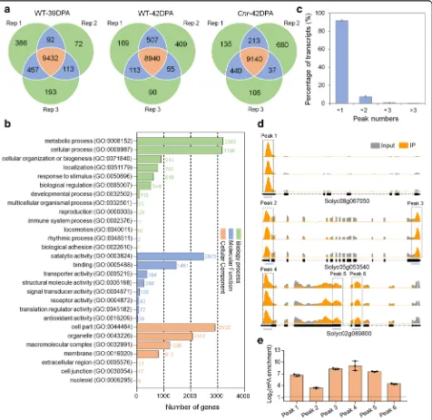
and 9023 gene transcripts, in the wild-type fruit at 39 DPA and 42 DPA, respectively, and 9140 m6A peaks within 9442 gene transcripts in the Cnr mutant at 42 DPA (Fig. 2a; Additional file 3: Table S2-S4). Gene Ontology (GO) enrichment analysis of m6A-containing transcripts revealed a potential function of m6A
Fig. 2Transcriptome-wide m6A methylation profiles in tomato fruit.aVenn diagrams showing the overlap of m6A peaks identified in three
independent m6A-seq experiments on wild-type (WT) fruit at 39 DPA and 42 DPA andCnrfruit at 42 DPA. Rep, replicate; DPA, days post-anthesis.
Only the peaks identified in all three biological replicates were regarded as confident peaks and used for subsequent data analysis.bGene Ontology (GO) analysis of the biological process, molecular function, and cellular component for the m6A-containing transcripts identified in
m6A-seq.cProportions of the m6A-modified transcripts containing different m6A peak numbers. Error bars represent the standard deviation of
three different m6A-seq experiments.dExamples of m6A-modified transcripts containing one m6A peak, two m6A peaks, and three m6A peaks in
WT fruit at 42 DPA. The black dot line rectangles indicate the positions of m6A peaks, which are named as peaks 1–6. The blue lines indicate the
positions of amplification fragments in the following m6A-immunoprecipitation (IP)-qPCR.eValidations of the m6A peaks shown indby
m6A-IP-qPCR. Data are presented as mean ± standard deviation (n= 3)
[image:4.595.62.538.172.635.2]modification in multiple signaling pathways and cellular processes (Fig.2b).
Based on these results, we estimated that the transcrip-tome of tomato fruit contains 0.5–0.6 m6
A peaks per ac-tively expressed transcript (Additional file 4: Table S5). These levels are comparable with those obtained in Arabidopsisor mammals [2,7,46]. Of the gene transcripts containing m6A modification, most (91.73%) contain one m6A peak, while 7.47% exhibit two m6A peaks, 0.69% exhibit three peaks, and 0.11% exhibit more than three peaks (Fig.2c).
We then validate the m6A-seq results with independ-ent m6A-immunoprecipitation (IP)-qPCR. Using this method, we verified the presence of m6A withinarginine N-methyltransferase (Solyc08g067050), dihydroxy-acid dehydratase (Solyc05g053540), and nuclear matrix constituent protein 1-like (Solyc02g089800) (Fig. 2d). These mRNAs were chosen for the validation of m6A presence in transcripts with a single methylation peak (Solyc08g067050) as well as those with multiple m6A peaks (Solyc05g053540 and Solyc02g089800). As ex-pected, we observed substantial enrichment of these genes after mRNA immunoprecipitation with the m6 A-specific antibody compared with the input control (Fig. 2e). These results indicated that our m6A-seq data were accurate and robust.
Collectively, these data demonstrated that m6A, which appears in a substantial fraction of the transcriptome, is a common feature of mRNA in tomato fruit, and m6 A-containing transcripts are related to a variety of bio-logical pathways.
m6A distribution and sequence motif in tomato fruit
We next characterized the distribution of m6A peaks in the whole transcriptome of tomato fruit. The meta-genomic profiles of m6A peaks in all three samples (wild-type fruit at 39 DPA and 42 DPA and Cnr epi-mutant at 42 DPA) indicated that m6A modifications were highly enriched around the stop codon and within the 3′ untranslated region (UTR) (Fig. 3a), consistent with the m6A distribution in Arabidopsis [31]. To confirm the distribution of m6A within the transcript, we divided the transcript into five non-overlapping segments: transcription start site (TSS), 5′ UTR, coding sequence (CDS), stop codon, and 3′
UTR. Each m6A peak was assigned to one of five
transcript segments. The stop codon segment (100-nucleotide window centered on the stop codon) ap-peared to be greatly enriched in m6A peaks, and 45.07 to 46.04% of the peaks from different samples fell into this segment (Fig. 3b). The enrichment of m6A peaks in the 3′ UTR was also revealed, which was comparable to that in the stop codon (Fig. 3b). After segment normalization by the relative fraction
that each segment occupied in the transcriptome, we observed that m6A is exclusively enriched around the stop codon and within the 3′ UTR, with stop codon
peaks being more pronounced than 3′ UTR peaks
(Fig. 3c). Overall, the distribution of m6A peaks did not display dramatic changes between the samples.
To identify the sequence motifs that are enriched within the m6A peaks in tomato fruit, hypergeometric
optimization of motif enrichment (HOMER; http://
homer.ucsd.edu/homer/) was applied [47]. Clustering of m6A peaks using HOMER did not identify previously established RRACH consensus sequence observed in mammals and yeasts [1, 7, 48], where R represents adenosine (A) or guanosine (G), underlined A indicates m6A, and H represents A, cytidine (C), or uridine (U), in our data set, but we did identify a UGUAYY sequence motif that was previously observed in Arabidopsis[31], where Y represents A, G, U, or C (Fig.3d). This demon-strated that the sequence motif for m6A methylation is conserved amongArabidopsisand tomato.
DNA hypermethylated mutantCnrshows overall increase in m6A mRNA methylation
To gain insight into the functional relationship
be-tween DNA methylation and mRNA m6A
methyla-tion, and the potential roles of m6A in the regulation of fruit ripening, we compared m6A methylomes be-tween the samples. A total of 401 transcripts with dif-ferential m6A levels (fold change ≥1.5; P value < 0.05) between 39 DPA and 42 DPA wild-type fruit were identified in all three biological replicates, among which 240 transcripts (Additional file 5: Table S6) exhibited higher m6A levels and 161 transcripts (Additional file 5: Table S7) displayed lower m6A levels in 39 DPA wild-type fruit compared to 42 DPA wild-type fruit (Fig. 4a). By contrast, we identified 1241 transcripts that exhibited differential m6A levels (fold change ≥1.5; P value < 0.05) between 42 DPA Cnr mutant fruit and 42 DPA wild-type fruit. A total of 1107 transcripts (Additional file 5: Table S8) dis-played higher levels of m6A enrichment in the Cnr mutant compared to the wild type, whereas only 134 transcripts (Additional file 5: Table S9) showed de-creased m6A levels (Fig. 4b), suggesting a global in-crease in m6A methylation. This is in accordance with the result of LC-MS/MS assay (Fig. 1c), showing
that m6A levels increased markedly in the Cnr
mutant.
m6A deposition has been reported to influence
mRNA abundance [6, 10, 29, 49]. To evaluate
whether there is a potential correlation between m6A mRNA methylation and gene transcript levels in to-mato fruit, RNA-seq analyses (Fig. 4c–f ) were
replicates (Additional file 1: Figure S3). Comparison of differentially expressed genes (fold change ≥1.5; P value < 0.05) (Additional file 6: Table S10-S11) with our list of transcripts showing altered m6A levels re-vealed that, among the 1107 transcripts with higher m6A levels in fruit of Cnr mutant compared to wild type, only 136 showed higher expression levels, whereas 349 exhibited lower expression levels (Fig. 4e;
Additional file 7: Table S12). Accordingly, among the 134 transcripts with lower m6A levels in the fruit of
Cnr mutant compared to wild type, 66 and 18
dis-played higher and lower expression levels, respectively (Fig. 4f; Additional file 7: Table S13). These data sug-gest that m6A methylation is generally negatively cor-related with the abundance of the transcripts. Similar results were observed in wild-type fruit between 39 DPA
Fig. 3Characteristics of m6A localization and sequence motif in tomato fruit.aMetagenomic profiles of peak summit distributions along the
transcripts composed of three rescaled non-overlapping segments (5′UTR, CDS, and 3′UTR). UTR, untranslated region; CDS, coding sequence.b
Pie charts depicting the fraction of m6A peak summits in five non-overlapping transcript segments. TSS, transcription start site.cRelative
enrichment of m6A peak summits in five non-overlapping transcript segments.a–cThe results for wild-type (WT) fruit at 39 DPA and 42 DPA and
Cnrfruit at 42 DPA. DPA, days post-anthesis.dSequence motif identified within m6A peaks by HOMER (http://homer.ucsd.edu/homer/)
[image:6.595.57.542.86.550.2]and 42 DPA (Fig. 4c, d; Additional file7: Table S14-S15). Notably, hundreds of induced and ripening-repressed genes, which exhibit significantly increased or decreased expression in 42 DPA wild-type fruit compared to 39 DPA wild-type fruit, show changed m6A levels dur-ing fruit ripendur-ing (Additional file 8: Table S16) or in the Cnrmutant (Additional file8: Table S17), implicating the involvement of m6A modification in the regulation of fruit ripening.
Transcripts of fruit-ripening genes exhibit increased m6A levels in theCnrmutant
In the m6A-seq analysis, we found that transcripts of several well-known fruit-ripening genes, including DEM-ETER-like DNA demethylase 2 (SlDML2), fruitfull 2 (FUL2), and never-ripe (NR), exhibit significantly increased m6A levels in the Cnr mutant (Fig. 5a; Additional file 5: Table S8). SlDML2 encodes a DNA demethylase [37], while FUL2and NRencode a
[image:7.595.60.538.89.461.2]box transcription factor and an ethylene receptor, re-spectively [50, 51]. The m6A peaks were enriched near the stop codon or within 3′ UTR in mRNAs of these genes, and the changes in m6A levels were observed in all three biological replications (Fig. 5a), indicating the reproducibility of our m6A-seq data. m6A-IP-qPCR con-firmed the results of m6A-seq and demonstrated that the mRNAs of SlDML2, FUL2, andNR displayed higher levels of m6A enrichment in the fruit of Cnr mutant compared with the wild-type (Fig. 5b). The transcript levels of these three genes decreased significantly in the
Cnr mutant as revealed by transcriptome analysis
(Fig. 5c), implying a negative correlation between m6A modification and mRNA abundance. It should be noted
that the mRNAs of SlDML2 and NR, but not FUL2,
exhibited lower levels of m6A enrichment, accompanied by higher transcript levels, in the fruit of wild type at 42
DPA compared with the fruit of wild type at 39 DPA (Additional file1: Figure S4).
SlALKBH2is a putative m6A RNA demethylase gene that declines in theCnrmutant
Having observing the changes in m6A levels in a large number of transcripts (Fig. 4a, b), including those of well-known fruit-ripening genes (Fig.5a), in theCnr mu-tant, we next examined the underlying mechanisms. SinceCnr is an epimutant that displays DNA hyperme-thylation, the variation in m6A might result from DNA methylation-mediated expression alteration of m6A methylation machinery, i.e., RNA methyltransferases and demethylases [15]. We speculate that the substantial increase in m6A levels in the Cnr mutant is mainly caused by downregulation of RNA demethylase genes
Fig. 5Changes in m6A levels in transcripts of specific ripening-related genes in theCnrmutant.aIntegrated Genome Browser (IGB) tracks
displaying m6A-seq read distributions inDEMETER-like DNA demethylase 2(SlDML2),fruitfull 2(FUL2), andnever-ripe(NR) transcripts. The black dot line rectangles indicate the position of m6A peaks with significantly increased m6A enrichment (fold changes≥1.5 andPvalue < 0.05) inCnrfruit
compared to wild-type (WT) fruit.bValidations of the m6A enrichment by m6A-immunoprecipitation (IP)-qPCR.cGene expression level of
SlDML2,FUL2, andNRrevealed by RNA-seq.b,cError bars represent the standard deviation of three independent experiments. Asterisks indicate significant differences (*P< 0.05, **P< 0.01, ***P< 0.001; Student’sttest)
[image:8.595.58.542.88.436.2]because DNA methylation is usually negatively corre-lated with target gene expression.
Based on the sequence of m6A RNA demethylase
(ALKBHs) in animal and Arabidopsis [26, 29], we searched for the ALKBH candidates in tomato genome. A total of eightALKBHgenes were identified by screen-ing the Sol Genomics Network (SGN) tomato database. They were namedSlALKBH1toSlALKBH8according to their location on the chromosomes (Additional file 9: Table S18). All the tomato ALKBHs contain a highly conserved AlkB domain (Additional file 1: Figure S5) with Fe (II) binding sites and alpha-ketoglutaramate binding sites (Additional file 1: Figure S6). Phylogenetic analysis indicated that some tomato ALKBHs shared high similarity with each other (Fig.6a; Additional file10:
Table S19), such as SlALKBH3 and SlALKBH4,
suggesting gene duplications. Three tomato ALKBHs (SlALKBH2, 3, and 4) exhibit high similarity with ArabidopsisALKBHs (Fig.6a), which have been demon-strated to participate in plant development and defense response [29, 52]. Transcriptome analysis indicated that, among the eight tomato ALKBH genes, only SlALKBH2 increased dramatically during fruit ripening but declined in the Cnr mutant (Fig.6b), and this was confirmed by quantitative RT-PCR analysis (Fig.6c). These data suggest that SlALKBH2, which was chosen for further analysis,
might be regulated by DNA methylation and involved in fruit ripening. It is noteworthy that the expression of the potential m6A RNA methyltransferase genes (MAT1-3) is not altered substantially during fruit ripening or in theCnr mutant (Additional file1: Figure S7).
DNA methylation regulatesSlALKBH2transcript in tomato fruit
To determine whether SlALKBH2 expression is regu-lated by DNA methylation, we examined the changes in DNA methylation patterns in SlALKBH2promoter dur-ing fruit ripendur-ing, as well as in the Cnr mutant, using the Tomato Epigenome Database (http://ted.bti.cornell. edu/epigenome/). A differentially methylated region (DMR) was found in the 5′region ofSlALKBH2at 979– 1080 bp upstream of the start codon (Fig.7a). This DMR becomes demethylated during ripening but remains hypermethylated in the fruit of Cnr mutant (Fig. 7a; Additional file 1: Figure S8). Interestingly, the hyperme-thylation ofSlALKBH2promoter was also observed in the fruit of sldml2 mutant (Fig.7b), suggesting thatSlDML2 might participate in the regulation of SlALKBH2 DNA demethylation.
To confirm that SlALKBH2 transcription is regu-lated by DNA methylation, the promoter activity of SlALKBH2 was assessed with a transient expression
Fig. 6Expression analyses of tomatoALKBHgenes reveal the involvement ofSlALKBH2in fruit ripening.aPhylogenetic analysis of tomato m6A
RNA demethylases (ALKBHs). Phylogenetic tree of the tomato ALKBHs is shown. Included in the tree are all ALKBHs fromArabidopsisand two well-characterized ALKBHs, human ALKBH5 and mouse ALKBH5. The phylogenetic tree was produced using MEGA (version 5.2). Bootstrap values from 1000 replications for each branch are shown. Tomato proteins are indicated in red. Species names are abbreviated as follows: At,Arabidopsis thaliana; Sl,Solanum lycopersicum; Hs,Homo sapiens; Mm,Mus musculus. The accession numbers are indicated in parentheses.bHeat map analysis showing the gene expression ofSlALKBH1-8in wild-type (WT) fruit at 39 DPA and 42 DPA andCnrfruit at 42 DPA by RNA-seq. Data are presented as the mean of three independent biological replicates. Asterisks indicate significant differences (*P< 0.05, **P< 0.01, ***P< 0.001; Student’sttest). NS, no significance; DPA, days post-anthesis; FPKM, fragments per kilobase of exon per million mapped fragments.cGene expression ofSlALKBH2in WT fruit at different ripening stages andCnrfruit at 42 DPA as determined by quantitative RT-PCR analysis. TheACTIN
[image:9.595.57.540.418.613.2]system in Nicotiana benthamiana. The SlALKBH2 promoter was cloned into the dual-luciferase reporter plasmid (Fig. 7c), which contains a firefly luciferase (Fluc) reporter gene and a renilla luciferase (Rluc) ref-erence gene. We found that, although weaker than
the CaMV 35S promoter, the SlALKBH2 promoter
has the ability to activate Fluc expression (Add-itional file 1: Figure S9). The relative Fluc activity (Fig. 7d) and Fluc transcript level (Fig. 7e) were
in-creased when SlDML2 was co-expressed with the
Fig. 7SlALKBH2is transcriptionally regulated by DNA methylation.aThe 5mC levels in the differentially methylated region (DMR) ofSlALKBH2
promoter in wild-type (WT) andCnrmutant fruit based on the Tomato Epigenome Database (http://ted.bti.cornell.edu/epigenome/).bThe 5mC levels in the DMR ofSlALKBH2promoter in WT andsldml2mutant fruit based on the DNA methylome database [38].a,bThe numbers indicate the cytosine positions relative to the start codon. Black represents the methylation frequency of cytosines at the indicated positions. DPA, days post-anthesis.cSchematic of the dual-luciferase system used for promoter activity assay. TheSlALKBH2promoter was cloned into the dual-luciferase reporter vector to activate the expression of firefly luciferase (Fluc). The renilla luciferase (Rluc) driven by the CaMV 35S promoter served as an internal control. LB, left border; RB, right border; Ter, terminator.d–fCo-expression of SlDML2 (SlDML2-HA) with the dual-luciferase reporter vector in theNicotiana benthamianaleaves increased the relative Fluc activity (d), facilitated theFlucgene expression (e), and reduced the 5mC level inSlALKBH2promoter (f) compared with the empty plasmid control (HA).dThe representative image from a total of six images (left panel). The Fluc activity was normalized against the Rluc activity, followed by normalization against the control (right panel). Data are presented as means ± standard deviation (n= 6). Asterisks indicate significant differences (***P< 0.0001; Student’sttest).eGene expression was determined by quantitative RT-PCR analysis. Error bars represent the standard deviation of three independent experiments. Asterisks indicate significant differences (*P< 0.05; Student’sttest).fThe box plot showing 5mC levels of all cytosines (n= 41) in the DMR analyzed by Sanger bisulfite sequencing. The plus sign represents the average level in each box
[image:10.595.59.540.85.503.2]dual-luciferase reporter plasmid, concomitant with a decline in DNA methylation level of SlALKBH2 pro-moter (Fig. 7f ). Together, these data demonstrated that SlALKBH2 transcription is regulated by DNA methylation and SlDML2 is involved in this process.
SlALKBH2 is an active m6A RNA demethylase that locates in the endoplasmic reticulum
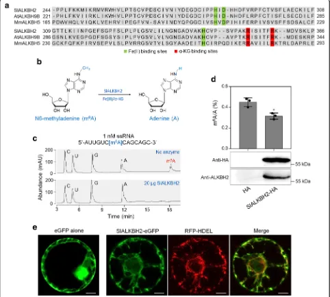
Sequence alignment revealed that SlALKBH2 contains a highly conserved AlkB domain as that of
Arabidop-sis ALKBH9B (AtALKBH9B) and mouse ALKBH5
(MmALKBH5B) (Fig. 8a). To examine whether
SlALKBH2 acts as an active m6A demethylase for
oxidative demethylation of m6A to adenosine (A) (Fig. 8b), full-length SlALKBH2 was expressed in Escherichia coli as fusion proteins with a His-tag. The recombinant proteins were purified and used for demethylation assay using a synthetic 14 nucleotide-long m6A-modified ssRNA as a substrate (Fig. 8c).
High-performance liquid chromatography (HPLC)
analysis of the nucleosides digested from the substrate indicated that almost all of the methyls in m6A were effectively removed by recombinant SlALKBH2 in vitro (Fig. 8c), demonstrating that SlALKBH2 exhib-ited strong demethylation activity toward m6A in vitro.
To further verify the demethylation activity of
SlALKBH2, the SlALKBH2 CDS was fused with a
HA-tag and transiently expressed in N. benthamiana leaves. Immunoblot analysis showed that SlALKBH2 was suc-cessfully expressed (Fig. 8d). Detection of the overall mRNA m6A levels by LC-MS/MS indicated that the ex-pression of SlALKBH2 led to reduced m6A levels com-pared with the control (empty plasmid; Fig. 8d), indicating that SlALKBH2 possesses m6A demethylation activity.
To determine the intracellular localization of
SlALKBH2, its CDS was introduced into a plasmid to generate a translational fusion with an enhanced green fluorescent protein (eGFP) at the C-terminus. The construct was agroinfiltrated into the N. benthamiana leaves, and then the mesophyll protoplasts were isolated and used for fluorescence microscopy. Confocal laser
scanning microscopy showed that eGFP-tagged
SlALKBH2 (SlALKBH2-eGFP) displayed a strong signal in the endoplasmic reticulum (ER), while the eGFP-only control produced a fluorescent signal throughout the cell, except the vacuolar lumen (Fig.8e). The fluorescent signals of SlALKBH2-eGFP co-localized with those of His-Asp-Glu-Leu (HDEL)-tagged red fluorescent protein (RFP-HEDL), which was used as a marker for ER loca-tion [53], confirming the intracellular localization of SlALKBH2 in ER (Fig.8e).
SlALKBH2-mediated m6A demethylation stabilizesSlDML2 mRNA
We next sought to explore whether SlALKBH2 could
directly bind to mRNAs of SlDML2, NR, and FUL2,
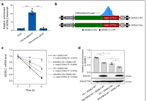
which show differential m6A methylation in our m6 A-seq analyses (Fig. 5a), using RNA immunoprecipitation (RIP). A polyclonal antibody that specifically recognized SlALKBH2 (Additional file 1: Figure S10) was used to immunoprecipitate SlALKBH2-bound mRNAs, and the result revealed a direct interaction between SlALKBH2 andSlDML2transcript (Fig. 9a). No interaction between
SlALKBH2 and NR or FUL2 transcript was observed
(Additional file 1: Figure S11), indicating that the m6A mRNA demethylation of these two genes was mediated by other components of the m6A pathway instead of SlALKBH2.
m6A methylation has been demonstrated to decrease mRNA stability, especially when m6A is located at the stop codon or 3′ UTR [6, 10, 29, 49]. As SlDML2 exhibits m6A modification within the 3′ UTR, we set out to determine if the m6A methylation affectsSlDML2
mRNA stability. The cDNA fragment of SlDML2
composed of CDS and 3′ UTR was introduced into
pCambia2300 vector (Fig. 9b), which was subsequently agroinfiltrated into theN. benthamianaleaves for transi-ent expression. The SlDML2 mRNA stability was mea-sured by monitoring the degradation rate of mRNA after treatment with transcription inhibitor actinomycin D. As shown in Fig.9c,SlDML2mRNA degraded quickly after
actinomycin D treatment. When SlALKBH2 was
co-expressed with SlDML2inN. benthamiana, the
degrad-ation rate of SlDML2 mRNA decreased, concomitant
with a significant decrease in m6A abundance of SlDML2 (Fig. 9d). Importantly, a mutated form of SlDML2 in which the potential m6A modification site identified in m6A-seq was mutated from A to C (Fig.9b) degraded slower than the intact SlDML2 (Fig. 9c).
Co-expression of the mutated form of SlDML2 with
SlALKBH2 further decreased the degradation rate of SlDML2 mRNA (Fig. 9c). Together, these data suggest that m6A modification promotes mRNA degradation of SlDML2, and SlALKBH2-mediated m6A demethylation stabilizesSlDML2mRNA.
SlALKBH2 is required for normal tomato fruit ripening
using Agrobacterium infection of leaf explants [55, 56]. Among transgenic plants in the second generation, we
isolated three distinct homozygous mutant lines
(slalkbh2-23, slalkbh2-25, and slalkbh2-28) through dir-ect sequencing of PCR products from genomic DNA
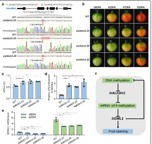
flanking the target sites. These homozygous mutants carry 1-bp insertion (slalkbh2-23and slalkbh2-28) or 5-bp deletion (slalkbh2-25) caused by target T2 in the fourth exon of SlALKBH2 (Fig. 10a), and no editing events were found around the sequence of target T1/3.
Fig. 8SlALKBH2 is a demethylase for mRNA m6A demethylation in tomato.aSequence alignment of the highly conserved AlkB domain in SlALKBH2,ArabidopsisALKBH9B (AtALKBH9B), and mouse ALKBH5 (MmALKBH5B). Fe (II) binding sites and alpha-ketoglutaramate (α-KG) binding sites are highlighted by green and red rectangles, respectively.bA proposed reaction mechanism of oxidative demethylation of
N6-methyladenosine (m6A) to adenosine (A) by SlALKBH2.cRecombinant SlALKBH2 protein directly demethylates the m6A modification in m6A-containing ssRNA in vitro. The digested substrates cytidine (C), uridine (U), guanosine (G), A, and m6A were analyzed by HPLC.dSlALKBH2 demethylates m6A modification in mRNA in vivo. Endogenous mRNA was isolated fromNicotiana benthamianaleaves transiently expressing the SlALKBH2-HA fusion protein or the empty plasmid control (HA) and used for LC-MS/MS assay. Data are presented as mean ± standard deviation (n= 3). Asterisks indicate significant differences (*P< 0.05; Student’sttest). Immunoblot analysis was performed to detect SlALKBH2 expression using both anti-HA and anti-ALKBH2 antibodies.eSubcellular localization showing that SlALKBH2 locates in the endoplasmic reticulum (ER). His-Asp-Glu-Leu (HDEL) represents an ER retention signal peptide. Protoplasts of theN. benthamianaleaves transiently expressing eGFP alone or co-expressing ALKBH2-eGFP and HDEL-RFP were isolated and observed under a Leica confocal microscope (Leica, DMI600CS). Scale bar = 50μm
[image:12.595.57.539.90.524.2]All mutants were predicted to cause premature stop codon within the following 10-bp sequence of editing sites. We did not find any off-target editing events in the seven potential off-target genes that were predicted by CRISPR-P (version 2.0, http://crispr.hzau.edu.cn/ CRISPR2/) (Additional file1: Figure S12).
By comparing the fruit of the wild-type and slalkbh2 mutants at 39, 42, 47, and 52 DPA, we found that slalkbh2-23, slalkbh2-25, and slalkbh2-28 mutant lines showed similar and obvious ripening-delayed phenotypes (Fig.10b). A visible color change was observed at 42 DPA in the wild-type fruit, whereas theslalkbh2mutant toma-toes remained green at this stage (Fig. 10b). At 47 DPA,
the wild-type fruit had a homogenous orange color, while the fruit from theslalkbh2mutants was only just starting to change color. This indicates that SlALKBH2 is indis-pensable for normal tomato fruit ripening. LC-MS/MS was subsequently performed to assay the total mRNA m6A levels in the wild type andslalkbh2 mutants at 39 DPA, and the result indicated that mutation ofSlALKBH2 led to a significantly higher mRNA m6A levels (Fig.10c). Meanwhile, the m6A-IP-qPCR assay showed thatslalkbh2 mutants exhibited higher m6A abundance in the transcript of SlDML2 compared to the wild type at this stage (Fig. 10d). By contrast, the mRNA level of SlDML2 declined in the slalkbh2 mutants (Fig. 10e). These data
Fig. 9SlALKBH2 protein bindsSlDML2transcript and promotes its stability by m6A demethylation.aRNA immunoprecipitation (RIP) assay
showing that SlALKBH2 protein bindsSlDML2transcript. For the RIP assay, the protein-RNA complexes were extracted from wild-type tomato fruit at 42 days post-anthesis and subjected to immunoprecipitation with anti-ALKBH2 polyclonal antibody or rabbit IgG (negative control). Data are presented as mean ± standard deviation (n= 6). Asterisks indicate significant differences (***P< 0.001; Student’sttest).bSchematic of the transient expression system used forSlDML2mRNA stability assay. The intact or mutatedSlDML2cDNA fragment, which is composed of coding sequence (CDS) and 3′untranslated region (UTR), was cloned into pCambia2300 vector driven by the CaMV 35S promoter. The potential m6A
modification site identified in m6A-seq was mutated from adenosine (A) to cytidine (C) using site-directed mutagenesis kit and highlighted in
[image:13.595.59.541.87.419.2]reveal that SlALKBH2 is necessary for m6A regulation during tomato fruit ripening. SlALKBH2 might participate in the regulation of fruit ripening by modulatingSlDML2
mRNA stability through m6
[image:14.595.58.542.87.548.2]A demethylation. Notably, the regulation of m6A is complicated, and other factors in addition toSlALKBH2might play roles in this process.
Fig. 10SlALKBH2 is necessary for normal tomato fruit ripening.aGenotyping of mutations mediated by CRISPR/Cas9 gene-editing system in
slalkbh2-23,slalkbh2-25, andslalkbh2-28mutants. Diagram showing the single guide RNAs (sgRNAs) containing different target sequences (T1, T2, and T3), which were designed to specifically target the exons ofSlALKBH2. The red letters indicate the protospacer adjacent motif (PAM). The transgenic plants in the second generation were genotyped by sequencing genomic regions flanking the target sites. Red arrows indicate the editing sites. Two mutants (slalkbh2-23andslalkbh2-28) have a homozygous 1-bp insertion, and one (slalkbh2-25) has a homozygous 5-bp deletion caused by target T2 in the fourth exon ofSlALKBH2.bRipening phenotype ofslalkbh2mutants. Fruit from wild-type (WT) andslalkbh2
mutants (slalkbh2-23,slalkbh2-25, andslalkbh2-28) at 39, 42, 47, and 52 days post-anthesis (DPA) are shown.cLC-MS/MS assay showing the amount of mRNA m6A in WT andslalkbh2mutant fruit at 39 DPA. Data are presented as mean ± standard deviation (n= 3).dm6A-IP-PCR assay
showing the relative m6A enrichment inSlDML2mRNA in WT andslalkbh2mutant fruit at 39 DPA.eSlDML2gene expression in WT andslalkbh2
mutant fruit at 39 and 42 DPA. TheACTINgene was used as an internal control.d,eError bars represent the standard deviation of three independent experiments. Asterisks indicate significant differences (*P< 0.05, **P< 0.01; Student’sttest).fModel for the relationship between DNA methylation and m6A mRNA methylation in fruit ripening. DNA methylation negatively regulatesSlALKBH2to mediate overall m6A mRNA
methylation. The m6A modification promotesSlDML2mRNA decay, thereby affecting DNA methylation and fruit ripening
Discussion
DNA methylation has been elucidated to play an essential role in the regulation of fruit ripening [35–39]. It is un-clear whether mRNA m6A modification, which is consid-ered as an mRNA“epitranscriptome”[3,57], participates in this process. In the present study, we show that m6A methylation represents a widespread mRNA modification in tomato and correlates with fruit ripening. The m6A modification is primarily located around the stop codon and within the 3′UTR of coding genes, and the sequence motif was conserved with that inArabidopsis. The mRNA m6A methylation in tomato fruit is mediated by endoplas-mic reticulum-located m6A RNA demethylase SlALKBH2 during ripening. We demonstrate that DNA methylation regulates the transcription of SlALKBH2, which in turn functions on m6A demethylation of SlDML2mRNA and modulates its stability. Our findings uncover the interplay between DNA and RNA methylation and reveal a novel layer of gene regulation in fruit ripening.
m6A RNA demethylase geneSlALKBH2is regulated by DNA methylation and required for normal fruit ripening
m6A modification could be dynamically regulated by both RNA methyltransferases and demethylases, which catalyze the m6A formation and removal, respectively [3, 9, 15]. Substantial insights have been made into the physiological functions of RNA methyltransferases in mammals and plants [6, 18, 29, 30, 58]. Furthermore, a recent study unveiled that the activity of RNA methyltransferase METTL3 in mammals was regulated by SUMOylation [59]. By contrast, the biological importance and the regulatory mechanisms underlying RNA demethylation remain largely unknown. In the model plantArabidopsis, there are five potential RNA demethylases, among which ALKBH10B functions in floral transition [29], while ALKBH9B modulates infection of alfalfa mosaic virus [52]. We carried out an extensive search of the tomato genome and identified eight putative RNA demethylases (SlALKBH1 to 8) that contain AlkB domain. Gene expres-sion analysis indicated thatSlALKBH2increased dramat-ically in ripening fruit that undergoes genome-wide loss of DNA methylation but declined in the fruit of ripening-deficient mutantCnrthat displays DNA hypermethylation (Fig. 6). This negative correlation between SlALKBH2 transcription and DNA methylation status led us to speculate that SlALKBH2 is regulated by DNA methyla-tion. As expected, we found that the promoter region of SlALKBH2 contains an obvious differentially methylated region (DMR) and demethylation ofSlALKBH2increased its transcript level (Fig.7). Further analysis indicated that SlALKBH2 possesses RNA demethylation activity (Fig.8) and mutation ofSlALKBH2delays fruit ripening (Fig.10). Notably, DMRs were also found in the promoters of other putative RNA demethylase genes (Additional file1: Figure
S8), but the transcription of these genes changes slightly during fruit ripening (Fig. 6), suggesting that they might be dispensable for tomato ripening.
The primary function of DNA methylation was thought to regulate gene expression at transcriptional level [40–42]. However, recent researches revealed that DNA methylation could also impact gene expression at post-transcriptional level via regulation of mRNA alter-native splicing [43, 44]. Moreover, it was demonstrated that some long non-coding RNA (lncRNA) promoters were targeted by DNA methylation [60], indicating that DNA methylation could regulate gene expression at multiple levels directly or indirectly. Our observation that SlALKBH2 was regulated by DNA methylation re-vealed that DNA methylation could impact gene expres-sion through regulation of mRNA m6A modification.
Modulation of DNA demethylase geneSlDML2by SlALKBH2-mediated m6A demethylation
m6A methylation affects gene expression by modulation of RNA metabolism [8–15]. It was reported that m6A methylation negatively affects the stability of target mRNAs and subsequent protein synthesis, thus acting as a negative regulator of gene expression [6,10,29,49]. In the m6A RNA methylome analyses, we found that hun-dreds of ripening-induced and ripening-repressed genes showed differential m6A levels between the samples (39 DPA wild type vs. 42 DPA wild type or 42 DPA wild type vs. 42 DPA Cnr mutant) (Additional file 8: Table S16-S17), and m6A deposition usually correlated with the decrease in gene expression (Fig. 4). Interestingly, the transcript ofSlDML2, a DNA demethylase gene [37], ex-hibited higher m6A level in the fruit ofCnrmutant,
con-comitant with a decline in SlDML2 mRNA level,
compared with the wild type (Fig. 5). This suggests that m6A methylation might participate in the regulation of SlDML2 mRNA abundance. To verify this speculation, we firstly assessed whether m6A modification promotes SlDML2mRNA degradation. We found that the
degrad-ation rate of SlDML2 mRNA was decreased when the
m6A sites were mutated (Fig.9). Furthermore, the RNA
demethylase SlALKBH2, which binds SlDML2 mRNA
and mediates its m6A demethylation, could stabilize SlDML2 mRNA (Fig. 9). We then mutated SlALKBH2 and observed that mutation of SlALKBH2 led to the increase in m6A level ofSlDML2 transcript, accompan-ied by the decline in SlDML2 mRNA level (Fig. 10). Together, these findings indicated that the mRNA abun-dance ofSlDML2was regulated by SlALKBH2-mediated m6A demethylation.
introduce and remove this mark, respectively [40]. In plants, active DNA demethylation is initiated by a subfamily of bifunctional 5-methylcytosine DNA glyco-sylases/lyases that includeArabidopsisproteins repressor of silencing 1 (ROS1) [61], Demeter (DME), and Demeter-like proteins 2 and 3 (DML2 and DML3) [62–64]. Recently, active DNA demethylation was revealed to be regulated by an RNA-binding protein ROS3 and a histone acetyltransferase IDM1 that are required for the recruit-ment of ROS1 to the chromatin [65, 66]. However, it re-mains uncertain whether DNA demethylation is regulated at post-transcriptional level. Data from this study provide
evidence that SlDML2, the close homolog of the
Arabidopsis DNA demethylase gene ROS1 [37, 38], is regulated by SlALKBH2-mediated m6A modification. SlDML2 was reported to be responsible for ripening-induced DNA demethylation in tomato [38]. Hundreds of ripening-related genes could be activated by SlDML2, and loss-of-function sldml2 mutant exhibits dramatic inhib-ition of fruit ripening [38]. Considering the importance of SlDML2 in fruit ripening, we suggest that SlALKBH2 regulates ripening, at least partially, by targeting SlDML2 and mediating its mRNA stability. It should be noted that SlALKBH2 might influence fruit ripening by concurrently targeting transcripts of other ripening-related genes. The SlALKBH2-mediated m6A modification of these tran-scripts and their molecular link to fruit ripening deserve further research. Based on our results and previous studies, we propose a model for the correlation
be-tween DNA methylation and m6A mRNA methylation
in fruit ripening (Fig. 10f ).
In conclusion, our findings reveal that DNA methyla-tion regulates m6A methylation by targeting RNA demethylase gene SlALKBH2, which in turn influences
DNA methylation via DNA demethylase gene SlDML2
by a feedback loop to affect fruit ripening. Considering the multiple roles of DNA methylation and m6A methy-lation, the regulation we describe here may have an essential function in many cellular contexts.
Methods Plant materials
Seeds of tomato (Solanum lycopersicum cv. Ailsa Craig), including wild type and the ripening-deficient mutant Colorless non-ripening (Cnr) in the cv. Ailsa Craig back-ground, were obtained from the Tomato Genetics Re-source Center (TGRC, https://tgrc.ucdavis.edu/policy. aspx). The plants were grown under standard culture con-ditions in a greenhouse, which was supplied with regular fertilizer and supplementary lighting when required. Flowers were tagged at the anthesis to accurately deter-mine the age of fruit through development and ripening. Wild-type fruit were harvested at immature green (IM), mature green (MG), breaker (Br), orange ripe (OR), and
red ripe (RR), which were on average 17, 39, 42, 47, and 52 days post-anthesis (DPA), respectively, based on the size, shape, color, and the development of seed and locular jelly in the fruit [67]. The fruit of Cnr and slalkbh2 mutants were harvested at the equivalent ripening stages, as determined by the DPA. The pericarp tissues were col-lected immediately after harvesting, frozen in liquid nitro-gen, and then stored at−80 °C until use.
Global DNA methylation assay
Global 5mC levels in tomato genomic DNA was deter-mined as previously described with minor modifications [68]. In brief, DNA was extracted from the pericarp tis-sues using Sureplant DNA kit (Cwbiotech, CW2298), with the disruption of total RNA according to the manu-facturer’s protocols. The extracted DNA was detected in 1% agarose gel and quantified by a SimpliNano spectro-photometer (GE Healthcare, 29-0617-11). Then, 100 ng of purified and integrated DNA for each measurement was used to perform 5mC assay by MethylFlash™ meth-ylated DNA quantification kit (Epigentek, P-1034). 5mC levels in different DNA samples were relatively quanti-fied using both the negative control and positive control, which contain 0% 5mC and 50% 5mC, respectively, following the manufacturer’s instructions.
Quantitative analysis of mRNA m6A by LC-MS/MS
Total RNAs were extracted from tomato pericarps or N. benthamianaleaves following the method of Moore et al. [69]. mRNAs were isolated from total RNAs by using Dynabeads mRNA purification kit (Life Technologies, 61006). Two hundred nanograms of mRNAs was digested with 1 unit of Nuclease P1 (Wako, 145-08221) in 50μL re-action buffer (10 mM ammonium acetate, pH 5.3, 25 mM NaCl, 2.5 mM ZnCl2) at 37 °C for 6 h. Then, 5.5μL 1 M
fresh NH4HCO3 and 1 unit of alkaline phosphatase
(Sigma-Aldrich, P6774) were added and incubated at 37 °C for another 6 h. The digested samples were centri-fuged at 15,000g for 5 min, and the supernatants were used to LC-MS/MS analysis. The nucleosides were sepa-rated by UPLC (Waters, ACQUITY) equipped with a ACQUITY UPLC HSS T3 column (Waters) and detected by MS/MS using a Triple Quad Xevo TQ-S (Waters) mass spectrometer in positive ion mode by multiple reaction monitoring. The mobile phase consists of buffer A (5 mM ammonium acetate) and buffer B (100% acetonitrile). Nu-cleosides were quantified using the nucleoside-to-base ion mass transitions of m/z 268.0 to 136.0 (A) and m/z 282.0 to 150.1 (m6A). Standard curves were generated by run-ning a concentration series of pure commercial A (Target-Mol, T0853) and m6A (TargetMol, T6599). Contents of nucleosides in samples were calculated by fitting the peak areas to the standard curves. The m6A/A ratio was
calculated accordingly. The experiment was performed with three independent biological replicates.
m6A-seq
The m6A-seq was performed as previously described [45]. Briefly, total RNAs were extracted from the pericarp tis-sues of wild-type fruit at 39 DPA and 42 DPA and Cnr fruit at 42 DPA. The integrity and concentration of ex-tracted RNAs were detected by using an Agilent 2100 bioanalyzer (Agilent, G2939A) and a SimpliNano spectro-photometer (GE Healthcare, 29-0617-11), respectively. Then, mRNAs were isolated from intact total RNAs using Dynabeads mRNA purification kit (Life Technologies, 61006) and fragmented into ~ 100 nucleotide-long frag-ments by incubation for 5 min at 94 °C in the RNA fragmentation buffer (10 mM Tris-HCl, pH 7.0, 10 mM ZnCl2). The fragmentation reaction was stopped by the addition of 50 mM EDTA, and then the fragmented mRNAs were purified by phenol-chloroform extraction and ethanol precipitation.
For the m6A-seq, 5μg of fragmented mRNAs was in-cubated with 10μg of anti-m6A polyclonal antibody (Synaptic Systems, 202003) at 4 °C for 2 h in 450μL of immunoprecipitation (IP) buffer containing 10 mM Tris-HCl, pH 7.4, 150 mM NaCl, 0.1% NP-40 (v/v), and 300 U mL−1 RNase inhibitor (Promega, N2112S). The mix-ture was then immunoprecipitated by incubation with 50μL of Dynabeads Protein-A (Life Technologies, 10002A) at 4 °C for another 2 h. After washing twice with high-salt buffer consisting of 50 mM Tris-HCl, pH 7.4, 1 M NaCl, 1 mM EDTA, 1% NP-40 (v/v), and 0.1% SDS (w/v) and twice with IP buffer, the bound mRNAs were eluted from the beads by incubation with 6.7 mM N6-methyladenosine (Sigma, M2780) in IP buffer and re-covered with phenol-chloroform extraction and ethanol
precipitation. Then, 50 ng of immunoprecipitated
mRNAs or pre-immunoprecipitated mRNAs (input control) was used for library construction with NEBNext ultra RNA library prepare kit for Illumina (NEB, E7530). High-throughput sequencing was performed on the Illumina HiSeq X sequencer with a paired-end read length of 150 bp according to the standard protocols. The sequencing was carried out with three independent biological replicates, and each RNA sample was prepared from the mix of at least 30 tomato fruits to avoid individual difference among fruits.
m6A-seq data analysis
The quality of raw sequencing reads in m6A-seq was assessed using FastQC tool (version 0.11.7) [70]. Adap-tors and low-quality bases with a score < 20 located in the 3′-end were trimmed from all raw reads by Cutadapt software (version 1.16) [71]. After trimming, reads
containing ambiguous nucleotides or with a length < 18 nucleotides were filtered out by Trimmomatic (version 0.30) [72]. The remaining reads were analyzed by using FastQC tool once again to ensure sufficient quality as-sessment. Then, read alignment was performed with Burrows Wheeler Aligner (BWA; version 0.30) [73] by using the tomato build_SL3.0 as a reference genome, and the ITAG3.2_release as a reference annotation (ftp://ftp.solgenomics.net/tomato_genome/). Mapping quality (MAPQ) of all aligned reads was concurrently calculated, and only uniquely mapped reads with a MAPQ ≥13 were remained for the subsequent analysis for each sample [45].
MACS software (version 2.0.10) [74] was used for the m6A peak identification in each anti-m6A immunopre-cipitation sample with the corresponding input sample serving as a control. A stringent cutoff threshold for MACS-assigned false discovery rate (FDR) < 0.05 was used to obtain high-confidence peaks. Only the peaks consistently called in all three independent biological samples were considered as confident peaks and used for subsequent analysis. PeakAnnotator (version 2.0) [75] was applied to annotate confident peaks to the tomato ITAG3.2_release annotation file. Differentially methylated peaks between the samples were determined using the m6A site differential algorithm [76] with a criterion of P value < 0.05 and enrichment fold change ≥1.5. The m6A-enriched motifs were identified by HOMER (version 4.7; http://homer.ucsd.edu/homer/) [47]. All peaks mapped to mRNAs were used as the target sequences, and the exon sequences except for the peak-containing sequences were used as the background sequences. The motif length was restricted to six nucleo-tides. Visualization analysis of m6A peaks was carried out using Integrated Genome Browser (IGB, version 9.0.2) [77]. Gene Ontology (GO) analysis of m6A-modified genes was performed on Gene Ontology Consortium (http:// www.geneontology.org/). GO term with a Bonferroni-corrected P value < 0.05 in individual genes was consid-ered to be statistically significant.
RNA-seq
m6A-IP-qPCR
m6A-IP-qPCR was performed as previously described with some modifications [80]. Briefly, 5μg of purified mRNAs were fragmented into ~ 300 nucleotide-long fragments by 30 s incubation at 94 °C in the RNA fragmentation buffer (10 mM Tris-HCl, pH 7.0, 10 mM ZnCl2). The fragmenta-tion reacfragmenta-tion was stopped by the addifragmenta-tion of 50 mM EDTA, followed by phenol-chloroform extraction and ethanol precipitation to purify the fragmented mRNAs.
The fragmented mRNAs were resuspended in 250μL
DEPC-treated water; 5μL was used as the input sample. Then, 100μL of fragmented mRNAs were incubated with 5μg of anti-m6A polyclonal antibody at 4 °C for 2 h in 450μL of IP buffer containing 10 mM Tris-HCl, pH 7.4, 150 mM NaCl, 0.1% NP-40 (v/v), and 300 U mL−1RNase inhibitor (Promega, N2112S). The mixture was then immunoprecipitated by incubation with 20μL of Dyna-beads Protein-A (Life Technologies, 10002A) at 4 °C for another 2 h. After washing twice with high-salt buffer con-taining 50 mM Tris-HCl, pH 7.4, 1 M NaCl, 1 mM EDTA, 1% NP-40 (v/v), and 0.1% SDS (w/v) and twice with IP buffer, the bound mRNAs were eluted from the beads by incubation with 6.7 mM N6-methyladenosine (Sigma, M2780) in IP buffer at 4 °C for 1 h and then recovered with phenol-chloroform extraction and ethanol precipita-tion. The immunoprecipitated mRNA fragments were
re-suspended in 5μL DEPC-treated water. Then, the
immunoprecipitated mRNA and pre-immunoprecipitated mRNA (input mRNA) were reverse transcribed with ran-dom hexamers using M-MLV reverse transcriptase (Takara, 2640A) and submitted to PCR amplification as quantitative RT-PCR below. m6A enrichment in specific gene regions was determined by using the cycle threshold (CT) 2(−ΔCT)method [81]. The value for the immunopreci-pitated sample was normalized against that for ACTIN (Solyc03g078400), which did not show any obvious mRNA m6A peak from m6A-seq data, as an internal control, and then normalized against that for the input.
All primers used for m6A-IP-qPCR are listed in
Additional file 11: Table S20. Each experiment has three biological replicates and each with three technical repeats.
Quantitative RT-PCR analysis
Total RNAs were extracted from tomato pericarps or N. benthamianaleaves as described above. Extracted RNAs were treated with DNase I (Takara, D2215) and then used to synthesize cDNA by reverse transcription with an oligo (dT)18primer using the Moloney murine leukemia virus (M-MLV) reverse transcriptase (Takara, 2640A). Quantitative RT-PCR was conducted on the StepOnePlus Real-Time RT-PCR Sys-tem (Applied BiosysSys-tems) using the SYBR green PCR master mix (Applied Biosystems, 4367659). PCR amplification with the gene-specific primers listed in Additional file 11: Table S21 was performed with the following program in a volume
of 20μL: 95 °C for 10 min, followed by 40 cycles of 95 °C for 15 s and 60 °C for 30 s. Relative mRNA levels were quan-tified by using the cycle threshold (CT) 2(−ΔCT) method [81]. Tomato ACTIN (Solyc03g078400) or N. benthamiana ACTIN (Niben101Scf03410g03002) was used to normalize the expression values. Each experi-ment contained three biological replicates and each with three technical repeats.
Tomato ALKBHs identification and phylogenetic analysis
For tomato AlkB homolog (ALKBHs) identification, the protein sequences of known Arabidopsis ALKBHs [29], human ALKBH5 [82], and mouse ALKBH5 [26] were used in BLAST searches against the Sol Genomics Net-work (SGN) tomato database (https://www.sgn.cornell. edu/) with default parameters. Obtained protein se-quences were used for further searching under the same conditions to avoid omissions. The conserved domain of all identified sequences was analyzed on pfam (http:// pfam.xfam.org/), and only the protein containing an AlkB domain (PF13532) was remained and considered as a tomato ALKBH. For phylogenetic analysis, the se-quences of tomato ALKBHs (Additional file1: Supplemen-tary text) were aligned with the sequences of Arabidopsis ALKBHs, human ALKBH5, and mouse ALKBH5 using Clustal X software (version 2.1) with standard parameters. The alignment result was manually edited by the Genedoc program and then imported into MEGA software (version 5.2) to construct a phylogenetic tree using the neighbor-joining statistical method with 1000 bootstrap replicates.
SlALKBH2promoter activity assay
For promoter activity assay, the SlALKBH2 promoter fragment (~ 2000 bp upstream of the start codon) was cloned from tomato genomic DNA using the primers F (5′-GTTAACACATAAATGGTAGCTATTCAC-3′) and R (5′-CCTGATTTTAATTTCTCCGATCAAC-3′). The amplified fragment was inserted into the dual-luciferase reporter vector pGreenII-0800-LUC [83], which contains a promoterless firefly luciferase (Fluc) reporter gene and a renilla luciferase (Rluc) reference gene driven by the
CaMV 35S promoter. Meanwhile, the CDS of SlDML2
without the stop codon was amplified from tomato cDNA using the primers F (5′-ATGGAAACAGGCCA AGGCAG-3′) and R (5′-GGAGGCTACTCCTTTGTC TTC-3′) and then ligated into the pCambia2300-MCS-HA vector. The constructed plasmids were transformed into Agrobacterium tumefaciens strain GV3101. The Agrobacterium was grown at 28 °C for 24 h in Luria-Bertani (LB) medium supplemented with 50μg mL−1 kanamycin, 50μg mL−1 gentamycin, and 50μg mL−1 ri-fampicin. Then, the cells were harvested and resus-pended in the infiltration medium (10 mM MES, pH 5.6,
10 mM MgCl2, 100μM acetosyringone) to a final
OD600nm of 0.3. The Agrobacterium harboring the
re-porter vector was then coinfiltrated into the N.
benthamianaleaves with theAgrobacteriumcarrying the SlDML2-expressing vector (pCambia2300-SlDML2-HA) or the control vector (pCambia2300-HA) at 1:1 ratio. After incubation at 22 °C for 36 h, the agroinfiltrated leaves were collected and the activity of cytosol-synthesized Fluc was detected after spraying 1 mM luciferin and displayed by chemiluminescence with pseudo-color. The Fluc activity was also quantitatively analyzed using a dual-luciferase assay kit (Promega, E1910). The analysis was executed using the Ultra-Sensitive and Versatile Single Tube Luminometer (Pro-mega, E5311) according to the manufacturer’s instructions. At least six measurements were contained for each assay.
5mC assay for tomatoALKBHpromoters
The 5mC levels of promoters of tomatoALKBHs in the fruit of wild type at various ripening stages and the fruit of Cnr or sldml2 mutant were analyzed on the base of tomato epigenome database (http://ted.bti.cornell.edu/ epigenome/) or tomato DNA methylome database pro-duced by Lang et al. [38].
The 5mC levels ofSlALKBH2promoter inN. benthami-anawere assessed as previously described with minor mod-ifications [84]. In brief, genomic DNA was extracted from the agroinfiltrated N. benthamiana leaves, and 500 ng of purified DNA was treated with bisulfite to produce muta-tions from cytosine (C) to thymine (T) using EZ DNA methylation-gold kit (ZYMO Research, D5005). Then, 100 ng of mutated DNA was used as templates for PCR amplifi-cation with the primers F (5′-GTCAACTTAGATGATA CGTAGAGACATTG-3′) and R (5′-CACAACCATGTA CACACATGG-3′). The PCR products were cloned into pClone007 vector (TSINGKE, NMBV-007S), and at least 20 positive clones were detected by Sanger bisulfite sequencing. The sequencing results were analyzed on Kismeth (http://katahdin.mssm.edu/) to calculate the 5mC level based on the ratio of C-T mutations in each C site.
Preparation of polyclonal antibodies
For SlALKBH2-specific antibody preparation, the
full-length CDS of SlALKBH2 was amplified from tomato
cDNA using the primers F (5′-ATGGCCGGAGATTA TAG-3′) and R (5′-TTATCTGCGACTTCTACGGC-3′) and inserted into the pET30a (+)-His-MCS vector (Merck KGaA). The resulting plasmid was transformed intoE. coli BL 21 (DE3) competent cells for the expression of recombinant protein. The bacteria were cultured at 37 °C for overnight in LB medium containing 50μg mL−1 kana-mycin and then diluted 1:100 in 50 mL of fresh LB medium to continue culture until the OD600nmreached at approximately 0.5. Then, isopropyl-1-thio-β-D-galactopyr-anoside (IPTG) was added to a final concentration of 1
mM to induce the expression of recombinant SlALKBH2 protein at 28 °C for 5 h. After induction, the bacterial cells were collected and dissolved in 5 mL 1× NTA binding buffer (50 mM Tris-HCl, pH 8.0, 300 mM NaCl, 10 mM imidazole). The cells were then broken by ultrasonication, followed by centrifugation at 4 °C, 10,000gfor 10 min. The supernatant was mixed with 1 mL Ni-NTA His Bind Resin (Merck KGaA, 70666-4). Then, the mixture was incubated at 4 °C for 1 h. After washing three times with wash buffer (50 mM Tris-HCl, pH 8.0, 300 mM NaCl, and 20 mM imidazole), the bind resin was incubated with 1 mL elution buffer (50 mM Tris-HCl, pH 8.0, 300 mM NaCl, 250 mM imidazole) at 4 °C for 10 min to elute recombinant SlALKBH2 protein. The SlALKBH2 protein was further purified by 10% SDS-PAGE and used to immunize rabbits at Abmart (http://www.ab-mart.com.cn). SlALKBH2 poly-clonal antibody was affinity-purified from antisera using the AminoLink Plus Coupling Resin (Thermo Scientific, 20501) according to the standard purification protocols.
SlALKBH2 demethylation activity assay in vitro
The demethylation activity of SlALKBH2 protein in vitro was measured following the method of Jia et al. [25] with minor modifications. Briefly, 20μg of recombinant SlALKBH2 protein prepared as described above and 1 nM of m6A-containing ssRNA, AUUGUC [m6A] CAGC AGC (synthesized at GenScript) were added to 100μL of the reaction buffer (50 mM HEPES, pH 7.0, 100 mM
KCl, 2 mM MgCl2, 2 mM L-ascorbic acid, 300μM
α-ketoglutarate, 150μM (NH4)2Fe(SO4)2·6H2O, 300 U
mL−1RNase inhibitor). The reaction was carried out at room temperature for 6 h and then quenched by adding 5 mM EDTA followed by heating at 95 °C for 10 min. ssRNA was isolated from the reaction mix by using MiRNeasy mini kit (Qiagen, 217004) and digested by nu-clease P1 (Wako, 145-08221). The digested substrates were analyzed on a HPLC system equipped with a 2489 UV/Vis detector (Waters, e2695), and the wave length for detection was set at 266 nm. Separation was per-formed at 22 °C on an Inertsil ods-3, C18, 5μm analyt-ical column (4.6 × 250 mm). The mobile phase consists of buffer A (25 mM NaH2PO4) and buffer B (100% acetonitrile). The analysis was performed at a 1 mL min−1flow rate with the following buffer A/B gradient: 15 min 95%/5%, 5 min 90%/10%, and 1 min 100%/0%.
SlALKBH2 demethylation activity assay in vivo
plasmid (pCambia2300-ALKBH2-HA) and the empty plasmid (pCambia2300-HA) were transformed into A. tumefaciens strain GV3101. The Agrobacterium was cultured at 28 °C for 24 h in LB medium. Then, the cells were harvested and resuspended in the infiltration
medium (10 mM MES, pH 5.6, 10 mM MgCl2, 100μM
acetosyringone) to a final OD600nmof 0.3 for infiltratingN. benthamianaleaves. After incubation at 22 °C for 36 h,N. benthamiana leaves were harvested and used for mRNA isolation and protein extraction. For in vivo demethylation activity assay, the abundance of mRNA m6A in N. benthamiana leaves with or without SlALKBH2 expres-sion was detected by LC-MS/MS assay as described above. The experiment was performed with three independent biological replicates.
Subcellular localization
For subcellular localization analysis, the SlALKBH2 CDS with the removal of stop codon was amplified from to-mato cDNA using the primers F (5′-ATGGCCGGAGAT TATAG-3′) and R (5′-TCTGCGACTTCTACGGC-3′) and then ligated into the pCambia2300-MCS-eGFP vec-tor. The constructed plasmid was transformed into A. tumefaciensstrain GV3101, which was subsequently used to infiltrateN. benthamianaleaves as described above for the expression of SlALKBH2-eGFP fusion protein. After incubation for 48 h, the mesophyll protoplasts were iso-lated from the agroinfiltrated leaves and observed under a Leica confocal microscope (Leica, DMI600CS). For an ac-curate localization, the RFP-HDEL fusion protein, a marker of the endoplasmic reticulum (ER), was co-expressed with the SlALKBH2-eGFP by using pBIN2-RFP-HDEL vector, which was kindly provided by Dr. Jinxin Lin (College of Biological Science and Technology, Beijing Forestry University). His-Asp-Glu-Leu (HDEL) is an ER retention signal peptide.
Western blot analysis
For western blot analysis, proteins were separated by 10% SDS-PAGE and then electrotransferred to an Immobilon-P PVDF membrane (Millipore, IPVH00010) using a semi-dry transfer unit (Amersham, TE77). The membrane was blocked with 5% non-fat milk in PBST buffer for 2 h at room temperature. The immunoblotting was conducted by incubation with anti-ALKBH2 (1: 10000), HA (1:5000), His (1:5000), or anti-ACTIN (1:5000) at room temperature for 2 h, followed by incubation with HRP-conjugated anti-rabbit IgG secondary antibody (1:10000) at room temperature for another 2 h. Immunoreactive bands were visualized by using the enhanced chemiluminescence detection kit as mentioned above. All commercial antibodies were pur-chased from Abmart (http://www.ab-mart.com.cn/).
RNA immunoprecipitation
RIP was performed as previously described with minor modifications [29]. Briefly, tomato pericarps from wild-type fruit at 42 DPA were fixed with 1% formaldehyde on ice for 30 min under a vacuum. The fixation was ter-minated with 150 mM glycine for another 5 min. The fixed tissues (2 g) were ground and then homogenized in 2 mL of lysis buffer containing 50 mM HEPES, pH 7.5, 150 mM KCl, 2 mM EDTA, 0.5% NP-40 (v/v), 0.5 mM DTT, 2 mM EDTA, 300 U mL−1RNase Inhibitor, and 1× cocktail protease inhibitor (Sigma, 04693132001). The homogenates were incubation at 4 °C for 1 h and then centrifuged at 15,000gfor 30 min. Two hundred microli-ters of the supernatant was retained as the input sample. The remainder was subjected to immunoprecipitation (IP) with anti-ALKBH2 polyclonal antibody and rabbit IgG bound to Dynabeads Protein-A (Life Technologies, 10002A). Obtained IP samples and input samples were then heated at 55 °C for 10 min to reverse the RNA-protein cross-link. The immunoprecipitated RNA and input RNA were purified by phenol-chloroform extrac-tion followed by ethanol precipitaextrac-tion. Equal amounts of RNA from each sample were reverse transcribed with an oligo (dT)18 primer using M-MLV reverse transcriptase (Takara, 2640A). Relative enrichment of individual gene was determined by quantitative RT-PCR using the primers listed in Additional file 11: Table S21. The ex-periment contained three biological replicates and each with three technical repeats.
mRNA stability assay
The mRNA stability assay was performed with a transient expression system in N. benthamiana. In brief, the cDNA fragment of SlDML2composed of full-length CDS and 3′ UTR was amplified from tomato cDNAs. A mutated form of the amplified sequence with mutations from adenylate (A) to cytidine (C) in the potential m6A site located in the 3′UTR ofSlDML2cDNA was constructed using the QuikChange II XL site-directed mutagenesis kit (Agilent Technologies, 200518) following the manufacturer’s instructions. The two resulting fragments were then separately inserted into the pCambia2300-MCS-HA vector for the expression of intact or mutated SlDML2 transcript. The constructing plasmids were introduced into A. tumefaciens strain GV3101. After cultivation, theAgrobacteriumwere coinfiltrated into theN. benthamiana leaves with the Agrobacterium carrying the SlALKBH2-expressing vector (pCambia2300-SlALKBH2-HA) or the control vector (pCambia2300-(pCambia2300-SlALKBH2-HA) at 1:1 ratio in a final OD600nm of 0.6. After incubation for 24 h, leaf disks were taken from the infection parts of theN. benthamiana leaves and transferred onto the sterile water containing 10μg mL−1 actinomycin D (Sigma, A4262). After 1 h of incubation, six leaf disks were collected and considered as time 0 controls, and subsequent samples were harvested