Antimicrobial, Anticancer Activities and DNA
Fragmentation of Cardiospermum Halicacabum l.
S. Divya1, S. Aswini2, N. Bowthriya3, D.Vinothkumar4 and N.G. RameshBabu5
1, 2, 3, 4, 5
Department of Biotechnology, Adhiyamaan College of Engineering (Autonomous), Hosur-635109, Tamil Nadu, India
Abstract: Methanolic extract of Cardiospermum halicacabum was examined for its antimicrobial, anticancer and DNA fragmentation properties. The methanolic extract of C. halicacabum was evaluated for its antifungal activity against the medicinally important fungi viz., Candida albicans, Trichoderma viride, Penicillium chrysogenum, Trichophyton interdigitale and Rhizopus microspores with agar disc diffusion method. Among this, C. albicans and T. interdigitale showed higher zone of inhibition and lesser activity against the control (Amphotericin-B). The methanolic extract of C. halicacabum was also evaluated for its antibacterial activity against the medicinally important bacteria viz., Vibrio parahaemolyticus, Shigella flexneri, Pseudomonas aeruginosa, Serratia marcescens and Micrococcus luteus. Among this, V. parahaemolyticus and S. marcescens showed higher zone of inhibition and lesser activity against the control (Ampicillin). It is indicated that the plant might be a good antimicrobial source. The anti-cancer potential of leaves extract of C. halicacabum against Hep-G2 cell line was observed. The anticancer activity was evaluated by methyl thiazoal tetrazolium assay method. DNA fragmentation caused by apoptosis event was evaluated through DNA extraction. DNA was extracted from treated cells showed the formation of DNA laddering. The results suggested that C. halicacabum extracts might inhibit Hep-G2 hepatocellular carcinoma cell growth via apoptosis and also it was found to be most efficient in inhibiting cell growth and inducing apoptosis. From the results, the present study indicates the anti-cancer potential of C. halicacabum leaves extract.
Keywords - Anticancer, antimicrobial, Cardiospermum halicacabum, Hep-G2 cell line, invitro, Rhizopus microsporus.
I. INTRODUCTION
Ancient Indian literature incorporates a remarkably broad definition of medicinal plants to the potential sources of medicinal substances. Nature has bestowed on us a very rich botanical wealth. A large number of diverse types of plants grow in different parts of the world such as Brazil, Central Africa, India, Philippines, etc. Herbal medicines play an important role in health care programs in the developing countries. India is rich in all the three levels of biodiversity, namely species diversity, genetic diversity and habitat diversity. In India, thousands of species are known to have medicinal value and the use of different parts of several medicinal plants to cure specific ailments has been in vogue since ancient times. Herbal medicine is still used by about 75-80% of the whole population, mainly in developing countries (Swammy and Tan, 2000). Therefore, green plants are the symbol of a reservoir of resourceful chemotherapeutics that provide important source of natural antimicrobials (Balandrin et al., 1985). The use of plants by man to treat common ailments since times memorial and many of the traditional medicines are still included as part of the habitual treatment of various maladies (Henrich et al., 2004). Natural products from microorganisms have been the primary source of antibiotics. But with the increasing acceptance of herbal medicine as an alternative form of health care, the screening of medicinal plants for active compounds has become very important because these may serve as promising sources of novel antibiotics (Koduru et al., 2006).
A. Habit and Habitat
various diseases such as skin diseases (rashes, itching, skin irritation, etc.), dandruff, rheumatoid arthritis, gastrointestinal diseases, respiratory tract diseases, urogenital diseases, etc. (Venkatesh Babu and Krishnakumari, 2006).
B.Medicinal properties of Cardiospermum halicacabum
It has been suggested that the aqueous and ethanolic extracts from plants used in allopathic medicine are potential source of antiviral, antitumoral and antimicrobial agents (Chung et al., 1995).C. halicacabum possesses various phytochemicals and active biomolecules, which play a major role in the treatment of cancer. Many plants have been examined to identify new and effective anticancer compounds, as well as to elucidate the mechanism of cancer prevention and apoptosis (Swammy and Tan, 2000). halicacabum belongs to the family Sapindaceae, commonly known as Balloon vine or Love in a puff. C. halicacabum is the combination of the Latin words cardio, meaning heart, and sperma, meaning seed and refers to the white heart-shaped pattern on the seed. Halicacabum is derived from the Latin word halicacabus, a plant with inflated fruits (Pieroni et al., 2002).
Cancer is a manifestation of critical alteration in cell physiology that results in uncontrolled malignant growth. It is a dynamic process that involves many complex factors that causes extensive morbidity and wide mortality in the human population and the costs to society of this dreadful disease are prodigious (Peter 1986). About 80% of the world’s rural people rely on medicinal plants for healthcare because plant drugs are easily available and accessible to most people in the sense of price compared to modern allopathic drugs (Sarker, 1996 and WHO, 2002).The discovery and development of vin blastine and vincristine alkaloids from Catharanthus roseus, etoposide (VM 26) and teniposide (VP 16-213) from Podophyllum, irinotecan or camptothecin from Camptotheca acuminate, paclitaxel from Taxes and several other natural compounds from different sources as efficacious anticancer agents provided convincing evidence that plants’ secondary metabolites could be a potential source of anticancer agents and cancer chemopreventives (Cragget al., 1996; Kinghorn, 2000; Malika et al., 2011). There is a necessity for research to search new compounds with cytotoxic activity for the treatment of cancer. The available anticancer drugs are often unsatisfactory due to the problem of causing cytotoxicity to the normal cells along with cancer cells. Plants are considered as valuable sources of bioactive compounds with antioxidant activity, which produce certain substances that have effects on living animal cells. DNA fragmentation is the separation or breaking of DNA strands into pieces. It can be done intentionally by laboratory personnel or by cells or can occur spontaneously. In this regard, three traditional medicinal plants are taken to assess their apoptosis inducing capacity. Though, some sparse reports of cytotoxicity of the plant extracts were found, no reports regarding their genotoxicity were available. Therefore, the present study probably reports for the first time the genotoxic potentiality of the organic fractions of these medicinal plants. For this, the methanolic extracts of whole plants have been subjected to fractionation in organic solvents. The cytotoxic activity of plant extract fractions have been assessed by MTT assay. Agarose gel electrophoresis was deployed to test the apoptosis inducing capability of the extract (Subhabrata Paul et al., 2015).Hence, this study aims to explore the antimicrobial activity of C. halicacabum and to investigate the anticancer potential of the leaf extracts of C. halicacabum against Hep-G2 cell lines and also to identify the apoptotic effect of solvent leaf extracts against cell by MTT assay.
II. MATERIALS AND METHODS
A. Extraction of sample
C. halicacabum was purchased from the local market in Chennai, Tamil Nadu, India. The sample was washed with distilled water for 10min to remove any adherent particles, shade dried and powdered. 50gof leaf powder was weighed and extracted with 300ml of methanol by continuous hot percolation with the help of soxhlet apparatus for 72hrs. On completion the extracts were filtered and concentrated using rotary evaporator under reduced pressure and controlled temperature of 100ºC – 110ºC. The concentrates were stored in the refrigerator.
B. Antimicrobial assay
2) Preparation of inoculum: Each organism was recovered by subculturing on fresh media. Loop full inoculums of each organism was suspended in 5ml of nutrient broth and incubated overnight at 37ºC. These overnight cultures were used as inoculums.
3) Preparation of discs: Discs usually consist of absorbent paper impregnated with the compound methanolic extract. It is convenient to use Whatman No.1 filter paper for preparing the discs. Dry discs of 6mm diameter were prepared from Whatman No.1 filter paper and sterilized in an autoclave. These dry discs were used for this assay.
III. PROCEDURE
A. Antimicrobial assay
Circular discs of 6mm diameter were prepared from Whatman No.1 filter paper and sterilized in an autoclave. For bacterial strains, each paper disc was impregnated with 0.2 mg/l of test compound (methanolic extract) in the respective solvent overnight and placed on nutrient agar plates seeded with the test bacterium. The plates were incubated at 37º C for 24hrs. After 24 hrs, the zone of inhibition around each disc was measured and recorded. Each concentration was tested four times to ensure the reliability of the result. Ampicillin was used as (Positive control). The negative control was prepared with the solvent used for the extraction. For fungal stains, the petriplates were prepared with Sabouraud Dextrose Agar medium and inoculated on the surface with a spoon suspension of 106CFU/ml. Sterile paper disc of 6mm diameter impregnated with the extract at a concentration of 100 mg/ml were placed over the test plates. Amphotericin-B 10 mcg per disc was used as the standard. The plates were incubated at 30ºC for 48hrs. The diameter of growth inhibition zone around each disc was measured against each concentration after 48hrs.
B. Anticancer activity
1) Cell line and culture:Cell line was obtained from National Centre for Cell Sciences (NCCS), Pune. The cells were maintained
in Dulbecco’s Modified Eagle’s Medium (DMEM) supplemented with 10% Fetal Bovine Serum, penicillin (100 U/ml), and
streptomycin (100 μg/ml) in a humidified atmosphere of 50 μg/ml CO2 at 37°C.
2) In vitro assay for anticancer activity:Cells (1 × 10⁵/well) were plated in 24-well plates and incubated at37ºCwith 5% CO2. After the cell reached the confluence, the various concentrations of the samples were added and incubated for 24hrs. After incubation, the sample was removed from the well and washed with phosphate-buffered saline (pH 7.4) or DMEM without serum. 100µl/well (5mg/ml) of 0.5% 3-(4,5-dimethyl-2-thiazolyl)-2,5-diphenyl-tetrazolium bromide (MTT) was added and incubated for 4 hours. After incubation, 1 ml of dimethyl sulfoxide was added in all the wells .The absorbance at 570nm was measured with UV- Spectrophotometer using DMSO as the blank. Readings were noted and the concentration required for a 50% inhibition (IC50) was determined graphically. The % cell viability was calculated using the following formula.
A570 of treated cells
% Cell viability = X 100 A570 of control cells
Graphs are plotted using the % of Cell Viability at Y-axis and concentration of the sample in X-axis. Cell control and sample control is included in each assay to compare the full cell viability assessments.
C. DNA fragmentation
1) HepG2 cells were plated in 6 well plate and kept in CO2 incubator to attain confluency. 2) Cells were treated with the samples. Control was maintained devoid of sample. 3) Plate was incubated for 24 hrs of time at 37οC in 5% CO2.
4) After incubation, cells were harvested using TPVG and cell suspension was dispensed in eppendorf. 5) Centrifuge cells at 200xg at 40C for 10 min.
6) Add to the pellet 0.5 ml of TTE Solution and vortex vigorously. This procedure allows the release of fragmented chromatin from nuclei, after cell lysis (due to the presence of Triton X- 100 in the TTE solution) and disruption of the nuclear structure (following Mg++ chelation by EDTA in the TTE Solution).
7) To separate fragmented DNA from intact chromatin, centrifuge tubes at 20,000 rpm for 10 min at 40C. 8) Carefully remove the supernatants and add 500µl of TTE solution into the pellet.
9) Add 500µl of Ice-cold NaCl and vortex vigorously. The addition of the salt should be able to remove histones from DNA. 10) Add 700µl of ice-cold isopropanol and vortex vigorously.
11) Allow precipitation to proceed overnight at -200C.
14) Centrifuge tubes at 20,000 rpm for 10 min at 40C.
15) Dissolve DNA by adding to each tube, 20-50 µl of TE solution and place the tubes at 40C.
16) Mix the samples of DNA with loading buffer by adding 10x loading buffer to a final concentration of 1X. The addition of loading buffer to samples allows to load in wells more easily and to monitor the run of samples.
17) Run the electrophoresis in standard TE buffer after setting the voltage to the desired level. During electrophoresis it is possible to monitor the migration of samples by following the migration of bromophenol blue dye contained in the loading dye.
18) Stop the electrophoresis when the dye reaches about 3 cm from the end of the gel. 19) To visualize DNA, place the gel on a UV Transilluminator.
IV. RESULTS AND DISCUSSION
A. Antimicrobial activity
The antimicrobial activity of methanolic leaves extract of C. halicacabum from disc diffusion method has been summarized in the Table 1 & 2, Fig 1 & 2. The inhibitory action was observed in terms of diameter of inhibition zone formed around each disc caused by the diffusion of antimicrobial substances from the paper disc into the surrounding medium. Among bacteria, the zone of inhibition was higher in V. Parahaemolyticus (2.6±0.15µg/ml), in S. marcescens (2.4±0.17µg/ml). Moderate inhibition was noted in S.flexneri (1.6±0.16µg/ml). The minimum zone of inhibition was noted against M. luteus (1.1±0.15µg/ml). Among fungi, the zone of inhibition was higher in T. interdigitale (1.7±0.14µg/ml), in C. albicans (1.9±0.09µg/ml). Moderate inhibition was noted in T. viride (1.3±0.12µg/ml). The zone of inhibition was not found in R. microsporus.
Table1
The antibacterial result for methanolic extract
Microorganisms
Zone of inhibition (mm)
Antibiotic (1mg/ml) Concentration(µg/ml)
100 75 50
Vibrio parahaemolyticus
2.6±0.15 2.2±0.11 1.9±0.10 3.6±0.06 Shigella flexneri
1.6±0.16 1.4±0.12 1.3±0.08 2.3±0.09 Pseudomonas aeruginosa
1.7±0.14 1.7±0.13 1.7±0.12 3.0±0.09 Serratia marcescens
2.4±0.17 1.9±0.15 1.8±0.13 3.1±0.15 Micrococcus luteus
[image:5.612.55.550.331.724.2]1.1±0.15 1.1±0.10 1.0±0.06 2.0±0.12
Table 2
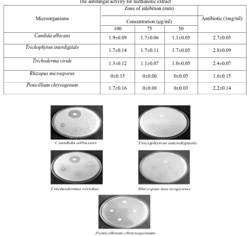
The antifungal activity for methanolic extract
Microorganisms
Zone of inhibition (mm)
Concentration (µg/ml) Antibiotic (1mg/ml)
100 75 50
Candida albicans 1.9±0.09 1.7±0.06 1.1±0.05 2.7±0.05
Trichophyton interdigitale
1.7±0.14 1.7±0.11 1.7±0.05 2.8±0.09
Trichoderma viride
1.3±0.12 1.1±0.07 1.0±0.05 2.4±0.07
Rhizopus microsporus 0±0.15 0±0.06 0±0.05 1.6±0.15
Penicillium chrysogenum
1.7±0.16 0±0.08 0±0.03 2.2±0.14
Figure 2. The antifungal activity of Methanolic Extract
B. Anticancer activity
Table 3
Anticancer effect of cardiospermum halicacabum on hep-g2 cell line
S.No Concentration (µg/ml) Dilution rate Absorbance values Cell viability (%)
1 1000 Neat 0.154 24.73
2 500 1:1 0.192 30.42
3 250 1:2 0.224 37.72
4 125 1:4 0.268 43.23
5 62.5 1:8 0.283 50.35
6 31.2 1:16 0.322 57.29
7 15.6 1:32 0.355 63.16
8 7.8 1:64 0.396 70.46
9 Cell control - 0.562 100
[image:7.612.126.517.133.689.2]C. DNA fragmentation
The DNA fragmentation activity of C. halicacabum leaves extract was observed in figure 3. The DNA in the control was not damaged while the DNA in the sample wells was damaged. DNA fragmentation caused by apoptosis event was evaluated through DNA extraction. DNA extracted from treated cells showed the formation of DNA laddering. The results shows that C. halicacabum extracts inhibit the Hep-G2 hepatocellular carcinoma cell growth via apoptosis and also it was found to be most efficient in inhibiting cancer cell growth and inducing apoptosis. From the results, the present study indicates the anti-cancer potential of C. halicacabum leaves extract.
V. CONCLUSION
The present study conclusively demonstrate that Cardiospermum halicacabum is a good source of various phytochemicals. The C. halicacabum has antimicrobial activity against pathogenic microorganisms. It was also suggested that C. halicacabum has anticancer activity against cancer cells and it can inhibit the growth of cancer cells. And also the DNA fragmentation activity of C. halicacabum via apoptosis also indicates that it has good anticancer activity. The whole plant contain highly medicinal values for many treatments. But the major role of leaves is the treatment of cancer.
REFERENCES
[1] Balandrin MF, Klocke JE,Wutule ES. and Bollinger WH. 1985. Natural plant chemicals: Sources of industrial and medicinal materials. Science, 228: 1154-1160.
[2] Chung TH, Kim JC. and Kim MK. 1995. Investigation of Korean plant extracts for potential phototherapeutic agents against B virus Hepatitis. Phytotherapy Res.9: 429-434.
[3] Cragg GM, Simon JE, Jato JG.and Snader KM. 1996. Drug discovery and development at the National Cancer Institute: Potential for New Pharmaceutical Crops. In: Janick J (ed) Progress in New Crops, ASHS Press, Arlington, VA.554-560.
[4] Cragg GM and Newman DJ. 2005. Biodiversity: A continuing source of novel drug leads. Pure Appl Chem.77:7-24. [5] Dubey NK, Kumar R. and Tripathi P. 2004. Global promotion of herbal medicines: India`s opportunity. Curr Sci.86:37-41.
[6] Henrich M, Barnes J, Gibbons S. and Williamson EM. 2004. Fundamentals of Pharmacognosyphytotherapy. Cyrchill Livingstone, Edinburgh.
[7] Jeyadevi R, Sivasudha T, Rameshkumar A, James Harnly and Long-Ze Lin. 2013. Phenolic profiling by UPLC–MS/MS and hepatoprotective activity of Cardiospermum halicacabum against CCl
4induced liver injury in Wistar rats. J of Functional Foods.289-98.
[8] Joshi SK, Dhstms BD, Bhatia CR, Singh RV. and Thakur RS. 1992. The Wealth of India Raw Materials Vol. III, New Delhi; Council of Scientific Ind. Res. Pub. 270-271.
[9] Kinghorn AD. 2000. Plant secondary metabolites as potential anticancer agents and cancer chemopreventives. Molecules.5:285-288. [10] Koduru S, Grierson DS. and Afolayan AJ (2006) Antimicrobial activity of Solanum aculeastrum. Pharm Biol, 44(4):283–286.
[11] Malika S, Cusido RM, Mirjalili MH, Moyano E, Palazon J. and Bonfill M. 2011. Production of the anticancer drug taxol in Taxus baccata suspension cultures: A review. Process Biochemistry. 46:23-34.
[12] Peter C Nowel. 1986. Mechanisms of Tumor Progression. Cancer Research.46: 2203-2207.
[13] Pieroni A, Nebel S, Quave C, Münz, H. and HeinrichM. 2002. Ethnopharmacology of Liakra: traditional weedy vegetables of the Arbëreshë of the Vulture area in southern Italy. Journal of Ethnopharmacology. 81(2): 165- 185.
[14] Sarker S. 1996. Medicinal plants and the law. Centre for Environmental law, WWF, India.
[15] Subhabrata Paul, Shuvechha Chakraborty, Anita Mukherjee. And Rita Kundu. 2015. Evaluation of cytotoxicity and DNA damaging activity of three plant extracts on cervical cancer cell lines, International Journal of Pharmaceutical Sciences Review and Research. 31(1): 183-189.
[16] Swammy SMK and Tan BKH. 2000. Journal of Ethnopharmacology. 70:1-7.
[17] Venkatesh Babu KC and Krishnakumari S. 2006.Cardiospermum halicacabum suppresses the production of TNF-alpha and Nitric oxide by Human Peripheral Blood Mononuclear cells. African J of Biomedical Res. 9:95-99.