Role of K+ ATP channels and adenosine in the
regulation of coronary blood flow during
exercise with normal and restricted coronary
blood flow.
D J Duncker, … , Y Ishibashi, R J Bache
J Clin Invest.
1996;
97(4)
:996-1009.
https://doi.org/10.1172/JCI118524
.
Regulation of coronary vasomotor tone during exercise is incompletely understood. We
investigated the contributions of K+ ATP channels and adenosine to the coronary
vasodilation that occurs during exercise in the normal heart and in the presence of a
coronary artery stenosis. Dogs that were chronically instrumented with a Doppler flow
probe, hydraulic occluder, and indwelling catheter on the left anterior descending coronary
artery were exercised on a treadmill to produce heart rates of approximately 200 beats/min.
By graded inflation of the occluder to produce a wide range of coronary stenosis severities,
we determined the coronary pressure-flow relation. K+ atp channel blockade with
intracoronary glibenclamide (10-50 microgram/kg per min) decreased coronary blood flow
during exercise at coronary pressures within and below the autoregulatory range, indicating
that coronary K+ ATP channel activation is critical for producing coronary vasodilation with
either normal arterial inflow or when flow is restricted by a coronary artery stenosis.
Adenosine receptor blockade with intravenous 8-phenyltheophylline (5 mg/kg) had no effect
on coronary flow at pressures within the autoregulatory range but decreased flow at
pressures < 55 mmHg. In contrast, in the presence of K+ ATP channel blockade, the
addition of adenosine receptor blockade further decreased coronary flow even at coronary
pressures in the autoregulatory range, indicating increased importance of the vasodilator
influence of endogenous adenosine during exercise when […]
Research Article
Find the latest version:
996 D.J. Duncker, N.S. van Zon, Y. Ishibashi, and R.J. Bache
J. Clin. Invest.
© The American Society for Clinical Investigation, Inc. 0021-9738/96/02/0996/14 $2.00
Volume 97, Number 4, February 1996, 996–1009
Role of K
1ATP
Channels and Adenosine in the Regulation of Coronary Blood Flow
during Exercise with Normal and Restricted Coronary Blood Flow
Dirk J. Duncker, Noëmi S. van Zon, Yutaka Ishibashi, and Robert J. Bache
From the Cardiovascular Division, Department of Medicine, University of Minnesota Medical School, Minneapolis, Minnesota 55455
Abstract
Regulation of coronary vasomotor tone during exercise is incompletely understood. We investigated the contributions
of K1
ATP channels and adenosine to the coronary
vasodila-tion that occurs during exercise in the normal heart and in the presence of a coronary artery stenosis. Dogs that were chronically instrumented with a Doppler flow probe, hy-draulic occluder, and indwelling catheter on the left ante-rior descending coronary artery were exercised on a
tread-mill to produce heart rates of z 200 beats/min. By graded
inflation of the occluder to produce a wide range of coro-nary stenosis severities, we determined the corocoro-nary
pres-sure–flow relation. K1
ATP channel blockade with
intra-coronary glibenclamide (10–50 mg/kg per min) decreased
coronary blood flow during exercise at coronary pressures within and below the autoregulatory range, indicating that
coronary K1
ATP channel activation is critical for producing
coronary vasodilation with either normal arterial inflow or when flow is restricted by a coronary artery stenosis. Aden-osine receptor blockade with intravenous 8-phenyltheophyl-line (5 mg/kg) had no effect on coronary flow at pressures within the autoregulatory range but decreased flow at
pres-sures , 55 mmHg. In contrast, in the presence of K1
ATP
channel blockade, the addition of adenosine receptor block-ade further decreased coronary flow even at coronary pres-sures in the autoregulatory range, indicating increased importance of the vasodilator influence of endogenous
aden-osine during exercise when K1
ATP channels are blocked.
In-tracoronary adenosine (50 mg/kg per min) increased
coro-nary flow at perfusion pressures both within and below the
autoregulatory range. In contrast, selective K1
ATP channel
activation with intracoronary pinacidil (0.2–5.0 mg/kg per
min) increased flow at normal but not at lower coronary
pressures (, 55 mmHg). This finding demonstrates that not
all K1
ATP channels are activated during exercise at
pres-sures in the autoregulatory range, but that most K1
ATP
channels are recruited as pressures approach the lower end
of the autoregulatory plateau. Thus, K1
ATP channels and
endogenous adenosine play a synergistic role in maintaining vasodilation during exercise in normal hearts and distal to a coronary artery stenosis that results in myocardial
hypoper-fusion during exercise. (J. Clin. Invest. 1996. 97:996–1009.)
Key words: coronary blood flow • myocardial ischemia •
myocardial oxygen consumption • regional myocardial
sys-tolic wall thickening
Introduction
In the normal heart, coronary blood flow is tightly coupled to metabolic demands to maintain a consistently high level of oxy-gen extraction by the myocardium. This close coupling, which is especially apparent during exercise, has been suggested to depend on messengers released from the myocardium or vas-cular endothelium. However, specific blockers of established endogenous vasodilators such as adenosine (1), prostacyclin (2), and nitric oxide (3) have not been found to impair coro-nary blood flow during exercise, indicating that these vasodila-tors are not mandatory for regulation of coronary vasomotor tone during exercise with normal coronary arterial inflow. In contrast, these vasodilator mechanisms can contribute signifi-cantly to regulation of coronary vasomotor tone during exer-cise in the presence of myocardial hypoperfusion. Thus, block-ade of block-adenosine (4, 5) or nitric oxide (6) decreased coronary blood flow distal to a coronary artery stenosis that resulted in myocardial hypoperfusion during exercise.
Recent evidence indicates that hyperpolarization of the vascular smooth muscle cell membrane caused by opening of
K1
ATP channels contributes to regulation of coronary
vasomo-tor tone (7). Thus, blockade of vascular smooth muscle K1
ATP
channels decreased coronary blood flow both under basal con-ditions (8–11) and during exercise with normal arterial inflow (11), and decreased coronary reactive hyperemia in response to a brief ischemic stimulus (8, 11). Studies in anesthetized open-chest dogs have suggested that the number of activated
K1
ATP channels increases progressively in response to
restric-tion of coronary blood flow produced by a coronary artery stenosis (12, 13). However, no study has compared the role of
K1
ATP channels in regulation of coronary vasomotor tone
dur-ing exercise in the presence of myocardial hypoperfusion with exercise during normal arterial inflow in the same animals.
The coronary pressure–flow relation describes the blood flow response over a range of perfusion pressures within and below the autoregulatory range, allowing a comprehensive as-sessment of regulation of coronary vasomotor tone. The be-havior of coronary blood flow over a range of perfusion pres-sures is relevant to the clinical situation in which an arterial stenosis can result in decreased perfusion pressure. To
deter-mine the contribution of K1
ATP channels to the regulation of
coronary blood flow under conditions of normal and restricted
arterial inflow, we studied the effects of the selective K1
ATP
Address correspondence to Dr. Robert J. Bache, Cardiovascular Di-vision, Department of Medicine, University of Minnesota Medical School, Box 508, UMHC, 420 Delaware Street S.E., Minneapolis, MN 55455. Phone: 612-625-2454; FAX: 612-626-4411. Dr. Dirk J. Duncker’s present address is Experimental Cardiology, Thorax-center, Erasmus University Rotterdam, POB 1738, 3000 DR Rotter-dam, The Netherlands.
channel antagonist glibenclamide and the selective agonist pi-nacidil on the coronary pressure–flow relation in awake exer-cising dogs. Since it has been demonstrated that, in the canine coronary circulation, adenosine produces coronary
vasodila-tion in part via activavasodila-tion of K1
ATP channels (8), we also
as-sessed the interaction between endogenous adenosine and
K1
ATP channels during exercise.
Methods
Studies were performed in 25 adult mongrel dogs weighing 20–27 kg and trained to run on a motor-driven treadmill. All experiments were performed in accordance with the Guiding Principles in the Care and Use of Laboratory Animals as approved by the Council of the Amer-ican Physiological Society, and with the prior approval of the Animal Care Committee of the University of Minnesota.
Surgical preparation
After sedation with acepromazine (0.5 mg/kg, i.m.), dogs were anes-thetized with sodium pentobarbital (30–35 mg/kg, i.v.), intubated, and ventilated with a mixture of oxygen (30%) and room air (70%). Res-piratory rate and tidal volume were set to keep arterial blood gases within physiologic limits. A left thoracotomy was performed through the fifth intercostal space and the heart was suspended in a pericar-dial cradle. A polyvinyl chloride catheter (3.0 mm o.d.) filled with hep-arinized saline was inserted into the left internal thoracic artery and advanced into the ascending aorta. Similar catheters were introduced into the right atrium through the atrial appendage and the left ventri-cle through the apical dimple. A solid state micromanometer (model P5; Konigsberg Instruments, Inc., Pasadena, CA) was also introduced into the left ventricle through the area of the apex. Approximately 1.5 cm of the proximal left anterior descending coronary artery (LAD)1
was dissected free, and a Doppler flow probe (Craig Hartley Method-ist Hospital, Houston, TX) was positioned around the artery. Imme-diately distal to the flow probe, a hydraulic occluder (3.0 mm o.d.) was placed around the vessel. A silicone catheter (0.3 mm, i.d.) bonded to a larger silicone catheter (1.6 mm i.d.) was introduced into the LAD immediately distal to the hydraulic occluder. Two pairs of 5-MHz miniature piezoelectric crystals to measure myocardial wall thickening were implanted in the myocardial region perfused by the LAD and the control region perfused by the left circumflex coronary artery (LCX), respectively. The pericardium was then loosely closed, and the catheters and electrical leads were tunneled subcutaneously to exit at the base of the neck. The chest was closed in layers, and the pneumothorax was evacuated. Catheters were flushed daily with hep-arinized saline.
Hemodynamic measurements
Studies were performed 2–3 wk after surgery with animals exercising on a motor-driven treadmill. Recordings of phasic and mean aortic pressure were obtained with pressure transducers (P23XL; Gould Inc., Cleveland, OH) positioned at mid-chest level. Left ventricular pressure was measured with the micromanometer calibrated with the fluid-filled left ventricular catheter. Left ventricular dP/dt was ob-tained via electrical differentiation of the left ventricular pressure sig-nal. Coronary Doppler shift was measured with a Doppler flowmeter system (Craig Hartley). Data were recorded on an eight-channel di-rect writing oscillograph (Coulbourn Instruments, Inc., Lehigh Val-ley, PA).
Regional myocardial function measurements
Regional myocardial wall thickening was measured by sonomicrome-try (model 120; Triton Technology, Inc., San Diego, CA) using two
pairs of 5-MHz ultrasonic crystals. End-diastolic wall thickness (EDT) was measured at the onset of positive LVdP/dt and end-sys-tolic wall thickness (EST) was measured 20 ms before peak negative LVdP/dt. Percent myocardial systolic wall thickening (SWT) was computed as follows:
SWT (%) 5 (EST 2 EDT)/EDT 3 100
Experimental protocols
To test the magnitude and selectivity of the agonists and antagonists used in the present study, dose response studies were performed in a total of seven dogs standing quietly in a sling.
Magnitude and selectivity of K1ATP channel blockade produced
by glibenclamide. In six resting dogs, the magnitude and selectivity of
glibenclamide as a K1
ATP channel blocker was assessed. For this
pur-pose we measured the increases in coronary blood flow produced by the K1
ATP channel opener pinacidil (0.25, 0.5, 1, and 2.5 mg/kg per
min) infused directly into the coronary artery of five dogs. After washout of pinacidil, an infusion of glibenclamide was started into the coronary artery in a dose of 10 mg/kg per min at a rate of 0.3 ml/min. While the glibenclamide infusion was continued, the pinacidil infu-sions were repeated (0.5, 1, and 2.5 mg/kg per min), and coronary blood flow measurements were obtained. The infusion of glibencla-mide was then increased to 50 mg/kg per min at a rate of 1.5 ml/min, and the pinacidil infusions were repeated (0.5, 1, 2.5, and 5 mg/kg per min). On separate days, we studied the effects of glibenclamide on the increases in coronary blood flow produced by nitroprusside (0.6, 1.5, and 3.0 mg/kg per min; n5 5) and adenosine (1, 2.5, 5, 10, 25, and 50 mg/kg per min; n5 6).
Glibenclamide had no effect on the increase in coronary flow caused by nitroprusside but markedly decreased the coronary vasodi-lation by pinacidil (Fig. 1). Thus, glibenclamide in a dose of 50 mg/kg per min caused 8565% inhibition of the increase in coronary flow caused by 2.5 mg/kg per min pinacidil. The coronary blood flow re-sponses to adenosine were also markedly inhibited, although slightly less than the pinacidil-induced hyperemia as the increase in flow pro-duced by 25 mg/kg per min adenosine was decreased by 7565%. Con-versely, a 10-fold higher dose of adenosine and an z 20-fold higher
dose of pinacidil were required to elicit a 10-ml/min increase in coro-nary blood flow in the presence of glibenclamide compared with con-trol conditions (Fig. 1).
Magnitude and selectivity of adenosine receptor blockade
pro-duced by 8-phenyltheophylline. In four resting dogs (two of which
were also studied in the above-described glibenclamide protocol), the magnitude and selectivity of adenosine receptor blockade produced by 8-phenyltheophylline was determined. For this purpose, we mea-sured the increases in coronary blood flow caused by intracoronary infusions of adenosine (0.5–5 mg/kg per min), nitroprusside (0.6–3.0 mg/kg per min), and pinacidil (0.25–2.5 mg/kg per min), administered in random order. After completion of these measurements, 8-phenyl-theophylline was administered into the right atrial catheter in a dose of 5 mg/kg over a period of 5 min. 10 min after completion of drug ad-ministration, intracoronary infusions of adenosine (2.5–25 mg/kg per min), nitroprusside (0.6–3.0 mg/kg per min), and pinacidil (0.5–5 mg/ kg per min) were repeated. The increases in coronary blood flow pro-duced by intracoronary infusions of pinacidil and nitroprusside were not altered by 8-phenyltheophylline, but those produced by adeno-sine were markedly attenuated (Fig. 2).
Exercise protocols
Exercise studies were performed in a total of 20 dogs (two of which were also studied in the dose response studies). In several of the dogs, more than one protocol was performed; in these animals exercise pro-tocols were executed in random order.
Control group. In 11 dogs, the reproducibility of the coronary
pressure–flow relation and coronary pressure–systolic wall thickening (pressure–function) relations during treadmill exercise were studied. With the dogs standing on the treadmill, resting measurements of
sys-1. Abbreviations used in this paper: LAD, left anterior descending
998 D.J. Duncker, N.S. van Zon, Y. Ishibashi, and R.J. Bache
temic and coronary hemodynamic variables and regional wall thick-ness were obtained. Dogs underwent a 5-min period of warm-up ex-ercise during which the speed and grade of the treadmill were gradually increased until a heart rate of z 200 beats/min was
achieved. This usually required treadmill exercise at 6.4 km/h and 5% incline. Animals were subsequently allowed to rest on the treadmill for 10–15 min, and exercise was then restarted at the predetermined level. After 2 min of exercise, when hemodynamic variables had reached a steady state, the occluder was inflated with saline using a micrometer-driven syringe to produce progressively increasing sever-ity of coronary artery stenosis until the LAD was totally occluded. At each level of stenosis, a minimum of 15 s was allowed (during which hemodynamic data reached a new stable level) before systemic and coronary hemodynamics were measured. The occluder was then de-flated, exercise was discontinued, and the animals were allowed to rest. A total of 10–20 coronary pressure–flow and pressure–function data points were obtained under conditions varying from no stenosis to total coronary artery occlusion. In total, animals underwent three exercise periods (each lasting 8–14 min) separated by 90 min of rest. Five animals received intracoronary infusions of saline during the second (0.3 ml/min) and third (1.5 ml/min) run, whereas six dogs did not receive an infusion of saline. The data from these two groups were not different and have therefore been analyzed together.
The coronary pressure–flow relation and the coronary pressure–
function relation during exercise were characterized by a plateau at coronary pressures . 80 mmHg (Fig. 3). As coronary pressure de-creased below 80 mmHg, coronary flow and systolic wall thickening decreased progressively in parallel with coronary pressure, reaching zero flow at a coronary pressure of 3662 mmHg; systolic wall thick-ening was 2263% during total coronary artery occlusion. The sec-ond and third exercise period produced pressure–flow and pressure– function relations that were identical to those measured during the first exercise period, demonstrating excellent reproducibility of these relations during three consecutive exercise periods (Fig. 3).
Glibenclamide group. The effects of K1
ATP channel blockade on
the coronary pressure–flow and pressure–function relations during exercise were studied in 11 dogs (of which 8 dogs were also studied in the control group). With dogs standing on the treadmill, resting mea-surements of systemic and coronary hemodynamic variables and re-gional wall thickness were obtained. Then animals were exercised, and the coronary pressure–flow and pressure–function relations were determined under control conditions, as described above. After 90 min of rest, an infusion of glibenclamide was started into the coronary artery in a dose of 10 mg/kg per min, delivered at a rate of 0.3 ml/min. 5 min later, resting measurements were obtained, and the exercise protocol was repeated. After another 90 min of rest, the exercise pro-tocol was repeated in the presence of glibenclamide, infused in a dose of 50 mg/kg per min (1.5 ml/min).
8-Phenyltheophylline group. The effects of adenosine receptor
blockade on the coronary pressure–flow and pressure–function rela-tions during exercise were studied in seven dogs (of which two ani-mals were also studied in the control group). With dogs standing on the treadmill, resting measurements of systemic and coronary hemo-dynamic variables and regional wall thickness were obtained. Ani-mals were then exercised, and the coronary pressure–flow and pres-sure–function relations were determined under control conditions, as described above. After 90 min of rest, 8-phenyltheophylline was
ad-Figure 1. Effects of glibenclamide on increases in coronary blood
[image:4.612.59.294.58.193.2]flow from baseline produced by intracoronary infusions of pinacidil (n 5 5), adenosine (n 5 6), and nitroprusside (n 5 5) in awake resting dogs. Shown are the blood flow responses during control (circles); glibenclamide, 10 mg/kg per min, intracoronary (squares); and glib-enclamide, 50 mg/kg per min, intracoronary (triangles). Data are mean6SEM.
Figure 2. Effects of 8-phenyltheophylline (5 mg/kg, intravenous) on
[image:4.612.317.495.407.652.2]increases in coronary blood flow from baseline produced by intracor-onary infusions of pinacidil (n 5 4), adenosine (n 5 4), and nitroprus-side (n 5 4) in awake resting dogs. Shown are the blood flow re-sponses during control (circles) and 8-phenyltheophylline, 5 mg/kg, intravenous (squares). Data are mean6SEM.
Figure 3. Coronary pressure–flow and –function relations in dogs
un-dergoing treadmill exercise. The relation between LAD coronary pressure and blood flow is displayed in the upper panel (n 5 11); the relation between LAD coronary pressure and systolic thickening in the anterior left ventricular wall is displayed in the lower panel (n 5 7). Shown are the relations during three consecutive control periods, control 1 (open circles), control 2 (closed circles), and control 3
[image:4.612.57.297.540.671.2]ministered intravenously in a dose of 5 mg/kg infused over 5 min, and 10 min later the exercise protocol was repeated.
Glibenclamide and 8-phenyltheophylline group. The effects of
K1
ATP channel blockade on the coronary pressure–flow and
pres-sure–function relations during exercise were studied in the absence and presence of adenosine receptor blockade in six dogs (of which two animals were also studied in the control group and three in the 8-phenyltheophylline group). With the dogs standing on the tread-mill, resting measurements of systemic and coronary hemodynamic variables and regional wall thickness were obtained. Animals were then exercised, and the coronary pressure–flow and pressure–func-tion relapressure–func-tions were determined under control condipressure–func-tions as described above. After 90 min of rest, an infusion of glibenclamide (50 mg/kg per min) was begun into the coronary artery catheter delivered at a rate of 1.5 ml/min. 5 min after beginning the infusion, resting mea-surements were obtained, and the exercise protocol was repeated. After completion of the exercise protocol, the glibenclamide infusion was discontinued, and the animals were allowed to rest for 90 min. 8-Phenyltheophylline was then administered intravenously in a dose of 5 mg/kg infused over 5 min. 10 min later, the intracoronary infu-sion of glibenclamide (50 mg/kg per min) was restarted, and 5 min later the exercise protocol was repeated.
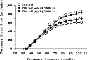
Pinacidil group. The effects of K1
ATP channel activation on the
coronary pressure–flow and pressure–function relations during exer-cise were studied in 11 dogs (of which 6 dogs were also studied in the control group, 3 in the glibenclamide group, and 2 in the glibencla-mide and 8-phenyltheophylline group). With dogs standing on the treadmill, resting measurements of systemic and coronary hemody-namic variables and regional wall thickness were obtained. The ani-mals were then exercised, and the coronary pressure–flow and pres-sure–function relations determined under control conditions as previously described. After 90 min of rest, an infusion of pinacidil into the coronary artery was started in a dose of 0.2 mg/kg per min, delivered at a rate of 0.3 ml/min. 5 min later, resting measurements were obtained, and the exercise protocol was repeated. After another 90 min of rest, the exercise protocol was repeated in the presence of pinacidil infused in a dose of 1.0 mg/kg per min, delivered at a rate of 1.5 ml/min.
High-dose pinacidil group. The effects of a high dose of pinacidil
on the coronary pressure–flow and pressure–function relations were studied in six dogs (of which two animals were studied in the control group, one in 8-phenyltheophylline group, and three in the pinacidil group). Ultrasonic crystals were implanted in five of these animals, but reliable tracking of the signal in the LAD region could be ob-tained in only three of these animals. Animals were exercised, and the coronary pressure–flow relation was determined under control conditions as described above. After 90 min of rest, the exercise pro-tocol was repeated during infusion of pinacidil into the coronary ar-tery in a dose of 5 mg/kg per min, delivered at a rate of 0.6 ml/min.
High-dose adenosine group. The effects of maximal vasodilation
with adenosine on the coronary pressure–flow and pressure–function relations were studied in six dogs (of which two animals were studied in the control group, two in the pinacidil group, and two in the high-dose pinacidil group). Ultrasonic crystals were implanted in four of these animals, but reliable tracking of the signal in the LAD region could be obtained in only two of these animals. Animals were exer-cised, and the coronary pressure–flow relation was determined under control conditions as described above. After 90 min of rest, the exer-cise protocol was repeated during infusion of adenosine into the coro-nary artery in a dose of 50 mg/kg per min, delivered at a rate of 0.6 ml/ min. Maximal vasodilation was demonstrated by the absence of reac-tive hyperemia in response to a 15-s coronary artery occlusion and by the lack of a further increase in coronary flow in response to an in-crease in adenosine infusion rate.
Data analysis
Heart rate, left ventricular and aortic and coronary pressures, coro-nary Doppler shift, and regional wall thickness of the LAD-perfused
region and the LCX-perfused control region were measured from the strip chart recordings. Coronary blood flow was computed from the Doppler shift using the equation Q 5 2.5 ? Df ? d2, where Q is the
cor-onary blood flow (ml/min), Df is the Doppler shift (KHz), and d is the internal diameter of the coronary artery (mm) within the flow probe (14).The factor 2.5 is a constant derived from the speed of sound in tissue (C 5 1.5 ? 105 cm/s), the frequency of the sound beam emitted
(f0 5 10 MHz), the cosine of the angle at which the sound beam is
emitted (458), and unit conversion factors: (C ? p/4 ? 3)/(2f 0 ? cos 458).
Since in the chronically instrumented animals the flow probe adheres to the coronary artery, the internal diameter of the flow probe is equal to the external diameter of the artery. To obtain the inner di-ameter of the coronary artery, we subtracted the arterial wall thick-ness, which in our experience is z 20% of the external diameter of
the coronary artery. In this way, errors in the computation of the cor-onary internal diameter would affect control and intervention condi-tions equally.
Optimal curve fitting of the coronary pressure–flow and pres-sure–function data was obtained with a fourth-order polynomial (y 5
a 1 bx 1 cx2 1 dx3 1 ex4), and coronary flows were computed at
sev-eral coronary pressures within the range of coronary pressures mea-sured during each protocol.
Statistical analysis was performed using two-way (experimental condition and drug treatment) ANOVA for repeated measures. When a significant effect of exercise was observed, comparisons within drug treatment groups were made using one-way ANOVA fol-lowed by Scheffe’s post hoc test. When a significant difference be-tween drug treatments was observed, comparisons bebe-tween treat-ment groups were made with Wilcoxon signed rank test. Statistical significance was accepted at P , 0.05 (two tailed). All data are pre-sented as mean6SEM.
Drugs
Pinacidil was dissolved in deionized water, pH 6.0–6.5. Glibenclamide was dissolved in deionized water, pH 8.0–8.5. 8-Phenyltheophylline was dissolved in dimethyl sulfoxide and deionized water, pH 10.0– 11.0. Adenosine was dissolved in physiologic saline. Sodium nitro-prusside was dissolved in 5% dextrose. All drugs were infused directly into the coronary artery catheter, with the exception of 8-phenylthe-ophylline. Because of the high pH (10–11) of the solution, 8-phenyltheophylline was administered intravenously to avoid inter-ference with coronary vasomotor tone regulation and to prevent pre-cipitation due to mixing with other drug solutions during infusion via the single coronary artery catheter.
Results
Glibenclamide group
Systemic hemodynamics. Exercise increased heart rate from
12264 at rest to 19366 beats/min (P, 0.01), mean arterial
pressure from 9563 to 10663 mmHg (P, 0.01), left
ventricu-lar systolic pressure from 11565 to 13465 mmHg (P, 0.01),
and LVdP/dtmax from 2,4006170 to 4,1406280 mmHg/s (P,
0.01), but did not significantly alter left ventricular end-dia-stolic pressure (Table I). While exercise was continued, pro-gressive coronary artery occlusion caused a marked increase in
left ventricular end-diastolic pressure from 962 mmHg during
exercise with normal coronary arterial inflow to 1662 mmHg
during exercise with total coronary artery occlusion (P, 0.05).
This was accompanied by a slight decrease in left ventricular
systolic pressure to 12265 mmHg (P, 0.05) and a small
in-crease in heart rate to 21765 beats/min (P, 0.05).
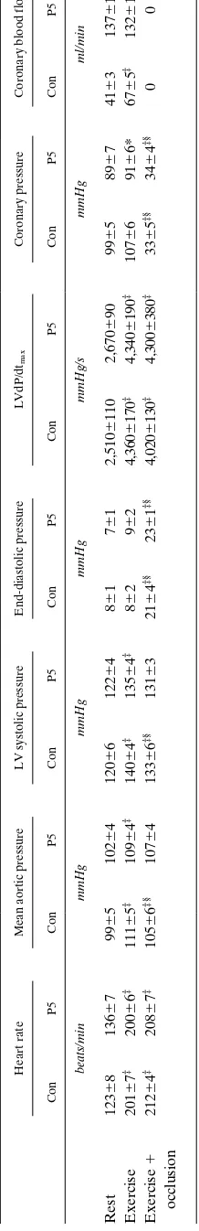
1000
[image:6.612.47.739.71.277.2]D.J. Duncker, N.S. van Zon, Y. Ishibashi, and R.J. Bache
Table I. Hemodynamic and Contractile Function Data during K1
ATP Channel Blockade
Heart rate Mean aortic pressure LV systolic pressure End-diastolic pressure LVdP/dtmax
Con G10 G50 Con G10 G50 Con G10 G50 Con G10 G50 Con G10 G50
beats/min mmHg mmHg mmHg mmHg/s
Rest 12264 12667 14466* 9563 9865 9765 11565 11967 11165 661 861 961* 2,4006170 2,4106250 2,2406170
Exercise 19366‡ 19367‡ 19065‡ 10663‡ 10464‡ 10464‡ 13465‡ 12865*‡ 12666*‡ 962 1061‡ 1362‡ 4,1406280‡ 3,6506280*‡ 3,3706210*‡
Exercise 21766‡§ 21467‡§ 20766*‡§ 10264‡ 10064 10163 12865‡§ 12265§ 11964*‡ 1662‡§ 1662‡§ 1762‡§ 4,0306280‡ 3,6606320*‡ 3,6706210‡§
1 occlusion
Coronary pressure Coronary blood flow EDT EST SWT
Con G10 G50 Con G10 G50 Con G10 G50 Con G10 G50 Con G10 G50
mmHg ml/min mm mm %
Rest 9564 9764 9465 4764 4564 4064* 8.660.7 8.660.7 8.860.9 10.260.7 10.060.8 9.761.0 1962 1663 1062*
Exercise 10463‡ 10464‡ 10364 7166‡ 6365*‡ 5566*‡ 8.860.8 8.960.8 8.961.0 10.960.9‡ 10.561.0 10.061.1* 2363 1864* 1263*
Exercise 3163‡§ 3363*‡§ 3763*‡§ 0 0 0 8.460.9§ 8.560.8§ 8.660.1‡§ 8.360.6‡§ 8.360.7‡§ 8.460.7‡§ 063‡§ 2163‡§ 2263‡§
1 occlusion
Values are mean6SEM. n5 11; n5 7 for left ventricular end-diastolic (EDT) and end-syotolic (EST) wall thickness and systolic wall thickening (SWT) in the LAD region.
*P , 0.05 vs corresponding control measurements, ‡P , 0.05 vs rest, §P , 0.05 exercise vs exercise 1 occlusion, Con control; G10, glibenclamide, 10 mg/kg per min i.c.; G50, glibenclamide, 50 mg/kg per min i.c.
Table II. Hemodynamic Data during Adenosine Receptor Blockade
Heart rate Mean aortic pressure LV systolic pressure End-diastolic pressure LVdP/dt max Coronary pressure Coronary blood flow
Con 8PT Con 8PT Con 8PT Con 8PT Con 8PT Con 8PT Con 8PT
beats/min mmHg mmHg mmHg mmHg/s mm Hg Ml/min
Rest 12567 13767 9964 10565 12364 13064 861 962 3,1206230 3,2706310 9463 9466 49610 5069
Exercise 20365* 20467* 11265* 1,13565* 14465* 14264* 1062 1162 5,4806360* 5,3606440* 9966 9666 78613* 76612*
Exercise 1 20966* 21466* 11064* 10866*‡ 13864*‡ 13666* 1964*‡ 1963*‡ 4,9606270*‡ 5,0606420* 3364*‡ 3665*‡§ 0 0
occlusion
[image:6.612.51.756.434.542.2]pressure and dP/dtmax during exercise (Table I). During
exer-cise in the presence of a total coronary artery occlusion, heart
rate, left ventricular systolic pressure, and dP/dtmax were
slightly lower in the presence of glibenclamide.
Coronary hemodynamics. Control exercise increased
coro-nary pressure from 9564 mmHg at rest to 10463 mmHg (P,
0.05), and coronary blood flow from 4764 ml/min at rest to
7166 ml/min (P, 0.01) (Table I). Progressive coronary artery
occlusion decreased coronary pressure to 3163 mmHg during
exercise in the presence of total coronary artery occlusion. Glibenclamide had no effect on coronary artery pressure at
rest or during exercise. In contrast, glibenclamide 50 mg/kg per
min decreased coronary blood flow at rest from 4764 ml/min
to 4064 ml/min (P , 0.05) and from 7166 to 5566 ml/min
during exercise (P, 0.01) (Table I). Glibenclamide caused a
dose-dependent increase in the coronary pressure at zero flow
during exercise to 3763 mmHg (P, 0.05) (Table II). Since
the extravascular determinants of pressure at zero flow (left ventricular end-diastolic pressure and heart rate) were not al-tered by glibenclamide, the increase in pressure at zero flow must have been due to an increase in vasomotor tone. Glib-enclamide decreased coronary blood flow during exercise at all coronary pressures, so that absolute flow reductions were similar at all pressures (Fig. 4). Thus, the decrease in coronary blood flow caused by glibenclamide was independent of coro-nary pressure.
Myocardial function. Control exercise had no effect on
end-diastolic wall thickness but increased end-systolic wall thickening in both the myocardial region perfused by the LAD
and the control region perfused by the LCX. As a result,
sys-tolic wall thickening increased from 1962% at rest to 2363%
during exercise in the LAD region (Table I), and from 1762%
to 2362% in the circumflex control region (not shown). In the
LAD region, glibenclamide had no effect on end-diastolic wall thickness but decreased end-systolic wall thickening. Conse-quently, glibenclamide decreased systolic wall thickening in
the LAD region to 1062% at rest and to 1263% during
exer-cise (both P , 0.05), with no effect on wall thickening in the
control region. The coronary pressure–function relation dur-ing exercise paralleled the changes observed in the pressure– flow relation (Fig. 4). Systolic wall thickening in the control re-gion was maintained during progressive occlusion of the LAD (not shown).
8-Phenyltheophylline group
Systemic hemodynamics.8-Phenyltheophylline (5 mg/kg, i.v.)
had no effect on any systemic hemodynamic variable at rest or during exercise (Table II).
Coronary hemodynamics. 8-Phenyltheophylline had no
ef-fect on either coronary pressure or coronary blood flow at rest or during exercise with normal arterial inflow. In contrast, 8-phenyltheophylline caused a small but significant decrease in coronary blood flow at coronary pressures of 50 mmHg and
below (Fig. 5), and increased the Pzf from 3364 to 3665
mmHg (P, 0.05) (Table II).
Myocardial function. 8-Phenyltheophylline had no effect
on systolic wall thickening at rest or during exercise with nor-mal arterial inflow. The coronary pressure–function relation during exercise paralleled the changes observed in the pres-sure–flow relation (not shown). Systolic wall thickening in the control region was maintained during progressive occlusion of the LAD.
8-Phenyltheophylline and glibenclamide group
Systemic hemodynamics. The addition of
8-phenyltheophyl-line (5 mg/kg i.v.) to glibenclamide (50 mg/kg per min, i.c.) did
not produce further changes in hemodynamics compared with glibenclamide alone, except for a slightly lower heart rate dur-ing exercise in the presence of a total coronary artery occlusion (Table III).
Coronary hemodynamics. The addition of
8-phenylthe-ophylline to glibenclamide further decreased coronary blood flow compared with glibenclamide alone, and this reached
lev-Figure 4. Coronary pressure–flow and –function relations in dogs
un-dergoing treadmill exercise. The relation between LAD coronary pressure and blood flow is displayed in the upper panel (n 5 11); the relation between LAD coronary pressure and systolic thickening in the anterior left ventricular wall is displayed in the lower panel (n 5 6). Shown are the relations during control (open circles); glibencla-mide, 10 mg/kg per min, intracoronary (closed circles); and glibencla-mide, 50 mg/kg per min, intracoronary (squares). Data are
[image:7.612.58.239.394.642.2]mean6SEM. *P , 0.05 versus corresponding control measurement.
Figure 5. Coronary pressure–flow relation in dogs undergoing
[image:7.612.317.491.563.682.2]1002 D.J. Duncker, N.S. van Zon, Y. Ishibashi, and R.J. Bache
els of significance during exercise (Table III). The addition of 8-phenyltheophylline decreased coronary blood flow at all cor-onary pressures during exercise (Fig. 6), so that the absolute flow reductions were similar at each pressure. Thus, the de-crease in coronary flow produced by 8-phenyltheophylline in the presence of glibenclamide was independent of coronary pressure. Glibenclamide increased coronary zero flow
pres-sure from 3564 to 4667 mmHg during exercise (P, 0.05); the
addition of 8-phenyltheophylline further increased coronary
pressure at zero flow during exercise to 5568 mmHg (P5
0.06) (Table III).
Myocardial function. The addition of
8-phenyltheophyl-line had no effect on end-diastolic wall thickness but further decreased end-systolic wall thickening in the LAD region (Ta-ble III). Consequently, systolic wall thickening in the LAD
re-gion decreased from 1163% during exercise in the presence of
glibenclamide to 562% (P , 0.05), with no effect on wall
thickening in the control region. The coronary pressure–func-tion relapressure–func-tion during exercise paralleled the changes observed in the pressure–flow relation (Fig. 6). Systolic wall thickening in the control region was maintained during progressive occlu-sion of the LAD (not shown).
Pinacidil group
Systemic hemodynamics. Pinacidil (0.2–1.0 mg/kg per min, i.c.)
[image:8.612.66.270.60.737.2]had no effect on any of the systemic hemodynamics either at
Figure 6. Coronary pressure–flow and –function relations in dogs
un-dergoing treadmill exercise. The relation between LAD coronary pressure and blood flow is displayed in the upper panel (n 5 6); the relation between LAD coronary pressure and systolic thickening in the anterior left ventricular wall is displayed in the lower panel (n 5 6). Shown are the relations during control (open circles); glibencla-mide, 50 mg/kg per min, intracoronary (closed circles); and glibencla-mide, 50 mg/kg per min, intracoronary, with 8-phenyltheophylline, 5 mg/kg, intravenous (squares). Data are mean6SEM, n 5 6. *P , 0.05 versus corresponding control measurement. †P , 0.05 versus
corre-sponding glibenclamide measurement.
Table III. Hemodynamic and Contractile Function Data during Combined K
1 ATP
Channel Blockade and Adenosine Receptor Blockade
Heart rate
Mean aortic pressure
LV systolic pressure
End-diastolic pressure LVdP/dt max Con G 8PT 1 G Con G 8PT 1 G Con G 8PT 1 G Con G 8PT 1 G Con G 8PT 1 G beats/min mmHg mmHg mmHg mmHg/s Rest 117 6 7 131 6 7 144 6 79 6 6 4 101 6 4 102 6 8 121 6 5 120 6 5 118 6 89 6 11 1 6 11 0 6 1 2,650 6 280 2,460 6 160 2,540 6 150 Exercise 194 6 4* 201 6 7* 190 6 9* 110 6 5* 107 6 4 105 6 8 141 6 4* 132 6 4* ‡ 126 6 8* 12 6 11 3 6 21 5 6 2* 4,670 6 480* 4,170 6 490* ‡ 3,890 6 300* ‡ Exercise 1 occlusion 196 6 4* 199 6 6* 184 6 10* i 107 6 5* 108 6 5 108 6 6 130 6 4 § 128 6 6* 125 6 72 1 6 2* § 20 6 3* § 20 6 2* § 4,330 6 380* 3,990 6 310* 3,770 6 260* ‡ Coronary pressure
Coronary blood flow
End-diastolic thickness End-systolic thickness Systolic thickening Con G 8pt 1 G Con G 8PT 1 G Con G 8PT 1 G Con G 8PT 1 G Con G 8PT 1 G mmHg mmHg Rest 95 6 49 9 6 4 100 6 94 3 6 33 3 6 52 6 6 3 7.3 6 0.2 7.2 6 0.3 7.1 6 0.2 8.5 6 0.2 7.6 6 0.3 ‡ 7.4 6 0.4 ‡ 15 6 36 6 2 ‡ 5 6 3 ‡ Exercise 104 6 4* 103 6 5 104 6 76 8 6 6* 47 6 8* ‡ 33 6 5* ‡ i 7.3 6 0.2 7.1 6 0.3 7.0 6 0.3 8.6 6 0.5 7.7 6 0.3 ‡ 7.3 6 0.3 ‡ i 19 6 41 1 6 3* ‡ 5 6 2 ‡ i Exercise 1 35 6 4* § 46 6 7* ‡§ 55 6 8* ‡§ ¶ 0 0 0 6.6 6 0.3* § 6.8 6 0.4 § 6.8 6 0.2 § 6.8 6 0.4* § 6.9 6 0.4 6.9 6 0.4 2 6 1* § 1 6 1 § 1 6 2 occlusion
Values are mean
6 SEM. n 5 6; n 5
5 for left ventricular end-diastolic (EDT) and end-systolic (EST) wall thickness and systolic wall thickening (SWT) in the LAD region. *
P
,
0.05 vs rest,
‡P
,
0.05 vs corresponding control
measurement,
§P
,
0.05 exercise vs exercise
1 occlusion, iP , 0.05, ¶P ,
0.06 vs corresponding glibenclamide measurement.
Con
, control;
G
, glibenclamide, 50
m
g/kg per min i.c.; 8PT
1
G
5
8-phenyltheophylline, 5 mg/kg i.v.
1
glibenclamide, 50
m
rest or during exercise with normal arterial inflow or coronary artery occlusion (Table IV).
Coronary hemodynamics. Pinacidil had no effect on
coro-nary artery pressure at rest or during exercise with normal
ar-terial inflow. Coronary blood flow increased from 4162 ml/
min under control conditions to 7065 ml/min during pinacidil,
1.0 mg/kg per min, at rest (P, 0.01) and from 6363 to 8566
ml/min during exercise (P, 0.01) (Table IV). Progressive
cor-onary artery occlusion decreased corcor-onary pressure to 3363
mmHg during control exercise in the presence of total coro-nary artery occlusion. Pinacidil did not alter the corocoro-nary pres-sure at zero flow during exercise (Table IV). Pinacidil in-creased coronary blood flow during exercise at perfusion
pressures . 65 mmHg but had no effect on coronary flow at
pressures , 65 mmHg (Fig. 7).
Myocardial function. Pinacidil had no effect on systolic
wall thickening in either the LAD region or the control region at rest or during exercise (not shown). The coronary pressure– function relations in the LAD region during exercise was also not affected by pinacidil.
High-dose pinacidil group
Systemic hemodynamics. The high dose of pinacidil (5 mg/kg
per min, i.c.) had no effect on any of the systemic hemodynam-ics either at rest or during exercise with normal arterial inflow or coronary artery occlusion (Table V).
Coronary hemodynamics.The high dose of pinacidil caused
a mild decrease in coronary artery pressure which reached sig-nificance during exercise with normal arterial inflow.
Coro-nary blood flow increased from 4163 ml/min under control
conditions to 137613 ml/min during pinacidil, 5.0 mg/kg per
min (P, 0.05), at rest, and from 6765 to 132610 ml/min
dur-ing exercise (P, 0.05) (Table V). The high dose of pinacidil
did not alter the coronary pressure at zero flow during exer-cise. The high dose of pinacidil increased coronary blood flow
during exercise at coronary pressures . 55 mmHg but had no
effect on coronary flow at pressures , 55 mmHg (Fig. 8).
Myocardial function. The high dose of pinacidil had no
ef-fect on systolic wall thickening in either the LAD region or the control region at rest or during exercise (not shown). The cor-onary pressure–function relation in the LAD region during ex-ercise was also not affected by the high dose of pinacidil (not shown).
Table IV. Hemodynamic Data during K
1 ATP
Channel Activation
Heart rate
Mean aortic pressure
LV systolic pressure
End-diastolic pressure LVdP/dt max Con P 0.2 P 1.0 Con P 0.2 P 1.0 Con P 0.2 P 1.0 Con P 0.2 P 1.0 Con P 0.2 P 1.0 beats/min mmHg mmHg mmHg mmHg/s Rest 115 6 3 117 6 6 116 6 5 100 6 3 101 6 4 102 6 3 122 6 4 119 6 4 123 6 46 6 18 6 28 6 1 2,560 6 160 2,490 6 190 2,610 6 200 Exercise 186 6 5* 185 6 5* 190 6 5* 110 6 4* 108 6 4* 110 6 4* 141 6 4* 136 6 3* 137 6 3* 10 6 2* 8 6 29 6 2 4,210 6 320* 4,140 6 320* 4,070 6 330* Exercise 1 occlusion 240 6 4* ‡ 270 6 6* ‡ 209 6 6* ‡ 106 6 3 104 6 4 106 6 3 130 6 4 128 6 4 ‡ 128 6 3* ‡ 16 6 4* ‡ 18 6 3* ‡ 17 6 3* ‡ 4,050 6 280* 4,050 6 350* 3,990 6 300* Coronary pressure
Coronary blood flow
Con P 0.2 P 1.0 Con P 0.2 P 1.0 mmHg ml/min Rest 99 6 39 8 6 3 100 6 34 1 6 25 1 6 3 § 70 6 5 § Exercise 108 6 5* 105 6 5 104 6 56 3 6 3* 69 6 4* § 85 6 6* § Exercise 1 occlusion 33 6 3* ‡ 32 6 3* ‡ 33 6 3* ‡ 00 0
Values are mean
6 SEM. n 5 11; * P ,
0.05 vs rest,
‡P
,
0.05 exercise vs exercise
1
occlusion,
§P
,
0.05 vs corresponding control measurements.
Con
, control;
P 0.2
, pinacidil, 0.2
m
g/kg per min i.c.;
P 1.0
,
pinacidil, 1.0
m
[image:9.612.316.490.561.679.2]g/kg per min i.c.
Figure 7. Coronary pressure–flow relation in dogs undergoing
tread-mill exercise. Shown are the relations during control (open circles); pinacidil, 0.2 mg/kg per min, intracoronary (closed circles); and pinaci-dil, 1.0 mg/kg per min, intracoronary (squares). Data are mean6SEM,
1004 D.J. Duncker, N.S. van Zon, Y. Ishibashi, and R.J. Bache
High-dose adenosine group
Systemic hemodynamics.Adenosine (50 mg/kg per min, i.c.)
had no effect on any of the systemic hemodynamics either at rest or during exercise with normal arterial inflow or coronary artery occlusion (Table VI).
Coronary hemodynamics. The high dose of adenosine
caused a decrease in coronary artery pressure at rest and dur-ing exercise with normal arterial inflow. Coronary blood flow
increased from 5064 ml/min under control conditions to
17065 ml/min during adenosine at rest (P, 0.05), and from
7465 to 14367 ml/min during exercise (P, 0.05) (Table VI).
Adenosine tended to decrease the coronary pressure at zero flow during exercise, but this did not reach levels of statistical significance. In contrast to the high dose of pinacidil, adeno-sine increased coronary blood flow during exercise at coronary pressures as low as 30 mmHg, indicating that coronary vasodi-lator reserve was present even during myocardial hypoperfu-sion (Fig. 9).
Myocardial function. Adenosine had no effect on systolic
wall thickening in either the LAD region or the control region at rest or during exercise. The downward limb of the coronary pressure–function tended to parallel the pressure–flow rela-tion, but in the two animals this did not reach levels of statisti-cal significance (not shown).
Discussion
The present study has yielded several new findings. (a) K1
ATP
channel blockade decreased coronary blood flow during
exer-cise with normal and restricted arterial inflow. (b) Adenosine
receptor blockade reduced coronary flow only at coronary pressures that were associated with severe myocardial
hypo-perfusion. (c) In contrast, in the presence of K1
ATP channel
blockade adenosine blockade aggravated coronary hypoperfu-sion, even at coronary pressures within the autoregulatory
range. (d) K1
ATP channel activation caused increments in
coro-nary flow at normal but not at lower corocoro-nary pressures. (e)
Adenosine produced an increase in flow during both normal and reduced coronary pressures. The implications of these findings will be discussed in detail.
Adenosine. Previous studies have demonstrated that
[image:10.612.68.177.69.742.2]en-dogenous adenosine is not obligatory for maintaining coronary blood flow during normal arterial inflow. Thus, studies in anes-thetized open-chest dogs (15–19) and awake dogs (1, 20) failed to demonstrate an effect of intracoronary adenosine deami-nase (15–17), or adenosine receptor blockade with intravenous aminophylline (18, 19) or 8-phenyltheophylline (1, 20), on
Figure 8. Coronary
pressure–flow relation in dogs undergoing treadmill exercise. Shown are the relations during control (open
circles) and pinacidil, 5
mg/kg per min, intracor-onary (closed circles). Data are mean6SEM,
n 5 6. *P , 0.05 versus corresponding control measurement.
Table V. Hemodynamic Data during K
1 ATP
Channel Activation
Heart rate
Mean aortic pressure
LV systolic pressure
End-diastolic pressure
LVdP/dt
max
Coronary pressure
Coronary blood flow
Con P5 Con P5 Con P5 Con P5 Con P5 Con P5 Con P5 beats/min mmHg mmHg mmHg mmHg/s mmHg ml/min Rest 123 6 8 136 6 79 9 6 5 102 6 4 120 6 6 122 6 48 6 17 6 1 2,510 6 110 2,670 6 90 99 6 58 9 6 74 1 6 3 137 6 13* Exercise 201 6 7 ‡ 200 6 6 ‡ 111 6 5 ‡ 109 6 4 ‡ 140 6 4 ‡ 135 6 4 ‡ 8 6 29 6 2 4,360 6 170 ‡ 4,340 6 190 ‡ 107 6 69 1 6 6* 67 6 5 ‡ 132 6 10* Exercise 1 212 6 4 ‡ 208 6 7 ‡ 105 6 6 ‡§ 107 6 4 133 6 6 ‡§ 131 6 32 1 6 4 ‡§ 23 6 1 ‡§ 4,020 6 130 ‡ 4,300 6 380 ‡ 33 6 5 ‡§ 34 6 4 ‡§ 00 occlusion
Values are mean
6 SEM. n 5 6; * P ,
0.05 vs corresponding control measurements,
‡P
,
0.05 vs rest,
§P
,
0.05 exercise vs exercise
1
occlusion.
Con
, control;
P5
, pinacidil, 5
m
basal coronary blood flow. McKenzie et al. (21) examined the effect of exercise on myocardial adenosine production. They found that treadmill exercise in dogs resulted in a fivefold in-crease in myocardial adenosine content, with a doubling of the coronary arterio-venous adenosine content difference. In dogs with chronically implanted pericardial catheters, graded exer-cise was associated with progressive increases of pericardial fluid adenosine concentrations (22–24), with a positive correla-tion between adenosine concentracorrela-tions and coronary blood flow. However, Bache et al. (1) reported that adenosine recep-tor blockade with 8-phenyltheophylline or increased adeno-sine catabolism with adenoadeno-sine deaminase did not alter exer-cise-induced increases in coronary blood flow. Taken together, these findings indicate that, although myocardial adenosine production increases during exercise, it is not obligatory for maintenance of coronary blood flow at rest or during exercise in the normal heart. This is in agreement with the present study, in which adenosine receptor blockade did not alter cor-onary blood flow at rest or during exercise with normal arterial inflow. In contrast, when coronary pressure was reduced, adenosine blockade significantly decreased blood flow during exercise. Similarly, adenosine receptor blockade has been pre-viously reported to decrease coronary blood flow distal to a coronary artery stenosis (4, 5). These findings suggest that re-ductions of coronary artery pressure sufficient to cause myo-cardial hypoperfusion result in augmented adenosine produc-tion, which contributes to coronary vasodilation.
K1
ATP channels. Several studies in anesthetized and awake
dogs have investigated the role of K1
ATP channels in
maintain-ing coronary blood flow at rest and durmaintain-ing exercise with nor-mal arterial inflow. Using extracorporeally perfused canine hearts, Aversano et al. (8) found that intracoronary
glibencla-mide in doses of 0.8 and 3.7 mmol/min had no effect on basal
coronary blood flow in open-chest dogs. Doses of
glibencla-mide of 0.8 and 3.7 mmol/min administered into the coronary
artery of 25–30-kg dogs correspond to intracoronary infusions
of z 14.4 and 66.5 mg/kg per min (mol wtglibenclamide5 494),
which are in the dose range used in the present study. In con-trast, Imamura et al. (10) reported that intracoronary
glib-enclamide in a dose of 50 mg/kg per min caused a 55%
de-crease in basal coronary blood in open-chest dogs. We
observed that glibenclamide in a dose of 50 mg/kg per min
caused a 20–30% decrease in coronary blood flow in awake resting dogs (11, present study). In addition, coronary blood flow during exercise with normal arterial inflow was
signifi-cantly decreased after K1
[image:11.612.316.556.57.200.2]ATP channel blockade. The reason for
Table VI. Hemodynamic Data during Maximum Vasodilation with Adenosine
Heart rate
Mean aortic pressure
LV systolic pressure
End-diastolic pressure
LVdP/dt
max
Coronary pressure
Coronary blood flow
Con Ado Con Ado Con Ado Con Ado Con Ado Con Ado Con Ado beats/min mmHg mmHg mmHg mmHg/s mmHg ml/min Rest 120 6 8 125 6 88 6 6 58 5 6 5 109 6 5 111 6 47 6 17 6 1 2,330 6 130 2,690 6 120 86 6 56 2 6 2* 50 6 4 170 6 5* Exercise 203 6 8 ‡ 200 6 11 ‡ 103 6 5 ‡ 94 6 6 132 6 5 ‡ 125 6 5 ‡ 9 6 21 0 6 2 ‡ 4,310 6 330 ‡ 3,860 6 230 ‡ 95 6 47 0 6 3* ‡ 74 6 5 ‡ 143 6 7* ‡ Exercise 1 214 6 5 ‡ 204 6 9 ‡ 97 6 6 ‡ 98 6 6 ‡ 123 6 6 ‡§ 127 6 5 ‡ 23 6 1 ‡§ 20 6 4 ‡§ 4,120 6 420 ‡ 3,830 6 220 ‡ 26 6 3 ‡§ 23 6 3 ‡§ 00 occlusion
Values are mean
6 SEM. n 5 7; ‡P ,
0.05 vs rest,
§P
,
0.05 exercise vs exercise
1
occlusion, *
P
,
0.05 vs corresponding control measurements.
Con
, control;
Ado
, adenosine, 50
m
[image:11.612.69.177.75.737.2]g/kg per min i.c.
Figure 9. Coronary
pressure–flow relation in dogs undergoing treadmill exercise. Shown are the relations during control (open
circles) and adenosine,
1006 D.J. Duncker, N.S. van Zon, Y. Ishibashi, and R.J. Bache
the differences in the response of basal coronary flow to simi-lar intracoronary doses of glibenclamide between our study and previous reports is not clear but might be related to the different experimental conditions, i.e., awake (11, present study) versus open-chest (10) versus open-chest extracorpo-real perfusion (8).
Studies in anesthetized (10) and awake dogs (11) demon-strated that the decrease in coronary blood flow produced by
K1
ATP channel blockade with glibenclamide was associated
with a decrease in regional systolic wall thickening. When cor-onary blood flow was restored to preglibenclamide levels with nitroprusside (which itself was devoid of any direct effect on systolic wall thickening), contractile performance recovered as well (10, 11), suggesting that glibenclamide caused a primary decrease in coronary flow with a secondary decrease in con-tractile function. Furthermore, the decrease in coronary blood flow caused by glibenclamide caused metabolic changes sug-gestive of ischemia, including a decrease in myocardial phos-phorylation potential and creatine phosphate with an increase of inorganic phosphate in open-chest dogs (9). Thus, both me-chanical and metabolic evidence support the concept that
K1
ATP channel blockade produced coronary vasoconstriction
that resulted in ischemia-induced contractile dysfunction.
In the present study, K1
ATP channel blockade decreased
coronary blood flow both at coronary pressures in the autoreg-ulatory range and at pressures that had resulted in hypoperfu-sion and loss of systolic wall thickening. These findings are in
agreement with previous studies that indicate that K1
ATP
chan-nels contribute not only to basal coronary tone but also to is-chemic coronary vasodilation. Thus, studies in anesthetized
(10) and awake dogs (11) have shown that K1
ATP channel
blockade not only decreases basal coronary blood flow but also blunts the reactive hyperemia in response to a brief coro-nary artery occlusion. Previous studies in open-chest dogs have
suggested that progressively more K1
ATP channels become
ac-tivated as coronary pressure is decreased. Thus, using intravi-tal microscopy and stroboscopic epiillumination, Komaru et al. (12) reported that superfusion of the subepicardial vasculature
with glibenclamide (1025 M) had no effect on the diameter of
small arterioles (, 100 mm) at coronary artery pressures of 90–
100 mmHg. In contrast, the arteriolar dilation that occurred in response to progressive coronary artery occlusion was abol-ished by glibenclamide. Similarly, in extracorporeally perfused
dog hearts, intracoronary glibenclamide (10 mg/kg per min)
had no effect on coronary blood flow at perfusion pressures
equal to or greater than aortic pressure (. 100 mmHg) but
sig-nificantly decreased flow at pressures , 80 mmHg (13). In
contrast to these studies in open-chest anesthetized dogs, in the present study glibenclamide decreased coronary blood flow in intact exercising dogs both at coronary pressures below the autoregulatory range and at normal pressures, so that the absolute decrease in flow was similar at all coronary pressures.
The data suggest that, although a significant number of K1
ATP
channels are open at coronary artery pressures in the autoreg-ulatory range, progressively more channels are activated as pressure decreases. This hypothesis is supported by the finding that pinacidil increased coronary flow at normal pressures but not at pressures below the autoregulatory range, suggesting that, as perfusion pressure reaches the lower limit of
autoregu-lation, most of the K1
ATP channels have become activated. In
contrast to the effects of pinacidil, adenosine increased flow at coronary pressures within but also below the autoregulatory
range, suggesting that the lack of an increase in flow by pinaci-dil at very low perfusion pressures was not due to exhaustion of vasodilator reserve. Interestingly, Cobb and co-workers (25) reported that the addition of a dose of pinacidil that was with-out effect on basal coronary blood flow enhanced total reac-tive hyperemia flow after 30–60-s coronary artery occlusions, suggesting that brief coronary artery occlusions do not cause
maximal recruitment of K1
ATP channels.
Interaction between adenosine and K1
ATP channels. In the
present study, adenosine receptor blockade had no effect on coronary blood flow at rest or during exercise with normal
ar-terial inflow. However, when the K1
ATP channels were blocked
with glibenclamide, adenosine blockade markedly aggravated myocardial hypoperfusion and further depressed myocardial systolic wall thickening. This suggests that endogenous aden-osine released from the myocardium can oppose the coronary vasoconstrictor effect of glibenclamide. The finding that
aden-osine opposed the hypoperfusion produced by K1
ATP channel
blockade during exercise but not under resting conditions
sug-gests that, after K1
ATP channel blockade, exercise caused
fur-ther deterioration of the oxygen supply–demand balance, thereby augmenting the release of adenosine into the myocar-dial interstitial space. This hypothesis is supported by the find-ings of Berne and co-workers (26), who observed greater increases in interstitial adenosine concentrations in isovolumi-cally beating hearts than in empty beating hearts when both underwent similar coronary blood flow reductions. Moreover, these authors observed excellent inverse correlations between the myocardial oxygen supply–demand balance and interstitial adenosine concentrations (26). Therefore, it is likely that, in
the present study, impaired coronary vasodilation after K1
ATP
channel blockade caused further deterioration of the oxygen supply–demand balance during exercise. The consequent aug-mented release of adenosine into the myocardial interstitial space provided an alternate pathway for coronary vasodilation in response to exercise. This interpretation is supported by the report of Samaha et al. (9) that the addition of adenosine re-ceptor blockade caused further deterioration of coronary blood flow and myocardial phosphorylation potential
com-pared with K1
ATP channel blockade alone in open-chest dogs.
Adenosine receptor blockade had no effect on coronary blood flow during exercise with normal arterial inflow, but de-creased coronary flow at pressures below the autoregulatory plateau. In contrast, after glibenclamide, adenosine blockade further compromised myocardial perfusion during exercise at perfusion pressures both within and below the autoregulatory
range. Thus, while in the presence of unimpaired K1
ATP
chan-nel activity, adenosine plays a role in the regulation of coro-nary vasomotor tone only at lower corocoro-nary pressures, adeno-sine also becomes important for the control of flow at
coronary pressures in the autoregulatory range when K1
ATP
channels are blocked.
Mechanism of interaction between adenosine and K1
ATP
channels. In the present study, the increases in coronary blood
flow produced by intracoronary infusions of nitroprusside, adenosine and pinacidil were measured to determine the
selec-tivity and magnitude of K1
ATP channel and adenosine receptor
va-sodilation produced by the K1
ATP channel opener pinacidil
was selectively inhibited by glibenclamide but was not affected by 8-phenyltheophylline. In contrast, the vasodilation pro-duced by adenosine was attenuated by both 8-phenyltheophyl-line and by glibenclamide. This observation is in agreement with previous reports that adenosine produces coronary
va-sodilation via activation of K1
ATP channels (8, 28, 29).
Al-though glibenclamide inhibited the dilation caused by intracor-onary adenosine, endogenous adenosine continued to exert a vasodilator effect during exercise even in the presence of glib-enclamide. Inhibition of adenosine-induced vasodilation by glibenclamide has been reported to be competitive; i.e., the maximum response elicited by adenosine is not decreased by glibenclamide (29). Consequently, it is possible that, in the
presence of K1
ATP channel blockade, adenosine accumulated
in sufficient concentrations during exercise to compete with
glibenclamide and cause opening of K1
ATP channels. It has
been proposed that, under conditions of normal coronary arte-rial inflow, an increase in metabolic demands causes opening
of vascular smooth muscle K1
ATP channels (7). Although
regu-lation of these channels in vascular smooth muscle is incom-pletely understood, a decrease in ATP or an increase in ADP levels near the sarcolemma of vascular smooth muscle cells (perhaps in response to decreased periarteriolar oxygen ten-sion) is believed to activate these channels (7). Adenosine
re-ceptors, which can activate K1
ATP channels via a G-protein (7),
do not appear to play a role in the channel activation process under normal arterial inflow conditions, since blockade of aden-osine receptors does not interfere with the exercise-induced in-crease in coronary blood flow (1). However, when vascular
smooth muscle K1
ATP channels are inhibited by glibenclamide,
a compensatory increase in interstitial adenosine concentra-tion occurs, which can result in adenosine receptor–mediated
activation of the K1
ATP channel. The present findings suggest
that, when adenosine concentrations are relatively low, e.g.,
under resting conditions, the K1
ATP channel blocking
(vaso-constrictor) effects of glibenclamide predominate over the
K1
ATP channel activating (vasodilator) actions of adenosine. It
is possible that, when exercise in the presence of glibenclamide causes further deterioration of the myocardial oxygen supply– demand balance, adenosine concentrations increase to levels that can effectively compete with glibenclamide, resulting in activation of the channels and coronary vasodilation (26, 30).
It is also possible that adenosine opposed the vasoconstric-tion caused by glibenclamide in part via a different pathway
not involving activation of K1
ATP channels. Adenosine can
cause vasodilation via stimulation of vascular smooth muscle
adenosine A2 receptors, which activate adenylyl cyclase (31–
33). In support of this hypothesis, in the present study, glib-enclamide was slightly more effective at blocking the vasodila-tion produced by pinacidil than that caused by adenosine, suggesting that adenosine could have mediated part of its ef-fect through another vasodilator pathway, likely adenylyl cy-clase activation. Furthermore, pinacidil failed to produce an increase in coronary flow at very low perfusion pressures, whereas adenosine increased flow at coronary pressures both within and below the autoregulatory range. These findings in-dicate that adenosine produced coronary vasodilation in part
via K1
ATP channel activation but also via other mechanisms.
Methodological considerations.The coronary pressure–flow
relation describes the blood flow response over a range of per-fusion pressures within and below the autoregulatory range.
The behavior of coronary blood flow over a range of perfusion pressures is relevant to the clinical situation in which an arte-rial stenosis can result in decreased perfusion pressure. Deter-mination of the entire pressure–flow relation allows examina-tion of blood flow responses over a wide range of poststenotic pressures produced by varying degrees of coronary artery stenosis, allowing a comprehensive assessment of regulation of coronary vasomotor tone. In addition to coronary vasomotor tone, other factors can influence coronary blood flow at a given arterial pressure. Extravascular compressive forces that act on the coronary vasculature can impede coronary blood flow; these forces are highest during systole, when the con-tracting myocardium compresses the intramural vasculature (34). Consequently, an increase in heart rate, which results in an increase in the relative time spent in systole each cardiac cy-cle, will result in an increase in averaged extravascular com-pressive forces (34, 35). In addition, left ventricular intracavi-tary pressure during diastole can impede coronary blood flow. Therefore, to be able to interpret the changes in coronary ar-tery blood flow as reflections of changes in coronary vasomo-tor tone, it is mandavasomo-tory that systemic hemodynamic variables are similar between sequential exercise trials. In the present study, hemodynamic variables (in particular left ventricular end-diastolic pressure and heart rate) were very similar be-tween exercise trials, indicating that differences in systemic he-modynamic profile cannot account for the observed differ-ences in coronary blood flow.
Another factor that can influence the coronary artery pres-sure–flow relation is collateral blood flow. As coronary artery pressure distal to the stenosis drops, collateral blood flow can contribute to total myocardial tissue flow, resulting in underes-timation of myocardial tissue flow when measuring blood flow on a proximal coronary artery. In the present study, we did not measure myocardial tissue blood flow. However, Messina et al. (36) reported that collateral blood flow begins to contribute significantly to myocardial tissue blood flow when intercoro-nary pressure gradients exceed 70 mmHg. In the present study, during exercise mean aortic pressure was in the range of 100– 110 mmHg while the pressure at zero flow was in the range of 30–40 mmHg, which would suggest that the collateral driving pressure was probably not sufficient to result in a considerable contribution of collateral blood flow to total myocardial tissue flow. Furthermore, the observation that changes in coronary
pressure–flow relation produced by K1
ATP channel
(ant)ago-nists and adenosine receptor (ant)ago(ant)ago-nists were accompanied by changes in the coronary–function relation (which is deter-mined by myocardial tissue perfusion) indicates that the changes in the coronary pressure–flow relation measured on a proximal coronary artery did reflect changes in myocardial tis-sue blood flow, and were not compensated for by an increase in collateral blood flow. Finally, the finding that the coronary pressure–flow and pressure–function relations were nearly identical during three consecutive control runs without a right-ward shift of the relations also indicates that there was no sig-nificant collateral blood flow recruitment after the first exer-cise trial.