Akt1 in the cardiovascular system: friend or
foe?
Brian T. O’Neill, E. Dale Abel
J Clin Invest.
2005;
115(8)
:2059-2064.
https://doi.org/10.1172/JCI25900
.
Akt is an important signaling molecule that modulates many cellular processes such as cell
growth, survival, and metabolism. Akt activation has been proposed as a potential strategy
for increasing cardiomyocyte survival following ischemia. In mammalian cells, 3 distinct
isoforms of Akt exist, but their precise roles in cardiovascular biology were previously
unknown. Three separate studies published in this issue of the
JCI
now provide important
new insight into the central role of Akt1 in the regulation of angiogenesis and the
maladaptive or deleterious consequences of chronic unregulated Akt activation in the heart
(see the related articles beginning on pages 2108, 2119, and 2128). Here we discuss the
implications of these exciting new studies.
Commentary
Find the latest version:
Commentaries
Akt1 in the cardiovascular system: friend or foe?
Brian T. O’Neill and E. Dale Abel
Division of Endocrinology, Metabolism and Diabetes and Program in Human Molecular Biology and Genetics, University of Utah School of Medicine, Salt Lake City, Utah, USA.
Akt is an important signaling molecule that modulates many cellular
pro-cesses such as cell growth, survival, and metabolism. Akt activation has been
proposed as a potential strategy for increasing cardiomyocyte survival
fol-lowing ischemia. In mammalian cells, 3 distinct isoforms of Akt exist, but
their precise roles in cardiovascular biology were previously unknown. Three
separate studies published in this issue of the
JCI
now provide important new
insight into the central role of Akt1 in the regulation of angiogenesis and the
maladaptive or deleterious consequences of chronic unregulated Akt
activa-tion in the heart (see the related articles beginning on pages 2108, 2119, and
2128). Here we discuss the implications of these exciting new studies.
The serine/threonine kinase Akt (also known as protein kinase B [PKB]) plays a central role in the regulation of cellular growth, survival, and metabolism across many species. In mammalian cells, there are 3 distinct Akt isoforms (Akt1, -2, and -3; also known as PKBα, -β, and -γ), which are the products of distinct genes (1). Gene knockout studies have provided evidence that these isoforms may serve distinct roles with varying degrees of functional overlap. For example, germline deletion of Akt1 in mice leads to a predominant phenotype of reduced size for multiple organs, which is more the result of reduced cell size than reduced cell numbers. In contrast, Akt2 KO mice do not exhibit striking reductions in cell size, but develop insulin resistance and diabetes due to impaired insulin signaling in the liver and skeletal muscle and impaired survival of pancreatic β cells (2–4). Akt3 KO mice exhibit a predominantly neurologi-cal phenotype with reduced brain size (5). While mice with individual Akt isoform KOs are viable, evidence for functional over-lap of these isoforms comes from studies in mice with combinatorial KOs. Animals with multiple gene deletions exhibit much more severe phenotypes (which are not com-patible with long-term survival) than those animals with single gene deletions (6). Akt isoforms can be activated in response to var-
ious growth factors and hormones, includ-ing insulin, IGF-1, VEGF, and β-adrenergic stimulation. In the case of insulin, the insu-lin receptor tyrosine kinase phosphorylates and activates insulin receptor substrate–1 (IRS-1) and IRS-2, which then activate PI3K. PI3K activation generates phosphoinosit-ide-3,4,5 triphosphate, which mediates the phosphorylation and activation of Akt. Once activated, Akt phosphorylates various intracellular signaling intermediates that regulate a diverse array of cellular processes (reviewed in ref. 7) (Figure 1A).
In the cardiovascular system, Akt plays an important role in the regulation of cardiac hypertrophy, angiogenesis, and apoptosis (7). The observation that acute activation of Akt in cardiomyocytes in vivo and in vitro protects against apoptosis after isch-emia/reperfusion (I/R) injury has raised hopes that Akt activation could represent an important therapeutic strategy for lim-iting myocardial injury following ischemia (8) (Figure 1A). Moreover, activation of Akt in mesenchymal stem cells may enhance cardiac function in heart failure models to a greater extent than that observed in non-transduced stem cells (9, 10). Although the results of transgenic overexpression studies suggest that Akt overexpression may pro- duce a compensated state of cardiac hyper-trophy, other work suggests that this might
be deleterious over the long term (11–13). Two separate reports in this issue of the
JCI, the first by Nagoshi et al. (14) and the second by Shiojima et al. (15), provide important new insights into the potential mechanisms by which Akt overexpression in the heart may become maladaptive.
Another unresolved issue in the cardio-vascular system was the specific role of the 3 Akt isoforms. For example, while it was clear that Akt mediates many of the proan-giogenic effects of secreted peptides such as VEGF, it remained unknown which specif-ic isoforms were responsible for mediating these effects in vivo (7). Similarly, while an essential role for PI3K/Akt signaling in the regulation of physiological (e.g., exercise-induced) hypertrophy is well established (16, 17), it was not previously known which specific Akt isoform(s) were responsible. In this issue of the JCI, the study by Ackah et al. (18) now provides very strong evidence that Akt1 is the specific Akt isoform that mediates adaptive angiogenesis.
Mechanisms responsible for maladaptation in the heart when Akt is chronically overexpressed
How much Akt is too much? Two studies in this issue describe distinct ways in which chronic unregulated Akt signaling in car-diomyocytes in vivo ultimately becomes maladaptive (14, 15). Both of these studies used mouse models with transgenic over-expression of an activated (myristoylated) form of Akt1. Shiojima and colleagues (15) used a tetracycline-regulated system that increased myocardial Akt kinase activity by approximately 15-fold, while Nagoshi and colleagues (14) used a transgenic mouse with constitutive Akt1 activation in which Akt kinase activity was increased to at least the extent of the Shiojima et al. study. The model reported by Shiojima and colleagues developed severe cardiac dysfunction fol-lowing long-term Akt activation (15), while cardiac dysfunction in mice reported by Nagoshi et al. was less severe (14). Two other independent transgenic lines of myo-cardial Akt1 overexpression were previously reported (12, 13). High-level overexpression (greater than 80-fold) of a constitutively Nonstandard abbreviations used: I/R, ischemia/
reperfusion; IRS, insulin receptor substrate; mTOR, mammalian TOR; PKB, protein kinase B; S6K, S6 kinase; TOR, target of rapamycin.
Conflict of interest: The authors have declared that no conflict of interest exists.
Citation for this article: J. Clin. Invest. 115:2059–2064 (2005). doi:10.1172/JCI25900.
commentaries
active Akt1 mutant, T308D/S473D, pre-cipitated age-related LV dysfunction (13), whereas overexpression of another mutant Akt1, E40K, was associated with increased basal cardiac contractility but impaired inotropic response to dobutamine (12). Taken together, these models suggest that
Akt1 overexpression in the heart may pro-duce a broad phenotypic spectrum in the unstressed heart, but in all cases there was some evidence of cardiac maladaptation. In contrast, a PI3K transgenic mouse with 4-fold increase in the ratio of phosphory-lated Akt/total Akt did not develop cardiac dysfunction (19). Moreover, in physiologi-cal contexts, Akt activation is increased 1.5-
[image:3.585.55.536.88.509.2]to 2-fold in response to exercise training (16) and 6-fold by transgenic overexpres-sion of IGF-1 (20) or the IGF-1 receptor (17). In these situations cardiac dysfunc-tion does not occur. Thus, interpretation of the phenotypes of transgenic models of Akt1 overexpression should always be tempered by the fact that the degree of
Figure 1
commentaries
transgene overexpression often far exceeds physiologically achieved levels.
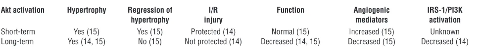
The studies by Nagoshi et al. (14) and Shiojima et al. (15) provide mechanistic insights into the deleterious consequences of long-term Akt1 overexpression in the heart (Figure 1B and Table 1). Shiojima and colleagues (15) report that short-term (less than 2-week) activation of Akt1 in the
heart leads to compensated cardiac hyper-trophy that is completely reversible after transgene expression is turned off. In con-trast, 6 weeks of transgene induction led to even greater LV hypertrophy and contractile dysfunction, both of which are associated with increased fibrosis and decreased capil- lary density. Thus, unlike short-term trans-gene activation in which VEGF expression is upregulated (in part by target of rapa- mycin–mediated [TOR-mediated] signal-ing) (Figure 1A), such adaptations are lost following longer-term activation of Akt (Figure 1B). This observation prompted Shiojima and colleagues to test the hypoth- esis that matched angiogenesis and cellu-lar growth is required for maintenance of compensated hypertrophy following Akt activation. Blocking VEGF signaling — by systemic administration of an adenovirus that expressed the soluble VEGF recep-tor — reduced capillary density following short-term Akt activation, and this precipi- tated LV dysfunction and increased myocar-dial injury. This model raises the intriguing
hypothesis that signaling cross-talk medi-ated by Akt1 is required for the coordinate regulation of myocardial growth and angio-genesis during cardiac hypertrophy. As Akt is activated in failing hearts and in response to pressure overload, it will therefore be important to determine whether Akt-medi- ated VEGF release and subsequent angio-genesis are impaired in these contexts.
The study by Shiojima et al. (15) raises additional questions, which future stud-ies will undoubtedly address: (a) Does increased injury account for all of the reduction in cardiac function in Akt1 transgenic mice, or could additional mechanisms such as altered myocardial excitation-contraction coupling or altered myocardial energetics exist?; (b) Does the fibrosis observed represent increased cell death via necrosis or apoptosis, or are pro-fibrotic signaling pathways activated? It is possible that if Akt-dependent pathways such as that resulting in VEGF production can be downregulated with chronic Akt expression; could this also be true of pro-survival/antiapoptotic pathways?; and (c) What is the mechanism responsible for the downregulation of TOR-mediated signal-ing to S6 kinase (S6K) and VEGF but not to myocyte growth that is seen in mice with chronic Akt activation? The results of S6K KO mouse studies suggest that S6K is not required for mediating pathological and physiological cardiac hypertrophy; how-ever, rapamycin clearly blocks Akt-induced cardiac hypertrophy and leads to regres-sion of pressure-overload hypertrophy (21, 22). Therefore, the additional mammalian TOR (mTOR) targets that mediate cardiac hypertrophy need to be identified.
Chronic Akt1 overexpression and the heart’s recovery from I/R injury
Nagoshi and colleagues report that in con-trast to mice with lower level and mosaic expression of a myristoylated Akt1 trans-gene (transgenic mouse line TG20), which their group previously reported to exhibit reduced infarct size following regional I/R
in vivo (11), a transgenic mouse line express-ing higher levels of Akt (transgenic mouse line TG564) failed to recover any function after global I/R in an isolated heart model and developed larger myocardial infarctions (14). The basis for myocardial injury is likely increased necrosis and not increased apop-tosis. The results of the Nagoshi et al. study suggest that downregulation of IRS-1/2– mediated PI3K activation is an important contributor to the increased susceptibility to I/R injury in this model. IRS-1/2 levels are reduced because of decreased expression of their respective genes as well as increased proteasomal degradation (Figure 1B). The I/R phenotype can be rescued by delivery of an adenovirus that harbors a constitutively active form of PI3K. These data support the notion of the presence of Akt-independent but PI3K-dependent cardioprotective signal- ing pathways that are lost when IRS-1–medi-ated signal transduction is impaired in the face of long-term Akt activation. This con-clusion is also supported by the observations by Nagoshi et al. that IRS-1 is depleted to a much lesser extent in the transgenic mouse line TG20, which was previously reported to exhibit enhanced recovery from I/R injury in
vivo (11), and that transgenic mice that over-express IGF-1 (which does not downregulate IRS-1 content despite an approximate 6-fold increase in Akt activation) have enhanced recovery from ischemic injury (14).
The major activation of IRS-1/2 signaling to PI3K in vivo occurs via tyrosine kinase receptors such as the insulin and IGF-1 receptors. Nagoshi et al. also show that in heart samples from humans with dilated
cardiomyopathy, Akt activation is associ-Table 1
Divergence in long-term versus short-term effects of Akt signaling
Akt activation Hypertrophy Regression of I/R Function Angiogenic IRS-1/PI3K
hypertrophy injury mediators activation
Short-term Yes (15) Yes (15) Protected (14) Normal (15) Increased (15) Unknown
Long-term Yes (14, 15) No (15) Not protected (14) Decreased (14, 15) Decreased (15) Decreased (14)
References corresponding to these results are cited in parentheses.
[A third study] in this issue
of the JCI . . . now provides
very strong evidence that
Akt1 is the specific Akt
isoform that mediates
adaptive angiogenesis.
How much Akt is too
[image:4.585.53.537.112.164.2]commentaries
ated with reduced IRS-1 levels (14), which raises the intriguing possibility that loss of IRS-1–mediated PI3K signal transduction may contribute to the cardiac injury in these patients. Thus these results underscore the importance of signaling to IRS-1/2 via these growth factors/hormones in mitigating the myocardial response to ischemic injury. Results obtained from 3 other mouse mod-els with impaired PI3K/Akt signaling also support the paradigm that growth factors signaling to PI3K are critical modulators of cardiac injury in response to stress: mice with cardiomyocyte-restricted KO of insu- lin receptors exhibit reduced cardiac perfor-mance and increased injury in response to pressure overload (23); mice with muscle-specific KO of
phosphoinositide-depen-dent kinase 1 that fail to phosphorylate Akt on Thr308 (although Ser473 is hyper-phosphorylated) die prematurely from a dilated cardiomyopathy (24); and mice with a dominant-negative PI3K transgene do not develop exercise-induced hypertrophy, but develop LV dysfunction following pressure-overload hypertrophy (16). Taken together, these data indicate that proximal signaling inputs to PI3K and Akt (such as insulin) play important regulatory roles in the adaptation of the heart to diverse stresses such as isch-emia and pressure-overload hypertrophy.
The current study by Nagoshi et al. (14) raises a number of questions and hypotheses that should be examined in future studies. First, what are the PI3K-dependent but Akt-independent pathways that are responsible
for cardioprotection, and are these pathways activated by alternative signaling pathways such as G protein–coupled receptors that may activate PI3K independently of IRS-1/2? It is unlikely that Akt-mediated protective pathways are not important, as shown by the impaired functional recovery in domi-nant-negative Akt transgenic or Akt1–/– mice
following I/R injury. It would therefore be interesting to determine the consequence of PI3K activation in Akt1-null hearts on their recovery from ischemic injury.
Second, Nagoshi and colleagues show that IRS-1 levels are increased in Akt1–/–
mice. Conversely, Akt1 overexpression pro-motes proteasomal degradation of IRS-1 that is not mediated via the mTOR pathway
(14). What, then, are the specific mecha-Figure 2
[image:5.585.45.539.78.427.2]commentaries
nisms that lead to IRS-1 degradation in this model? There is much evidence that serine phosphorylation of IRS-1 mediates its pro-teasomal degradation and that there are a variety of serine/threonine kinases that can mediate this process (25). It will therefore be important to determine whether Akt is directly responsible for the serine phos- phorylation of IRS-1 or whether the respon-sibility lies in an Akt-dependent kinase. Third, what are the mechanisms that lead to increased cell death in Akt transgenic mice following I/R? The report by Shiojima et al. (15) raises the intriguing possibility that decreased capillarity could limit capil-lary recruitment during reperfusion, which could contribute to increased injury. Anoth-er possibility is that changes in energetics that occur on the basis of chronic Akt acti- vation may precipitate more rapid deple-tion of high energy phosphates or impair ATP regeneration upon reperfusion. In this regard, studies in E40K Akt transgenic mice suggest that 1 mechanism for increased contractility in these mice is increased activ- ity of the sarcoplasmic endoplasmic reticu-lum Ca2+ ATPase, which would increase the rate of ATP utilization (26). Finally, it would be of interest to deter- mine the time course for IRS-1 degrada-tion in vivo using the inducible Akt model. A critical experiment would be to deter-mine the extent of IRS-1/PI3K activation in the initial 2-week window during which the phenotype of these mice is completely reversible and determine whether recovery from I/R injury is normal or impaired fol-lowing short-term Akt activation. In summary, these 2 transgenic mouse models (14, 15) define important Akt-dependent feedback mechanisms that lead to cardiac maladaptation. These include feedback inhibition of VEGF syn-thesis, which impairs angiogenesis, as well as inhibition of IRS-1/2 expression and IRS-mediated PI3K activation (Figure 1B). Akt has many more substrates within the cell. No doubt there will be a growing list of other signaling pathways that may be inhibited by long-term unregulated Akt signaling, such as the recently reported increase in insulin receptor threonine phosphorylation by Akt1-mediated path- ways that leads to impaired insulin signal-ing (27). Stay tuned.Akt1 and adaptive angiogenesis
A persuasive case for a role of the Akt1 iso- form in the regulation of postnatal adap-tive angiogenesis and arteriogenesis (as
opposed to developmental angiogenesis) is made in this issue of the JCI by Ackah and colleagues (18). In contrast to the expres- sion seen in the heart and intact blood ves-sels, which express both Akt1 and Akt2, there appears to be low-level expression of Akt2 in endothelial and vascular smooth
muscle cells. Thus in the vasculature, Akt2 might be predominantly expressed in adventitial fibroblasts. Ackah et al. (18) demonstrate that Akt1, but not Akt2, medi-ates the angiogenic response to hind-limb ischemia and VEGF stimulation in vivo (Figure 2). Moreover, Akt1 is required for the homing of endothelial precursor cells to areas of ischemia in vivo. Akt1 is also required for basal and agonist-mediated NO release from mouse lung endothelial cells (MLECs) and for the migration of MLECs (which express Akt1) and mouse lung fibroblasts (which express both Akt1 and Akt2) in response to chemoattractants in vitro. In light of the evidence provided by Shiojima and colleagues showing that a coordinated balance between angiogen-esis and cellular hypertrophy is essential for an adaptive response to physiological hypertrophy (15), the results of Ackah et al. (18) raise interesting questions regarding the potential role of Akt1-mediated angio-genesis in the failed induction of cardiac hypertrophy by exercise in mice express-ing a dominant-negative PI3K transgene (16) or in the increased injury observed in cardiomyocyte-specific insulin receptor knockout (CIRKO) mice following pressure overload (23). Does impaired VEGF-medi- ated angiogenesis therefore retard physi-ological hypertrophy or promote injury in the hearts of mice with impaired PI3K/Akt signaling? This will be an important ques-tion to pursue in future studies. In light of the novel specific roles for Akt1 now defined in the vasculature and heart, it will be important to identify the specific downstream mediators of these effects. Akt continues to take center stage, and these recent studies have provided important new insights into the regula-tion of cardiac and vascular biology by
this family of signaling molecules (14, 15, 18). The studies have important implica-tions for therapeutic strategies that may be based upon modulating Akt activity in the heart and vasculature in an effort to mitigate the consequences of heart failure and myocardial or other organ ischemia. In this installment of “Akt in the cardio-vascular system,” Akt1 has clearly emerged as the star of Act 1. We are sure that Act 2 will be equally interesting. Acknowledgments B.T. O’Neill is supported by a Physician- Scientist Training Award from the Ameri- can Diabetes Association. E.D. Abel is sup-ported by grants HL 73167, HL 70070, and HL 70525 from the National Institutes of Health and is an Established Investigator of the American Heart Association. Address correspondence to: E. Dale Abel, Division of Endocrinology, Metabolism and Diabetes, Program in Human Molecu-lar Biology and Genetics, 15 North 2030 East, Building 533 Room 3410B, Salt Lake City, Utah 84112, USA. Phone: (801) 585-0727; Fax: (801) 585-0701; E-mail: dale. abel@hmbg.utah.edu. 1. Bellacosa, A., Testa, J.R., Moore, R., and Larue, L. 2004. A portrait of AKT kinases: human cancer and animal models depict a family with strong individ-ualities [review]. Cancer Biol. Ther. 3:268–275. 2. Chen, W.S., et al. 2001. Growth retardation and
increased apoptosis in mice with homozygous dis-ruption of the Akt1 gene. Genes Dev. 15:2203–2208. 3. Cho, H., Thorvaldsen, J.L., Chu, Q., Feng, F., and Birnbaum, M.J. 2001. Akt1/PKBalpha is required for normal growth but dispensable for mainte-nance of glucose homeostasis in mice. J. Biol. Chem.
276:38349–38352.
4. Cho, H., et al. 2001. Insulin resistance and a diabetes mellitus-like syndrome in mice lacking the protein kinase Akt2 (PKB beta). Science. 292:1728–1731. 5. Easton, R.M., et al. 2005. Role for Akt3/protein
kinase Bgamma in attainment of normal brain size.
Mol. Cell. Biol. 25:1869–1878.
6. Peng, X.D., et al. 2003. Dwarfism, impaired skin development, skeletal muscle atrophy, delayed bone development, and impeded adipogenesis in mice lacking Akt1 and Akt2. Genes Dev. 17:1352–1365.
7. Oudit, G.Y., et al. 2004. The role of phosphoinosit-ide-3 kinase and PTEN in cardiovascular physiology and disease [review]. J. Mol. Cell. Cardiol. 37:449–471. 8. Matsui, T., et al. 2001. Akt activation preserves car-diac function and prevents injury after transient cardiac ischemia in vivo. Circulation. 104:330–335. 9. Gnecchi, M., et al. 2005. Paracrine action accounts
for marked protection of ischemic heart by Akt-modified mesenchymal stem cells. Nat. Med.
11:367–368.
10. Mangi, A.A., et al. 2003. Mesenchymal stem cells modified with Akt prevent remodeling and restore performance of infarcted hearts. Nat. Med.
9:1195–1201.
11. Matsui, T., et al. 2002. Phenotypic spectrum caused by transgenic overexpression of activated Akt in the heart. J. Biol. Chem. 277:22896–22901.
commentaries
12. Condorelli, G., et al. 2002. Akt induces enhanced myocardial contractility and cell size in vivo in transgenic mice. Proc. Natl. Acad. Sci. U. S. A.
99:12333–12338.
13. Shioi, T., et al. 2002. Akt/protein kinase B pro-motes organ growth in transgenic mice. Mol. Cell. Biol. 22:2799–2809.
14. Nagoshi, T., et al. 2005. PI3K rescues the detrimen-tal effects of chronic Akt activation in the heart during ischemia/reperfusion injury. J. Clin. Invest.
115:2128–2138. doi:10.1172/JCI23073.
15. Shiojima, I., et al. 2005. Disruption of coordinated cardiac hypertrophy and angiogenesis contributes to the transition to heart failure. J. Clin. Invest.
115:2108–2118. doi:10.1172/JCI24682.
16. McMullen, J.R., et al. 2003. Phosphoinositide 3-kinase(p110alpha) plays a critical role for the induction of physiological, but not pathological, cardiac hypertrophy. Proc. Natl. Acad. Sci. U. S. A.
100:12355–12360.
17. McMullen, J.R., et al. 2003. The insulin-like growth factor 1 receptor induces physiological heart growth via the phosphoinositide 3-kinase(p110alpha) pathway. J. Biol. Chem. 279:4782–4793.
18. Ackah, E., et al. 2005. Akt1/protein kinase Bα is critical for ischemic and VEGF-mediated angio-genesis. J. Clin. Invest. 115:2119–2127. doi:10.1172/ JCI24726.
19. Shioi, T., et al. 2000. The conserved phosphoinosit-ide 3-kinase pathway determines heart size in mice.
EMBO J. 19:2537–2548.
20. Yamashita, K., et al. 2001. Reperfusion-activated Akt kinase prevents apoptosis in transgenic mouse hearts overexpressing insulin-like growth factor-1.
Circ. Res. 88:609–614.
21. McMullen, J.R., et al. 2004. Deletion of ribosomal S6 kinases does not attenuate pathological, physi- ological, or insulin-like growth factor 1 receptor- phosphoinositide 3-kinase-induced cardiac hyper-trophy. Mol. Cell. Biol. 24:6231–6240.
22. McMullen, J.R., et al. 2004. Inhibition of mTOR signaling with rapamycin regresses established cardiac hypertrophy induced by pressure overload.
Circulation. 109:3050–3055.
23. Hu, P., et al. 2003. Minimally invasive aortic band-ing in mice: effects of altered cardiomyocyte insulin signaling during pressure overload. Am. J. Physiol. Heart Circ. Physiol. 285:H1261–H1269.
24. Mora, A., et al. 2003. Deficiency of PDK1 in cardiac muscle results in heart failure and increased sensi-tivity to hypoxia. EMBO J. 22:4666–4676. 25. White, M.F. 2002. IRS proteins and the common
path to diabetes. Am. J. Physiol. Endocrinol. Metab.
283:E413–E422.
26. Kim, Y.K., et al. 2003. Mechanism of enhanced car-diac function in mice with hypertrophy induced by overexpressed Akt. J. Biol. Chem. 278:47622–47628. 27. Morisco, C., et al. 2005. Akt mediates the cross-talk
between beta-adrenergic and insulin receptors in neonatal cardiomyocytes. Circ. Res. 96:180–188.
A new direction for gene therapy: intrathymic
T cell–specific lentiviral gene transfer
Ruth Seggewiss and Cynthia E. Dunbar
National Heart, Lung, and Blood Institute, NIH, Bethesda, Maryland, USA.
Reports of neoplasia related to insertional activation of protooncogenes by
retroviral vectors have raised serious safety concerns in the field of gene
ther-apy. Modification of current approaches is urgently required to minimize
the deleterious consequences of insertional mutagenesis. In this issue of the
JCI
, Adjali and colleagues report on their treatment of SCID mice lacking the
70-kDa protein tyrosine kinase, ZAP-70, with direct intrathymic injection
of a ZAP-70–expressing T cell–specific lentiviral vector, which resulted in
T cell reconstitution (see the related article beginning on page 2287). Using
lentiviral vectors and in situ gene transfer may represent a safer approach
than using retroviral vectors for ex vivo gene transfer into HSCs, avoiding
3 factors potentially linked to leukemogenesis, namely HSC targets, ex vivo
transduction and expansion, and standard Moloney leukemia virus–based
retroviral vectors.
Conflict of interest: The authors have declared that no conflict of interest exists.
Citation for this article: J. Clin. Invest. 115:2064–2067 (2005). doi:10.1172/JCI26401.
SCID and its current standard of therapy
SCID disorders are a family of genetically determined conditions characterized by a block in T cell development and function, variably associated with defects in other hematopoietic lineages. The genetic and phenotypic heterogeneity among these disorders is considerable. Affected children present with severe opportunistic infections due to a lack of functional mature T cells. The most serious forms of SCID are fatal in the first years of life (1). The only curative standard of therapy is an allogeneic bone
marrow transplant from an HLA-matched, related donor. If this is not possible, bone marrow transplantation from an HLA- matched, unrelated or a parental haploi-dentical donor is performed. The survival rate after HLA-matched transplantation is greater than 75% but falls markedly with the use of alternative donors, including haploidentical family donors and unrelat- ed donors, and chronic disabling complica-tions such as graft-versus-host disease are common. Some children reach full func-tional immunity following transplantation, but others fail to have their B cell function restored or can experience progressive loss of T cell immunity and even complete graft failure over time. These poor outcomes are particularly common when children are transplanted with non–HLA-matched
donor cells, for instance from a parent or an unrelated donor (2, 3).
Leukemia induction after successful oncoretroviral gene transfer in SCID-X1
These unsatisfying outcomes, particu- larly with alternative donor transplanta-tion, have stimulated intense interest in gene therapy approaches to SCID. The most common SCID subtype is SCID- X1, an X-linked recessive disease charac-terized by a block in T and natural killer cell differentiation due to mutations in the common γ chain of cytokine receptor complexes. Recently, complete immune reconstitution of young boys with SCID-X1 by transduction of their CD34+ bone marrow cells with an oncoretroviral vector encoding the common γ chain followed by reinfusion of transduced cells without conditioning therapy was reported (Figure 1A) (4, 5). These encouraging results rep- resented the first unequivocal demonstra-tion of the clinical efficacy of gene therapy. However, elation was short-lived, as 3 out of 11 enrolled children in this clinical trial have developed T cell leukemia linked to insertional mutagenesis, specifically, acti-vation of the LIM domain only 2 (known as