Neph1 and nephrin interaction in the slit
diaphragm is an important determinant of
glomerular permeability
Gang Liu, … , Yashpal S. Kanwar, Sumant S. Chugh
J Clin Invest.
2003;
112(2)
:209-221.
https://doi.org/10.1172/JCI18242
.
Neph1
-deficient mice develop nephrotic syndrome at birth, indicating the importance of this
protein in the development of a normal glomerular filtration barrier. While the precise
subcellular localization of Neph1 remains unknown, its relationship with other components
of the glomerular filtration barrier is of great interest in this field. In this paper, we localize the
expression of Neph1 to the glomerular slit diaphragm by immunogold electron microscopy
in rodents and describe its direct interaction with two other components of the slit
diaphragm, nephrin and ZO-1. Both native and recombinant Neph1 associate with each
other as dimers and multimers and interact with nephrin via their extracellular segments.
Disruption of the Neph1-nephrin interaction in vivo by injecting combinations of individual
subnephritogenic doses of anti-Neph1 and anti-nephrin results in complement- and
leukocyte-independent proteinuria with preserved foot processes. This disruption modestly
reduces Neph1 and nephrin protein expression in podocytes and dramatically reduces
ZO-1 protein expression via the interaction of ZO-ZO-1 PDZ domains with the cytoplasmic tail of
Neph1, independent of changes in mRNA expression of all three genes. The interaction
between nephrin and Neph1 is specific and not shared by either protein with P-cadherin,
another integral slit diaphragm protein. The interaction between nephrin and Neph1
therefore appears to be an important determinant of glomerular permeability.
Article
Aging
Find the latest version:
Introduction
A large number of genes with relevance to the biology of the podocyte and slit diaphragm have recently been characterized. Some encode transcriptional factors and are thought to regulate the expression of podocyte pro-teins (1–3), but the vast majority encode for structural components of the podocyte foot process or the slit diaphragm. Whereas the localization of ZO-1 (4) and the mAb 5-1-6 antigen (5) to the cytoplasmic and extra-cellular aspects, respectively, of the slit diaphragm were in themselves stellar observations, the discovery of nephrin (6) opened the field for the identification of other novel proteins. The rapid discovery of CD2 AP (7), podocin (8), α-actinin 4 (9), P-cadherin (10), and FAT (11) as components of the podocyte foot process/slit diaphragm complex, the recent identifica-tion of filtrin (12), and the recharacterizaidentifica-tion of mAb
5-1-6 as an anti-nephrin Ab (13) are just some of the important developments over the past 4 years.
This study addresses the characterization of Neph1, a gene recently mutated in mouse embryonic stem cells using a high-throughput mutagenesis method (14). Mutant mice generated from these stem cells develop nephrotic syndrome at birth, underscoring the importance of Neph1 in the development of a normal glomerular permeability barrier. Electron microscopy of the kidneys from the mutant mice revealed the absence of normal foot process develop-ment, and Lac-Z staining of these sections revealed the presence of Neph1-β-geo fusion transcripts in sev-eral cell types, including podocytes. A single 9-kb tran-script was noted by Northern blot of several organs, with high expression in the kidney. Whereas the human Neph1 cDNA sequence published by Do-noviel et al. (14) encoded for five Ig-like domains and a transmembrane domain, the mouse counterpart had only four Ig-like domains and lacked a trans-membrane domain, suggesting that it was a partial clone. A more recent paper (15) included both full-length mouse and human sequences, and described the interaction of Neph1 with podocin. Whereas the expression of Neph1 in the podocyte is not in doubt, its localization in the slit diaphragm has been inferred from the nephrotic phenotype of Neph1–/–mice and its
protein-protein interactions (16).
Neph1 and nephrin interaction in the slit diaphragm
is an important determinant of glomerular permeability
Gang Liu,
1Beenu Kaw,
2Jayson Kurfis,
1Syed Rahmanuddin,
1Yashpal S. Kanwar,
3and Sumant S. Chugh
11Division of Nephrology, Department of Medicine, Feinberg School of Medicine, Northwestern University,
Chicago, Illinois, USA
2Department of Medicine, Illinois Masonic Medical Center, Chicago, Illinois, USA
3Department of Pathology, Feinberg School of Medicine, Northwestern University, Chicago, Illinois, USA
Neph1-deficient mice develop nephrotic syndrome at birth, indicating the importance of this protein in the development of a normal glomerular filtration barrier. While the precise subcellular localiza-tion of Neph1 remains unknown, its relalocaliza-tionship with other components of the glomerular filtralocaliza-tion barrier is of great interest in this field. In this paper, we localize the expression of Neph1 to the glomerular slit diaphragm by immunogold electron microscopy in rodents and describe its direct interaction with two other components of the slit diaphragm, nephrin and ZO-1. Both native and recombinant Neph1 associate with each other as dimers and multimers and interact with nephrin via their extracellular segments. Disruption of the Neph1-nephrin interaction in vivo by injecting com-binations of individual subnephritogenic doses of anti-Neph1 and anti-nephrin results in comple-ment- and leukocyte-independent proteinuria with preserved foot processes. This disruption mod-estly reduces Neph1 and nephrin protein expression in podocytes and dramatically reduces ZO-1 protein expression via the interaction of ZO-1 PDZ domains with the cytoplasmic tail of Neph1, inde-pendent of changes in mRNA expression of all three genes. The interaction between nephrin and Neph1 is specific and not shared by either protein with P-cadherin, another integral slit diaphragm protein. The interaction between nephrin and Neph1 therefore appears to be an important determi-nant of glomerular permeability.
J. Clin. Invest.112:209–221 (2003). doi:10.1172/JCI200318242.
Received for publication February 28, 2003, and accepted in revised form May 6, 2003.
Address correspondence to:Sumant S. Chugh, Division of Nephrology, Department of Medicine, Feinberg School of Medicine, Northwestern University, 320 East Superior Avenue, Searle 10-475, Chicago, Illinois 60611, USA.
Phone: (312) 503-3072; Fax: (312) 503-0622; E-mail: s-chugh@northwestern.edu.
Conflict of interest:The authors have declared that no conflict of interest exists.
In this paper, we provide evidence for the subcellular localization of Neph1 in the glomerular slit diaphragm by immunogold electron microscopy. We present evi-dence that Neph1 interacts with nephrin in vitro and in vivo, and that this heterologous protein-protein interaction is an important factor in maintaining the normal permeability characteristics of the slit diaphragm. We also show the association of PDZ domains of ZO-1 with the cytoplasmic tail of Neph1 and characterize a novel model of the functional prop-erties of the slit diaphragm.
Methods
Cloning of mouse Neph1 cDNA. Using primers designed from the published incomplete clone, we identified a full-length Neph1 cDNA by 3′rapid amplification of cDNA ends (RACE) and RT-PCR in the following man-ner. Total RNA was extracted from 4-week-old male C57BL/6 mouse kidney using TRIzol reagent (Invitro-gen Corp., Carlsbad, California, USA), and cDNA tem-plates were generated by reverse transcription using SuperScript II (Invitrogen Corp.). Using primers at either end of the published sequence, PCR fragments were cloned into pCRII (Invitrogen Corp.), sequenced, and compared with the published sequence. Using primers K7 (5′- GCAGGAGGGGGCTGTGACTAGCACG-GAGC-3′) and K8 (nested primer 5′- GGGGGCTGTGAC-TAGCACGGAGCTGC-3′) from this sequence, 3′RACE was performed using the GeneRacer kit (Invitrogen Corp.) per the manufacturer’s instructions. A 2,673-bp RACE product was cloned into pCRII, and both sense and antisense strands were sequenced up to the polyA tail. Upon combining both sets of clones, a stop codon was noted 2,367 bp downstream of the open reading frame. Using primers overlapping the start and stop codons, several full-length cDNA clones were generat-ed, sequencgenerat-ed, and compared for sequence identity. Protein translation, domain and motif analysis, and sequence comparisons were done using software on the BLAST (17), Scansite (18), and ExPASy (19) web-sites. Signal peptide prediction was done using SignalP version 1.1 software on the CBS World Wide Web Pre-diction server (20) and transmembrane domain pre-diction was done on the “DAS” Transmembrane Pre-diction server (21).
Since several experiments in rats were anticipated, we also cloned, sequenced, and assembled a partial rat Neph1 cDNA sequence using two primer pairs from the mouse sequence (K281, 5′-GTACAGTGGACCAAGGACGGGCTGG -3′and K283, 5′-CTGCATGCGCTGCTGGAATCGCTGGCC -3′; K282, 5′-GTCTGATGACGCTTCCTATGAGTGCCAGG-3′ and K284, 5′-CTCATAGTTCTCGTAGGACAGCTGGCTGG -3′) and rat kidney cDNA. The rat cDNA sequence and its predicted protein sequence were used primarily to design real-time PCR primers and peptides for Ab generation, respectively.
Raising polyclonal Ab’s. The extracellular segments of several mouse podocyte proteins were analyzed by hydrophobicity plots using Kyte-Doolittle curves on
the ProtScale web site (22); the most hydrophilic por-tions specific to the protein were selected for peptide synthesis. Peptide sequences that were chosen were homologous or nearly homologous (maximum of one AA difference) with corresponding rat sequences. The Neph1 peptide sequences that were synthesized had perfect homology with both mouse and rat Neph1 pro-teins. Prior to immunization, 15 ml of preimmune serum was obtained from each rabbit. Two KLH-con-jugated peptides (of 15 AAs each) for each protein were used to immunize two rabbits each to raise polyclonal Ab’s. Initial immunization was done with complete Freund’s adjuvant, whereas boosters at days 20, 40, and 60 were given with incomplete Freund’s adjuvant. ELISA titers for antigen-specific reactivity were checked after the first bleed, at day 70, using peptide coated wells. The following Ab’s were raised: anti–α -dystro-glycan (PNQRPELKNHIDRVD, STTTTTRRPTKKP-RTP), anti–β-dystroglycan (RRIADENGKPRPAFS, TEVPDRDPEKSSEDD), anti-nephrin (DLQDPRY-TEHKYHQG, FPRYSLEGDSAKGEF), anti–aminopep-tidase A (YVQPNQKETAEYAA, YTLEQYQKT-SLAQEK), and anti-Neph1 (EDAHESRYETNVDYS, RSMNEAIPNGKETSI). These Ab’s were raised by Gen-emed Synthesis Inc. (South San Francisco, California, USA) on a fee-for-service basis.
Rabbit anti–ZO-1 and rat anti–P-cadherin with reac-tivity against the extracellular domain of P-cadherin (10) were purchased from Zymed Laboratories Inc. (South San Francisco, California, USA). Other Ab’s purchased included mouse anti–rat CD45 (Cedarlane Laboratories Ltd., Hornby, Ontario, Canada), goat anti–mouse CD2 AP (Santa Cruz Biotechnology Inc., Santa Cruz, California, USA), goat anti-rabbit IgG cou-pled with 10-nm gold particles (Electron Microscopy Services, Fort Washington, Pennsylvania, USA), rabbit anti-goat IgG coupled with 15-nm gold particles (Elec-tron Microscopy Services), and goat anti–rat C3 (ICN Biomedical Inc., Aurora, Ohio, USA). Secondary Ab’s for immunofluorescence and Western blotting were purchased from Sigma-Aldrich (St. Louis, Missouri, USA). Rabbit anti–rat albumin Ab was obtained from Research Diagnostics Inc. (Flanders, New Jersey, USA). General laboratory reagents were obtained from Sigma-Aldrich. Reagents for electron microscopy were purchased from Electron Microscopy Services. Labora-tory animals were purchased from Harlan Sprague Dawley Inc. (Indianapolis, Indiana, USA).
very little measurable baseline proteinuria. For the induction of proteinuria, all Ab’s or antisera were heat-treated at 56°C for 30 minutes to inactivate comple-ment prior to intravenous injection into rats or mice. To study in vivo interactions between Neph1 and nephrin or P-cadherin, dose-response studies with anti-Neph1, anti-nephrin, and anti–P-cadherin were first performed by injecting rats via the tail vein with up to 500 µl of antisera (anti-nephrin, anti-Neph1, or preimmune serum) or up to 100 µg of anti–P-cad-herin. The goal of this study was to define the opti-mum subnephritogenic dose of these Ab’s beyond which a mild increase in slit diaphragm permeability would occur. The rats were placed in metabolic cages with access to water but no food for an 18-hour urine collection after injection. Proteinuria was measured using a protein assay kit from Bio-Rad Laboratories Inc. (Hercules, California, USA) based on the Bradford method; readings were multiplied by 1.33 to adjust for a 24-hour collection. Wherever proteinuria was noted, urine samples were appropriately diluted in PBS, resolved by SDS PAGE, and stained with Gel-Code Blue (Pierce Biotechnology Inc., Rockford, Illi-nois, USA). Samples were also processed for Western blot with an anti–rat albumin Ab. To test for an in vivo association between two proteins, the highest subnephritogenic dose of one of two Ab’s or a combi-nation of 50% of that dose of both Ab’s was intra-venously injected into three different groups of ani-mals. In the anti-Neph1 and anti-nephrin study, two groups of rats were injected with 300 µl of either anti-Neph1 or anti-nephrin, while a third group received a combination of 150 µl of each Ab. A fourth group (control) received 300 µl of preimmune serum from the same rabbits. For the anti-Neph1 and anti–P-cad-herin study, rats received either 300 µl of anti-Neph1, 75 µg of anti–P-cadherin, a combination of 50% of these doses, or 300 µl of preimmune serum. We also intravenously injected increasing doses up to 250 µl of preimmune serum, anti-nephrin, anti–α-dystro-glycan, anti–β-dystroglycan, and anti–aminopeptidase A (anti-APA) separately into 8- to 12-week-old female CD1 mice, and analyzed the collected urine for pro-teinuria. Since rat glomeruli can be isolated by sieving with over 90% purity, most of the in vivo studies that required measurement of glomerular protein and mRNA expression were conducted in rats.
In all rodent studies, kidneys were removed after euthanasia and appropriate portions were used for fresh glomerular isolation for protein or total RNA extraction, or preserved for electron microscopy (with 2.5% glutaraldehyde), light microscopy (with 3.7% formaldehyde), or frozen sections (snap-frozen and preserved in 2-methylbutane).
Expression of FLAG-tagged proteins. To study their relative roles in protein-protein interactions, cDNA fragments corresponding to both extracellular and intracellular seg-ments of Neph1 were cloned separately into pCAL-n-FLAG (Stratagene, La Jolla, California, USA) in the
following manner. A 1,424-bp fragment of mouse Neph1 cDNA corresponding to the extracellular segment was cloned into pCAL-n-FLAG between the EcoRI and HindI-II sites using primers K227 ( GCGCGCGAATTCCC-CGGGACTCAGACTCGCTTCAGCC) and K230 ( GCGCG-CAAGCTTCTATAACACCTCTCGCTCTTCCAGCTGG). A 717-bp fragment corresponding to the intracellular seg-ment was cloned between the same restriction enzyme sites using primers K229 ( GCGCGCGAATTCCTCTACC-GACGTCGCAAAGGCAGTC) and K233 ( GCGCGCAAG-CTTTCTACACATGAGTCTGCATGCGCTGCTGG). A seg-ment of mouse ZO-1 containing the three PDZ domains was also cloned between the BamHI and HindIII sites of pCAL-n-FLAG using primers K238 ( GCTATAGGATCCAA-GATGTCCGCCAGGGCCGCGG) and K239 ( GCGCTTAA-GCTTTGCGGCGATAAACGTCCTTCTTC) and a cDNA template derived from mouse kidney total RNA by reverse transcription.
After verifying the accuracy of the plasmids by sequencing, protein expression was induced with iso-propyl-β-D-thiogalactoside (IPTG) in BL21-Gold (DE3) E. coli(Stratagene) according to the product manual. Protein expression was verified by resolving preinduced and induced cultures by SDS PAGE under reducing and nonreducing conditions and staining with GelCode Blue, as well as transferring the proteins to nitrocellu-lose for Western blot with anti-FLAG (all expressions) or anti-Neph1 (extracellular segment only). The puri-fied recombinant proteins were column-puripuri-fied, dia-lyzed against PBS, and stored at –80°C. To generate non–FLAG-tagged recombinant protein, the FLAG domain and the calmodulin-binding peptide were cleaved from the recombinant protein using enteroki-nase as recommended in the product manual for pCAL-n-FLAG. Non–FLAG-tagged and FLAG-tagged recom-binant proteins were used in competition experiments.
Western blot and coimmunoprecipitation studies. For glomerular studies involving the characterization of rat/mouse Neph1 and for coimmunoprecipitation studies, a standard protocol described previously was used (23). Autoradiographs were scanned into Adobe Photoshop 5.0, and band densitometry was measured using Gel-Pro Analyzer 3.1 software (Media Cybernet-ics Inc., Silver Spring, Maryland, USA).
SDS PAGE. Following transfer onto nitrocellulose membranes, Western blot studies were conducted with anti-Neph1, anti-nephrin, and anti-ZO-1.
For coimmunoprecipitation studies involving FLAG-tagged recombinant proteins and native glomerular protein extracts, the amount of FLAG-tagged protein to be added to the glomerular extract was determined in the following manner. Thirty micrograms of the glomerular extract was resolved by SDS PAGE along with different concentrations of recombinant protein and stained with GelCode Blue (Pierce Biotechnology Inc.). We then determined the recombinant protein band that appeared to be roughly equivalent in intensi-ty to corresponding bands at the same molecular weight in the membrane extract. The protein concentration of the selected band determined the amount of the FLAG-tagged protein added to the glomerular extract.
Deglycosylation studies. To study whether native mouse Neph1 from glomerular extracts was actually glycosylated as predicted, we denatured anti-Neph1
immunoprecipitates with 0.1 M 2-mer-captoethanol and 0.1% SDS, followed by overnight incubation at 37°C with either PNGase F or control. The proteins were then resolved by reducing SDS PAGE and Western blot with anti-Neph1 Ab.
Affinity absorption of anti-Neph1 Ab with Neph1 peptides and recombinant extracellular segment of Neph1. We coupled 3 mg of each of the two peptides used to generate the anti-Neph1 Ab with 1 ml of CNBr-activat-ed Sepharose 4B (Amersham Pharmacia Biotech, Uppsala, Sweden) according to the manufacturer’s instructions to make an affinity column that was used to absorb out reactivity to these peptides from the anti-Neph1 Ab. Depletion of anti-Neph1 activity was assessed by Western blot and immunofluorescence. A similar strategy was adopted for using nephrin peptides to absorb out reactivity to nephrin from the anti-nephrin Ab. A second affinity column for mouse Neph1 was made with 3 mg of the recombinant extracellular segment of Neph1 after the FLAG tag and the calmod-ulin-binding peptide tag were removed using enterokinase, and was tested in a similar manner to the first affinity column.
Real-time PCR for Neph1, nephrin, and ZO-1. Real-time PCR was used to study rat glomerular mRNA expression of nephrin, Neph1, and ZO-1 in the groups injected with anti-nephrin, anti-Neph1, or a combi-nation of the two as described above. Glomeruli were isolated from each rat by sieving, and 2 µg of total RNA was used to generate cDNA by reverse transcription. Three cDNA templates were generated from each rat so that every animal was assayed in triplicate. The following primers and TaqMan probes for real-time PCR were designed using Primer Express ver-sion 2.0 software (Applied Biosystems, Foster City, Cali-fornia, USA). Rat nephrin: H7 forward primer 5′
-TTCAGCAAGGAGACCTTCAAGAA-3′, H8 reverse primer
5′-GCCCCTCAATCCACAGCTT-3′, probe 6FAM- TCACT-CACCTTGAATGTGA-MGB; rat ZO-1: H13 forward primer 5′-GAGCTACGCTTGCCACACTGT-3′, H14 reverse primer 5′-TCGGATCTCCAGGAAGACACTT-3′, probe
6FAM-ACCCTAAAACTTGGCAAAA-MGB; rat Neph1: H3 forward primer 5′-TTGCTGCCTTAGTGTTCTTTCTCTAC -3′, H4 reverse primer 5′-CCTCAACGTCACATCCTTTCG-3′, probe 6FAM-ACGTCGCAAAGGCAGT-MGB.
[image:5.576.63.332.58.279.2]Forward and reverse primers were designed in adja-cent exons to avoid false amplification by possible con-taminating genomic DNA. All real-time PCR products from individual primer pairs were sequenced to ensure the accuracy of the PCR reaction. Control 18S RNA probe (VIC-MGB) and corresponding primers were pur-chased from Applied Biosystems. Real-time PCR was
Figure 1
performed in a 96-well format on the ABI Prism 7000 (Applied Biosystems) in multiplex mode (i.e., study primers/probe plus 18S control primers/ probe) using preset cycling parameters specified by the manufacturer. Appropriate nontemplate con-trols were included in each experiment. Prior to the run, threshold settings were set at 0.2. After the run was concluded, the data was reanalyzed to obtain Ct values with a threshold setting of 0.75 in ∆Rn versus Cycle mode for all groups. ∆Ct values for each indi-vidual reading were calculated by subtracting the corresponding 18S Ct values from each study group reading, mean ± SE was calculated for each group, and differences between combination and individual Ab groups were analyzed by Student ttest. In addi-tion, we subtracted the ∆Ct of the individual Ab-injected groups from that of the combination inject-ed group to obtain the ∆∆Ct and calculated the difference in mRNA expression between these groups using the formula 2–∆∆Ct(see Applied
Biosys-tems ABI 7000 user bulletin no. 2 for sequence detec-tion at http://docs.appliedbiosystems.com/search. taf). A fourfold difference in mRNA expression, equivalent to a mean difference of two cycles by
real-time PCR, was used as a criterion to define a signifi-cant difference between the groups.
Imaging studies. For immunofluorescence studies, a standard protocol recently described in detail was used (23). The positive control for complement staining with anti–rat C3 Ab was day 5 kidney of a rat injected with sheep anti–rat Fx1A. Positive control for leukocyte staining with anti-CD45 was kidney from a rat inject-ed with whole sheep anti-rat nephrotoxic serum 24 hours prior to euthanasia.
For immunogold electron microscopy of perfused rat kidney and for transmission electron microscopy, stan-dard protocols described elsewhere were used (25, 26). The positive control for the procedure of immunogold localization of Neph1 was staining for CD2 AP in rat glomeruli. In addition, other rats were injected sepa-rately with 150 µl of either anti-Neph1 or anti-nephrin 1 hour prior to perfusion fixation, followed by staining with 10 nm gold–coupled goat anti-rabbit IgG to assess for binding of injected Ab’s to the slit diaphragm.
[image:6.576.79.515.358.649.2]Statistical analysis to study the difference in protein expression between different experimental groups was done using the unpaired Student ttest in Microsoft Excel 2000. A Pvalue of < 0.05 was taken as significant.
Figure 2
Results
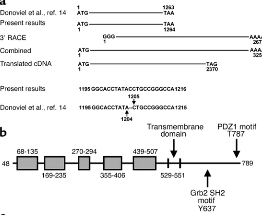
Cloning of mouse Neph1 cDNA. Cloning and sequencing by our group of the mouse cDNA fragment published by Donoviel et al. (14) revealed a single base pair dis-parity (absence of C after 1,204 in the Donoviel et al. sequence) which had led to a premature stop codon after 1,260 bp (Figure 1a). Combining our correspon-ding sequence with the 2,673-bp 3′RACE product revealed a 3,255-bp clone with a stop codon 2,367 bp downstream of the open reading frame, encoding for a 789-AA protein. This sequence has been submitted to GenBank (accession number AY243095). The 2,107-bp partial rat cDNA sequence also obtained had over 95% homology at the nucleotide level and 98% homology at the protein level with mouse Neph1 cDNA (GenBank accession number AY271309).
Structural analysis of the mouse Neph1 gene. A BLAST search of the mouse genome with our cDNA sequence revealed the presence of 15 exons spanning a 57.4-kb region of chromosome 3, with intron 1 being dispro-portionately long (42.7 kb). Perfect identity between the predicted exons and our mouse cDNA sequence was noted. Our sequence, as well as the exons in the genom-ic sequence, differ from another recently submitted sequence by four base pairs (present study and Sellin et al., ref. 15) 1,064 C/T; 1,324 A/G, 2,008 A/G, 2,179 T/C).
Characterization of the mouse Neph1 protein. Domain analy-sis of the predicted protein sequence revealed a signal peptide (AAs 1–47), five IgG-like domains with the mid-dle domain overlapping with a PKD-like domain, an
RGD sequence (AAs 437–439), a transmembrane domain (AAs 529–551), and a cytoplasmic tail (552–789) (Figure 1b). The most significant motifs in the cytoplasmic tail include the Grb2 SH2 site at 637 and the PDZ1 motif at 787. Another motif on the cytoplasmic tail is the Itk SH2 at 672. However, the presence of Itk kinases in the podocyte has not yet been described, although this does not exclude potential binding by a member of the Tec kinase family at that site.
[image:7.576.63.356.55.363.2]Western blot studies of native rodent glomerular pro-tein suggest that Neph1 exists as a monomer of about 110 kDa under reducing conditions (Figure 2, a and b) and as a dimer and perhaps even as a multimer under nonreducing conditions (Figure 2c). These observations are confirmed by Western blot studies under nonre-ducing conditions with the recombinant protein that show dimeric bands of the extracellular and intracellu-lar segments (Figure 2e), and a multimeric band of the extracellular segment. Association among Neph1 mol-ecules is also favored by the coimmunoprecipitation of native Neph1 by an anti-FLAG Ab from a mixture of native mouse glomerular proteins and the FLAG-tagged recombinant extracellular domain (see Figure 5b, lane 11). The 110-kDa size of rodent Neph1 on reducing Western blot is identical to that noted by Sellin et al. (15), but differs from the 90-kDa protein noted by Bar-letta et al. (27). Since the predicted molecular weight of the protein, excluding the signal peptide, is 82 kDa, slower migration on Western blot may be related to posttranslational modification of the protein. Indeed,
Figure 3
there are four potential N-glycosylation sites (at AAs 78, 172, 329, and 503), seven potential O-glycosylation sites (at AAs 56, 256, 349, 597, 698, 700, and 703), and sever-al potentisever-al phosphorylation sites in Neph1 (Figure 1c). Indeed, deglycosylation of immunoprecipitated mouse Neph1 with PNGase F causes the protein to migrate at approximately 90 kDa (Figure 2d).
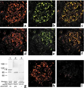
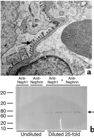
Localization of Neph1 in adult and developing rodent glomeruli. Dual immunofluorescence staining of adult rat (Figure 3, a–c) and mouse glomeruli reveals an interrupted capillary loop pattern that is identical to, and colocalizes well with, the staining pattern for CD2 AP, suggesting that Neph1 is present in podocytes. Similar colocalization of Neph1 and CD2 AP was noted in the day 15 developing mouse kidney in the precapillary loop stage (Figure 3, d–f). In this immunofluorescence experiment, CD2 AP was used merely as a podocyte marker, since the limited resolu-tion of immunofluorescence does not differentiate slit diaphragm proteins from other podocyte proteins present in the vicinity. Anti-Neph1 Ab depleted of immunoreactivity by absorption over a Neph1 pep-tide affinity column (Figure 3g) or recombinant Neph1 extracellular segment (blots not shown) failed to stain rodent glomeruli by immunofluorescence (Figure 3, h and i). Similar results were noted with anti-nephrin absorbed with a nephrin peptide column (data not shown). Immunogold electron microscopy, which is the technique of choice for precise subcellu-lar localization, reveals most of the 10-nm gold parti-cles to be present in the slit diaphragm, thereby estab-lishing Neph1 as a slit diaphragm protein (Figure 4b). Some gold particles were also present on the foot processes in the vicinity of the slit diaphragm. The positive control for this study shows most of the 15-nm gold particles representing CD2 AP to be present on the cytoplasmic aspect of the slit diaphragm (Fig-ure 4a). In a different experiment, both anti-Neph1 (Figure 4c) and anti-nephrin (Figure 4d) Ab’s injected into separate rats 1 hour prior to perfusion fixation localized almost exclusively to the slit diaphragm by immunogold staining.
Overall, in a survey of ten glomeruli per study for binding of gold particles, more than 95% of the gold particles in the anti-Neph1– and anti-nephrin–injected rats were present in the slit diaphragm, compared with 75–80% in sections stained with the anti-Neph1 Ab in noninjected rats. The rest of the particles in both groups were equally divided between the foot process surface in close proximity to the slit diaphragm and the podocyte cytoplasm. In glomeruli stained with anti–CD2 AP, about 85% of the gold particles were pres-ent at the cytoplasmic aspect of the slit diaphragm, and the rest were present in the podocyte cytoplasm away from the slit diaphragm. Whereas Donoviel et al. had noted the presence of Neph1-β-geo fusion transcripts in podocytes, parietal epithelial cells, and mesangial cells, glomerular expression of native Neph1 as noted by our group appears to be restricted to podocytes.
[image:8.576.323.507.255.565.2]Association of Neph1 with nephrin. We conducted coim-munoprecipitation experiments to study potential inter-actions between three slit diaphragm proteins: Neph1, nephrin, and P-cadherin. Coimmunoprecipitation stud-ies under nonreducing conditions using anti-Neph1 and anti-nephrin Ab’s, along with the corresponding preim-mune antisera, revealed that nephrin coimmunoprecipi-tates with anti-Neph1 and vice versa (Figure 5a). Fur-thermore, we were able to coimmunoprecipitate the recombinant extracellular segment of mouse Neph1 with anti-nephrin, and vice versa (Figure 5b), but not the recombinant intracellular part (blots not shown), indi-cating that the Neph1-nephrin interaction occurs pri-marily at the level of the extracellular segments of these proteins. We were also able to demonstrate competition between FLAG-tagged and non–FLAG-tagged (unFLAG)
Figure 4
proteins during coimmunoprecipitation studies with anti-Neph1 and anti-nephrin, as shown in Figure 5b. Lane 4 and lane 5 (unFLAG/FLAG-tagged ration, 5:1;
n= 3 readings/group) show anti-Neph1 coimmunopre-cipitates; densitometry readings were 2,235 ± 29 (lane 4) and 927 ± 26 (lane 5), densitometric units, P< 0.01. Lane 6 and lane 7 (unFLAG/FLAG ratio, 10:1) show anti-nephrin coimmunoprecipitates; densitometry readings were 1,272 ± 20 (lane 6) and 49 ± 3 (lane 7) densitometric units, P< 0.01). We were unable to coimmunoprecipitate P-cadherin with either anti-nephrin or anti-Neph1. As negative controls for the method of coimmunoprecipi-tation, we performed similar studies between slit diaphragm proteins (using anti-nephrin and anti-Neph1) and focal adhesion complex proteins (using anti–α- and anti–β-dystroglycan) or podocyte surface proteins (using anti-APA), all of which were negative (blots not shown).
In vivo study of the Neph1-nephrin association. We ration-alized that disruption of slit diaphragm function would result in proteinuria and that targeting two interacting proteins critical to the barrier function
[image:9.576.59.314.52.311.2]would cause this disruption more efficiently than tar-geting them alone. To study a functional association between slit diaphragm proteins, we first injected anti-nephrin, anti-Neph1 (Table 1 and Figure 6a), and anti–P-cadherin Ab’s (Table 2) separately into rats in increasing amounts to find a threshold nephritogenic dose that would cause mild proteinuria compared with preimmune serum injection. Dose-response curves with individual Ab’s helped us identify an optimum subnephritogenic dose in rats for anti-Neph1 (300 µl, Figure 6a and Table 1), anti-nephrin (300 µl, Figure 6a and Table 1), and anti–P-cadherin (75 µg, Table 2). Sim-ilarly, in mouse experiments, the optimum subnephri-togenic doses were 200 µl for anti-APA and 150 µl for anti-nephrin and anti-Neph1. We were unable to induce any proteinuria in mice beyond baseline with anti–α-dystroglycan and anti–β-dystroglycan. Of vari-ous subnephritogenic combinations tested, we were able to induce heterologous-phase proteinuria within the first 18 hours with anti-Neph1 and anti-nephrin; this proteinuria declined over the next 2 days (Table 3
Figure 5
[image:9.576.56.536.623.702.2]Composite Western blots showing the association of native (a) and recombinant (b) mouse Neph1 and nephrin. (a) Native Neph1 from mouse glomerular extracts coimmunoprecipitates with anti-nephrin (lane 2), and nephrin with anti-Neph1 (lane 3) under nonreducing conditions. All lanes, including control immunoprecipitations (lane 1 and lane 4), show the promi-nent band of nonreduced rabbit IgG at 150–160 kDa and traces of heavy chain (50 kDa) and light chain (25 kDa). (b) Lane 1 shows the absence of FLAG-reactive proteins in native mouse glomerular extracts. In lanes 2–11, native glomerular extracts were combined with FLAG-tagged recombinant extra-cellular Neph1 and also supplemented with an excess of recombinant extracellular Neph1 digested with enterokinase to remove the FLAG tag (labeled “unFLAG”) in lane 5 (unFLAG/FLAG ratio, 5:1) and lane 7 (unFLAG/FLAG ratio, 10:1). Coimmunoprecipitation of recombinant Neph1 by anti-Neph1 (lane 4) and anti-nephrin (lane 6); coimmunoprecipi-tation of nephrin (lane 9) and native Neph1 (lane 11) by anti-FLAG; and competition between FLAG-tagged and non–FLAG-tagged protein (lane 5 and lane 7) are demon-strated (see text for details). GE, glomerular extract.
Table 1
Urinary protein excretion in response to anti-Neph1 and anti-nephrin
Preimmune serum Anti-Neph1 Anti-nephrin
Dose injected Proteinuria Dose injected Proteinuria Dose injected Proteinuria
Baseline 0.87 ± 0.19 Baseline 0.87 ± 0.19 Baseline 0.87 ± 0.19
100 µl 0.65 ± 0.05 100 µl 0.80 ± 0.05 100 µl 0.70 ± 0.10
300 µl 0.65 ± 0.02 300 µl 0.80 ± 0.40 300 µl 0.80 ± 0.09
500 µl 1.30 ± 0.10 500 µl 2.36 ± 0.38A,B 500 µl 1.84 ± 0.04A,B
and Figure 6b). This experiment was repeated with older rats and revealed similar results. The proteinuria was comprised primarily of albumin, as was obvious from the approximately 70-kDa band seen on GelCode Blue–stained gels in the combination-injected group (Figure 7b); this was confirmed to be albumin by West-ern blot (blots not shown). The following combina-tions did not result in significant proteinuria beyond baseline: anti-Neph1 and anti–P-cadherin; anti-nephrin and anti–P-cadherin; anti-nephrin and anti-APA; and anti-Neph1 and anti-APA.
Light microscopy of the kidney (hematoxylin and eosin stain) in all four groups of the anti-Neph1 and anti-nephrin experiment at 24 hours after injection showed the glomeruli to be normocellular, and stain-ing for complement and leukocytes was negative by immunofluorescence with appropriate positive con-trols (images not shown). Transmission electron microscopy revealed preserved foot processes in all four groups, as exemplified by an electron micrograph from proteinuric rats at 24 hours (Figure 7a). Immunofluo-rescence for bound rabbit IgG (anti-Neph1, anti-neph-rin, or combination) revealed a change from the normal
interrupted linear pattern to a cluster-like distribu-tion in the three experimental groups starting 12 hours after intravenous injection of Ab (images not shown); this has been observed with several podocyte surface proteins by several investigators, including us, and appears to be unrelated to proteinuria (23). The induction of complement- and leukocyte-inde-pendent proteinuria by a combination of individual-ly subproteinuric Ab doses indicates disruption of an in vivo association between nephrin and Neph1 in the slit diaphragm, without interference with foot process integrity.
As anticipated, we were unable induce proteinuria in rats using a combination of 150 µl of anti-nephrin and 150 µl of the Neph1 peptide column unbound frac-tion, reiterating the specific role played by anti-Neph1 Ab’s in this process: baseline proteinuria, 0.85 ± 0.15 mg/24 hours; Ab injected, 0.95 ± 0.17 mg/24 hours (mean ± SE,n= 4 rats weighing 100 g, P> 0.05). Simi-lar results were noted when 150 µl of anti-Neph1 was affinity-absorbed over a recombinant extracellular Neph1 column and injected in combination with 150 µl of anti-nephrin: baseline, 0.85 ± 0.15 mg/24 hours; antibody injected, 0.92 ± 0.15 mg/24 hours, mean ± SE (n= 4 rats weighing 100 g each, P> 0.05). Similarly, when the anti-nephrin Ab used in the combination experiment was absorbed over a nephrin peptide affin-ity column, no significant proteinuria was induced by the combination: baseline, 0.85 ± 0.15 mg/24 hours; antibody injected, 0.90 ± 0.12 mg/24 hours (n= 4 rats weighing 100 g, mean ± SE, P> 0.05).
Association of Neph1 with ZO-1. The presence of a PDZ-binding motif on the cytoplasmic tail of Neph1, and the absence in the literature of the direct inter-action of any other slit diaphragm protein with ZO-1, prompted us to look for a potential association between Neph1 and ZO-1. As also recently shown by Huber et al. (28), we were able to coimmunoprecipi-tate the FLAG-tagged PDZ domains of ZO-1 with anti-Neph1, and vice versa, indicating that Neph1 and ZO-1 associate with each other on the cytoplas-mic aspect of the slit diaphragm (Figure 8).
[image:10.576.81.272.49.339.2]Protein and mRNA expression in Neph1– and anti-nephrin–induced slit diaphragm dysfunction. Analysis of the glomerular content of nephrin, Neph1, and ZO-1
Figure 6
The induction of heterologous-phase proteinuria in rats by combi-nations of anti-Neph1 and anti-nephrin in individual subnephrito-genic doses. (a) A dose-response plot of each individual Ab or preim-mune serum to assess the optimum subnephritogenic dose. Some increase in permeability is noted at 500 µl for both study Ab’s com-pared with preimmune serum (P< 0.05). (b) Induction of heterolo-gous-phase proteinuria in rats by a combination of 150 µl each of anti-Neph1 and anti-nephrin, whereas 300 µl of each individual anti-body and preimmune serum do not cause proteinuria.
Table 2
Urinary protein excretion in response to anti–P-cadherin
Control serum Anti–P-cadherin
Dose injected Proteinuria Dose injected Proteinuria Baseline 1.61 ± 0.09 Baseline 1.61 ± 0.09 100 µl 1.56 ± 0.05 50 µg 1.60 ± 0.60 150 µl 1.63 ± 0.10 75 µg 1.65 ± 0.40 200 µl 1.66 ± 0.03 100 µg 2.40 ± 0.05A
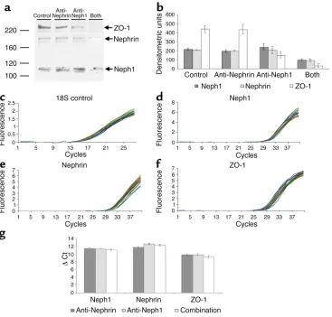
[image:10.576.305.529.620.695.2]proteins 24 hours after injection of the Ab’s revealed modest reduction of nephrin and Neph1 in the com-bination group by densitometric analysis (Table 4 and Figure 9, a and b). In addition to being substantially reduced in the combination group, ZO-1 protein was also modestly reduced in the anti-Neph1 group.
The results of real-time PCR are illustrated in Figure 9, c–g, and the ∆Ct values and 2–∆∆Ctvalues are
sum-marized in Table 5. The ∆Ct values of the combination versus individual Ab–injected groups were not signifi-cantly different by Student ttest, and the 2–∆∆Ctvalues
between the same groups were also less than 2, indicat-ing that altered gene expression did not account for the pathogenesis of proteinuria.
Discussion
Over two decades of research has provided interesting insights into the structure and function of the glomerular capillary loop (29–31). More recent studies have addressed the relative role of podocyte versus base-ment membrane proteins in maintaining the perme-ability barrier (24). The paper on Neph1 by Donoviel et al. (32) describing nephrotic syndrome at birth in Neph1 mutant mice provides conclusive evidence that Neph1 plays a significant role in the normal develop-ment and function of the glomerular capillary loop. The paper by Sellin et al. (15) describes the interaction of Neph1 with podocin in vitro, the distribution of Neph1 in a glomerular capillary loop pattern, and the transactivation of AP-1 in the presence of Tec kinases. Our current study provides conclusive evidence that Neph1 is a slit diaphragm protein, as shown by immunogold localization in the slit diaphragm.
The coimmunoprecipitation studies using anti-Neph1, anti-nephrin, and anti-FLAG Ab’s with glomerular extracts and recombinant proteins were highly suggestive of an interaction between Neph1 and nephrin at the level of their slit diaphragm spanning extracellular segments. We therefore decided to disrupt this interaction using an in vivo approach. The occur-rence of complement- and leukocyte-independent pro-teinuria by a combination of Neph1 and anti-nephrin Ab’s in individually subnephritogenic doses is highly suggestive that this interaction exists in vivo, and is at least partially altered by this Ab combination approach. Moreover, the induction of heterologous-phase proteinuria by the combination of anti-Neph1
and anti-nephrin was not associated with foot process effacement or differ-ence in the mRNA expression of ZO-1, nephrin, and Neph1 between protein-uric and nonproteinprotein-uric subgroups, indicating that it largely affected slit diaphragm permeability due to altered protein-protein interaction.
It is important to note that the dose-response curves performed as a prel-ude to these studies were intended to identify an optimum subnephrito-genic dose of individual Ab’s, and not to study the full extent of the nephritogenic potential of the indi-vidual Ab’s. Indeed, both Neph1 and anti-nephrin start increasing slit diaphragm permeability individually (compared with preimmune serum con-trols) at a dose of 500 µl in rats. It is possible that future experiments directed toward investigating the nephritogenic potential of these individual Ab’s may show more proteinuria at higher doses.
[image:11.576.59.348.81.131.2]The choice of an in vivo approach to test the Neph1-nephrin association over an in vitro approach using monolayers of cultured glomerular epithelial cells was made for several reasons. The slit diaphragm is the final barrier that restricts passage of plasma proteins into the urine (29). We have now known for several years that
Table 3
Urinary protein excretion in response to subnephritogenic doses of Ab’s
Preimmune serum Anti-Neph1 Anti-nephrin Anti-Neph1 (150 µl) + (300 µl) (300 µl) (300 µl) anti-nephrin (150 µl)
Day 1 0.65 ± 0.02 0.80 ± 0.60 0.80 ± 0.10 7.73 ± 2.60A
Day 3 0.67 ± 0.04 0.90 ± 0.30 0.38 ± 0.20 5.10 ± 2.07
[image:11.576.341.494.390.613.2]Urinary protein excretion over 24 hours in response to injection of subnephritogenic doses of Ab’s alone or in combination (n= 4 male rats/group, 100 g each, baseline proteinuria 0.87 ± 0.19). Data are presented as mean ± SE, in mg/24 hours. ASignificantly different from corresponding val-ues of other three groups, P< 0.05.
Figure 7
certain Ab’s directed against podocyte proteins injected intravenously induce complement- and leukocyte-inde-pendent proteinuria (5, 33, 34). We therefore rational-ized that Ab’s directed against the extracellular seg-ments of slit diaphragm proteins would bind their targets and induce slit diaphragm dysfunction that would be detectable as proteinuria. The classic example of this is mAb 5-1-6, which induces proteinuria in rats in certain doses after binding to an epitope on the extra-cellular segment of nephrin. An in vitro approach to this issue would be less than ideal, since there are no cul-tured glomerular epithelial cells that form foot process-es and slit diaphragms with morphological similarity to their in vivo counterparts. In addition, cultured cells tend to form tight junctions, which are not seen in dif-ferentiated podocytes and would make the interpreta-tion of monolayer permeability data more difficult.
The association of ZO-1 with the cytoplasmic tail of Neph1, and the interaction of nephrin with Neph1 in the slit diaphragm, also offer valuable insights into changes in podocyte proteins during proteinuria. The study complements the astute observation of Kawachi et al. (24) that the induction of proteinuria with mAb 5-1-6, an anti-nephrin Ab, results in reduced amounts of glomerular ZO-1 by immunostaining and Western blot. In the anti-Neph1/anti-nephrin model discussed above, glomerular ZO-1 was unchanged compared with controls when rats were injected with anti-nephrin at a dose that did not cause proteinuria. Some reduction of glomerular ZO-1 content was noted with the injection of anti-Neph1 alone, testifying further to the direct association of ZO-1 with Neph1. The most dramatic reduction, however, was observed when the Ab combi-nation induced heterologous-phase proteinuria. This indicates that the interaction between nephrin and Neph1 helps to partially stabilize the association between Neph1 and ZO-1, and that disruption of this association results in a rapid reduction of glomerular ZO-1. This allows us to postulate that a nephrin/ Neph1/ZO-1 macromolecular complex exists in the slit diaphragm and may be altered in some forms of pro-teinuria. Whether our anti-nephrin Ab given alone in higher doses reduces glomerular ZO-1 will be a subject of future investigations designed to examine the nephritogenic phase of this Ab. It would also be inter-esting to conduct similar studies with mAb 5-1-6.
A substantial amount of novel data presented in the Results section allows us to propose further refinements in the organization of Neph1 molecules in the slit diaphragm. The existence of Neph1 homodimers and multimers, the coimmunoprecipitation of nephrin with native Neph1, the selective coimmunoprecipitation of nephrin with the recombinant extracellular Neph1, and the induction of complement- and leukocyte-independ-ent heterologous-phase proteinuria by the combination of anti-nephrin and anti-Neph1 Ab’s in subnephrito-genic doses all help in generating possible configura-tions. Neph1 homodimers/multimers may be arranged side by side in the slit diaphragm or adhere to each other from adjacent foot processes. It is also likely that Neph1 forms additional heterodimers with nephrin molecules either side by side or approaching each other from adja-cent foot processes. Even though both the homodimer-ic association of Neph1 and a heterodimerhomodimer-ic association with nephrin are both likely to be important in main-taining the permeability characteristics of the slit diaphragm, the occurrence of proteinuria after treat-ment with subnephritogenic combinations of Ab’s rather than with individual Ab’s would suggest that the latter may be more significant. In addition to the data presented above, the likelihood of a Neph1-nephrin interaction is also supported by the interaction in
[image:12.576.60.318.56.181.2]Drosophilabetween Hibris and Dumbfounded, two pro-teins with structural similarity to nephrin and Neph1, respectively, during myoblast fusion (35). Also, an asso-ciation between nephrin and Neph1 has very recently also been shown by two other studies (27, 36).
Table 4
Densitometric analysis of protein expression
n Control Anti- Anti- Anti-nephrin + nephrin Neph1 anti-Neph1
Neph1 3 216 ± 12 194 ± 14 243 ± 34 98 ± 12A
Nephrin 3 205 ± 3 197 ± 5 205 ± 41 92 ± 12B
ZO-1 5 440 ± 40 436 ± 56 153 ± 36B 33 ± 22C,D
Expression of Neph1, nephrin, and ZO-1 in rat glomeruli extracted from exper-imental and control groups (mean ± SE densitometric units). ASignificantly different from corresponding control, anti-nephrin, and anti-Neph1 groups,
[image:12.576.304.537.607.669.2]P< 0.01. BSignificantly different from corresponding control and anti-nephrin groups, P< 0.01. CSignificantly different from corresponding anti-Neph1 group, P< 0.05. DSignificantly different from corresponding control and anti-nephrin groups, P< 0.001.
Figure 8
involved in adhesion at all, but may have a sensing role (37). Also, two genes related to Neph1, named Neph2and
Neph3, have now been shown to be expressed in human kidney cortex by RT PCR (15). Precise cellular and sub-cellular localization is still pending. Further studies are required to investigate other protein-protein associations in the slit diaphragm that may also be important in main-taining the permeability status of this important structure. We have not as yet investigated the potential
[image:13.576.128.495.50.401.2]interac-tions of Neph1 or nephrin with FAT, a member of the cadherin superfamily that also spans the slit diaphragm (11). Interestingly, FAT also has a PDZ-binding motif that was not shown to bind ZO-1 in a recent paper also addressing the association between Neph1 and ZO-1 (28). Moreover, the data appears to suggest that very large cadherins, such as the FAT family members, may not be
Table 5
∆Ct values for mRNA expression of individual groups and 2–∆∆Ctvalues of difference between combination and individual groups by real-time PCR
Anti-nephrin ∆Ct Anti-Neph1 ∆Ct Combination ∆Ct Combination minus Combination minus anti-nephrin 2–∆∆Ct anti-Neph1 2–∆∆Ct
Neph1 11.46 ± 0.12 11.34 ± 0.13 11.07 ± 0.15 1.42 ± 0.24 1.22 ± 0.09
Nephrin 11.74 ± 0.18 12.62 ± 0.19 12.27 ± 0.22 0.83 ± 0.25A 1.13 ± 0.05
ZO-1 9.85 ± 0.19 9.86 ± 0.20 9.20 ± 0.30 1.99 ± 0.59 1.67 ± 0.24
[image:13.576.57.534.649.710.2]Data are presented as mean ± SE, n= 9 per measurement. AAnti-nephrin minus combination was 1.60 ± 0.45. P> 0.05 for ∆Ct between all combination and corresponding individual Ab groups.
Figure 9
Acknowledgments
This work is supported by NIH grants DK-61275 to S.S. Chugh, DK-28492 and DK-60635 to Y.S. Kanwar, and the Carl W. Gottschalk Award of the American Society of Nephrology to S.S. Chugh. We thank Richard Quigg for providing rat kidney specimens injected with sheep anti–rat Fx1A and David Salant for providing sheep anti-rat nephrotoxic serum.
1. Quaggin, S.E., et al. 1999. The basic-helix-loop-helix protein pod1 is crit-ically important for kidney and lung organogenesis. Development.
126:5771–5783.
2. Rohr, C., et al. 2002. The LIM-homeodomain transcription factor Lmx1b plays a crucial role in podocytes.J. Clin. Invest. 109:1073–1082. doi:10.1172/JCI200213961.
3. Miner, J.H., et al. 2002. Transcriptional induction of slit diaphragm genes by Lmx1b is required in podocyte differentiation. J. Clin. Invest. 109:1065–1072. doi:10.1172/JCI200213954.
4. Schnabel, E., Anderson, J.M., and Farquhar, M.G. 1990. The tight junc-tion protein ZO-1 is concentrated along slit diaphragms of the glomeru-lar epithelium. J. Cell. Biol.111:1255–1263.
5. Orikasa, M., Matsui, K., Oite, T., and Shimizu, F. 1988. Massive protein-uria induced in rats by a single intravenous injection of a monoclonal antibody. J. Immunol.141:807–814.
6. Kestila, M., et al. 1998. Positionally cloned gene for a normal glomeru-lar protein-nephrin is mutated in congenital nephrotic syndrome.
Mol. Cell.1:575–582.
7. Shih, N.Y., et al. 1999. Congenital nephrotic syndrome in mice lacking CD2-associated protein. Science.286:312–315.
8. Boute, N., et al. 2000. NPHS2, encoding the glomerular protein podocin, is mutated in autosomal recessive steroid-resistant nephrotic syndrome.
Nat. Genet. 24:349–354.
9. Kaplan, J.M., et al. 2000. Mutations in ACTN4, encoding alpha-actinin-4, cause familial focal segmental glomerulosclerosis. Nat. Genet.24:251–256.
10. Reiser, J., Kriz, W., Kretzler, M., and Mundel, P. 2000. The glomerular slit diaphragm is a modified adherens junction. J. Am. Soc. Nephrol. 11:1–8.
11. Inoue, T., et al. 2001. FAT is a component of glomerular slit diaphragms.
Kidney Int.59:1003–1012.
12. Ihalmo, P., Palmen, T., Ahola, H., Valtonen, E., and Holthofer, H. 2003. Filtrin is a novel member of nephrin-like proteins. Biochem. Biophys. Res. Commun.300:364–370.
13. Topham, P.S., et al. 1999. Nephritogenic monoclonal antibody 5-1-6 is directed against the extracellular domain of rat nephrin. J. Clin. Invest. 104:1559–1566.
14. Donoviel, D.B., et al. 2001. Proteinuria and perinatal lethality in mice lacking NEPH1, a novel protein with homology to NEPHRIN. Mol. Cell. Biol.21:4829–4836.
15. Sellin, L., et al. 2003. NEPH1 defines a novel family of podocin interact-ing proteins. FASEB J.17:115–117.
16. Mundel, P., and Shankland, S.J. 2002. Podocyte biology and response to injury. J. Am. Soc. Nephrol.13:3005–3015.
17. BLAST. www.ncbi.nlm.nih.gov/BLAST. 18. Scansite. http://scansite.mit.edu/.
19. ExPASy Proteomics tools. http://ca.expasy.org/tools/#primary. 20. SignalP V1.1 World Wide Web Prediction Server.
www.cbs.dtu.dk/serv-ices/SignalP.
21. DAS — Transmembrane Prediction Server. www.sbc.su.se/∼miklos/DAS/. 22. ExPASy ProtScale. http://ca.expasy.org/cgi-bin/protscale.pl. 23. Chugh, S., et al. 2001. Aminopeptidase A: a nephritogenic target antigen
of nephrotoxic serum. Kidney Int.59:601–613.
24. Kawachi, H., et al. 1997. Slit diaphragm-reactive nephritogenic MAb 5-1-6 alters expression of ZO-1 in rat podocytes. Am. J. Physiol. 273:F984–F993.
25. Wada, J., and Kanwar, Y.S. 1998. Characterization of mammalian translocase of inner mitochondrial membrane (Tim44) isolated from diabetic newborn mouse kidney. Proc. Natl. Acad. Sci. U. S. A.95:144–149. 26. Kanwar, Y.S., et al. 1996. D-glucose–induced dysmorphogenesis of
embryonic kidney. J. Clin. Invest.98:2478–2488.
27. Barletta, G.M., Kovari, J.A., Verma, R.K., Kerjaschki, D., and Holzman, L.B. 2003. Nephrin and Neph1 co-localize at the podocyte foot process intercellular junction and form cis hetero-oligomers. J. Biol. Chem. 278:19266–19271.
28. Huber, T.B., et al. 2003. The carboxyl terminus of neph family members binds to the PDZ domain protein zonula occludens-1. J. Biol. Chem. 278:13417–13421.
29. Kanwar, Y.S., and Venkatachalam, M.A. 1992. Ultrastructure of the glomerulus and juxta-glomerular apparatus. In Handbook of renal physi-ology. E.E. Windhager, editor. Oxford University Press. New York, New York, USA. 3–40.
30. Wieslander, J., et al. 1984. Goodpasture antigen of the glomerular base-ment membrane: localization to noncollagenous regions of type IV col-lagen. Proc. Natl. Acad. Sci. U. S. A.81:3838–3842.
31. Abrahamson, D.R., and Caulfield, J.P. 1982. Proteinuria and struc-tural alterations in rat glomerular basement membranes induced by intravenously injected anti-laminin immunoglobulin G. J. Exp. Med. 156:128–145.
32. Hamano, Y., et al. 2002. Determinants of vascular permeability in the kidney glomerulus. J. Biol. Chem.277:31154–31162.
33. Assmann, K.J.M., van Son, J.P.H.F., Dijkman, H.B.P.M., and Koene, R.A.P. 1992. A nephritogenic rat monoclonal antibody to mouse aminopepti-dase A. J. Exp. Med.175:623–635.
34. Mentzel, S., van Son, J.P., Dijkman, H.B., Wetzels, J.F., and Assmann, K.J. 1999. Induction of albuminuria in mice: synergistic effect of two mon-oclonal antibodies directed to different domains of aminopeptidase A.
Kidney Int.55:1335–1347.
35. Dworak, H.A., Charles, M.A., Pellerano, L.B., and Sink, H. 2001. Charac-terization of Drosophila hibris, a gene related to human nephrin. Devel-opment.128:4265–4276.
36. Gerke, P., Huber, T.B., Sellin, L., Benzing, T., and Walz, G. 2003. Homo-dimerization and heteroHomo-dimerization of the glomerular podocyte pro-teins nephrin and NEPH1. J. Am. Soc. Nephrol.14:918–926.