The forkhead transcription factor Foxo1 links
insulin signaling to Pdx1 regulation of
pancreatic
bb
cell growth
Tadahiro Kitamura, … , Karen C. Arden, Domenico Accili
J Clin Invest. 2002;
110(12)
:1839-1847.
https://doi.org/10.1172/JCI16857
.
Diabetes is caused by an absolute (type 1) or relative (type 2) deficiency of
insulin-producing
b
cells. The mechanisms governing replication of terminally differentiated
b
cells
and neogenesis from progenitor cells are unclear. Mice lacking insulin receptor substrate-2
(Irs2) develop
b
cell failure, suggesting that insulin signaling is required to maintain an
adequate
b
cell mass. We report that haploinsufficiency for the forkhead transcription factor
Foxo1 reverses
b
cell failure in Irs2
–/–mice through partial restoration of
b
cell proliferation
and increased expression of the pancreatic transcription factor pancreas/duodenum
homeobox gene-1 (Pdx1). Foxo1 and Pdx1 exhibit mutually exclusive patterns of nuclear
localization in
b
cells, and constitutive nuclear expression of a mutant Foxo1 is associated
with lack of Pdx1 expression. We show that Foxo1 acts as a repressor of Foxa2-dependent
(Hnf-3
b
–dependent) expression from the Pdx1 promoter. We propose that insulin/IGFs
regulate
b
cell proliferation by relieving Foxo1 inhibition of Pdx1 expression in a subset of
cells embedded within pancreatic ducts.
Article
Metabolism
Find the latest version:
Introduction
Type 2 diabetes results from com-bined defects of insulin action and pancreatic βcell function (1, 2). Clas-sically, the two abnormalities have been considered as separate entities. However, the recent demonstration that insulin/IGF signaling plays a
role in insulin secretion and β cell proliferation has led to a critical reassessment of this view (3). For example, inactivation of insulin receptor (Insr) (4) or IGF-1 receptor (Igf1r) (5, 6) leads to impaired insulin secretion, while inactivation of insulin receptor substrate-2 (Irs2) impairs β cell proliferation (7, 8). These observations indicate that peripheral insulin resistance and β
cell failure may share a common pathogenesis. However, the mecha-nism by which insulin signaling reg-ulates β cell function remains unclear. Forkhead transcription fac-tors of the Foxosubfamily, previous-ly known as Fkhr (9), can promote (10) or repress (11) gene expression. Insulin inhibits Foxo through Akt-mediated phosphorylation and nuclear exclusion, and Foxo1 muta-tions affect insulin sensitivity in mice (12). Here we report the
identifica-tion of Foxo1 as an effector of insulin action in pancreatic βcells, and pro-pose a model in which Foxo1 links insulin signaling to regulation of
βcell mass.
Methods
Reagents. We maintained and trans-fected kidney epithelial cells and β
cells according to standard protocols (13). We purchased insulin anti-body from Linco Research Inc. (St. Charles, Missouri, USA), anti-Hemagglutinin (12CA5), and anti–5-bromo-2-deoxyuridine (anti-BrdU) antibodies from Sigma-Aldrich (St. Louis, Missouri, USA); anti–c-Myc antibody (9E10) from Roche Molecu-lar Biosystems (Indianapolis, Indi-ana, USA); anti-Foxo1 and anti– phospho-Foxo1S253from Cell
Signal-ing Technology Inc. (Beverly, Massa-chusetts, USA); and anti-Foxa2 mon-oclonal antibody from the Develop-mental Studies Hybridoma Bank at the University of Iowa (Iowa City, Iowa, USA). We described previously the anti-Foxo1 antiserum used for gel shift assays (13) and the antibody against pancreas/duodenum home-obox gene-1 (Pdx1) (12). Expression vectors and Foxo1 adenoviruses have been described previously (13). All primer sequences are available upon request.
Animal production and phenotypic analysis. We have describedInsr, Irs2, and Foxo1mutant mice (12). We meas-ured glucose and insulin as indicated in previous publications (12).
Real-time RT-PCR and Northern analy-ses of gene expression. We isolated mRNA using the Micro-Fast Track 2.0 kit (Invitrogen Corp., San Diego,
The forkhead transcription factor Foxo1
links insulin signaling to Pdx1 regulation
of pancreatic
β
cell growth
Tadahiro Kitamura,
1Jun Nakae,
1Yukari Kitamura,
1Yoshiaki Kido,
1William H. Biggs III,
2Christopher V.E. Wright,
3Morris F. White,
4Karen C. Arden,
2and Domenico Accili
11Naomi Berrie Diabetes Center, Department of Medicine, College of Physicians & Surgeons of Columbia University,
New York, New York, USA
2Ludwig Institute for Cancer Research, University of California San Diego, San Diego, California, USA 3Vanderbilt University Medical Center, Nashville, Tennessee, USA
4Joslin Diabetes Center and Howard Hughes Medical Institute, Harvard University, Boston, Massachusetts, USA
Diabetes is caused by an absolute (type 1) or relative (type 2) defi-ciency of insulin-producing βcells. The mechanisms governing repli-cation of terminally differentiated βcells and neogenesis from pro-genitor cells are unclear. Mice lacking insulin receptor substrate-2 (Irs2) develop β cell failure, suggesting that insulin signaling is required to maintain an adequate βcell mass. We report that hap-loinsufficiency for the forkhead transcription factor Foxo1 reverses β
cell failure in Irs2–/–mice through partial restoration of βcell
prolif-eration and increased expression of the pancreatic transcription fac-tor pancreas/duodenum homeobox gene-1 (Pdx1). Foxo1 and Pdx1 exhibit mutually exclusive patterns of nuclear localization in βcells, and constitutive nuclear expression of a mutant Foxo1 is associated with lack of Pdx1expression. We show that Foxo1 acts as a repressor of Foxa2-dependent (Hnf-3β–dependent) expression from the Pdx1 promoter. We propose that insulin/IGFs regulate βcell proliferation by relieving Foxo1 inhibition of Pdx1expression in a subset of cells embedded within pancreatic ducts.
J. Clin. Invest.110:1839–1847 (2002). doi:10.1172/JCI200216857.
Rapid Publication
Received for publication September 9, 2002, and accepted in revised form October 22, 2002.
Address correspondence to: Domenico Accili, Berrie Research Pavilion, 1150 St. Nicholas Avenue, Room 238A, New York, New York 10032, USA.
Phone: (212) 851-5332; Fax: (212) 851-5331; E-mail: da230@columbia.edu.
Conflict of interest: The authors have declared that no conflict of interest exists.
Nonstandard abbreviations used: insulin receptor (Insr); IGF-1 receptor (Igf1r); insulin receptor substrate-2 (Irs2); 5-bromo-2-deoxyuridine (BrdU); pancreas/duodenum homeobox gene-1 (Pdx1); glucokinase (Gck); Fas ligand (FasL); Bcl-2 interacting mediator (BimL); secreted alkaline phosphatase (SEAP);
California, USA). We carried out Northern blots according to stan-dard methods and semiquantitative RT-PCR using the GeneAmp RNA PCR kit (Applied Biosystems, Foster City, California, USA) with amplifi-cation primers corresponding to
Foxo1, Foxo3, Foxo4, Pdx1, Irs2, insulin, glucagon, pancreatic polypeptide, and
β-actinsequences. We performed real-time PCR using primers encoding
insulin 2, Pdx1, Slc2a2, glucokinase
(Gck), Foxa2, Bad, Bcl-2 interacting medi-ator (BimL), Fas ligand (FasL), and
p27kip. For these experiments, we
iso-lated mRNA from handpicked islets of similar sizes derived from three 4-week-old animals for each genotype and amplified it using a LightCycler PCR instrument and LightCycler RT-PCR kit (Roche Molecular Biosys-tems). We carried out each reaction in triplicate using a standard curve with the relevant cDNA for each primer set.
Immunohistochemical and morphomet-ric analysis of pancreatic islets. We isolat-ed and fixisolat-ed pancreata from 8-week-old wild-type, Irs2–/–, Irs2–/–Insr+/–, and
Irs2–/–Foxo1+/– mice overnight in 2%
paraformaldehyde solution, embed-ded them in paraffin, and immunos-tained consecutive 5-µm sections for
βand αcells using anti-insulin and anti-glucagon antibodies, respective-ly. We performed morphometry using NIH Image 1.60 software (NIH, Bethesda, Maryland, USA) as de-scribed (14). We expressed results as percentage of total surveyed pancreat-ic area occupied by βand αcells.
Immunofluorescence. We incubated frozen islet sections with anti-insulin, anti-Pdx1, and anti-Foxo1 antibodies at dilutions of 1:1,000, 1:5,000 and 1:30, respectively. We visualized immune complexes using a CY3-conjugated anti–guinea pig IgG for anti-insulin antiserum, and FITC-conjugated secondary anti-rab-bit IgG for anti-Pdx1 and anti-Foxo1 antisera. For colocalization of Foxo1 and Pdx1, we costained two adjacent sections with insulin and Pdx1, or insulin and Foxo1 antibodies, respec-tively. Thereafter, we acquired images using a SPOT-RT digital camera (Morrell Instruments, Melville, New York, USA) and identified matching
islets in the two sections by overlay-ing the insulin immunostainoverlay-ing. We rendered Pdx1 immunoreactivity as mauve pseudocolor and scored cells in which we could unambiguously identify the nucleus on both sections for either Pdx1 or Foxo1 immunore-activity. We analyzed a total of ten sections and 50 islets from three mice for each genotype. We fixed trans-fected βTC-3 cells in 2% parafor-maldehyde and permeabilized them in 0.2% Triton X-100 for immunoflu-orescence as described previously (13). We visualized endogenous Pdx1 with anti-Pdx1 polyclonal antibody and FITC-conjugated anti-rabbit IgG. We detected transiently ex-pressed c-Myc–Foxo1 in the same sec-tions with a monoclonal anti–c-Myc antibody followed by CY3-conjugat-ed anti-mouse IgG.
Detection and quantitation of β cell replication. We injected BrdU intraper-itoneally (0.1 g/kg) into 2-week-old wild-type, Irs2–/–, and Irs2–/–Foxo1+/–
mice. After 4 hours, we removed and fixed pancreata in PBS (pH 7.4) con-taining 2% paraformaldehyde and embedded them in paraffin. We immunostained the sections with mouse anti-BrdU antibody (1:1,000). We incubated the sections sequen-tially with biotinylated anti-mouse IgG reagent (Vector Laboratories Inc., Burlingame, California, USA), VECTASTAIN Elite ABC reagent and DAB (Vector Laboratories Inc.) for visualization. For double staining with BrdU and insulin bodies, we used a guinea pig anti-insulin antibody. We performed quantitation of βcell and acinar cell replication by counting cells in more than 50 islets stained with anti-insulin antibody for each genotype (for BrdU-labeled βcells) and more than 50 different exocrine fields for each genotype (for BrdU-labeled exocrine cells). We expressed the results as a “labeling index,” i.e., per-centage of total cells in surveyed islets and exocrine fields that were BrdU-positive.
Luciferase and secreted alkaline phos-phatase assays. The sequence used for these experiments spanned nucleo-tides –2,233 to –2,097 of the Pdx1 pro-moter and includes a single consensus
forkhead binding site (15). We carried out cotransfections in βTC-3 cells at 70–80% confluence with Pdx1 /lucifer-ase reporter gene (pGL3-PDX1-PH2) and control pCMV5 vector or pCMV5-Foxa2, pCMV5–ADA-Foxo1, and pCMV5–wild-type Foxo1. We used secreted alkaline phosphatase (SEAP) (20 ng) as a control for trans-fection efficiency. We measured luciferase and SEAP activity accord-ing to the manufacturer’s specifica-tions (Great EscAPe SEAP kit; Clon-tech Laboratories Inc., Palo Alto, California, USA). In cotransfection experiments, we added recombinant adenovirus encoding ADA-Foxo1to cells at different moi’s 12 hours after transfection with Foxa2. Forty-eight hours later, we determined luciferase activity in triplicate samples and nor-malized it by SEAP activity.
Gel shift assays. We isolated nuclei of LLC or βTC-3 cells by sequential extraction in NaCl (16). In some exper-iments, we transduced cells with Foxo1 adenovirus 48 hours prior to nuclei isolation. We incubated double-stranded 32P-labeled oligonucleotide
probes (20,000 cpm; 5 fmol) corre-sponding to the consensus forkhead binding region in the Pdx1 PH2 domain (5′- AGTGCTAAGCAAACATC-CTG-3′) with nuclear extracts for 20 minutes at room temperature in the presence of 10 mM Tris-HCl (pH 7.5), 50 mM NaCl, 1 mM MgCl2, 0.5 mM
Results
RNA expression studies indicate that the three Foxo isoforms differ with respect to tissue localization, with Foxo1 representing the predominant mRNA isoform in liver (17), lung, and spleen, and Foxo4 in heart, skeletal muscle, and kidney (13). Foxo3 mRNA distribution mirrored that of Foxo1, albeit at lower levels (Figure 1a). Foxo1 is also the main member of the Foxo subfamily in cultured βcells (βTC-3 line) (Figure 1b, lane 2) and murine islets. Its levels decline over the first 6 months of life, as do Irs2 and Pdx1 lev-els. Foxo3 is expressed at lower levels and Foxo4 is undetectable in islets (not shown). Although a decrease in Pdx1 expression with age has been reported in rat islets (18), the present
observation suggests that the down-regulation of Pdx1 mRNA occurs earlier than previously thought. It re-mains to be seen, however, whether this is a specific difference between rats and mice or a reflection of inter-strain variability in mice. Immunohis-tochemistry of pancreatic islets with anti-Foxo1 and anti-insulin antisera demonstrates colocalization of Foxo1 and insulin to βcells. The subcellular localization of Foxo1 in βcells is het-erogeneous, with some cells showing exclusive cytoplasmic or nuclear immunoreactivity, and some cells showing diffuse immunoreactivity (Figure 1d).
To address whether Foxo1 partici-pates in βcell function, we tested the ability of a loss-of-function Foxo1
mutation to rescue βcell failure in
Irs2–/– mice by crossing Irs2–/– and
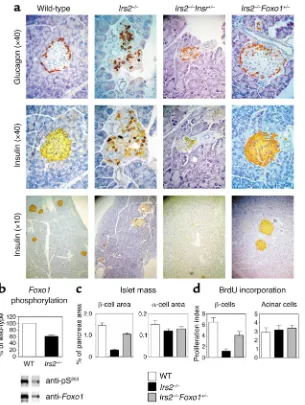
Foxo1+/– mice (12). In Irs2–/– mice,
insulin-immunoreactive β cell area was about 70% smaller than in the wild- type at 8 weeks of age (Figure 2, a and b), with only a slight decrease in glucagon-immunoreactive αcell area (Figure 2b). No changes were observed in Foxo1+/–mice (12). In
con-trast, in Irs2–/–Foxo1+/–mice, βcell area
was restored to about 80% of wild-type values. Irs2–/–Foxo1+/–mice have a
normal life span and do not develop diabetes (Table 1).
Akt-dependent phosphorylation inhibits Foxo1 activity. When Foxo1 was expressed in islets from Irs2–/–
[image:4.576.88.334.52.477.2]mice by adenoviral transduction, phosphorylation of the “gatekeeper”
Figure 1
(a) Tissue survey of Foxoisoform expression in mice. We hybridized multiple tissue Northern blots (filter membranes; Clontech Laboratories Inc.) with probes encoding the three Foxoisoforms, labeled with 32P at
Akt site at serine 253 was decreased by approximately 40% compared with wild-type islets (Figure 2b), suggest-ing that Foxo1 is in the Irs2 signalsuggest-ing pathway. Since insulin receptor sig-naling inhibits Foxo1, we tested the effect of a loss-of-function Insr muta-tion on diabetes in Irs2–/–mice. We
generated recombinant congenic mice carrying Irs2 and Insrtargeted null alleles on the same chromosome (the two genes are about 4 Mb apart near the centromeric end of chromo-some 8; ref. 19), and intercrossed them to produce Irs2–/–Insr+/– mice.
Insrhaploinsufficiency increased the severity and accelerated the onset of diabetes in Irs2–/–mice. Irs2–/–Insr+/–
mice developed diabetes at 6–8 weeks (Table 1) and invariably died by 10 weeks of age. Pancreatic islets were virtually undetectable by 8 weeks
(Figure 2a). Withers and colleagues have reported a similar phenotype in
Irs2–/–Igf1r+/–mice (20).
To distinguish between increased cell proliferation and decreased apop-tosis as mechanisms of βcell restora-tion in Irs2–/–Foxo1+/–mice, we
meas-ured BrdU incorporation and DNA fragmentation. BrdU incorporation was reduced by about 85% in islets of 2-week-old Irs2–/– mice and was
restored to approximately 67% of wild-type in Irs2–/–Foxo1+/–mice (P < 0.05 by
ANOVA). We observed no changes in exocrine acinar tissue (Figure 2c). In contrast to proliferation, we failed to detect apoptotic cells in any of the genotypes examined using TUNEL staining at ages 2, 3, and 4 weeks (data not shown). These data indi-cate that restoration of βcell mass in
Irs2–/–Foxo1+/–mice is due to increased
proliferation, rather than decreased apoptosis. Nevertheless, given that apoptosis in βcells is difficult to doc-ument (21), we cannot rule out its con-tribution to the observed phenotype. Unlike βcell mass, Foxo1 haploinsuf-ficiency did not affect glucose-stimu-lated insulin secretion from islets of
Irs2–/–Foxo1+/– mice, nor did
overex-pression of wild-type or mutant Foxo1 affect insulin secretion in cultured
βTC-3 cells (data not shown).
To examine the mechanism by which
Foxo1haploinsufficiency prevents dia-betes in Irs2–/– mice, we analyzed
expression of Foxo1target genes. They include the cell cycle inhibitor p27kip
(22) and the proapoptotic genes FasL
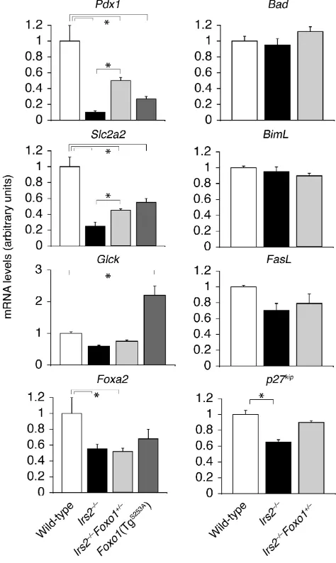
[image:5.576.66.371.51.456.2](23) and BimL (24). For these experi-ments we used islets of similar size derived from 4-week-old mice to exclude artifacts due to loss of βcells.
Figure 2
(a) Pancreatic histology in mice with target-ed null alleles of Irs2and Foxo1. We stained pancreatic sections from 8-week-old mice of the indicated genotypes with anti-insulin and anti-glucagon antibodies. (b) Foxo1 phos-phorylation in Irs2–/–islets. We transduced
islets from wild-type and Irs2–/–mice with
adenovirus encoding hemagglutinin-tagged wild-type Foxo1. Following immunoprecipi-tation with anti-hemagglutinin antibody, we carried out immunoblotting with anti–phos-pho-Foxo1S253. We then stripped and
reprobed the blots with anti-Foxo1 antibody. One of three experiments is shown, and mean phosphorylation ± SEM is summarized in the graph above (WT, white bar; Irs2–/–, black
bar), following scanning densitometry and normalization for total Foxo1 protein levels. (c) Islet morphometry. We quantitated βand
However, real-time RT-PCR analysis of islet RNA failed to detect differences in Bad, BimL, and FasL, while p27kipwas
decreased by 40% inIrs2–/–mice and by
30% in Irs2–/–Foxo+/–mice (Figure 3),
which is inconsistent with the possi-bility that Foxo1 inhibits βcell repli-cation by increasing p27kip. Thus,
changes in the expression of these genes do not appear to play a role in the observed phenotype. These data were confirmed by experiments in
βTC-3 cells, in which a dominant-neg-ative (δ256) Foxo1 adenovirus failed to inhibit p27kip, FasL, and BimL
expres-sion (data not shown).
In contrast, real-time RT-PCR demonstrated a greater than 80% decrease in the expression of Pdx1and its target gene Slc2a2 (glucose trans-porter Glut2) in Irs2–/– mice. The
decrease was also observed in islets of transgenic mice bearing a phosphory-lation-defective Foxo1S253Amutant in β
cells (12), and was partially reversed in
Irs2–/–Foxo1+/– mice to about 50% of
wild-type. Lesser changes (∼50%) were observed in Gcklevels, consistent with two recent studies showing that Gckis less sensitive than Slc2a2to reduction of Pdx1expression (25, 26). In contrast,
Gck expression was increased about twofold in Foxo1S253Atransgenics
(Fig-ure 3). The reasons for the differential regulation of Gckand Slc2a2in Irs2–/–
and Foxo1S253Amice are unclear. Foxa2
expression was also decreased by about 50% in Irs2–/–and Foxo1S253Amice, but
was not restored in Irs2–/–Foxo1+/–islets.
We next compared Pdx1expression by immunohistochemistry. In wild-type islets, more than 95% of insulin-immunoreactive cells scored (793 of 827) were Pdx1-positive (Figure 4a, green). In Irs2–/–mice, the percentage
of Pdx1-positive cells decreased to 38.7% (245 of 632); moreover, Pdx1 was frequently mislocalized to the cytoplasm. In Irs2–/–Foxo1+/–mice, the
percentage of Pdx1-positive cells was restored to greater than 90% (678 of 730) (P < 0.001 by ANOVA). These data support the real-time PCR analysis
and suggest a negative correlation between Foxo1 activity and Pdx1
expression levels.
[image:6.576.57.536.74.139.2]To investigate this relationship fur-ther, we analyzed the subcellular dis-tribution of Foxo1 in Pdx1-positive and Pdx1-negative βcells. Because the anti-Foxo1 and anti-Pdx1 antisera available for immunohistochemistry have been raised in rabbits, we could not perform double staining on the same tissue section. Therefore, we stained adjacent sections with anti-Foxo1 and anti-insulin, or anti-Pdx1
Table 1
Metabolic characteristics of 8-week-old mice
Genotype Body weight (g) Glucose (fasting) Glucose (fed) Insulin (fasting) Insulin (fed)
Wild-type (n = 13) 33.1 ± 0.6 96 ± 3 123 ± 5 0.9 ± 0.1 2.8 ± 0.2 Irs2–/–(n= 14) 31.2 ± 0.7 98 ± 4 191 ± 13A 2.0 ± 0.3 4.5 ± 0.4 Irs2–/–Foxo1+/–(n= 14) 31.8 ± 0.6 98 ± 4 157 ± 9 1.3 ± 0.1 4.4 ± 0.4
Irs2–/–Insr+/–(n= 10) 29.4 ± 0.4 250 ± 13A 440 ± 28A 0.4 ± 0.1 1.1 ± 0.1
[image:6.576.259.496.338.735.2]AP < 0.05 by one-factor ANOVA compared with wild-type.
Figure 3
Foxo1 haploinsufficiency restores Pdx1 expression in βcells of Irs2–/–mice. Real
and anti-insulin antisera and carefully matched the position of βcells using insulin immunostaining to outline the nuclear margins (Figure 4b). On aver-age, approximately 30% of cells showed matching nuclear “ghosts” in two adjacent 5-µm sections, consistent with the fact that the average βcell diameter is 10 µm and the nucleus occupies about 70% of the cell surface (6). In the vast majority of this subset of cells, Pdx1 and Foxo1 showed mutually exclusive nuclear localiza-tion. Thus, in approximately 80% of
Pdx1-positive cells examined, Foxo1 localized to the cytoplasm (Figure 4b, cells 1, 2, 7, and 8). By contrast, in about 80% of Pdx1-negative cells, Foxo1 localized to the nucleus (Figure 4b, cells 3, 4, 6, and 9). In addition, the occasional double-positive cells (such as cell 10 in Figure 4b) showed very weak Pdx1 immunoreactivity. These correlative findings were investi-gated further by expressing wild-type or a phosphorylation-defective Foxo1 mutant (ADA-Foxo1) that is constitu-tively nuclear (13) in βTC-3 cells.
Wild-type Foxo1 was basally phosphorylat-ed (data not shown) and was localizphosphorylat-ed to the cytoplasm of Pdx1-expressing cells. In contrast, cells expressing the ADA-Foxo1 mutant in the nucleus were Pdx1-negative (Figure 5a). More-over, we have previously shown that expression of a similar constitutively nuclear Foxo1 mutant in islets of transgenic mice results in greatly decreased Pdx1 levels (12).
In summary, Pdx1levels decreased in three models of increased Foxo1 activity: Irs2–/–mice, in which Foxo1
phosphorylation is decreased (Figure 2b); transgenic mice expressing the phosphorylation-defective, constitu-tively nuclear Foxo1S253A in β cells
(12); and βTC-3 cells transduced with ADA-Foxo1 adenovirus. Conversely,
Foxo1 haploinsufficiency partially reverses the decrease in Pdx1levels in
Irs2–/–mice. These data support the
hypothesis that Foxo1 inhibits Pdx1
and are consistent with the observa-tion that overexpression of Pdx1 res-cues diabetes in Irs2–/–mice (27).
To determine potential sites of inter-action between Foxo1 and Pdx1, we sur-veyed pancreatic Foxo1expression. The targeting vector used to ablate Foxo1
function contains a β-galactosidase (β-gal) cassette fused in-frame with
[image:7.576.62.353.54.539.2]Foxo1 exon 1 to enable detection of
Figure 4
(a) Immunohistochemical analysis of Pdx1 expression in βcells of wild-type, Irs2–/–, and
Irs2–/–Foxo1+/–mice. We costained
pancreat-ic sections with Pdx1 (green) and anti-insulin antibodies (red). We show a repre-sentative section. (b) Subcellular localization of Foxo1 in Pdx1-positive and Pdx1-negative
Foxo1expression using X-gal reactivity (12). Using this technique, we detected occasional Foxo1-positive cells in pan-creatic ducts and exocrine acini, in addi-tion to islets (Figure 5b, top row). In view of the potential role of duct-associ-ated cells in βcell neogenesis, we inves-tigated the colocalization of Foxo1 with insulin and Pdx1 in duct-associated cells. The majority of duct-associated Foxo1-positive cells did not display insulin or Pdx1 immunoreactivity (Fig-ure 5b, red arrows). However, all the insulin/Pdx1-positive duct-associated cells were Foxo1-positive (Figure 5b, black arrows). These data are consistent with the possibility that Foxo1 regulates
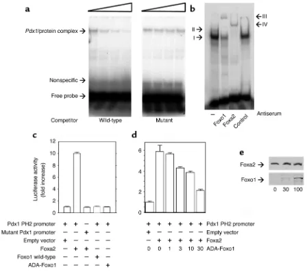
Pdx1expression in duct-associated cells. Next, we used gel shift and reporter gene assays to test the hypothesis that Foxo1 binds directly to the Pdx1 pro-moter and inhibits its transcription. A 6.5-kbp Pdx1 promoter contains ele-ments required for β cell–specific expression, including three potential forkhead binding sites (15, 28, 29).
Among the forkhead proteins, Foxa2 is a known activator of Pdx1, and Pdx1
expression is blunted in mice lacking Foxa2 in βcells (30). We carried out gel shift assays using nuclear extracts from LLC (Figure 6a) and βTC-3 cells (Figure 6b). When we used a probe car-rying the proximal consensus fork-head binding site of the Pdx1 promot-er (PH2 domain) (15), we detected a gel retardation complex that could be competed for by excess cold probe, but not by a mutant probe (Figure 6a). In insulin-producing βTC-3 cells, we detected at least two gel retardation complexes, indicated as I and II. Addi-tion of anti-Foxo1 antiserum caused a supershift to yield a distinct complex III. Addition of anti-Foxa2 monoclon-al antibody caused a supershift of complex A to yield complex IV. Addi-tion of nonimmune serum did not cause any supershift (Figure 6b).
We used cotransfection assays with a minimal Pdx1 promoter containing the proximal Foxa2 binding site to
study the effect of Foxo1 in Pdx1 tran-scription. Expression of Foxa2 in-creased Pdx1/luciferase activity ap-proximately tenfold, whereas Foxo1 failed to do so (Figure 6c). Coexpres-sion of constitutively active Foxo1 inhibited Foxa2-dependent Pdx1 tran-scription in a dose-dependent manner up to about 60%, consistent with the possibility that Foxo1 acts as a tran-srepressor of Foxa2-dependent Pdx1
transcription (Figure 6d). The ability of Foxo1 to inhibit Foxa2-dependent
Pdx1transcription was not due to the presence of an excess of Foxo1 protein, since we detected the inhibitory effect when Foxa2 levels exceeded those of Foxo1 (Figure 6e). We obtained similar data when we used the full-length
[image:8.576.57.536.374.634.2]Pdx1 promoter construct (data not shown). These data provide proof of principle that Foxo1 can act as a repressor of Pdx1 transcription in a transfection system. However, it is like-ly that the in vivo interaction between Foxo1 and Pdx1 is complex, and
Figure 5
depends on the relative abundance of Foxo1, the contributions of coactiva-tors and corepressors, and signals reg-ulating Foxo1 localization.
Discussion
Our genetic analysis indicates that
Foxo1is an effector of Irs2 signaling in pancreatic βcells. Foxo1inactivation leads to increased Pdx1expression and
β cell proliferation. Since Foxo1 is expressed in a subset of cells
embed-ded within pancreatic ducts, we pro-pose that, in quiescent duct-associated cells that are not committed to a βcell fate, Foxo1 acts as a transcriptional brake on Pdx1. We propose the follow-ing mechanism of Foxo1 regulation: small quantities of insulin are released in the pancreatic duct (31), where they activate signaling (32) in the Foxo1-positive duct cell subset, leading to Foxo1 nuclear exclusion and Pdx1
expression. In this hypothesis,
[image:9.576.62.510.49.446.2]pancre-atic islets of Langerhans are function-ally associated with ducts. While con-troversial (33), this assumption is sup-ported by a recent reassessment of the functional anatomy of the pancreas, showing that most islets are associat-ed with pancreatic ducts and provid-ing a morphological correlate for insulin release into pancreatic ducts (34). The implication of this model is that insulin is the main promoter of islet proliferation in hyperinsulinemic,
Figure 6
Foxo1 binds to the Pdx1promoter and inhibits Foxa2-induced Pdx1 transcription. (aand b) Gel shift assays. (a) We used nuclear extracts from kidney epithelial LLC cells transduced with Foxo1 adenovirus to test the specificity of binding to the Pdx1 probe. We chose this cell type because it does not express Foxa2 (13), which is abundant in βTC-3 cells. The concentrations of cold competitor used are indicated by the triangle and are 0, 100-, 300-, and 900-fold excess for both wild-type and mutant probes. (b) Antibody-induced supershift. We incubated
insulin-resistant patients, and that insulin resistance predisposes to βcell failure via impaired Foxo1 phosphory-lation. This model provides a unifying hypothesis for the commonest abnor-malities of type 2 diabetes: peripheral insulin resistance and impaired βcell compensation. Furthermore, it sug-gests that acute islet destruction in type 1 diabetes prevents significant islet regrowth, because there is no insulin to stimulate ductal neogenesis. This hypothesis is consistent with the observation that insulin administra-tion following streptozotocin-induced
β cell ablation is associated with robust islet regrowth (35, 36). Whether this mechanism is physiologically rel-evant in the context of ongoing autoimmunity is an open question. Finally, the notion that βcell neogen-esis from a subset of duct-associated cells is a compensatory response to insulin resistance, rather than a physi-ologic event, reconciles the proposed role of ductal neogenesis (37) with the recent demonstration that duct epithelial cells do not physiologically give rise to endocrine cells (38). We propose that Foxo1 is an important target for the development of drugs that improve βcell proliferation.
Acknowledgments
This work was supported by NIH grant DK-57539 and Juvenile Diabetes Research Foundation grant 200-893 (to D. Accili). T. Kitamura is the recip-ient of a Juvenile Diabetes Foundation Postdoctoral Fellowship. We thank N. Fleischer for the gift of βTC-3 cells, and Y. Liu for skilled technical assis-tance with immunohistochemistry.
1. Saltiel, A.R., and Kahn, C.R. 2001. Insulin signal-ing and the regulation of glucose and lipid metabolism. Nature. 414:799–806.
2. Bell, G.I., and Polonsky, K.S. 2001. Diabetes mel-litus and genetically programmed defects in beta-cell function. Nature. 414:788–791.
3. Accili, D. 2001. A kinase in the life of the βcell. J. Clin. Invest. 108:1575–1576. doi:10.1172/JCI200114454. 4. Kulkarni, R.N., et al. 1999. Tissue-specific
knock-out of the insulin receptor in pancreatic βcells creates an insulin secretory defect similar to that in type 2 diabetes. Cell. 96:329–339.
5. Kulkarni, R.N., et al. 2002. Beta-cell-specific dele-tion of the Igf1 receptor leads to hyperinsuline-mia and glucose intolerance but does not alter beta-cell mass. Nat. Genet. 31:111–115. 6. Xuan, S., et al. 2002. Defective insulin secretion in
pancreatic βcells lacking type 1 IGF receptor. J. Clin. Invest.110:1011–1019. doi:10.1172/JCI200215276. 7. Withers, D.J., et al. 1998. Disruption of IRS-2 causes type 2 diabetes in mice. Nature.
391:900–904.
8. Kubota, N., et al. 2000. Disruption of insulin receptor substrate 2 causes type 2 diabetes because of liver insulin resistance and lack of compensatory beta-cell hyperplasia. Diabetes.
49:1880–1889.
9. Kaestner, K.H., Knochel, W., and Martinez, D.E. 2000. Unified nomenclature for the winged helix/forkhead transcription factors. Genes Dev.
14:142–146.
10. Datta, S.R., Brunet, A., and Greenberg, M.E. 1999. Cellular survival: a play in three Akts. Genes Dev.
13:2905–2927.
11. Zhao, H.H., et al. 2001. Forkhead homologue in rhabdomyosarcoma functions as a bifunctional nuclear receptor-interacting protein with both coactivator and corepressor functions. J. Biol. Chem.276:27907–27912.
12. Nakae, J., et al. 2002. Regulation of insulin action and pancreatic beta-cell function by mutated alle-les of the gene encoding forkhead transcription factor Foxo1. Nat. Genet.32:245–253.
13. Nakae, J., Kitamura, T., Silver, D.L., and Accili, D. 2001. The forkhead transcription factor Foxo1 (Fkhr) confers insulin sensitivity onto glucose-6-phosphatase expression. J. Clin. Invest.
108:1359–1367. doi:10.1172/JCI200112876. 14. Kitamura, T., et al. 2001. Preserved pancreatic
beta-cell development and function in mice lack-ing the insulin receptor-related receptor. Mol. Cell. Biol. 21:5624–5630.
15. Marshak, S., et al. 2000. Functional conservation of regulatory elements in the pdx-1 gene: PDX-1 and hepatocyte nuclear factor 3beta transcription factors mediate beta-cell-specific expression. Mol. Cell. Biol.20:7583–7590.
16. Schreiber, E., Matthias, P., Muller, M.M., and Schaffner, W. 1989. Rapid detection of octamer binding proteins with ‘mini-extracts’, prepared from a small number of cells. Nucleic Acids Res.
17:6419.
17. Nakae, J., Park, B.-C., and Accili, D. 1999. Insulin stimulates phosphorylation of the forkhead tran-scription factor FKHR on serine 253 through a wortmannin-sensitive pathway. J. Biol. Chem.
274:15982–15985.
18. Stoffers, D.A., Heller, R.S., Miller, C.P., and Habener, J.F. 1999. Developmental expression of the homeodomain protein IDX-1 in mice trans-genic for an IDX-1 promoter/lacZ transcription-al reporter. Endocrinology. 140:5374–5381. 19. Sun, X.J., et al. 1997. The IRS-2 gene on murine
chromosome 8 encodes a unique signaling adapter for insulin and cytokine action. Mol. Endocrinol.11:251–262.
20. Withers, D.J., et al. 1999. Irs-2 coordinates Igf-1 receptor-mediated beta-cell development and peripheral insulin signaling. Nat. Genet.23:32–40.
21. Bonner-Weir, S. 2000. Life and death of the pan-creatic beta cells. Trends Endocrinol. Metab.
11:375–378.
22. Medema, R.H., Kops, G.J., Bos, J.L., and Burger-ing, B.M. 2000. AFX-like Forkhead transcription factors mediate cell-cycle regulation by Ras and PKB through p27kip1. Nature. 404:782–787. 23. Brunet, A., et al. 1999. Akt promotes cell survival
by phosphorylating and inhibiting a forkhead transcription factor. Cell. 96:857–868. 24. Dijkers, P.F., Medemadagger, R.H., Lammers, J.J.,
Koenderman, L., and Coffer, P.J. 2000. Expression of the pro-apoptotic Bcl-2 family member Bim is regulated by the forkhead transcription factor FKHR-L1. Curr. Biol. 10:1201–1204.
25. Brissova, M., et al. 2002. Reduction in pancre-atic transcription factor PDX-1 impairs glu-cose-stimulated insulin secretion. J. Biol. Chem.
277:11225–11232.
26. Kim, S.K., et al. 2002. Pbx1 inactivation disrupts pancreas development and in Ipf1-deficient mice promotes diabetes mellitus. Nat. Genet.
30:430–435.
27. Kushner, J.A., et al. 2002. Pdx1 restores βcell function in Irs2 knockout mice. J. Clin. Invest.
109:1193–1201. doi:10.1172/JCI200214439. 28. Sharma, S., et al. 1997. Hormonal regulation of
an islet-specific enhancer in the pancreat-ic homeobox gene STF-1. Mol. Cell. Biol.
17:2598–2604.
29. Wu, K.L., et al. 1997. Hepatocyte nuclear factor 3beta is involved in pancreatic beta-cell-specific transcription of the pdx-1 gene. Mol. Cell. Biol.
17:6002–6013.
30. Lee, C., et al. 2002. Foxa2 controls Pdx1 gene expression in pancreatic beta-cells in vivo. Dia-betes. 51:2546–2551.
31. Conlon, J.M., Rouiller, D., Boden, G., and Unger, R.H. 1979. Characterization of immunoreactive components of insulin and somatostatin in canine pancreatic juice. FEBS Lett.105:23–26.
32. Williams, J.A., and Goldfine, I.D. 1985. The insulin-pancreatic acinar axis. Diabetes. 34:980–986. 33. Pour, P. 1978. Islet cells as a component of
pan-creatic ductal neoplasms. I. Experimental study: ductular cells, including islet cell precursors, as primary progenitor cells of tumors. Am. J. Pathol.
90:295–316.
34. Bertelli, E., Regoli, M., Orazioli, D., and Ben-dayan, M. 2001. Association between islets of Langerhans and pancreatic ductal system in adult rat. Where endocrine and exocrine meet together? Diabetologia. 44:575–584.
35. Guz, Y., Nasir, I., and Teitelman, G. 2001. Regen-eration of pancreatic beta cells from intra-islet precursor cells in an experimental model of dia-betes. Endocrinology. 142:4956–4968. 36. Movassat, J., Saulnier, C., and Portha, B. 1997.
Insulin administration enhances growth of the beta-cell mass in streptozotocin-treated newborn rats. Diabetes. 46:1445–1452.
37. Bonner-Weir, S., et al. 2000. In vitro cultivation of human islets from expanded ductal tissue. Proc. Natl. Acad. Sci. USA. 97:7999–8004. 38. Gu, G., Dubauskaite, J., and Melton, D.A. 2002.
Direct evidence for the pancreatic lineage: NGN3+ cells are islet progenitors and are dis-tinct from duct progenitors. Development.