1773
Introduction
Preimplantation development is a mammalian-specific event, and is vital for successful implantation and pregnancy. This process involves many crucial events with cavity formation occurring during morula to blastocyst stages being one of the most important. Recently, several reports focusing on preimplantation development, especially cavity formation, have appeared (Fleming et al., 2001; Barcroft et al., 2003; Salas-Vidal and Lomeli, 2004). However, neither the detailed molecular mechanisms nor the crucial genes that regulate preimplantation developmental processes have been clarified. To address these issues, we have focused on intracellular signal transduction pathways that might regulate gene expression during cavity formation.
The mitogen-activated protein kinase (MAPK) cascades have central roles in diverse cellular functions (Sturgill and Wu, 1991; Ahn et al., 1992; Nishida and Gotoh, 1993; Davis, 2000; Ono and Han, 2000; Chang and Karin, 2001; Kyriakis and Avruch, 2001; Pearson et al., 2001). The MAPK pathways include the extracellular signal-regulated protein kinase (ERK) pathway, the Jun N-terminal kinase (JNK) pathway and the p38 pathway. Each member of MAPK is activated in response to various extracellular stimuli, and controls, mainly through regulating gene expression, various biological processes such
as cell proliferation, cell differentiation, cell cycle arrest, apoptosis, etc. As previous genetic and biochemical studies have revealed the crucial involvement of the MAP kinase family molecules in early embryonic development, it has been expected that they might act also in preimplantation development. However, difficulties in manipulating preimplantation embryos have delayed progression in this field.
Recent development of specific inhibitors of each MAPK pathway, however, enabled us to test our hypothesis that several of the MAPK family molecules may be involved in processes during preimplantation development. In this study, the use of the specific inhibitors has firstly shown that inhibition of the p38 pathway or the JNK pathway, but not the ERK pathway, leads to abnormality in mouse preimplantation development, especially in cavity formation. Immunostaining with anti-phospho-specific antibodies has then shown the existence of activated forms of p38 and JNK from four-cell to blastocyst stages. These results strongly suggest the involvement of JNK and p38 in mouse preimplantation development. As for p38, Natale et al. (Natale et al., 2004) reported, just after completion of this work, that p38 MAPK activity is required to support successful murine preimplantation development, in agreement with our results. Our experiments with actinomycin D then Mammalian preimplantation development involves several
crucial events, such as compaction and blastocyst formation, but little is known about essential genes that regulate this developmental process. Here, we have focused on MAP kinase signaling pathways as potential regulatory pathways for the process. Our results show that inhibition of the JNK pathway or of the p38 MAP kinase pathway, but not of the ERK pathway, results in inhibition of cavity formation, and that JNK and p38 are active during mouse preimplantation development. Our subsequent microarray analyses show that, of about 39,000 transcripts analyzed, the number of those genes whose expression level is sensitive to the inhibition of the JNK or the p38 pathway, but insensitive to the inhibition of the ERK pathway, is only
156. Moreover, of the 156 genes, expression of 10 genes (two genes upregulated and eight genes downregulated) is sensitive to either inhibition of the JNK or p38 pathways. These 10 genes include several genes known for their function in axis and pattern formation. Downregulation of some of the 10 genes simultaneously using siRNA leads to abnormality in cavity formation. Thus, this study has successfully narrowed down candidate genes of interest, detailed analysis of which will probably lead to elucidation of the molecular mechanism of preimplantation development.
Key words: JNK, Microarray analysis, p38, Preimplantaion development, Signal transduction
Summary
Requirement of the MAP kinase signaling pathways for mouse
preimplantation development
Momoko Maekawa1, Takuya Yamamoto1, Takuji Tanoue1,*, Yasuhito Yuasa2, Osamu Chisaka1and
Eisuke Nishida1,†
1Department of Cell and Developmental Biology, Graduate School of Biostudies, Kyoto University, Sakyo-ku, Kyoto 606-8502,
Japan
2Department of Molecular Oncology, Graduate School of Medicine and Dentistry, Tokyo Medical and Dental University, 1-5-45
Yushima, Bunkyo-ku, Tokyo 113-8519, Japan
*Present address: RIKEN Center for Developmental Biology, 2-2-3 Minatojima-Minamimachi, Chuo-ku, Kobe 650-0047, Japan
†Author for correspondence (e-mail: L50174@sakura.kudpc.kyoto-u.ac.jp)
Accepted 31 January 2005
Development 132, 1773-1783
Published by The Company of Biologists 2005 doi:10.1242/dev.01729
Research article
De
show that gene expression is essential for compaction and cavitation, in accordance with previous reports with another inhibitor (Khidir et al., 1995; Kidder and McLachlin, 1985). Moreover, recent microarray analyses during mouse preimplantation development revealed global gene expression changes, which could be associated with various signaling pathways (Hamatani et al., 2004; Wang et al., 2004). These observations prompted us to examine expression profiles of genes that are regulated by the JNK or the p38 pathway, but not by the ERK pathway. Our microarray analyses with Affymetrix GeneChips then show that of over 39,000 transcripts (45,000 probe sets) analyzed, there are only 156 transcripts (161 probe sets) whose expression is enhanced or decreased significantly (by at least twofold) by the inhibition of the JNK or the p38 pathway but is insensitive to the inhibition of the ERK pathway. Of the 156 genes, 10 genes are regulated by both the JNK and p38 pathways. These genes include several genes known for their function in axis and pattern formation. Targeting some of these 10 simultaneously by RNAi results in abnormality in cavity formation. Thus, our study reveals involvement of the MAPK pathways in mouse preimplantation development and identifies a limited number of genes that could be crucial for this developmental process.
Materials and methods
Embryo collection and cultureOne-cell stage embryos were released from swollen ampullae of ICR mice (Japan SLC), and follicle cells were removed with hyaluronidase (Sigma). Eggs were incubated in M2 medium (Sigma) plus hyaluronidase (about 300 µg/ml) at room temperature for several minutes until the cumulus cells fell off, and transferred to fresh M2 to rinse off the hyaluronidase. Two- to eight-cell stage embryos were flushed from oviducts using M2 medium. Embryos were cultured in M16 medium (Sigma) at 37°C in 5% CO2. PD98059, SB203580 and
SP600125 were purchased from Calbiochem. U0126 was from Promega and actinomycin D was from Sigma. In some experiments, the zona pellucida was removed by the method using acid tyrode. Embryos were transferred in acid tyrode (Sigma) at room temperature, and observed continuously under the stereomicroscope. As soon as the zona was dissolved, embryos were collected and transferred back to M16 medium.
Immunofluorescence confocal microscopy
Prior to fixation, the zona pellucida was removed by treatment with acid tyrode, and then embryos were washed twice in M16 medium. Then, the embryos were fixed overnight in 4% paraformaldehyde in PBS at 4°C and washed in 2% BSA in PBS. The fixed embryos were permeabilized and blocked by incubation for 1 hour in 2% BSA in PBS plus 0.01% Triton X-100 at room temperature. The embryos were then washed in 2% BSA in PBS and incubated with anti-phospho-p38 antibody (Promega) (1:250), anti-phospho-Jun (Ser 73) antibody (Cell Signaling) (1:500), anti-phospho-MAPKAPK-2 (Thr 334) antibody (Cell Signaling) (1:50), anti-phospho-HSP27 (Ser 82) antibody (Cell Signaling) (1:50), anti-Dkk-1 (H-120) antibody (Santa Cruz) (1:250), or anti-CDX1 antibody (Bai et al., 2002) (1:250) in 2% BSA in PBS for 16 hours at 4°C. Embryos were washed three times with 2% BSA in PBS, and incubated with anti-rabbit IgG secondary antibody in 2% BSA in PBS for 2 hours at room temperature. After three washes with 2% BSA in PBS, fluorescence was viewed with a BioRad confocal microscope (Radiance 2000).
Generation of microarray data
We used Affymetrix GeneChip for microarray analysis. Three
independent experiments were carried out: one using GeneChip Mouse Expression Set 430 (MOE 430) and two using GeneChip Mouse Genome 430 2.0 Array (Mouse 430 2.0) (Affymetrix). For each microarray experiment, we collected two sets of 40 embryos from four kinds of pools: wild-type embryos, SB203580-treated embryos, SP600125-treated embryos and U0126-treated embryos. Eight-cell stage embryos were treated with these drugs, and blastocyst stage embryos were collected and stored at –80°C for RNA extraction. Total RNA was isolated by following the manufacturer’s instructions (Isogen, Nippon Gene). Eighty embryos were used for one array. All amplifications started with 100 ng whole-embryo total RNA. Two rounds of amplifications were performed for each replicate following the protocol ‘Two-Cycle Target Labeling Assays’ by Affymetrix. For each replicate, 10 µg cRNA was fragmented and hybridized following Affymetrix instructions. The microarrays were then washed and stained using the GeneChip fluidics station, according to the manufacturer’s instructions.
Microarray data analysis
Hybridized arrays were scanned using an Affymetrix GeneChip Scanner. We used the GeneChip Operating Software 1.0 (Affymetrix) to analyze the data. For clustering, the first variation was the detection call with the statistical algorithms (Affymetrix software). The detection call indicates whether a transcript is reliably detected (present) or not detected (absent). Genes deemed absent in all four conditions in any one experiment were excluded from further analysis. Second, to generate change significance and change quantity metrics for every probe set, we used a comparison analysis (Affymetrix software). Those genes that are increased or decreased by at least twofold over baseline (control, wild-type embryos) with a statistical significance are judged as increased or decreased, respectively. The data generated from the above process were imported into GeneSpring 6.1 (Silicon Genetics, Redwood City, CA) for making a gene list. For hierarchical clustering, the cosine similarity method was used and the distance metric (1–correlation) was calculated (Fig. 3B). The array data have been deposited into the Gene Expression Omnibus (GEO) database (http://www.ncbi.nlm.nih.gov/geo/) (series Accession Number GSE2229; sample Accession Numbers GSM40799, GSM40801, GSM40802, GSM40803, GSM40865, GSM40866, GSM40867, GSM40868, GSM40869, GSM40870, GSM40871, GSM40872, GSM40873, GSM40874, GSM40875 and GSM40876).
RT-PCR
Embryos were obtained at the appropriate stages and stored in ISOGEN at –80°C. Total RNA was isolated by following the manual of ISOGEN (Nippon Gene). Forty embryos were used in isolation of RNA and reverse-transcription reaction with SuperScript II reverse transcriptase (Invitrogen) in a 40 µl reaction. Prepared cDNA was purified and subjected to quantitative PCR analysis by using Light Cycler (Roche Diagnostics) with SYBR Green PCR Kit (Qiagen). The sequences of the PCR primer pairs (5′ to 3′) that were used are as follows: G3PDH, CATCCACTGGTGCTGCCAAGGCTGT and ACAACCTGGTCCTCAGTGTAGCCCA; CD24a antigen, CTCC-TACCCACGCAGATTTA and TGGTGGTAGCGTTACTTGGA; Map2k6, GCAAACCATACATGGCTCCT and GCGTTCCCCAA-GAATCATAA; Tcstv1, GATGCAGTCCATTCCCAGTT and TGCC-TAAACAAAGTGCCTCA; Cdx1, CACTACAGCCGGTACATCAC and AGAAGGCCAGCATTAGTAGG; Glipr1, GGTTGTTTGGGCA-GACAGTT and TGGGTAATTTCCTGCTGGTC; and Aqp8, TTGC-TACCTTGGGGAACATC and CCAAATAGCTGGGAGATCCA. These primers, except for Tcstv1, were designed to contain intron. For Tcstv1, our result (no-RT negative control) verified that there was no contamination of genome (data not shown).
siRNA
Chemically synthesized 21 nucleotide siRNAs were commercially obtained (Japan Bio-service). These RNAs were designed to form 19
De
bp dsRNA with 2 nucleotide deoxynucleotide overhangs at both 3′ ends. The targeting sequences of the siRNA are as follows: siJNK1-1, GAAGCUCAGCCGGCCAUUUTT; siJNK1-2, GCGAGCCUACC-GAGAACUATT; siJNK2-1, GUCGUCCUUUUCAGAACCATT; siJNK2-2, CGCACGCAAAGAGAGCCUATT; siCdx1-1, CUACUAAUGCUGGCCUUCUTT; siCdx1-2, CAACGCCUA-GAGCUGGAAATT; siDkk1-1, CCAACGCGAUCAAGAACCUTT; siDkk1-2, GAACCACACUGACUUCAAATT; siFoxq1-1, GCAAG-GACAACUACUGGAUTT; siFoxq1-2, AGAUCAACGAGUACCU-CAUTT; siSox7-1, AAGCCGAGCUGUCGGAUGGTT; siSox7-2, ACCUUCUUCUCGUCCUCAUTT. siRNA duplexes were generated by mixing 20 µM sense and antisense ssRNA oligomers in the annealing buffer that has been previously described (Elbashir et al., 2001).
Microinjection
Microinjection was performed under inverted microscope using a mechanical micromanipulator (Narishige). We injected about 10 pl of 20 µM siRNA duplexes into the cytoplasm of one-cell stage embryo. The injected embryos were cultured in M16 medium.
Results and discussion
To see possible involvement of the MAPK signaling pathways in mouse preimplantation development, we examined the effect of the specific inhibitors of the four major MAPK pathways, the ERK1/2 pathway, the JNK pathway, the p38 pathway and the ERK5 pathway, on the development. The inhibitors were added at the eight-cell stage, and developmental processes were examined. MEK specific inhibitors U0126 and PD98059 (Davies et al., 2000; Dudley et al., 1995; Alessi et al., 1995; Favata et al., 1998; Kamakura et al., 1999; Mody et al., 2001), which inhibit the activation of ERK1/2 and ERK5, had no apparent effect on the preimplantation development at even higher concentrations, up to 50 µM. Apparent morphology of the inhibitor-treated embryos under a conventional stereomicroscope was indistinguishable from that of control embryos at the compaction stage, the morula stage and the blastocyst stage (data not shown). The indirect immunostaining pattern for junctional proteins, E-cadherin (adherens junction), ZO-1 (tight junction), Connexin-43 (gap junction) or occludin (tight junction) was essentially normal in the inhibitor-treated embryos (data not shown). Even when the fresh inhibitors were repeatedly added at every 7 hours, the embryos were indistinguishable from control embryos (data not shown). These results indicate that neither the ERK1/2 pathway nor the ERK5 pathway plays an essential role in mouse preimplantation development.
In contrast to the treatment with the ERK pathway-specific inhibitors, treatment with SB203580, a specific inhibitor of p38α and β MAPKs (Cuenda et al., 1995), caused a severe defect in blastocyst formation. The embryos treated with SB203580 at 20 µM were not distinguishable morphologically from control embryos until the cavitation started (Fig. 1C). They had undergone compaction normally. The indirect immunostaining pattern for E-cadherin, connexin-43, ZO-1 or occludin was also apparently normal (data not shown). However, the embryos treated with SB203580 did not form a cavity, or formed a much smaller cavity than did control embryos (Fig. 1C). The cavity formation was inhibited markedly even when SB203580 was added at the pre-cavitation stage (late morula) or after initiation of cavitation (Fig. 1C),
and the cavity was not formed or remained small. There are four isoforms in the p38 MAPK family, p38α, β, γand δ(Ono and Han, 2000; Pearson et al., 2001). p38αand βare thought to play redundant roles. γor δis different from αand βin their upstream activating kinases, downstream targets and inactivating MAPK-specific dual specificity phosphatases (Ono and Han, 2000; Pearson et al., 2001). Thus, functions of p38γand δappear to be different from those of p38α and β. SB203580 specifically inhibits p38αand β, but not p38δor γ. Therefore, our results here suggest that p38α and β are crucially involved in cavitation during preimplantation development. Possible roles of p38γ and δ in the preimplantation development, however, remain to be elucidated in the future studies.
Treatment with SP600125, an inhibitor of JNK1, 2 and 3 (Han et al., 2001; Bennett et al., 2001), also caused severe defects in the cavity formation. The inhibitor was added at the eight-cell stage. The overall morphology of embryos treated with the inhibitor was normal until cavitation started (Fig. 1C). However, the cavitation was severely impaired. Like the treatment with SB203580, defects in cavity formation were caused even when SP600125 was added at the pre-cavitation stage (32 cells) or after initiation of cavitation (Fig. 1C).
To eliminate the possibility that the sensitivity of the preimplantation stages to these drugs is merely a reflection that the cultured embryos are stressed, we performed uterine transfer experiments. Eight-cell stage embryos were treated with or without the MEK-specific inhibitor U0126, that has no effect on preimplantation development in culture (see Fig. 1), and then the blastocyst stage embryos were transferred back to 2.5-day p.c. pseudopregnant recipients. Both control embryos and U0126-treated embryos implanted and developed to term (data not shown), suggesting that our drug treatment in culture per se is not severely toxic.
Apparent phenotypes of the embryos treated with SP600125 are similar to, but not completely the same as, those treated with SB203580. Differences were seen in observations of time-dependent morphological changes of a single embryo under a stereomicroscope. Each embryo was cultured in separate dish, and was examined for its apparent phenotypic changes from the eight-cell stage when the inhibitor was added (Fig. 1D). The phenotypes of embryos treated with SB203580 can be categorized into two types: no cavity formation or a small cavity. When SB was added at eight-cell, morula or blastocyst, no cavity formation was found in about 90%, 60% or 20% of the embryos, respectively. The phenotypes of embryos treated with SP600125 can be roughly categorized into three types (see Fig. 1B): a small cavity (the first type), apparent multiple small cavities in an embryo instead of a single cavity (the second type) or a normal size cavity with abnormality in the inner-cell mass (the third type). In the third type, cells were partially detached from the inner-cell mass. As these three phenotypes are overlapping one another, exact classification was difficult. In the case of the SB203580 treatment, a much severer defect in cavitation was observed when the drug was added at the eight-cell stage. By contrast, in the case of the SP600125 treatment, marked defects in blastocyst were found even when the drug was added after initiation of cavitation. These results suggest that p38 and JNK are simultaneously, but slightly differently, involved in blastocyst formation during preimplantation development.
De
De
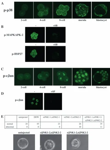
We then examined whether p38 and JNK are activated during mouse preimplantation development. Activation of p38α can be assessed by using anti-phospho-specific p38α antibody. Indirect immunofluorescence showed that p38α began to be activated from the four-cell stage and remained active during morula and blastocyst stages (Fig. 2A). The phosphorylated form of p38αwas detected in dots or patches near the plasma membrane (Fig. 2A). The activation of p38α showed no apparent asymmetry within the cells. As confirmation of the activation of p38, we investigated whether SB203580 inhibition of the kinase activity of p38 abolished the staining of phospho-MAPKAPK-2 and phospho-HSP27. MAPKAPK-2, MAP kinase-activated protein kinase 2, is a direct target of p38 MAPK (Rouse et al., 1994), and HSP27 is a substrate of MAPKAP kinase 2 (Landry et al., 1992; Rouse et al., 1994). Treatment with SB203580 markedly decreased the staining intensity of MAPKAPK-2 and phospho-HSP27 (Fig. 2B), confirming that p38 MAPK is activated during mouse preimplantation development. The activation of JNK can be assessed by using anti-phospho-specific antibody for Jun transcription factor. Jun is a well-known substrate of JNK. Indirect immunofluorescent staining showed that nuclear existence of phosphorylated Jun was detected from the four-cell stage to the blastocyst stage (Fig. 2C). Furthermore, to verify that the staining of phospho-Jun was caused by the activation of JNK, we examined whether treatment with SP600125, a JNK inhibitor, abolished this staining. The result, shown in Fig. 2D, demonstrated that the SP600125 treatment inhibited almost completely the phosphorylation of Jun, indicating that JNK is activated during the four-cell to the blastocyst stages. These results taken together show that both p38α and JNK are activated in mouse preimplantation development.
As SP600125 was recently reported not to be totally specific (Bain et al., 2003), we used the RNAi approach to inhibit the JNK pathway. We chemically synthesized four sets of complementary 21 nucleotide RNA oligonucleotides, each producing a 19 nucleotide RNA duplex. 1 and siJNK1-2 are two RNA duplexes for mouse JNK1. siJNKsiJNK1-2-1 and siJNK2-2 are for mouse JNK2. These RNA duplexes were injected into one-cell zygotes and the embryos were cultured
in vitro for three days. Embryos that were not injected or injected with distilled deionized water (DDW) developed normally (Fig. 2E). Likewise, siJNK1-1 and siJNK2-1 had no effect on preimplantation development. By contrast, embryos injected with siJNK1-2 and siJNK2-2 showed abnormality in cavity formation: apparent multiple cavities in an embryo instead of one cavity, a normal size cavity with abnormality in the inner-cell mass or a small cavity (Fig. 2E). These phenotypes are similar to those of SP600125-treated embryos. Importantly, the siJNK1-2 and siJNK2-2-injected embryos were not stained by anti-phospho-Jun (Ser 73) antibody, while uninjected or DDW-injected embryos were stained (data not shown). This suggests that the two siRNAs, siJNK1-2 and siJNK2-2, specifically inhibit the expression of JNK1 and JNK2. Taken together, these results clearly indicate that JNK plays an essential role in preimplantation development.
Phenotypes of mice carrying a single or doubly targeted deletion of the molecules comprising the MAPK cascades have previously been reported (Pearson et al., 2001). A defect in cavitation during preimplantation development, however, has not been observed in any of these mice. This may be due to redundancy of the function of multiple isoforms of the family members. Moreover, when deletion of one gene leads to embryonic lethality, preimplantation developmental processes may not be examined. Thus, possible roles of members of the MAPK family in mammalian preimplantation development have not been addressed. Our present results have revealed crucial involvement of two subfamily members of the MAPK family, p38 and JNK, in mouse preimplantation development. After completion of this work, a paper by Natale et al. (Natale et al., 2004) appeared, reporting requirement of p38 activity for mouse preimplantation development. Although there are subtle differences between our experiments and theirs, such as the period of the inhibitor treatment and the focused events in preimplantation development, the obtained conclusions are essentially the same.
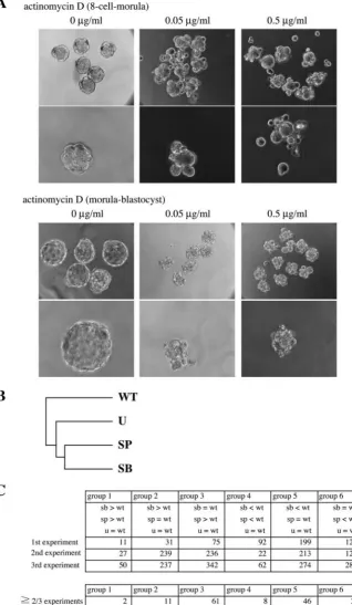
As the MAPK signaling pathways are known to regulate gene expression in the nucleus, we next asked whether gene transcription is required for preimplantation development. Previous experiments with α-amanitin showed that gene expression is necessary for preimplantation development (Khidir et al., 1995; Kidder and McLachlin, 1985). To confirm this, we examined the influence of transcription inhibitor, actinomycin D, on preimplantation development. Treatment of eight-cell stage embryos with actinomycin D (0.05 µg/ml or 0.5 µg/ml) completely inhibited compaction, and embryos were unable to develop into morula (Fig. 3A, left). When we added actinomycin D to morula stage embryos, dose-dependent effects were observed (Fig. 3A, right). At 0.05
µg/ml actinomycin D, the cavity formation was completely blocked in about 60% of embryos. In about 40% of embryos, the smaller cavity was formed. At 0.5 µg/ml actinomycin D, the complete inhibition of cavitation was observed in about 85% of embryos, and the remaining embryos showed a much smaller cavity. These results, together with the previous studies (Khidir et al., 1995; Kidder and McLachlin, 1985), suggest that both compaction and cavity formation require de novo synthesis of mRNAs. It is likely that the p38 and JNK signaling pathways are involved in these processes during preimplantation development through regulation of gene expression.
Fig. 1. SB203580 and SP600125 inhibit normal blastocoel
formation. (A) Schedule of inhibitor treatment and analysis of preimplantation embryos. The program of developmental processes is shown at the top. Arrowheads indicate timing of compaction and that of cavitation. Bars in scheme 1, 2, 3 and 4 indicate the duration of the inhibitor treatment. At the end of the bars (indicated by arrowheads), embryos were observed and the typical images are shown in B. The zona pellucida was removed before the inhibitor was added to M16 medium. The inhibitor was added at the early eight-cell stage, and embryos were examined before the cavitation in scheme 1. In scheme 2, the inhibitor was added at the early eight-cell stage, and embryos were examined at the blastocyst stage. In scheme 3, the inhibitor was added at the pre-cavitation stage. In scheme 4, the inhibitor was added after initiation of cavitation. (B) Schematic representation of the phenotypes of embryos treated with each inhibitor. (C) Embryos treated with 20 µM SB203580 (+SB) or 25 µM SP600125 (+SP) were compared with control embryos (Cont.). 1, 2, 3 and 4 correspond to those in A. (D) Time-dependent observations of control, SB203580-treated or SP600125-treated embryos were performed. The inhibitor (SB or SP) was added at the eight-cell stage.
De
Based on the above observations, we examined expression profiles of genes that could be regulated by the MAPK pathways during preimplantation development. We used Affymetrix GeneChips for microarray analysis. Three independent experiments were carried out. For each experiment, we collected eighty embryos from four kinds of pools: wild-type embryos, SB203580-treated embryos, SP600125-treated embryos and U0126-treated embryos. Eight-cell stage embryos were treated with these drugs, and blastocyst stage embryos were collected for RNA extraction, labeling and hybridization. The used array comprised over 39,000 transcripts (45,000 probe sets) and about 60% (about
[image:6.612.182.563.221.741.2]24,000 transcripts) were judged to be expressed in the wild-type sample by the GeneChip Operating Software 1.0 determination. In any of the SB203580-treated, SP600125-treated and U0126-SP600125-treated samples, the percentages of transcripts that were judged to be expressed were about 60%. Rather surprisingly, the expression level of most of the transcripts was not changed by the drug treatment. Only ~1-4% of transcripts showed the sensitivity to the inhibition of MAPK pathways; the expression level of these genes was increased or decreased by either of SB203580, SP600125 or U0126 at least by twofold compared with baseline (wild type). Hierarchical clustering was performed with those transcripts
Fig. 2.p38 and JNK are active
during mouse preimplantation development. (A) The activated form of p38 was detected by using anti-phospho-specific p38αMAP kinase antibody. Embryos at the four-cell, eight-cell, morula or blastocyst stage were stained with anti-phospho-specific p38αMAP kinase antibody. Fluorescence was viewed with a confocal
microscope. (B) Phosphorylation of MAPKAPK-2 or HSP27 with or without SB203580. The eight-cell stage embryos were treated with SB203580 for 1.5 hours, and then the embryos were fixed and stained with anti-phospho-specific MAPKAPK-2 (Thr 334) antibody or anti-phospho-specific HSP27 (Ser 82) antibody. (C) Activation of JNK was assessed by using anti-phospho-specific Jun (Ser 73) antibody. Embryos at the four-cell, eight-four-cell, morula and blastocyst stage were stained with anti-phospho-specific Jun (Ser 73) antibody. Fluorescence was viewed with a confocal microscope. (D) Phosphorylation of Jun was examined with or without SP600125. SP600125 was added at the eight-cell stage. After 2 hours of incubation, the embryo was fixed and stained with anti-phospho-specific Jun (Ser 73) antibody. (E) Effect of JNK1/2-targeting siRNAs on mouse preimplantation development. One-cell stage embryos were not injected or injected with DDW or dsRNA oligonucleotides as indicated, and cultured for 3 days. The numbers of morphologically normal or abnormal embryos were counted. Bright-field microscopic photographs are shown.
De
that were sensitive to these drugs by GeneSpring 6.1. The result shows that the effect of SB203580 is most similar to that of SP600125, and the effect of U0126 is not so similar to that of SB203580 or SP600125 (Fig. 3B), as was the case
for the effect of these drugs on
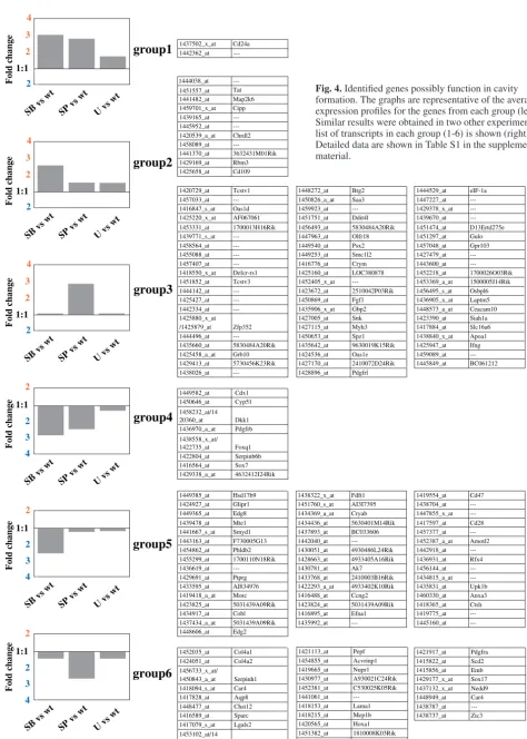
preimplantation development (see Fig. 1). Thus, we extracted 156 transcripts (161 probe sets) whose changes in mRNA levels were more than twofold in at least two out of the three independent experiments; 74 transcripts (groups 1, 2 and 3) showed an increased level of expression while the remaining 82 transcripts (groups 4, 5 and 6) showed a decreased level of expression by SB203580 or SP600125 treatment but not by U0126 treatment (Fig. 3C). We classified these 156 transcripts into six groups based on the sensitivity to the drugs (Fig. 3C, Fig. 4). In Fig. 4 (left), the average expression levels of genes in each group were shown. Expression levels of genes in group 1, 2, or 3 were increased by the inhibition of the p38 or the JNK pathway, but rather insensitive to the inhibition of the ERK pathway. By contrast, expression levels of genes in group 4, 5, or 6 were decreased by SB203580 or SP600125, but insensitive to U0126.
[image:7.612.50.369.78.626.2]Group 1 includes two transcripts, whose expression levels are increased at least twofold when compared with baseline by either of the SB203580 and SP600125 treatments. Group 2 (11 transcripts) shows at least a twofold increase only by SB203580. The effect of SP600125 was minimal in this group. Group 3 comprises 61 transcripts (62 probe sets). Transcripts in this population are increased at least twofold by SP600125 treatment, but insensitive to SB203580 treatment. Group 4 is interesting, as expression of genes in this group is decreased by either SB203580 or SP600125 treatment but is insensitive to U0126. Thus, it is likely that the function of the genes in this group is required for successful preimplantation development. This group contains eight transcripts (10 probe sets). Cdx1, caudal type homeobox 1, has been described as an intestine-specific transcription factor. Because many homeobox family genes play key roles in determining cell fate, Cdx1 may also have a role in preimplantation development. Foxq1, forkhead box Q1, is a member of the evolutionarily conserved winged helix (WH)/forkhead transcription factor gene
Fig. 3. Gene transcription is essential for preimplantation development. (A) Eight-cell stage
embryos were treated with actinomycin D (0.05 µg/ml or 0.5 µg/ml), and compared with control embryos (0 µg/ml) at morula stage (upper panel). In addition, morula stage embryos were treated with actinomycin D, and observed at blastocyst stage (lower panel). Low and high magnifications are shown. (B) Hierarchical clustering analysis. The transcripts used in this analysis were increased or decreased by SB203580, SP600125 or U0126 at least by twofold over baseline (wild type). (C) The transcripts were classified into six groups based on the sensitivity to the drugs as shown. The numbers of transcripts in each group in each experiment are shown (upper). The numbers of the transcripts that were classified into each group in at least two out of the three independent experiments are shown (lower).
De
g
ro
up
1
e g n a hc d l o Fg
ro
up
4
g
ro
up
2
g
ro
up
5
g
ro
up
3
g
ro
up
6
t w s v B S t w s v P S t w s v U x 1. 1 x 1. 1 2 1:1 2 3 4 e g n a hc d l o F e g n a hc d l o F e g n a hc d l o F e g n a hc d l o F e g n a hc d l o F 2 1:1 2 3 4 t w s v B S t w s v P S t w s v U x1.2x1.2 x 1. 1 x 1. 1 2 1:1 2 3 4 t w s v B S t w s v P S t w s v U 2 4 1:1 2 3 t w s v B S t w s v P S t w s v U 2 4 1:1 2 3 t w s v B S t w s v P S t w s v U 2 4 1:1 2 3 t w s v B S t w s v P S t w s v U 5730456K23Rik 1700013H16Rik 1420729_at Tcstv1 1457033_at ---1416847_s_at Oas1d 1425220_x_at AF067061 1453331_at 1439771_s_at ---1458564_at ---1455088_at ---1457407_at ---1418550_x_at Defcr-rs1 1451852_at Tcstv3 1444142_at ---1425427_at ---1442334_at ---1425880_x_at /1425879_at Zfp352 1444496_at ---1435660_at 5830484A20Rik 1425458_a_at Grb10 1429413_at 1438026_at---2510042P03Rik
9630019K15Rik 1448272_at Btg2 1450826_a_at Saa3 1459923_at ---1451751_at Ddit4l 1456493_at 5830484A20Rik 1447963_at Olfr18 1449540_at Psx2 1449253_at Smc1l2 1416776_at Crym 1425160_at LOC380878 1452405_x_at ---1423672_at 1450869_at Fgf1 1435906_x_at Gbp2 1427005_at Snk 1427115_at Myh3 1450653_at Spz1 1435642_at 1424536_at Oas1e 1427170_at 2410072D24Rik 1428896_at Pdgfrl
1444529_at eIF-1a 1447227_at ---1429378_x_at ---1439670_at ---1451474_at D13Ertd275e 1451297_at Gulo 1457048_at Gpr103 1427479_at ---1443600_at ---1452218_at 1700026O03Rik 1453369_a_at 1500005J14Rik 1456495_s_at Osbpl6 1436905_x_at Laptm5 1448573_a_at Ceacam10 1423390_at Siah1a 1417884_at Slc16a6 1438840_x_at Apoa1 1425947_at Ifng 1459089_at ---1445849_at BC061212 1444038_at 1451557_at Tat 1441482_at Map2k6 1459701_x_at Cipp 1439165_at ---1445952_at ---1420539_a_at Chrdl2 1458089_at ---1441370_at 3632431M01Rik 1429169_at Rbm3 1425658_at Cd109 ---5630401M14Rik 4930486L24Rik 4933405A16Rik 2410003B16Rik 5031439A09Rik 1438322_x_at Fdft1 1451760_s_at AI3I7395 1434369_a_at Cryab 1434436_at 1437893_at BC033606 1442040_at ---1430051_at 1428663_at 1430781_at Ak7 1433768_at 1422293_a_at 4933402K10Rik 1416488_at Ccng2 1423824_at 1416895_at Efna1 1435992_at ---1419554_at Cd47 1438704_at ---1447855_x_at ---1417597_at Cd28 1457377_at ---1452387_a_at Amotl2 1442918_at ---1436931_at Rfx4 1456144_at ---1434815_a_at ---1435831_at Upk1b 1460330_at Anxa3 1418365_at Ctsh 1419775_at ---1445160_at ---1449385_at Hsd17b9 1424927_at Glipr1 1449365_at Edg8 1439478_at Mte1 1441667_s_at Smyd1 1443163_at F730005G13 1454862_at Phldb2 1455299_at 1700110N18Rik 1436619_at ---1429691_at Ptprg 1433595_at AI834976 1419418_a_at Morc 1423825_at 5031439A09Rik 1434917_at Cobl 1437434_a_at 5031439A09Rik 1448606_at Edg2
1452035_at Col4a1 1424051_at Col4a2 1456733_x_at/ 1450843_a_at Serpinh1 1418094_s_at Car4 1417828_at Aqp8 1448477_at Chst12 1416589_at Sparc 1417079_s_at Lgals2 1453102_at/14 29310_at Flrt3
1810008K03Rik 1421113_at Pepf 1454855_at Acvrinp1 1419665_at Nupr1 1430977_at A930021C24Rik 1452381_at C530025K05Rik 1441061_at ---1418153_at Lama1 1418215_at Mep1b 1420565_at Hoxa1 1451382_at 1455019_x_at Ckap4
1421917_at Pdgfra 1415822_at Scd2 1415856_at Emb 1429177_x_at Sox17 1437132_x_at Nedd9 1448949_at Car4 1438787_at ---1438737_at Zic3 1449582_at Cdx1 1450646_at Cyp51 1458232_at/14 20360_at Dkk1 1436970_a_at Pdgfrb 1438558_x_at/ 1422735_at Foxq1 1422804_at Serpinb6b 1416564_at Sox7 1429338_a_at 4632412I24Rik 1437502_x_at Cd24a 1442362_at
---Fig. 4. Identified genes possibly function in cavity
formation. The graphs are representative of the average expression profiles for the genes from each group (left). Similar results were obtained in two other experiments. A list of transcripts in each group (1-6) is shown (right). Detailed data are shown in Table S1 in the supplementary material.
De
[image:8.612.64.538.76.740.2]family, and has been shown to have a unique and distinct function in differentiation and development of the hair shaft (Hong et al., 2001). Foxq1 is expressed throughout embryonic
development (Hong et al., 2001), but its role during embryogenesis remains unclear. Dkk1, Dickkopf-1, is a secreted protein that specifically inhibits Wnt/β-catenin signaling (Mao et al., 2001; Zorn, 2001), and Wnt-antagonizing function of Dkk1 is conserved in vertebrates (Shinya et al., 2000; Marvin et al., 2001). It was originally identified as a strong head inducer in Xenopus(Glinka et al., 1998). A recent report has shown that the expression of Dkk1 is regulated by Jun and that Dkk1 has a potential function in the process of vertebrate limb development (Grotewold and Ruther, 2002). It is possible that Dkk1 plays an important role in cavitation. Sox proteins, SRY box-containing transcription factors, are key players in the regulation of embryonic development and in the determination of different cell fates (Wegner, 1999; Soullier et al., 1999). Sox7 has been shown to be expressed rather ubiquitously during embryonic development, and has been suggested to function in several differentiation processes (Takash et al., 2001). In addition, SOX7 protein has the ability to significantly reduce Wnt/β -catenin-stimulated transcription (Takash et al., 2001). As the recent report has suggested that Wnt signaling may operate during mouse preimplantation development (Wang et al., 2004), it is notable that two genes known for Wnt-antagonizing activity, Dkk1 and Sox7, are identified as a member of the group 4 in our present study. Other genes in group 4 are Cyp51, Pdgfrb, Serpinb6b and one unknown gene. Group 5 contains 46 transcripts. Expression of transcripts in this group is halved at least by SB203580 treatment but insensitive to SP600125 treatment. Expression of transcripts in the last group (group 6), including 28 transcripts (30 probe sets), is halved at least specifically by SP600125 treatment. The most notable transcript in group 6 should be Hoxa1, homeobox A1, which is a transcription factor and known to function in patterning of the segments in the developing hindbrain. Because Hox genes, in general, play a role in the processes that control the AP identity of the embryo, Hoxa1 may have a role in preimplantation developmental processes.
[image:9.612.48.335.113.631.2]Representative transcripts in each group (CD24a in group 1, Map2k6 in group 2, Tcstv1 in group 3, Cdx1 in group 4, Glipr1 in group 5 and Aqp8 in group 6) were examined for their sensitivity to each drug by RT-PCR to confirm the microarray data (data not shown). The obtained results completely conform to the classification (see Fig. 3C, Fig. 4). Recently, two reports performing microarray analysis revealed that there are two major phases of stage-specific gene activity, which precede blastocoel formation (Hamatani et al., 2004; Wang et al., 2004). In addition Hamatani et al. (Hamatani et al., 2004) have classified genes in terms of their expression pattern (clusters 1 to 9). Our rough analysis has suggested that there exists little or no specific correlation between their clustering and our grouping (1 to 6); for example, any of the genes in our group 4 could not be classified into any of nine clusters. Thus, at present we are unable to characterize stage-specific expression patterns of our 156 transcripts identified. Further studies should identify transcriptional regulation mechanisms of these genes. In this context, it has been shown that
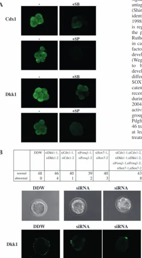
Fig. 5. Functional analysis of several genes in group 4. (A) Eight-cell stage
embryos were treated with or without SB203580 or SP600125 for 1 hour, and stained with anti-CDX1 antibody or anti-Dkk1 antibody. Fluorescence was viewed with a confocal microscope. (B) Effect of siRNA-based inhibition of genes in group 4. One-cell stage embryos were injected with dsRNA oligonucleotides as indicated, and cultured for 3 days. The numbers of morphologically normal or abnormal embryos were counted, and embryos were stained with anti-Dkk1 antibody. Bright-field (upper) and fluorescence (lower) micrographs of DDW-injected (left) and
siCdx1/siDkk1/siFoxq1/siSox7-injected (middle and right) embryos are shown.
De
the expression of Dkk1 is dependent on the Ap-1 family member Jun (Grotewold and Ruther, 2002). We were able to find ten possible Ap-1 sites in 1 kb upstream from the ORF of Dkk1 (data not shown). Other members of group 4 also have 5-19 possible Ap-1 sites. Moreover, several members of group 4 may have other transcription factor binding sites, e.g. MEF and CRE. Because Ap-1, MEF and CRE are known to be regulated by the p38 and JNK pathways, it is possible that the expression of genes in group 4 could be regulated directly by the p38 and JNK pathways.
Last, we performed two types of experiments to assess the possible function of several genes in group 4. First, we carried out immunofluorescent examination for Cdx1 and Dkk1 in eight-cell embryos. Both proteins were clearly detected at this stage, and their expression level was decreased by SB203580 or SP600125 treatment (Fig. 5A). This result is consistent with the p38- and JNK-dependent increase of the mRNA level of both genes. Next, siRNA experiments were performed to examine the function of Cdx1, Dkk1, Foxq1 and Sox7. siCdx1-1 and siCdxsiCdx1-1-2 are two RNA duplexes for mouse CdxsiCdx1-1. siDkk1-1 and siDkk1-2 are for mouse Dkk1. siFoxq1-1 and siFoxq1-2 are for mouse Foxq1. siSox7-1 and siSox7-2 are for mouse Sox7. These RNA duplexes were injected into one-cell zygotes and 3 days later, the embryos were examined. Embryos injected with DDW developed normally and were stained by anti-Dkk1 antibody (Fig. 5B). At this stage, Dkk1 protein appeared to localize at or near plasma membrane. siRNA of individual genes had little effect on preimplantation development. However, when all these siRNAs for the four genes were injected together, about one fifth of the injected embryos showed abnormality in cavity formation (Fig. 5B). These results suggest that at least several of the group 4 genes in combination may play a role in preimplantation development.
In summary, our microarray analyses, based on the finding of differential involvement of various MAPK signaling pathways in preimplantation development, have identified a small number of genes, the function of which could be important for developmental processes. This is a crucial step for our understanding of the molecular mechanisms of preimplantation development in mammals. Detailed functional analysis of these genes will be the next challenge.
We are grateful to Y. Matsubayashi for technical comments. This work was supported by grants from the Ministry of Education, Culture, Sports, Science and Technology of Japan (to E.N.).
Supplementary material
Supplementary material for this article is available at http://dev.biologists.org/cgi/content/full/132/8/1773/DC1
References
Ahn, N. G., Seger, R. and Krebs, E. G. (1992). The mitogen-activated protein kinase activator.Curr. Opin. Cell Biol.4, 992-999.
Alessi, D. R., Cuenda, A., Cohen, P., Dudley, D. T. and Saltiel, A. R. (1995). PD098059 is a specific inhibitor of the activation of mitogen-activated protein kinase kinase in vitroandin vivo. J. Biol. Chem.270, 27489–27494.
Bai, Y.-Q., Yamamoto, H., Akiyama, Y., Tanaka, H., Takizawa, T., Koike, M., Yagi, O. K., Saitoh, K., Takeshita, K., Iwai, T. and Yuasa, Y. (2002). Ectopic expression of homeodomain protein CDX2 in intestinal metaplasia and carcinomas of the stomach. Cancer Lett.176, 47-55.
Bain, J., McLauchlan, H., Elliott, M. and Cohen. P. (2003). The specificities of protein kinase inhibitors: an update.Biochem. J.371, 199-204.
Barcroft, L. C., Offenberg, H., Thomsen, P. and Watson, A. J. (2003). Aquaporin proteins in murine trophectoderm mediate transepithelial water movements during cavitation.Dev. Biol. 256, 342-354.
Bennett, B. L., Sasaki, D. T., Murray, B. W., O’Leary, E. C., Sakata, S. T., Xu, W., Leisten, J. C., Motiwala, A., Pierce, S., Satoh, Y., Bhagwat, S. S., Manning, A. M. and Anderson, D. W. (2001). SP600125, an anthrapyrazolone inhibitor of Jun N-terminal kinase. Proc. Natl. Acad. Sci. USA98, 13681-13686.
Chang, L. and Karin, M. (2001). Mammalian MAP kinase signalling cascades. Nature410, 37-40.
Cuenda, A., Rouse, J., Doza, Y. N., Meier, R., Cohen, P., Gallagher, T. F., Young, P. R. and Lee, J. C. (1995). SB 203580 is a specific inhibitor of a MAP kinase homologue which is stimulated by cellular stresses and interleukin-1. FEBS Lett.364, 229-233.
Davies, S. P., Reddy, H., Caivano, M. and Cohen, P. (2000). Specificity and mechanism of action of some commonly used protein kinase inhibitors. Biochem. J.351, 95-105.
Davis, R. J. (2000). Signal transduction by the JNK group of MAP kinases. Cell103, 239-252.
Dudley, D. T., Pang, L., Decker, S. J., Bridges, A. J. and Saltiel, A. R.
(1995). A synthetic inhibitor of the mitogen-activated protein kinase cascade. Proc. Natl. Acad. Sci. USA92, 7686-7689.
Elbashir, S. H., Harborth, J., Lendeckel, W., Yalcin, A., Weber, K. and Tuschl, T. (2001). Duplexes of 21-nucleotide RNAs mediate RNA interference in cultured mammalian cells. Nature411, 494-498.
Favata, M. F., Horiuchi, K. Y., Manos, E. J., Daulerio, A. J., Stradley, D. A., Feeser, W. S., van Dyk, D. E., Pitts, W. J., Earl, R. A., Hobbs, F. et al. (1998). Identification of a novel inhibitor of mitogen-activated protein kinase kinase. J. Biol. Chem.273, 18623-18632.
Fleming, T. P., Sheth, B. and Fesenko, I. (2001). Cell adhesion in the preimplantation mammalian embryo and its role in trophectoderm differentiation and blastocyst morphogenesis. Front. Biosci. 6, 1000-1007.
Glinka, A., Wu, W., Delius, H., Monaghan, A. P., Blumenstock, C. and Niehrs, C. (1998). Dickkopf-1 is a member of a new family of secreted proteins and functions in head induction. Nature391, 357-362.
Grotewold, L. and Ruther, U. (2002). The Wnt antagonist dickkopf-1 is regulated by Bmp signaling and c-Jun and modulates programmed cell death. EMBO J. 21, 966-975.
Hamatani, T., Carter, M. G., Sharov, A. A. and Ko, M. S. H. (2004). Dynamics of global gene expression changes during mouse preimplantation development. Dev. Cell6, 117-131.
Han, Z., Boyle, D. L., Chang, L., Bennett, B., Karin, M., Yang, L., Manning, A. M. and Firestein, G. S. (2001). c-Jun N-terminal kinase is required for metalloproteinase expression and joint destruction in inflammatory arthritis. J. Clin. Invest. 108, 73-81.
Hong, H.-K., Noveroske, J. K., Headon, D. J., Liu, T., Sy, M.-S., Justice, M. J. and Chakravarti, A. (2001). The winged helix/forkhead transcription factor Foxq1 regulates differentiation of hair in satin mice. Genesis29, 163-171.
Kamakura, S., Moriguchi, T. and Nishida, E. (1999). Activation of the protein kinase ERK5/BMK1 by receptor tyrosine kinases. Identification and characterization of a signaling pathway to the nucleus. J. Biol. Chem. 274, 26563-26571.
Khidir, M. A., Stachecki, J. J., Krawetz, S. A. and Armant, D. R. (1995). Rapid inhibition of mRNA synthesis during preimplantation embryo development: vital permeabilization by lysolecithin potentiates the action of
α-amanitin. Exp. Cell Res.219, 619-625.
Kidder, G. M. and McLachlin, J. R. (1985). Timing of transcription and protein synthesis underlying morphogenesis in preimplantation mouse embryos. Dev. Biol. 112, 265-275.
Kyriakis, J. M. and Avruch, J. (2001). Mammalian mitogen-activated protein kinase signal transduction pathways activated by stress and inflammation. Physiol. Rev.81, 807-869.
Landry, J., Lambert, H., Zhou, M., Lavoie, J. N., Hickey, E., Weber, L. A. and Anderson, C. W. (1992). Human HSP27 is phosphorylated at serines 78 and 82 by heat shock and mitogen-activated kinases that recognize the same amino acid motif as S6 kinase II.J. Biol. Chem.267, 794-803.
Mao, B., Wu, W., Li, Y., Stannek, P., Glinka, A. and Niehrs, C. (2001). LDL-receptor-related protein 6 is a receptor for Dickkopf proteins. Nature
411, 321-325.
Marvin, M. J., di Rocco, G., Gardiner, A., Bush, S. M. and Lassar, A. B.
De
(2001). Inhibition of Wnt activity induces heart formation from posterior mesoderm. Genes Dev. 15, 316-327.
Mody, N., Leitch, J., Armstrong, C., Dixon, J. and Cohen, P. (2001). Effects of MAP kinase cascade inhibitors on the MKK5/ERK5 pathway. FEBS Lett.
502, 21-24.
Natale, D. R., Paliga, A. J. M., Beier, F., D’Souza, S. L. A. and Watson, A. J. (2004). P38 MAPK signaling during murine preimplantation development. Dev. Biol.268, 76-88.
Nishida, E. and Gotoh, Y. (1993). The MAP kinase cascade is essential for diverse signal transduction pathways. Trends Biochem. Sci.18, 128-131.
Ono, K. and Han, J. (2000). The p38 signal transduction pathway: activation and function. Cell Signal.12, 1-13.
Pearson, G., Robinson, F., Beers Gibson, T., Xu, B. E., Karandikar, M., Berman, K. and Cobb, M. H. (2001). Mitogen-activated protein (MAP) kinase pathways: regulation and physiological functions. Endocr. Rev.22, 153-183.
Rouse, J., Cohen, P., Trigon, S., Morange, M., Alonso-Llamazares, A., Zamanillo, D., Hunt, T. and Nebreda, A. R. (1994). A novel kinase cascade triggered by stress and heat shock that stimulates MAPKAP kinase-2 and phosphorylation of the small heat shock proteins. Cell78, 1027-1037.
Salas-Vidal, E. and Lomeli, H. (2004). Imaging filopodia dynamics in the mouse blastocyst. Dev. Biol. 265, 75-89.
Shinya, M., Eschbach, C., Clark, M., Lehrach, H. and Furutani-Seiki, M.
(2000). Zebrafish Dkk1, induced by the pre-MBT Wnt signaling, is secreted from the prechordal plate and patterns the anterior neural plate. Mech. Dev.
98, 3-17.
Soullier, S., Jay, P., Poulat, F., Vanacker, J. M., Berta, P. and Laudet, V.
(1999). Diversification pattern of the HMG and SOX family members during evolution. J. Mol. Evol. 48, 517-527.
Sturgill, T. W. and Wu, J. (1991). Recent progress in characterization of protein kinase cascades for phosphorylation of ribosomal protein S6. Biochim. Biophys. Acta1092, 350-357.
Takash, W., Canizares, J., Bonneaud, N., Poulat, F., Mattei, M.-G., Jay, P. and Berta, P. (2001). SOX7 transcription factor: sequence, chromosomal localization, expression, transactivation and interference with Wnt signaling. Nucleic Acids Res. 29. 4274-4283.
Wang, Q. T., Piotrowska, K., Ciemerych, M. A., Milenkovic, L., Scott, M. P., Davis, R. W., Zernicka-Goetz, M. (2004). A genome-wide study of gene activity reveals developmental signaling pathways in the preimplantation mouse embryo. Dev. Cell 6, 133-144.
Wegner, M. (1999). From head to toes: the multiple facets of Sox proteins. Nucleic Acids Res. 27, 1409-1420.
Zorn, A. M. (2001). Wnt signaling: antagonistic Dickkopfs. Curr. Biol. 11, R592-R595.