5375
Introduction
Canonical Wnt signalling (Wnt/-catenin signalling) functions in many tissues and at several different developmental stages to trigger a wide variety of cellular reactions. It is currently unclear how the correct tissue- and stage-specific reaction is triggered in response to Wnt/-catenin signalling.
Early development of Xenopusis the best understood model system for tissue- and stage-specific Wnt signalling (Darken and Wilson, 2001; Hamilton et al., 2001; Roel et al., 2002; Schohl and Fagotto, 2003). Wnt/-catenin signalling mediates three separate responses during the early developmental stages leading to gastrulation. First, from cleavage stage to early blastula (stages 3-8), maternal Wnt/-catenin signalling establishes the dorsal axis of the embryo by lifting the transcription repression imposed by Tcf3 on dorsal genes such as siamois (Houston et al., 2002; Yang et al., 2002). This early function of Wnt/-catenin signalling is still reflected by the expression of later dorsal genes such as chordinin dorsal cells during gastrulation. Second, during slightly later blastula stages (stages 8.5-9.5), Wnt/-catenin signalling is also active all around the marginal zone (equatorial region), and is required upstream of zygotic FGF and nodal signals for
mesoderm induction (Schohl and Fagotto, 2003). The role of Wnt/-catenin signalling in mesoderm induction is revealed by the expression of the pan-mesoderm marker brachyury(Xbra). Third, subsequent to mesoderm induction, zygotic Wnt8/ -catenin signalling promotes ventral and lateral, but restricts dorsal, mesoderm development (Christian and Moon, 1993; Hamilton et al., 2001; Hoppler et al., 1996; Hoppler and Moon, 1998). This Wnt/-catenin signalling activity is best analysed during gastrulation by the expression of ventrolateral mesoderm marker Xpo, and the dorsolateral mesoderm marker
XmyoD. As nuclear -catenin is present all around the marginal zone during blastula stages (Schohl and Fagotto, 2002), the question arises how gene expression is regulated tissue- and stage-specifically downstream of Wnt/-catenin signalling.
Wnt/-catenin signalling is mediated by protein complexes of -catenin with individual members of the Tcf/Lef family of DNA-binding factors. The vertebrate Tcf/Lef family consists of four genes, Tcf1, Lef1, Tcf3 and Tcf4, which give rise to many different splice variants (e.g. van Noort and Clevers, 2002). Without -catenin, they all inhibit the transcription of target genes in association with co-repressors (e.g. Brantjes et al., 2001). Wnt/-catenin signalling stabilizes -catenin, which
Tcf/Lef transcription factors and -catenin mediate canonical Wnt signalling, which plays remarkably diverse roles in embryonic development, stem cell renewal and cancer progression. To investigate the molecular mechanisms allowing for these diverse yet specific functions, we studied the several distinct roles for Wnt/ -catenin signalling in early Xenopus development: establishing the dorsal body axis; regulating mesoderm induction; and subsequent ventrolateral patterning. Our previous experiments and the expression patterns of Tcf/Lef factors during these embryonic stages led us to examine whether different Tcf/Lef factors mediate these distinct events downstream of canonical Wnt/-catenin signalling. By manipulating gene expression with morpholino-driven gene knockdown and capped RNA-mediated rescue, we show that genes encoding different Tcf/Lef transcription factors mediate distinct responses to
Wnt signalling in early Xenopus development: Tcf1 and Tcf3 genes are non-redundantly required in mesoderm induction for mediating primarily transcriptional activation and repression, respectively; while ventrolateral patterning requires both Tcf1 and Lef1 genes to express sufficient levels of transcription-activating Tcf factors. Our investigation further identifies that motifs within their central domain, rather than their C-terminus, determine the particular molecular function of Tcf/Lef factors. These findings suggest that Tcf/Lef genes encode factors of different activities, which function together in antagonistic or synergistic ways to modulate the intensity and outcome of Wnt/-catenin signalling and to trigger tissue-specific responses.
Key words: Lef, Mesoderm, Signalling, Tcf, Wnt, Xenopus
Summary
Distinct roles for Xenopus Tcf/Lef genes in mediating specific
responses to Wnt/

-catenin signalling in mesoderm development
Fei Liu1,*, Olaf van den Broek2, Olivier Destrée2and Stefan Hoppler1,†
1Institute of Medical Sciences, University of Aberdeen, Aberdeen AB25 2ZD, UK
2Netherlands Institute for Developmental Biology (NIOB), Hubrecht Laboratorium, 3584CT Utrecht, The Netherlands
*Present address: Departments of Dermatology and Cell and Developmental Biology, University of Pennsylvania, M13 Stellar-Chance Laboratories, 422 Curie Boulevard, Philadelphia, PA 19104-6100, USA
†Author for correspondence (e-mail: s.p.hoppler@abdn.ac.uk)
Accepted 6 October 2005
Development 132, 5375-5385
Published by The Company of Biologists 2005 doi:10.1242/dev.02152
Research article
De
then forms a complex with Tcf/Lef factors to permit or actively promote activation of target gene transcription. Members of the Tcf/Lef family are highly homologous in the N-terminal  -catenin-binding domain and the high mobility group (HMG) DNA-binding domain, which is located more towards the C-terminus of the protein. Except for these two short domains, their amino acid sequences are diverse, and some functional motifs, including a CtBP-binding motif (Brannon et al., 1999), a p300 interacting domain (Hecht and Stemmler, 2003), or an E-tail motif (CRARF motif) (Atcha et al., 2003), are present only in certain isoforms of Lef/Tcfs. Analysis of knockout mice phenotypes indicated that Lef/Tcf gene function may not be fully interchangeable or redundant (Korinek et al., 1998; Reya et al., 2000). This finding could reflect a difference in the temporal or spatial expression pattern of Tcf/Lef genes, or indicate a functional difference in the protein products of these genes. Recent reports showed that different Tcf/Lef proteins when ectopically expressed have different activities (Gradl et al., 2002) and may exert distinct functions on different promoters of target genes (Hecht and Stemmler, 2003).
In Xenopus, all four members of the Tcf/Lef family were recently cloned. Tcf1 and Tcf3 are both maternally and zygotically expressed, while Lef1is expressed only after the onset of zygotic gene expression at the mid-blastula transition (MBT) (Molenaar et al., 1998; Roel et al., 2003). Maternal Tcf3
is ubiquitously present in early embryos, while zygotic expression of Tcf3 appears only much later in the anterior region of the late gastrula. Tcf1RNA is detected at high levels in the animal hemisphere of cleavage- and blastula-stage embryos; at early gastrula stages, Tcf1is highly expressed in the animal cap and most of the marginal zone except for a narrow domain around the blastopore. Low-level transcripts of
Lef1become detectable in the mid- and late blastula. In the early gastrula, we also detected an elevated expression of Lef1
in the ventrolateral marginal zone (data not shown). The expression of Xenopus Tcf4is reported to be detectable from late neurula stages in the midbrain region (Konig et al., 2000), but another investigation indicates that maternal Tcf4
expression is detected by RT-PCR (Houston et al., 2002). The functions of Lef1and Tcf1in the early development of
Xenopusembryos are still unclear. Considering the significant difference in structures and expression patterns between Lef1,
Tcf1and Tcf3, it is reasonable to assume that these Tcfs may be involved in mediating tissue-specific responses downstream of Wnt/-catenin signalling. In support of this notion, we have previously demonstrated that Wnt/-catenin signalling mediates tissue-specific Wnt signalling at different stages of early Xenopus development by engaging different Tcf-mediated nuclear mechanisms (Hamilton et al., 2001) and that constitutively repressing constructs of Tcf3 and Lef1 have the capacity to interfere specifically with Wnt/-catenin signalling-mediated processes in different tissues and at different stages of early Xenopus development (Roel et al., 2002).
Here we show by gene knockdown, rescue and overexpression experiments in Xenopus, that expression of genes encoding different Tcf/Lef transcription factors are required to mediate distinct responses to Wnt signalling. In particular, we show that Tcf1 and Tcf3 are non-redundantly required for mesoderm induction, and that for subsequent ventrolateral mesoderm patterning, both normal levels of Tcf1
and Lef1 gene expression are required. Further analysis indicates that different molecular functions of these Tcf/Lef factors are determined by LVPQ and SXXSS motifs in their central domains. This is the first systematic comparison of endogenous functions of Tcf/Lef genes in early Xenopus
mesoderm development, which yields interesting and novel conclusions that are important beyond the context of early
Xenopusdevelopment.
Materials and methods
Xenopus embryo manipulationsXenopus laevis embryos were harvested and staged by standard methods (Nieuwkoop and Faber, 1994). For phenotype analysis, embryos were injected with morpholino antisense oligonucleotides (MOs) in a total volume of 10 nl/cell into the lateral marginal zone (LMZ) of both blastomeres at the 2-cell stage or into the marginal zones of two dorsal blastomeres (DMZ) or two ventral blastomeres (VMZ) at the 4-cell stage and fixed in 3.7% formaldehyde-PBS at tailbud stages (approx. stage 35). For analysis by in-situ hybridization, embryos were injected with MOs and capped RNAs in 10 nl into one side of the LMZ at the 2-cell stage, then fixed at stages 10-11. A minimum of 35 embryos was analysed per individual experiment.
Whole-mount in-situ hybridization
Whole-mount RNA in-situ hybridization was performed (Harland, 1991) with modifications as described in McGrew et al. (McGrew et al., 1999). The digoxigenin-labelled antisense RNA probes used were Xbra (Smith et al., 1991), Xpo (Sato and Sargent, 1991), XmyoD (Frank and Harland, 1991) and Xenopus chordin(Sasai et al., 1994). All experiments were repeated independently at least once.
In-vitro transcription and translation (TNT)
TNT Quick Coupled (Promega, Madison, WI) in-vitro transcription and translation reactions (25 l reaction volume) were used to test the efficiency of MOs. One hundred nanograms of pCS2+-based vector DNA (Turner and Weintraub, 1994) (see also http://sitemaker.umich.edu/dlturner.vectors) encoding the 5⬘ sequences complementary to XlTcf1, XlLef1or XlTcf3MOs were used as gene-specific templates. Additionally, a control DNA vector template (100 ng) encoding Luciferase was used to monitor independently the enzymatic reactions and subsequent gel loading. MOs were added to the reactions (see below). The reactions were performed in the presence of 35S-Methionine to radioactively label the protein products. Following incubation, reactions were run on 10% acrylamide gels (Nu-PAGE, Invitrogen Life Technologies) and the results were visualized by exposure to radioactivity-sensitive film. MO titration (50-250 ng per reaction) produced gene-specific inhibition of protein synthesis to different extents. While keeping other conditions of the assay unchanged, we found 50, 100 and 150 ng to be sufficient for Tcf1 MO, Tcf3 MO and Lef1 MO, respectively, to inhibit protein synthesis of their specific target to undetectable levels. We chose to use MOs at 100 ng per reaction in the representative experiment shown in Fig. 1A.
MO and mRNA injections
MOs targeting Xenopus laevisTcf/Lef factors were designed by and purchased from Gene Tools (Philomath, OR). The MO sequences were: Lef1MO: 5⬘-CTC CAG AGA GCT GAG GCA TGG CTC C-3⬘; Tcf1MO: 5⬘-CGG CGC TGT TCA TTT GGG GCA T-3⬘; Tcf3 MO: 5⬘-CGC CGC TGT TTA GTT GAG GCA TGA-3⬘; Tcf4MO: 5⬘-CGC CAT TCA ACT GCG GCA TCT CTG C-3⬘ (Kunz et al., 2004); and control MO: 5⬘-CCT CTT ACC TCA GTT ACA ATT TAT A-3⬘. To examine the phenotypes produced by the knockdown of individual Tcfs, we injected MOs individually into the prospective
De
mesoderm (marginal zone) of Xenopus embryos. MO titration produced the phenotypic series of overt effects: none-mild-substantial-toxic at 10, 15, 20 and 25 ng/cell for the Tcf1 MO and 30, 45, 60 and 75 ng/cell for both Lef1 and Tcf3 MOs. We chose 15-20 ng/cell for Tcf1 MO and 60 ng/cell for Lef1 and Tcf3 MOs. Note that the observed relative efficiencies of the Tcf1, Tcf3 and Lef1 MOs in the embryo correspond well with those assayed in the in-vitro TNT reactions (see above).
Capped mRNA for microinjection were synthesized from plasmids containing the following subcloned cDNAs: XlLef1 and XlTcf3 in HA-tagged pT7TS constructs (Molenaar et al., 1998); XlTcf3⌬C in HA-tagged pT7TS (PmlI fragment of XlTcf3 in HA-pT7TS was deleted, linearized with XbaI); XtlTcf1 construct is a chimera of 5⬘-UTR and about 500 nucleotides of 5⬘ coding sequences from XtTcf1 with other domains from XlTcf1 in HA-tagged pT7TS (linearized with XbaI); XlTcf4A/4C in myc-tagged pCS2+ (Gradl et al., 2002); linearized with NotI); Xbra in pCS2+ (EcoRI + HpaI fragment of pXT6 (Smith et al., 1991) was inserted into EcoRI + StuI cut pCS2+, linearized with NotI). The following mutated XlTcf3 constructs were inserted into pCS2+myc vector and
linearized with Asp718 or NotI. In XTcf3⌬N, the binding domain for -catenin is disrupted (Molenaar et al., 1996). In XTcf3⌬grg⌬C (Gradl et al., 2002), the putative binding sites for Grg and CtBP transcriptional co-repressors are eliminated. In TVGR (Darken and Wilson, 2001), the -catenin-binding domain of XlTcf3 is replaced with the VP16 transcriptional activation domain (amino acids 411-490) and the hormone-binding domain of human Glucocorticoid Receptor is fused to the C-terminus. In Tcf3⌬LVPQ-258,259,263SA (Gradl et al., 2002) (here abbreviated to Tcf3⌬L-SA), the LVPQ motif is eliminated and Serine 258,259,263 are mutated to Alanine. Capped RNA was synthesized using the mMessage mMachine kit (Ambion), purified by passing over a ProbeQuant G-50 Micro Columns.
[image:3.612.45.348.77.519.2]All capped RNAs of each of the Tcf/Lef constructs were injected alone or with MOs into one side of the LMZ of embryos at the 2-cell stage. MO rescue experiments were always performed with mRNAs that lacked the target sequence recognized by the particular MO at the starting site of translation. The injection doses of these Tcf mRNAs were titrated to find a concentration that does not affect expression pattern of Xbra, Xpoor XmyoDwhen injected alone.
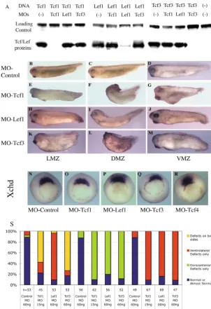
Fig. 1.MOs against Tcf/Lef factors produce different and specific phenotypes. (A) XlTcf1, XlLef1or XlTcf3 MOs specifically inhibit protein synthesis from its corresponding DNA construct in in-vitro transcription and translation assays, while not affecting significantly translation of other Tcf constructs or a control luciferase DNA construct. Injection of 60 ng control MO into LMZs of both blastomeres at the 2-cell stage, or the marginal zones of two dorsal blastomeres (DMZ) or two ventral blastomeres (VMZ) does not affect the phenotype significantly (B,C,D). Injection of 20 ng Tcf1 MO into the LMZ causes a severe developmental arrest phenotype in the majority of embryos, and in the rest (E) or when only 15 ng Tcf1 MO is injected it interferes with both dorsal and ventral development (S). Injection of 20 or 15 ng of Tcf1 MO into the DMZ causes a severe dorsal bend at approximately the position of hindbrain (F,S), and into the VMZ causes an anteriorized phenotype (G,S). Injection of 60 ng Lef1 MO into the LMZ interferes slightly with both dorsal and ventral development (H,S), into the DMZ causes a slight dorsal bend (I,S), and into the VMZ causes a mild defect in ventral tissue development and a significant defect of tail development (J,S). Injection of 60 ng Tcf3 MO into the LMZ interferes with both dorsal and ventral development (K,S), but to a lesser degree than 20 or 15 ng of Tcf1 MO does. Injection of 60 ng Tcf3 MO into the DMZ causes a complete headless phenotype (L,S), and into the VMZ causes significant ventral development defects in both anterior and posterior regions (M,S). (N-R) Vegetal view of chordin(Xchd) expression in stage 10.5 embryos, dorsal towards the top, injections into the right side. The expression pattern and level of Xchdare not significantly affected by injection of 60 ng control MO (N), 20 ng Tcf1MO (O), 60 ng Lef1MO (P), 60 ng Tcf3MO (Q) or 60 ng Tcf4MO (R). (S) Numerical summary illustrating penetrance of morphological phenotypes caused by Tcf/Lef MOs, indicating dorsoanterior defects (i.e. clearly identifiable defects in the dorsal axis and the head and neck region), ventrolateral defects and combinations of these defects (but note that the detailed nature and severity of defects vary between Tcf1 MO, Lef1 MO and Tcf3 MO experiments, as illustrated in panels B-M).
De
Results
Inhibition of Xenopus Tcf/Lef factors
In order to investigate the gene-specific functions of Tcf/Lef transcription factors (Tcfs) in mesoderm development, we designed MOs that inhibit mRNA translation initiation during
Xenopusembryonic development (Heasman et al., 2000). The efficiency of the designed MOs was tested in coupled in-vitro transcription and translation reactions. Our results show that each MO could specifically knock down the expression of the targeted Tcf/Lef gene to a considerable degree (Fig. 1A). Importantly, although the sequences of the Xenopus laevis Tcf1
and Tcf3genes in the targeted region differ by only six bases, there was no non-specific translation-blocking effect of Tcf1
and Tcf3MOs.
MO-mediated knockdown of different Tcfs during
Xenopus laevisdevelopment produced significantly different phenotypes. The induced phenotypes were also generally different if any given Tcf was tissue-specifically knocked down in dorsal tissue as opposed to lateral or ventral tissue. Targeted dorsal MO-mediated Tcf3 knockdown caused a complete headless phenotype (Fig. 1L), similar to that caused by the hdl (zebrafish Tcf3) mutation or hdl MO injection in zebrafish (Dorsky et al., 2003); dorsal Tcf1 knockdown caused a severe bend in the dorsal axis at approximately the position of the hindbrain (Fig. 1F), and dorsal Lef1
knockdown caused only a slight dorsal bend in the dorsal axis but also an apparently mild patterning defect in the forebrain region (Fig. 1I). Targeted ventral knockdown of Tcf1, Lef1
and Tcf3 affected ventral development to different extents (Fig. 1G,J,M), while Lef1 knockdown also affected tail development, consistent with the results of Lef1 gene knockdown in Xenopus tropicalis (Roel et al., 2002). Targeted lateral knockdown of Tcf1 with 20 ng Tcf1 MO, which was usually used throughout this investigation, caused a severe phenotype in 90% of embryos with much delayed gastrulation movements, typically followed by developmental arrest and widespread apparent cell death at late gastrula and early neurula control stages (not shown). In the remaining 10% of embryos it caused a combination of dorsolateral and ventrolateral phenotypes (Fig. 1E). Less complete lateral knockdown of Tcf1 with just 15 ng Tcf1 MO showed the same combination of dorsolateral and ventrolateral phenotypes in the vast majority of embryos (Fig. 1S). Generally, lateral targeted knockdown of Tcf1, Lef1and Tcf3
impaired both dorsal and ventral developments to a lesser degree than targeted knockdowns in the dorsal or ventral mesoderm (Fig. 1E,H,K,S). These similar yet distinct phenotypes indicate that the gene functions of Tcf1, Lef1and
Tcf3in early development of Xenopusembryos may be both overlapping and unique.
MO knockdown of Tcf/Lef gene expression does not affect the establishment of the dorsal axis
In our experiments, none of the Tcf/Lef MO appeared to either inhibit the development of dorsal trunk axis structures when targeted to the prospective dorsal side (Fig. 1F,I,L), or to induce axis duplication when targeted to one cell of the ventral side (data not shown), or even to affect the expression of the organizer gene chordin (Fig. 1N-R). This result may at first seem surprising, as maternal Tcf3is required for repression of
organizer gene expression in early Xenopusembryos (Houston et al., 2002) and MO-mediated -catenin knockdown inhibits the establishment of the dorsal axis (Heasman et al., 2000). Possible explanations for our results include: (1) that there are sufficient maternal Tcf proteins in eggs and early embryos before expression of Organizer genes, which last long enough in order to mediate Wnt/-catenin signalling function in establishing the dorsal axis; (2) that the MO-mediated knockdown is insufficient to inhibit the protein synthesis from maternally or zygotically expressed Tcf mRNA in early embryos (despite evidence that it is efficient at only slightly later stages, see below); or (3) that there is comprehensive redundancy between the different maternally or zygotically expressed Tcf proteins in early embryos (despite the fact that co-injections of MO targeting different Tcf genes does not affect axis development or chordin expression). We favour the first possibility and think it most likely that MOs can effectively knock down only zygotic expression of Tcf/Lef genes and is therefore insufficient to inhibit maternal Tcf expression, which functions to mediate dorsalizing Wnt signalling before the MBT (Darken and Wilson, 2001; Hamilton et al., 2001; Yang et al., 2002). We conclude that MOs are not suitable reagents to investigate Tcf requirements in dorsal axis establishment and have therefore focused our investigation on studying the function of Tcf/Lefs in later events of mesoderm induction and patterning.
Tcf1 and Tcf3 are non-redundantly required for mesoderm induction
It was reported recently that Wnt/-catenin signalling is required for early expression of pan-mesoderm markers, such as brachyury(Xbra), through FGF3 and Nodal signalling in the prospective mesoderm (Schohl and Fagotto, 2003). To investigate a potential role of Tcf/Lef molecules in mesoderm induction, we analysed MO-mediated knockdowns of each Tcf factor by detecting the expression of the early pan-mesoderm marker Xbra (Fig. 2). We found that Xbra expression was totally abrogated by Tcf1 knockdown (Fig. 2D,L) and significantly reduced by Tcf3 knockdown (Fig. 2G,M) in a dose-dependent manner (Fig. 2L,M,N), but it was not significantly affected by knockdowns of either Lef1 or Tcf4
(Fig. 2B,C,J). These results are consistent with the temporal expression pattern of these Tcf/Lef molecules, i.e. Tcf1 and
Tcf3are expressed before the beginning of Xbraexpression (at midblastula stages), while Lef1and Tcf4are mainly expressed later. To test whether the molecular functions of Tcf1 and Tcf3 proteins are interchangeable in this event, we attempted to rescue the effect of the Tcf1knockdown with mRNA-mediated Tcf3 overexpression and vice versa. The effects of overexpression of Tcf1 or Tcf3 on their own on Xbra expression were very dose-dependent (Fig. 2K). Absence of
Xbra expression in Tcf1 knockdown was rescued by an appropriate dose of Tcf1 mRNA but was not rescued by Tcf3 mRNA at any dose (Fig. 2E,F,L); reduced Xbraexpression in the Tcf3 knockdown was rescued by an appropriate dose of Tcf3 mRNA, but was not significantly rescued by Tcf1 mRNA at any dose (Fig. 2H,I,M). Furthermore, the combination of Tcf1 MO and Tcf3 MO attenuated their effects on Xbra expression (Fig. 2N). These results show that Tcf1and Tcf3are non-redundantly required for mesoderm induction for what appears to be antagonistic roles.
De
Both Tcf1 and Lef1 are required for ventrolateral mesoderm development
Zygotic Wnt8 signalling promotes ventral and lateral, but restricts dorsal, mesoderm development through a  -catenin-dependent pathway (Christian and Moon, 1993; Hamilton et al., 2001; Hoppler et al., 1996; Hoppler and Moon, 1998). To investigate the role of Tcf/Lef molecules in ventrolateral mesoderm development, we analysed the expression of ventrolateral mesoderm markers Xpo and XmyoDmRNA by in-situ hybridization after knockdowns of each Tcf factor. We found that XmyoD and Xpo expression were reduced significantly in knockdowns for Lef1, Tcf1or Tcf3, but were not affected by either Tcf4MO or control MO (Fig. 3C,D,F,K,P and C⬘,D⬘,F⬘,K⬘,P⬘). As the mesoderm induction is a pre-condition for later dorsoventral mesoderm patterning, and
because Xbra function is required for expression of later mesoderm markers, including both ventral and dorsal markers (Giovannini and Rungger, 2002), it is necessary to test whether the apparent requirement for Tcf1 and Tcf3 function for the expression of later regional mesoderm markers is only a consequence of their prior requirement for mesoderm induction or whether they are directly involved in dorsoventral mesoderm patterning. We rescued Xbra expression (by Xbra
mRNA injections) in these knockdowns to restore mesoderm induction. We found that Xbra mRNA did restore Xpo and
XmyoDexpression in the Tcf3knockdown (Fig. 3E,E⬘,P,P⬘) but did not rescue the expression of these two regional mesoderm markers in the knockdowns for Tcf1(Fig. 3G,G⬘,P,P⬘) or Lef1
(Fig. 3L,L⬘,P,P⬘). These results indicate that only Tcf1and Lef1
[image:5.612.103.552.60.558.2]are required for promoting ventrolateral mesoderm
Fig. 2.Tcf1and Tcf3are non-redundantly required for mesoderm induction. Vegetal view of brachyury(Xbra) expression in stage 10.5 embryos, dorsal towards the top, injections into the right side. Xbra expression is not affected by injection of 60 ng control MO (A), Lef1MO (B) or Tcf4MO (C), but is completely blocked by injection of 20 ng Tcf1MO (D) and significantly downregulated by injection of 60 ng Tcf3MO (G) in the injected tissue. Blocking of Xbraexpression by XlTcf1MO is rescued by co-injection of 0.3 ng XtlTcf1 mRNA (E), but is not rescued by injection of 0.5 ng HA-Tcf3 mRNA (F). Downregulation of Xbra expression by XlTcf3MO is rescued by co-injection of 0.5 ng HA-XlTcf3 mRNA (I), but is not rescued by co-injection of 0.3 ng XtlTcf1 mRNA (H). (J-N) Numerical summary illustrating
penetrance of effects of Tcf/Lef MOs and Tcf/Lef mRNA on Xbra expression in stage 10.5 embryos, indicating absent, reduced and normal or almost normal Xbra expression detected at the site of injection. Control MO, Lef1 MO and Tcf4 MO do not significantly affect Xbra expression (J). mRNA injection-mediated overexpression of relatively low amounts of Tcf1 or Tcf3 hardly affects Xbra expression, while higher amounts of either Tcf1 or Tcf3 inhibit Xbra expression more dramatically and in a dose-dependent way (K). Xbra expression is strongly inhibited by Tcf1 MO in the vast majority of injected embryos, but is rescued by co-injection of relatively low
amounts of Tcf1 mRNA; however, increasing amounts of Tcf1 mRNA result in an apparently less successful rescue, and Tcf3 mRNA fails to rescue Xbra expression altogether (L). Xbra expression is reduced by Tcf3 MO in the vast majority of injected embryos, but is dramatically rescued by relatively low amounts of Tcf3 mRNA and hardly rescued by relatively low amounts of Tcf1 mRNA; however, increasing amounts of Tcf3 or Tcf1 mRNA result in an apparently less successful rescue (M). At relatively low doses, Tcf1 MO or Tcf3 MO also inhibit Xbra expression to a lesser degree; however, Xbra expression is not further inhibited by co-injection of these two MOs, but is rescued to some extent (N).
De
development, independent of any role in mesoderm induction. To investigate the specificity of Tcf1 and Lef1 protein function in this process, we expressed Tcf1or Lef1mRNAs in either the
Tcf1knockdown or the Lef1knockdown. We found that Xpo
and XmyoDexpression in Tcf1or Lef1knockdown were both rescued by either Lef1 or Tcf1 mRNA (Fig. 3H,I,M,N and H⬘,I⬘,M⬘,N⬘,P,P⬘). Tcf3 was not only unable to rescue Xpoand
XmyoDexpression in Tcf1or Lef1knockdown, but appeared to downregulate their expression even further (Fig.
3J,J⬘,O,O⬘,P,P⬘). Moreover, overexpression of Tcf3 alone significantly downregulated Xpoand XmyoDexpression (data not shown). These results show that although normal level expression from both Tcf1 and Lef1 genes is required for ventrolateral mesoderm development, the molecular roles of their protein gene products in this particular process are interchangeable. In other words, the function of Lef1and Tcf1
[image:6.612.191.561.187.740.2]in ventrolateral mesoderm development is qualitatively redundant but quantitatively non-redundant.
Fig. 3.Tcf1and Lef1are required for ventrolateral mesoderm development. Vegetal view of Xpo (A-O) and XmyoD(A⬘-O⬘) expression in stage 10.5 embryos, dorsal towards the top, injections into the right side. Xpoand XmyoDexpression are not affected by injection of 60 ng control MO (B,B⬘) or Tcf4MO (C,C⬘), but are both significantly downregulated by injection of 60 ng Tcf3MO (D,D⬘), 20 ng Tcf1MO (F,F⬘) or 60 ng Lef1MO (K,K⬘). Co-injection of 0.1 ng Xbra mRNA rescues the downregulation of both Xpoand XmyoDexpression by Tcf3MO (E,E⬘), but does not rescue their downregulation caused by Tcf1MO (G,G⬘) or Lef1 MO (L,L⬘), indicating that while Tcf3 is required for ventrolateral mesoderm development only because it is required for normal mesoderm induction, which is a prerequisite for ventrolateral mesoderm development; Tcf1 and Lef1 are required for ventrolateral mesoderm development
independent of any requirement in mesoderm induction.
Downregulation of Xpoand XmyoDexpression by XlTcf1MO or XlLef1MO are both
significantly rescued by co-injection of either 0.3 ng XtlTcf1 mRNA (H,H⬘; M,M⬘) or HA-XlLef1 mRNA (I,I⬘; N,N⬘), but cannot be rescued by co-injection of 0.3 ng HA-XlTcf3 mRNA (J,J⬘; O,O⬘). (P,P⬘) Numerical summary illustrating penetrance of effects of Tcf/Lef MOs (as labelled) or the co-injection of these MOs with Tcf/Lef mRNAs or Xbra mRNA (as labelled), indicating reduced or absent Xpo expression and XmyoD expression, respectively.
De
Tcf3 is required as transcription repressor, while Tcf1 and Lef1 are required as transcription activator in mesoderm development
In the absence of Wnt/-catenin signalling, Tcf/Lef factors inhibit target gene expression by interacting with transcriptional co-repressors (e.g. Brantjes et al., 2001). In the presence of Wnt/-catenin signalling, Tcf/Lef mediated transcriptional repression is relieved and a -catenin-Tcf/Lef protein complex mediates transcriptional activation (Daniels and Weis, 2005). To investigate whether the revealed requirement for gene function of different Tcf/Lef factors is caused by insufficient Tcf/Lef-mediated transcriptional repression or loss of -catenin-mediated transcriptional activation, we expressed either a constitutive repressor form of Tcf3, Tcf3⌬N (Molenaar et al., 1996), a  -catenin-dependent active form of Tcf3, Tcf3⌬grg⌬C (Gradl et al., 2002), which lost its repressor function, or a constitutive activator form of Tcf3, TVGR: VP16-Tcf3⌬N-Glucocorticoid Receptor fusion protein (Darken and Wilson, 2001), which we induced to function from stage 9 through its Dexamethasone-regulated Glucocorticoid Receptor domain (Fig. 4A).
Normal level Xbraexpression in the Tcf1knockdown was significantly rescued by both TVGR and Tcf3⌬grg⌬C but was hardly rescued by Tcf3⌬N (Fig.
4B-E,R). Conversely, Xbraexpression in the Tcf3knockdown was perfectly restored by Tcf3⌬N but was only marginally rescued by either TVGR or Tcf3⌬grg⌬C (Fig. 4F-I,R). These results show that for mesoderm induction, Tcf1 gene function is mainly required to mediate the -catenin-dependent activation of target genes, while Tcf3gene function is mainly required for transcriptional repression of target genes.
Similarly, the effects of Lef1and Tcf1gene knockdown on
[image:7.612.295.559.204.742.2]Xpoexpression were rescued by active forms of Tcf3, such as TVGR and Tcf3⌬grg⌬C, but were not rescued and were even
Fig. 4.Tcf3is predominantly required as a transcription repressor in mesoderm induction, while Tcf1 and Lef1are predominantly required as transcription activators in mesoderm development. (A) Schematic representation of mutated XlTcf3 constructs used as molecular tools in this study. The -catenin binding domain, the DNA-binding HMG box, and the Grg- and CtBP-binding domains are as indicated. Tcf3⌬N represents a constitutive repressor form, Tcf3⌬grg⌬C a  -catenin-dependent active form and TVGR a constitutive activator form of XlTcf3. (B-I) Vegetal view of brachyury(Xbra) expression at stage 10.5 embryos, dorsal towards the top, injections into the right side. Blocking of Xbraexpression by Tcf1MO (B) is hardly rescued by co-injection of 0.3 ng Tcf3⌬N mRNA (C), but is significantly rescued by co-injection of 0.3 ng Tcf3⌬grg⌬C mRNA (D) or 1 pg TVGR mRNA (induced at stage 9) (E). Downregulation of Xbraexpression by XlTcf3MO (F) is rescued by co-injection 0.3 ng HA-XTcf3⌬N mRNA (G) but is hardly rescued by co-injection of 0.3 ng
HA-XTcf3⌬grg⌬C mRNA (H) or 1 pg TVGR mRNA (induced at stage 9) (I). (J-Q) Vegetal view of Xenopus posterior(Xpo) expression at stage 10.5 embryos, dorsal towards the top, injections into the right side. Downregulation of Xpo
expression by Lef1MO (J) is not rescued by co-injection of 0.3 ng XTcf3⌬N mRNA (K), but is rescued by co-injection of 0.3 ng XTcf3⌬grg⌬C mRNA (L) or 1 pg TVGR mRNA (M, induced at stage 9). Similarly, downregulation of Xpo expression by Tcf1MO (N) is not rescued by co-injection of 0.3 ng XTcf3⌬N mRNA (O), but is rescued by co-injection of 0.3 ng XTcf3⌬grg⌬C mRNA (P) or 1 pg TVGR mRNA (Q, induced at stage 9). (R) Numerical summary illustrating the penetrance of rescue effects of Tcf3 mutants mRNAs with distinct activity for downregulation of Xbraexpression caused by Tcf1/Tcf3 MOs, indicating absent and reduced Xbra expression at the site of injection. (S) Numerical summary illustrating the penetrance of rescue effects of mRNAs encoding different Tcf3 mutated constructs in Tcf1 MO- or Lef1 MO-injected embryos, indicating absent or reduced Xpo expression at the site of injection.
De
exacerbated by the constant repressor form of Tcf3, Tcf3⌬N (Fig. 4J-Q,S). These findings were confirmed in identical rescue experiments analysed by XmyoD expression (data not
[image:8.612.98.553.75.655.2]shown). These results show that in ventrolateral mesoderm patterning, Tcf1and Lef1are required to mediate the  -catenin-mediated activation of target genes.
Fig. 5. The central motifs are crucial for the repressive role of Tcf3 in mesoderm development. (A) Schematic representation of the wild-type and mutated constructs used in this study. The  -catenin binding domain, the HMG box, LVPQ and SXXSS motifs are as indicated. (B-M) Vegetal view of brachyury(Xbra) expression in stage 10.5 embryos, dorsal towards the top, injections into the right side. Blocking of Xbra expression by injection of 20 ng Tcf1MO (B) is hardly rescued by co-injection of 0.3 ng XTcf3⌬C (C) or 0.3 ng Tcf4A mRNA (D), but significantly rescued by 0.3 ng Tcf4C mRNA (E), 0.15 ng XTcf3⌬L-SA (F) or 0.3 ng Lef1 mRNA (G). By contrast, downregulation of Xbra expression by injection of 60 ng Tcf3MO (H) is rescued by co-injection of 0.3 ng XTcf3⌬C (I) or 0.3 ng Tcf4A mRNA (J), but is not rescued by 0.3 ng Tcf4C mRNA (K), 0.1 ng XTcf3⌬L-SA (L) or 0.3 ng Lef1 mRNA (M). (N-S) Vegetal view of Xpo expression in stage 10.5 embryos, dorsal towards the top, injections into the right side. Downregulation of Xpo expression by injection of XlLef1MO (N) is not rescued by co-injection of 0.3 ng XTcf3⌬C (O) or 0.3 ng Tcf4A mRNA (P), but is rescued by 0.3 ng Tcf4C mRNA (Q), 0.15 ng XTcf3⌬L-SA (R) or 0.3 ng HA-Lef1 mRNA (S). (T) Numerical summary illustrating the penetrance of rescue effects of mRNAs of Lef1, Tcf4 isoforms or Tcf3 mutated constructs in Tcf1 MO- or Tcf3 MO-injected
embryos, indicating reduced or absent Xbraexpression. (U) Numerical summary illustrating the penetrance of rescue effects of mRNAs of Lef1, Tcf4 isoforms or Tcf3 mutated constructs in Lef1 MO-injected embryos, indicating reduced or absent Xpoexpression.
De
Protein domains determining the specific functions of Tcf factors in mesoderm development
Which domain or motif is responsible for the different functions of Tcf/Lef factors in mesoderm development? The molecular structures of Tcf/Lef factors are highly conserved in the N-terminal -catenin-binding domain and the DNA-binding HMG box, but are highly diverse in the central domain and the C-terminus. At first, we focused on the C-terminus of Tcf3, which gives Tcf3 the unique property among all the
Xenopus Tcf factors of recruiting the transcriptional co-repressor CtBP. To test the role of this domain, we used a C-terminus deleted form of Tcf3, Tcf3⌬C (Pukrop et al., 2001) and native XenopusTcf4A, which has a central domain similar to that of Tcf3 but without the CtBP-binding domain in the C-terminus (Fig. 5A). Results from rescue experiments indicate that Tcf3⌬C and Tcf4A behave like Tcf3 in our analysis: they restored normal levels of Xbraexpression in Tcf3knockdown (Fig. 5H,I,J,T), but were unable to rescue significantly the effect of Tcf1 knockdown (Fig. 5B,C,D,T). Furthermore, Tcf3⌬C and Tcf4A, like Tcf3, were also unable to rescue, and downregulated even further, the levels of Xpo expression in
Lef1knockdown (Fig. 5N,O,P,U). These results show that the C-terminus of Tcf3 is not necessarily required for its inhibitive role in mesoderm induction. In the central domain of Tcf factors, there are three motifs identified: two short motifs, LVPQ and SXXSS, flanking a longer motif, usually referred to as Exon IVa (Pukrop et al., 2001). In XenopusTcfs all these three motifs are present in Tcf3, but they are absent in Tcf1 or Lef1. To test the role of the two short motifs, we used Tcf3 mutant constructs, which lack both LVPQ and SXXSS motifs (XTcf3⌬LVPQ-258,259,263SA, abbreviated to Tcf3⌬L-SA), as well as XenopusTcf4C, which has Exon IVa but not the two flanking motifs in its central domain (Gradl et al., 2002), and
Xenopus Lef1, which has none of these three motifs in its central domain (Molenaar et al., 1998). Our results show that these molecules behave like Tcf1 and Lef1: they rescued the effect of Tcf1knockdown (Fig. 5E,F,G,T) or Lef1knockdown (Fig. 5Q,R,S,U), but could not rescue the effect of Tcf3
knockdown (Fig. 5K,L,M,T). These results clearly indicate that the two LVPQ and SXXSS motifs determine the difference in function of these Tcf factors in mesoderm development.
Discussion
Wnt/-catenin signalling mediates both mesoderm induction and dorsoventral patterning of mesoderm during a short period between mid-blastula stage and early gastrula stage. The activation of early, pan-mesoderm genes, such as brachyury
(Xbra), is in a band all around the embryo in the marginal zone; but the activation of later, ventrolateral genes, such as Xpoand
XmyoD, is limited to the VMZ and LMZ. During this short period, differences of timing, location and intensity of nuclear -catenin are marginal and transient (Schohl and Fagotto, 2002), while Tcf/Lef factors are expressed in overlapping, but distinct, patterns (Molenaar et al., 1998; Roel et al., 2003).
To study the role of Tcf/Lef factors in early Xenopus
development, we used MOs to interfere with gene expression of individual Tcf/Lef factors. Our results suggest that the MOs chosen are powerful and reliable tools for studying the requirement for individual gene function in Xenopus
development. They suggest that Tcf/Lef genes have distinct
roles in early development, as well as some shared roles. Could the observed differences in effects caused by MOs targeting different Tcf/Lef genes be attributed to possible differences (due to perhaps inefficient MO action) in residual gene expression of gene products that could be claimed to be essentially interchangeable? We do not believe so, because in our experiments different amounts of MOs injected and different amounts of rescuing mRNA co-injected changed the proportion of affected embryos but did not change the nature of the effect itself, which remained to a remarkable extent different between different Tcf/Lef genes. These closely related Tcf/Lef factors undoubtedly share molecular function, and the genes encoding these factors share redundant roles in embryonic development. However, our investigation highlights the extent to which these closely related genes appear to have evolved in vertebrate evolution to diversify and specialize in both their molecular function and in their roles in embryonic development.
Qualitatively different functions of Tcf1 and Tcf3 are required for mesoderm induction
Our gene knockdown experiments show that Tcf1and Tcf3are non-redundantly required for mesoderm induction, while Lef1
is not required (see Fig. 6). This role of Tcf1is consistent with its high expression all around the marginal zone (Roel et al., 2003). We showed that the effect of Tcf1gene knockdown was predominantly caused by lack of transcription-activating  -catenin signalling, while the similar but seemingly milder effect of Tcf3gene knockdown was predominantly caused by insufficient repression. These results suggest either that these Tcf transcription factors, through as yet unknown mechanisms, regulate a different set of downstream genes, which are eventually responsible for mesoderm induction, or that a careful balance between repressive and activating Tcf factor function is required for the regulation of precise levels of
Tcf3
Tcf1
Lef1
mesoderm
induction
ventrolateral
mesoderm
patterning
target gene(s)
[image:9.612.333.559.471.600.2]target gene(s)
Fig. 6.Summary of findings about the required role of Tcf/Lef genes in Xenopusearly mesoderm development. Tcf3 gene function is predominantly required in a transcriptional repressor role for mesoderm induction; while Tcf1 gene function is also required for mesoderm induction, but predominantly in a transcriptional activator role. However, Tcf1 gene function has another required role, also mainly as an activator, in ventrolateral mesoderm patterning, independent of its role in mesoderm induction. Lef1 gene function is also required as an activator for ventrolateral mesoderm patterning. Perturbing Tcf3 gene function also affects ventrolateral mesoderm induction, but only in a roundabout way, i.e. because ventrolateral mesoderm patterning is dependent on prior mesoderm induction, which is dependent of normal Tcf3 function.
De
expression of a common set of downstream genes responsible for mesoderm induction. Whatever the precise downstream mechanisms, our current findings suggest that mesoderm induction is mediated where an appropriate intensity of Wnt/ -catenin signalling-mediated transcriptional activity is tightly regulated by the expression level and ratio of Tcf1and Tcf3.
Ventrolateral mesoderm development is promoted by Tcf1 and Lef1
Gene knockdown of Lef1 and Tcf1 affects ventrolateral mesoderm development, independent of mesoderm induction, and through a loss of -catenin-mediated transcriptional activation (see Fig. 6). Moreover, at doses that are not interfering with mesoderm induction, overexpression of repressive forms of Tcf factors (Tcf3, Tcf4A, Tcf3⌬N) represses ventrolateral mesoderm development, while active forms of Tcf factors (Tcf1, Tcf4C, Lef1) do not interfere with it (data not shown). These results show that Wnt8/-catenin signalling, which promotes ventrolateral mesoderm development, is mediated by the -catenin-dependent transcriptional activation function of Tcf1 and Lef1. Unlike the earlier mesoderm induction event, the repressive role of Tcf3 is not required, indicating that the mechanisms of Wnt/ -catenin signalling in these two events are different. It was reported recently that in zebrafish Wnt/-catenin signalling activates ventrolateral mesoderm genes directly in combination with BMP signalling (Szeto and Kimelman, 2004), where both BMP and Wnt signalling are active at submaximal levels. This finding fits well with our model that the suboptimal Wnt/ -catenin signalling required for ventrolateral mesoderm development is mediated only by the more active forms of Tcf factors (Tcf1 and Lef1), and would be interfered with by overexpression of more repressive forms of Tcf factors (i.e. Tcf3).
Repressive role of Tcf factors is mainly determined by LVPQ and SXXSS motifs
Our investigation into the role of Tcf/Lef genes suggests a requirement for two fundamentally different types of Tcf/Lef factors in Xenopus mesoderm development: a Tcf3-like predominant repressor and a Tcf1/Lef1-like predominant activator. In our experiments, mutation or deletion of two short motifs, LVPQ and SXXSS, in the central domain of Tcf3 dramatically changed the activity of Tcf3 from transcriptional repressor to activator, which made it function more like Tcf1 and Lef1 in mesoderm development. However, although Tcf3 is the only known XenopusTcf factor that interacts with the ubiquitously expressed transcription co-repressor CtBP through its C-terminus, deletion of the C-terminus from Tcf3 did not change its repressive nature significantly in mesoderm development. These results show that LVPQ and SXXSS motifs are crucial for the repressive function of Tcf3 in mesoderm development. LVPQ and SXXSS motifs do not appear to affect the interaction between Tcf/Lef factors and transcriptional co-repressor Grg (Pukrop et al., 2001), and the mechanism of repression via these two motifs is still unclear. One possible mechanism is that phosphorylation regulated by the SXXSS serine-rich motif could prevent the formation of a ternary complex between DNA, Tcf and -catenin, as has been shown for Xenopus Tcf4 (Pukrop et al., 2001). We tend to support this model, as it would explain the dominant role of
LVPQ and SXXSS motifs in determining the repressive function of Tcf factors in mesoderm development.
Tcf/Lef function in aberrant Wnt/-catenin signalling in colorectal cancer
The primary molecular cause of colorectal cancer (CRC) is thought to be the abnormal activation of the Wnt/-catenin signalling pathway. Wnt signalling in normal colon tissue is mainly mediated by Tcf4, which is absolutely required for maintenance of a mitotically active stem cell population in the intestine (van de Wetering et al., 2002). Tcf1is also present in normal colon tissues, but mainly functions as a tumour suppresser to prevent aberrant Wnt signalling in the gut [probably mainly present as truncated constitutively repressive isoforms (Roose et al., 1999)]. Human Tcf4 consists of various isoforms, among them the LVPQ, SXXSS or Exon IVa motifs are either present or absent (Duval et al., 2000). Our results here show that different Tcf/Lef factors trigger distinct effects in vivo, and point mutations in some crucial motifs are sufficient to change their functions completely. This finding suggests that mutation or aberrant expression of Tcf/Lef factors may contribute to the progression and maintenance of CRC and that these aberrantly expressed Tcf/Lef factors would be potential drug targets for treating and preventing CRC by specifically interfering with aberrant Wnt signalling.
Conclusion
Tcf/Lef genes encode a variety of DNA-binding factors with different molecular functions. Tcf/Lef factors mainly differ in the way they mediate the activation of target genes by Wnt/ -catenin signalling. Short polypeptide motifs within their central protein domain are crucial for determining this difference in function. The expression level and ratio of Tcf/Lef factors with these different functions modulates the intensity and outcome of Wnt/-catenin signalling-controlled gene expression in the embryo, which makes specific responses possible. These conclusions are important beyond the context of Xenopus
mesoderm development.
We thank Doris Wedlich and Dietmar Gradl for XenopusTcf4 and Tcf3 constructs, and Janet Heasman and Henrietta Standley for personal communication of unpublished results. This investigation was funded by the Association for International Cancer Research. SH acknowledges additional support from The Wellcome Trust and the BBSRC. O.v.d.B. and O.D. are supported by the IOP Genomics program (IGE01010), which is subsidized by the Dutch Ministry of Economic Affairs.
References
Atcha, F. A., Munguia, J. E., Li, T. W., Hovanes, K. and Waterman, M. L.
(2003). A new beta-catenin-dependent activation domain in T cell factor. J. Biol. Chem.278, 16169-16175.
Brannon, M., Brown, J. D., Bates, R., Kimelman, D. and Moon, R. T.
(1999). XCtBP is a XTcf-3 co-repressor with roles throughout Xenopus development. Development126, 3159-3170.
Brantjes, H., Roose, J., van De Wetering, M. and Clevers, H.(2001). All Tcf HMG box transcription factors interact with Groucho-related co-repressors. Nucleic Acids Res. 29, 1410-1419.
Christian, J. L. and Moon, R. T.(1993). Interactions between Xwnt-8 and Spemann organizer signaling pathways generate dorsoventral pattern in the embryonic mesoderm of Xenopus. Genes Dev.7, 13-28.
Daniels, D. L. and Weis, W. I. (2005). Beta-catenin directly displaces Groucho/TLE repressors from Tcf/Lef in Wnt-mediated transcription activation. Nat. Struct. Mol. Biol. 12, 364-371.
De
Darken, R. S. and Wilson, P. A.(2001). Axis induction by wnt signaling: Target promoter responsiveness regulates competence. Dev. Biol.234, 42-54.
Dorsky, R. I., Itoh, M., Moon, R. T. and Chitnis, A.(2003). Two tcf3 genes cooperate to pattern the zebrafish brain. Development130, 1937-1947.
Duval, A., Rolland, S., Tubacher, E., Bui, H., Thomas, G. and Hamelin, R.(2000). The human T-cell transcription factor-4 gene: structure, extensive characterization of alternative splicings, and mutational analysis in colorectal cancer cell lines. Cancer Res.60, 3872-3879.
Frank, D. and Harland, R. M.(1991). Transient expression of XMyoD in non-somitic mesoderm of Xenopus gastrulae. Development 113, 1387-1393.
Giovannini, N. and Rungger, D.(2002). Antisense inhibition of Xbrachyury impairs mesoderm formation in Xenopus embryos. Dev. Growth Differ.44, 147-159.
Gradl, D., Konig, A. and Wedlich, D. (2002). Functional diversity of Xenopus lymphoid enhancer factor/T-cell factor transcription factors relies on combinations of activating and repressing elements. J. Biol. Chem.277, 14159-14171.
Hamilton, F. S., Wheeler, G. N. and Hoppler, S.(2001). Difference in XTcf-3 dependency accounts for change in response to beta-catenin-mediated Wnt signalling in Xenopus blastula. Development128, 2063-2073.
Harland, R. M.(1991). In situ hybridization: an improved whole-mount method for Xenopus embryos. Methods Cell Biol.36, 685-695.
Heasman, J., Kofron, M. and Wylie, C. (2000). Beta-catenin signaling activity dissected in the early Xenopus embryo: a novel antisense approach.
Dev. Biol.222, 124-134.
Hecht, A. and Stemmler, M. P.(2003). Identification of a promoter-specific transcriptional activation domain at the C terminus of the Wnt effector protein T-cell factor 4. J. Biol. Chem.278, 3776-3785.
Hoppler, S. and Moon, R. T.(1998). BMP-2/-4 and Wnt-8 cooperatively pattern the Xenopus mesoderm. Mech. Dev.71, 119-129.
Hoppler, S., Brown, J. D. and Moon, R. T.(1996). Expression of a dominant-negative Wnt blocks induction of MyoD in Xenopus embryos. Genes Dev. 10, 2805-2817.
Houston, D. W., Kofron, M., Resnik, E., Langland, R., Destree, O., Wylie, C. and Heasman, J.(2002). Repression of organizer genes in dorsal and ventral Xenopus cells mediated by maternal XTcf3. Development 129, 4015-4025.
Konig, A., Gradl, D., Kuhl, M. and Wedlich, D.(2000). The HMG-box transcription factor XTcf-4 demarcates the forebrain-midbrain boundary.
Mech. Dev.93, 211-214.
Korinek, V., Barker, N., Moerer, P., van Donselaar, E., Huls, G., Peters, P. J. and Clevers, H.(1998). Depletion of epithelial stem-cell compartments in the small intestine of mice lacking Tcf-4. Nat. Genet. 19, 379-383.
Kunz, M., Herrmann, M., Wedlich, D. and Gradl, D.(2004). Autoregulation of canonical Wnt signaling controls midbrain development. Dev. Biol.273, 390-401.
McGrew, L. L., Hoppler, S. and Moon, R. T.(1999). Qualitative analysis of differential gene expression during early Xenopus embryogenesis by whole-mount in situ hybridization and RT-PCR. In A Comparative Methods Approach to the Study of Oocytes and Embryos(ed. J. D. Richter), pp. 316-340. Oxford: Oxford University Press.
Molenaar, M., van de Wetering, M., Oosterwegel, M., Peterson-Maduro, J., Godsave, S., Korinek, V., Roose, J., Destree, O. and Clevers, H.
(1996). XTcf-3 transcription factor mediates beta-catenin-induced axis formation in Xenopus embryos. Cell86, 391-399.
Molenaar, M., Roose, J., Peterson, J., Venanzi, S., Clevers, H. and Destree, O.(1998). Differential expression of the HMG box transcription factors XTcf-3 and XLef-1 during early xenopus development. Mech. Dev.75, 151-154.
Nieuwkoop, P. D. and Faber, J.(1994). Normal Table of Xenopus Laevis.
New York: Garland Publishing.
Pukrop, T., Gradl, D., Henningfeld, K. A., Knochel, W., Wedlich, D. and Kuhl, M.(2001). Identification of two regulatory elements within the high mobility group box transcription factor XTCF-4. J. Biol. Chem.276, 8968-8978.
Reya, T., O’Riordan, M., Okamura, R., Devaney, E., Willert, K., Nusse, R. and Grosschedl, R.(2000). Wnt signaling regulates B lymphocyte proliferation through a LEF-1 dependent mechanism. Immunity13, 15-24.
Roel, G., Hamilton, F. S., Gent, Y., Bain, A. A., Destree, O. and Hoppler, S.(2002). Lef-1 and Tcf-3 transcription factors mediate tissue-specific Wnt signaling during Xenopus development. Curr. Biol.12, 1941-1945.
Roel, G., van den Broek, O., Spieker, N., Peterson-Maduro, J. and Destree,
O. (2003). Tcf-1 expression during Xenopus development. Gene Expr. Patterns3, 123-126.
Roose, J., Huls, G., van Beest, M., Moerer, P., van der Horn, K., Goldschmeding, R., Logtenberg, T. and Clevers, H. (1999). Synergy between tumor suppressor APC and the beta-catenin-Tcf4 target Tcf1.
Science285, 1923-1926.
Sasai, Y., Lu, B., Steinbeisser, H., Geissert, D., Gont, L. K. and De Robertis, E. M. (1994). Xenopus chordin: a novel dorsalizing factor activated by organizer-specific homeobox genes. Cell79, 779-790.
Sato, S. M. and Sargent, T. D.(1991). Localized and inducible expression of Xenopus-posterior (Xpo), a novel gene active in early frog embryos, encoding a protein with a ‘CCHC’ finger domain. Development112, 747-753.
Schohl, A. and Fagotto, F.(2002). Beta-catenin, MAPK and Smad signaling during early Xenopus development. Development129, 37-52.
Schohl, A. and Fagotto, F.(2003). A role for maternal beta-catenin in early mesoderm induction in Xenopus. EMBO J.22, 3303-3313.
Smith, J. C., Price, B. M., Green, J. B., Weigel, D. and Herrmann, B. G.
(1991). Expression of a Xenopus homolog of Brachyury (T) is an immediate-early response to mesoderm induction. Cell67, 79-87.
Szeto, D. P. and Kimelman, D.(2004). Combinatorial gene regulation by Bmp and Wnt in zebrafish posterior mesoderm formation. Development131, 3751-3760.
Turner, D. L. and Weintraub, H. (1994). Expression of achaete-scute homolog 3 in Xenopus embryos converts ectodermal cells to a neural fate.
Genes Dev.8, 1434-1447.
van de Wetering, M., Sancho, E., Verweij, C., de Lau, W., Oving, I., Hurlstone, A., van der Horn, K., Batlle, E., Coudreuse, D., Haramis, A. P. et al.(2002). The beta-catenin/TCF-4 complex imposes a crypt progenitor phenotype on colorectal cancer cells. Cell111, 241-250.
van Noort, M. and Clevers, H.(2002). TCF transcription factors, mediators of Wnt-signaling in development and cancer. Dev. Biol.244, 1-8.
Yang, J., Tan, C., Darken, R. S., Wilson, P. A. and Klein, P. S.(2002). Beta-catenin/Tcf-regulated transcription prior to the midblastula transition.
Development129, 5743-5752.