INTRODUCTION
The cerebral cortex contains different subclasses of excitatory projection neurons derived from the dorsal telencephalon or pallium, as well as inhibitory interneurons originating from the ventral telencephalon or subpallium (Marin and Rubenstein, 2003; Molyneaux et al., 2007). The cerebral cortex is subdivided into different regions with divergent evolutionary histories, i.e. the archicortex, neocortex and paleocortex, and into areas with distinct functions, i.e. the motor, somatosensory, visual and auditory cortical areas (O’Leary et al., 2007; Rakic, 1988; Rash and Grove, 2006; Sur and Rubenstein, 2005). The cortex is also organised into six layers containing neurons with different morphological, molecular and physiological characteristics and unique patterns of connectivity. This cytoarchitecture is tightly regulated, with a defined number of neurons adopting specific laminar features in each zone, which is crucial for the proper activity of the cerebral cortex.
Cajal-Retzius (CR) cells are among the first neurons to be generated between E10.5 and E13.5 in mouse (Hevner et al., 2001; Takiguchi-Hayashi et al., 2004), and they die during the first postnatal weeks (Abraham and Meyer, 2003; del Rio et al., 1995; Derer and Derer, 1990; Marin-Padilla, 1990; Marin-Padilla, 1992; Zecevic and Rakic, 2001). This transient pioneer neuronal
population was discovered more than a century ago, in humans by G. Retzius and in lagomorphs by S. Ramon y Cajal, but the features and functions of these cells remain largely unknown. They appear to play a key role in the radial migration of cortical neurons and in the laminar organisation of the mouse and human cortex, largely through the production of the extracellular glycoprotein reelin (D’Arcangelo et al., 1995; Ogawa et al., 1995; Rice and Curran, 2001; Super et al., 2000).
CR cells populate the marginal zone (MZ) of the cortex evenly in the prospective neocortex, but accumulate at distinct locations such as at the olfactory piriform cortex. Various CR cell subpopulations have been identified and shown to differentially express several molecular markers, including reelin, calretinin (calbindin 2 – Mouse Genome Informatics) and p73a(Trp73 – Mouse Genome Informatics) (Bielle et al., 2005; Meyer et al., 2002; Takiguchi-Hayashi et al., 2004). CR cells have long been assumed to arise from the whole pallial ventricular zone (VZ) and to migrate radially to the cortical surface, similarly to other glutamatergic cortical neurons (del Rio et al., 1995; Hevner et al., 2003; Marin-Padilla, 1998). However, Meyer and colleagues identified restricted sites of generation of CR cells based on the expression of p73a, a transcription factor that is expressed by different CR cell subpopulations (Meyer et al., 2002). Consistently, fate mapping and cell lineage tracing studies have shown that CR cells arise from specific locations along the rostrocaudal and dorsoventral axes of the pallial VZ (Bielle et al., 2005; Garcia-Moreno et al., 2007; Imayoshi et al., 2008; Monuki et al., 2001; Takiguchi-Hayashi et al., 2004; Yoshida et al., 2006; Zhao et al., 2006). Four different sites of CR generation have been identified, comprising the pallial domain of the septum in the rostromedial (RM) pallium (Bielle et al., 2005), the ventral pallium (VP) laterally (Bielle et al., 2005), the prospective choroid plexus, and the cortical hem (CH) caudally (Garcia-Moreno et al., 2007; Imayoshi et al., 2008; Monuki et al., 2001; Takiguchi-Hayashi et Development 137, 293-302 (2010) doi:10.1242/dev.041178
1National Institute for Medical Research (NIMR), Medical Research Council (MRC), Department of Molecular Neurobiology, London NW7 1AA, UK. 2Biozentrum, Department of Cell Biology, University of Basel, and Friedrich Miescher Institute for Biomedical Research, 4056 Basel, Switzerland. 3Institut Jacques Monod, Program in Development and Neurobiology, CNRS UMR 7592 and Université Paris Diderot, Paris 75013, France. 4INSERM U784, Ecole Normale Supérieure, Département de Biologie, Paris 75005, France.
*Present address: IBDML, CNRS UMR6216, Marseille 13009, France
†Authors for correspondence (zimmer@ibdml.univ-mrs.fr;fguille@nimr.mrc.ac.uk)
Accepted 19 November 2009
SUMMARY
Cajal-Retzius (CR) cells play a key role in the formation of the cerebral cortex. These pioneer neurons are distributed throughout the cortical marginal zone in distinct graded distributions. Fate mapping and cell lineage tracing studies have recently shown that CR cells arise from restricted domains of the pallial ventricular zone, which are associated with signalling centres involved in the early regionalisation of the telencephalic vesicles. In this study, we identified a subpopulation of CR cells in the rostral telencephalon that expresses Er81, a downstream target of Fgf8 signalling. We investigated the role of the rostral telencephalic patterning centre, which secretes FGF molecules, in the specification of these cells. Using pharmacological inhibitors and genetic inactivation of Fgf8, we showed that production of Fgf8 by the rostral telencephalic signalling centre is required for the specification of the Er81+CR cell population. Moreover, the analysis of Fgf8gain-of-function in cultivated mouse embryos and of Emx2and Gli3mutant embryos revealed that ectopic Fgf8 signalling promotes the generation of CR cells with a rostral phenotype from the dorsal pallium. These data showed that Fgf8 signalling is both required and sufficient to induce rostral CR cells.Together, our results shed light on the mechanisms specifying rostral CR cells and further emphasise the crucial role of telencephalic signalling centres in the generation of distinct CR cell populations.
KEY WORDS: Cajal-Retzius cells, Fgf8 signalling, Forebrain development, Neuronal specification, Mouse
Role of Fgf8 signalling in the specification of rostral
Cajal-Retzius cells
Céline Zimmer1,*,†, Jun Lee2, Amélie Griveau3, Silvia Arber2, Alessandra Pierani3, Sonia Garel4
and François Guillemot1,†
D
E
V
E
LO
P
M
E
N
T
D
E
V
E
LO
P
M
E
N
al., 2004; Yoshida et al., 2006; Zhao et al., 2006). CR cells migrate tangentially from these focal sites to populate the entire cortical surface. Furthermore, fate mapping studies have shown that CR cells originating from different sources preferentially settle in distinct regions of the cortex (Bielle et al., 2005; Imayoshi et al., 2008; Takiguchi-Hayashi et al., 2004; Yoshida et al., 2006; Zhao et al., 2006). Indeed, ablation of the CH results in a substantial depletion of CR cells, except in the rostral cortex, thus demonstrating that CH-derived CR cells mainly populate the caudal cortex (Yoshida et al., 2006).
Intriguingly, the neuroepithelial domains that generate CR cells are closely associated with signalling centres involved in the early regionalisation of the telencephalic vesicles. These signalling centres secrete morphogens that provide positional and proliferative cues to the surrounding telencephalic neuroepithelium (O’Leary et al., 2007; Rash and Grove, 2006; Sur and Rubenstein, 2005). Recently, the signals produced by the CH in the caudomedial telencephalon, which include TGFbmolecules, were shown to be necessary for the generation of caudal CR cells (Friedrichs et al., 2008; Hanashima et al., 2007; Siegenthaler and Miller, 2008; Theil, 2005). Morphogens produced by the commissural plate (CoP) in the RM telencephalon and by the putative anti-hem in the lateral telencephalon might similarly participate in CR cell generation. In contrast to the substantial progress that has been made in understanding how caudal CR cell populations are specified, the mechanisms underlying the specification of rostral CR cells have remained poorly characterised.
FGF signalling has been shown to play an essential role in patterning the rostral telencephalon (Cholfin and Rubenstein, 2007; Cholfin and Rubenstein, 2008; Fukuchi-Shimogori and Grove, 2001; Fukuchi-Shimogori and Grove, 2003; Garel et al., 2003; Paek et al., 2009; Shimogori et al., 2004; Storm et al., 2006). Fgf8is expressed early on at the rostral tip of the neural tube (called the anterior neural ridge or ANR) (Crossley and Martin, 1995; Crossley et al., 2001; Shimamura and Rubenstein, 1997) and its expression persists after fusion of the ANR to form the CoP at the rostrodorsal midline of the telencephalon (Crossley et al., 2001). Fgf8 is the main secreted factor produced by the rostral organising centre, where it regulates the expression of Fgf17 and Fgf18 and is involved in patterning both the dorsal and ventral telencephalon, as well as in promoting cell survival (Bachler and Neubuser, 2001; Borello et al., 2008; Chi et al., 2003; Cholfin and Rubenstein, 2008; Fukuchi-Shimogori and Grove, 2001; Fukuchi-Fukuchi-Shimogori and Grove, 2003; Gimeno and Martinez, 2007; Gutin et al., 2006; Lee et al., 2000; Ohkubo et al., 2002; Shanmugalingam et al., 2000; Storm et al., 2006; Storm et al., 2003).
In this study, we have examined the mechanisms engaged in the specification of rostral CR cells and asked specifically whether Fgf8 signalling from the rostral patterning centre is involved in this process. We found that Er81, an ETS transcription factor downstream of Fgf8 signalling, is specifically expressed at early stages by CR cells in the rostral cortex and not by caudal CH-derived CR cells. These rostral Er81+CR cells derive largely from the RM
pallium, as shown by their persistence in Pax6mutants. We used pharmacological inhibitors and genetic inactivation of Fgf8 to demonstrate that the Fgf8 telencephalic signalling centre is required for the specification of Er81+CR cells. We have also used an Fgf8
gain-of-function approach in vitro and analysed Emx2and Gli3 mutant mouse embryos, which express Fgf8ectopically, to show that ectopic Fgf8 signalling promotes the generation of rostral-type CR cells from the dorsal pallium. Together, our results shed light on the mechanisms that specify rostral CR cells.
MATERIALS AND METHODS Mice
MF1, Parkes and F1 (CBA/CA ⫻C57Bl/10) mice were used. All transgenic mouse lines were genotyped as previously described: Dbx1nls-lacZ(Bielle et al., 2005), Pax6(Stoykova et al., 1996), Emx2(Pellegrini et al., 1996), Gli3
[Extra-toes/Extra-toes(Buscher et al., 1998)] Fgf8Null/Neo(Storm et al., 2003) and Fgf8TelKO[Foxg1Cre; Fgf8Flox/Null(Storm et al., 2006)]. At least three embryos were analysed per condition, unless specified otherwise. Midday of the day of vaginal plug discovery was considered as E0.5. All mouse experiments used protocols approved under the UK Animal (Scientific Procedures) Act.
Embryo culture and electroporation
E10.5 mouse embryos were dissected in Tyrode’s solution, cultivated for 1 hour in rat serum at 37°C, 65% O2/5% CO2 (rolling incubator, BTC Engineering), injected (Femtojet, Eppendorf) and electroporated (chamber, CUY520P20; electroporator, Nepa Gene ECM830; 50 V, five pulses of 50 milliseconds, 1 second interval). Vectors: 1 mg/ml for pCaggs::IRES-nls-GFP (gift from J. Briscoe, NIMR, London, UK), pCaggs::IRES-nls-lacZ
(gift from S. Price, UCL, London, UK) and pCDNA3::Fgfr1-DA(Freeman et al., 2003), or 0.8 mg/ml for pMiwIII::Fgf8 (b isoform, gift from A. Joyner, NYU, New York, USA); the vectors are referred to as nls-GFP,nls-lacZ,
Pgfr1-DAand Fgf8, respectively, in the figures. Twenty-four hours after electroporation, embryos were transferred into fresh rat serum at 37°C, 100% O2, and cultured for a further 24 hours. For cell cycle studies, 40 mM BrdU was added to the culture medium for 20 minutes.
Rostral telencephalic explants
E9.5 mouse embryos were dissected in Tyrode’s solution. Only 22- to 24-somite embryos were used (see Fig. S8 in the supplementary material) (Storm et al., 2003). FGF signalling inhibitors (10 mM, DMSO diluted): SU5402 (#572630, Calbiochem); UO126 (#19-147, Millipore). Cell proliferation was only weakly affected by these inhibitors and could not account for the observed phenotypes. At 2 DIV, explants were: (1) washed in cold PBS, fixed for 30 minutes and washed in PBS for immunochemistry; or, (2) fixed overnight, washed in PBS, dehydrated in successive PBS/ethanol baths and kept in 100% ethanol at –20°C, for whole-mount in situ hybridisation.
Histology
Embryos were collected in PBS, heads fixed for 1 hour for immunohistochemistry or 3 hours for in situ hybridisation, washed with PBS, transferred into 15% sucrose in phosphate buffer (PB) pH 7.2 overnight, embedded in 7.5% gelatin, 15% sucrose in PB at 42°C, frozen in –40°C isopentane and stored at –80°C. Sections (10 mm) were prepared using a Microm cryostat (Zeiss).
For in situ hybridisation, tissues were processed as described by Hirsch et al. for sections (Hirsch et al., 2007) and by Bielle et al. for whole-mount (Bielle et al., 2005). Probes: Erm(IMAGE 4036564), Pea3, Er81, Mash1
(C. Goridis, ENS, Paris, France), Ngn2, reelin (Y. Hayashizaki, RIKEN, OSC, Kanagawa, Japan), p73a(IMAGE 6812399), Fgfr1(J. Partanen, University of Helsinki, Finland), Spry2(G. Martin, UCSF, San Francisco, USA) and Foxg1 (J. Mason, University of Edinburgh, UK). For immunohistochemistry, frozen sections were air dried, washed in PBS at 42°C to remove the gelatin and processed for immunofluorescence. Primary antibodies: mouse anti-reelin (1/375, #MAB5364, Chemicon), mouse anti-p73a(1/200, #MS762PO, LabVision), mouse (#6B3) or rabbit (#7699) anti-calretinin (both at 1/2000, Swant), goat anti-b-galactosidase (1/1000, #ab12081, Abcam), sheep anti-GFP (1/750, #47451051, Biogenesis), mouse anti-TUJ1 (1/1000, #MMS435P, Babco), rat anti-BrdU (1/1000, #OBT0030CX, Serotec; denaturation in 2N HCl for 30 minutes at 37°C, washes with 0.1 M sodium borate pH 8.0), mouse anti-Pax6 (1/20, Developmental Studies Hybridoma Bank), rabbit Er81 and rabbit anti-Pea3, rabbit anti-Tbr1 (gift from R. Hevner, University of Washington, Seattle, USA) and rabbit anti-Lhx2 (gift from E. Monuki, University of California, Irvine, USA). Fluorescent secondary antibodies were Alexa 488 (Millipore) or Cy3 or Cy5 (Jackson ImmunoResearch) conjugated. Note that to perform the co-detection of Er81 and Tbr1, rabbit anti-Tbr1 was directly
D
E
V
E
LO
P
M
E
N
T
D
E
V
E
LO
P
M
E
N
labelled with Cy5 using the Zenon Kit (Molecular Probes). For explant immunostaining, 1 hour blocking at room temperature was followed by incubation with antibody overnight at 4°C and washing in PBS.
Image analysis, quantifications and statistics
Images were captured using a ProgRes C14 camera (Jenoptik) linked to an Axioplan II microscope (Zeiss), a QImaging camera linked to MZ16/MZ16F scopes (Leica), or a Radiance 2100 confocal microscope (BioRad). Images were processed with Openlab (Perkin Elmer), ImageJ (NIH), Photoshop (Adobe) or FreeHand (Adobe). Quantifications were performed on confocal photographs (200 mm ⫻200 mm; stack of 2 or 3 mm) using Photoshop. For example, in Fig. 2 the number of Er81+cells was counted by marking the cells with a dot on a transparent layer linked to the Er81 staining layer in Photoshop. Then each Er81+cell was assessed for its expression of either reelin, calretinin or p73a, using another transparent layer linked to each of these stainings. The same principle was used to perform all counts. In Fig. 2, four stacks per area from two wild-type embryos were analysed. For electroporated embryos, two to three stacks were analysed per embryo. Because of electroporation variability, results were normalised for each embryo before being gathered for statistical analysis using a paired Student’s t-test (see tables in the supplementary material). For quantification in mutant embryos, three to four stacks were analysed per area.
RESULTS
Rostral CR cells express Er81, a downstream target of FGF signalling
Genetic ablation of the CH-derived CR cell subpopulation has revealed the extent of the remaining CR cell subpopulations in the rostral telencephalon (Yoshida et al., 2006). The signalling activity of the CH during telencephalic patterning is balanced by the activity of the rostral CoP (O’Leary et al., 2007; Rash and Grove, 2006; Sur and Rubenstein, 2005). We thus hypothesised that rostral CR cells, which are spared by the ablation of the CH, might be induced by FGF signals emanating from the CoP.
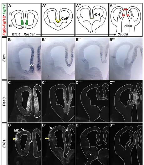
To address this issue, we first mapped the domains of Fgf8 signalling activity by characterising the expression patterns of the ETS transcription factorsErm,Pea3 andEr81(also known as Etv5, Etv4and Etv1, respectively), which are effectors of Fgf8 signalling (Cholfin and Rubenstein, 2008; Fukuchi-Shimogori and Grove, 2003). We conducted our analysis at different rostrocaudal levels of the mouse telencephalon at E11.5 (Fig. 1B-D). At rostral levels, Erm and Pea3were expressed medially throughout the dorsoventral extent of the telencephalic VZ (Fig. 1B,B⬘,C,C⬘), whereas Er81 expression was restricted to the MZ of the whole cortex at this level (Fig. 1D,D⬘). More caudally, Ermand Pea3were expressed at low levels in the CH (Fig. 1B⬙,C⬙), whereas Er81was absent (Fig. 1D⬙). Further caudally, expression of all three genes was absent from the telencephalon (Fig. 1B,C,D). FGF signalling activity thus correlated well with Fgf expression domains and was elevated within the RM pallium, which includes the pallial septum (Fig. 1A-A). Er81expression was detected in the rostral cortex, where it started to decline at E12.5 and was absent at E13.5 (data not shown), suggesting a transient FGF signalling activity in postmitotic neurons.
We characterised the Er81-expressing cells of the rostral cortex by co-immunolabelling for Er81 and cell type-specific markers at E11.5. All Er81+cells expressed Tbr1, a T-box transcription factor
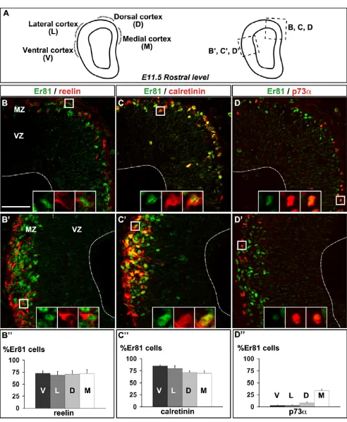
that labels both CR and preplate neurons (see Fig. S1C-F in the supplementary material) (Hevner et al., 2001). Using reelin and calretinin as specific CR cell markers (Alcantara et al., 1998; del Rio et al., 1995), we further determined that ~75% of Er81+cells were
positive for reelin or calretinin and had a similar distribution in the different cortical areas analysed (Fig. 2B-C⬙; see Fig. S2 in the
supplementary material). Using p73a as a marker of CR cells derived from the pallial septum (A.G. and A.P., unpublished) (Hanashima et al., 2007), we also observed that some cells expressed both Er81 and p73a, representing more than 30% of the Er81+
population in the medial cortex (Fig. 2D-D⬙). These data suggest that rostral CR cells are likely to represent two distinct CR cell types, one p73a+and one p73a–. Together with the spatial analysis of Er81
expression (Fig. 1D,D), these data showed that rostral CR cells mainly express reelin, calretinin and the ETS transcription factor Er81 at E11.5. The co-expression of Er81 with reelin or calretinin can thus be used to discriminate rostral CR cells from CH-derived CR cells, which express neither Er81 nor calretinin at early developmental stages (E10.5-E12.5).
The pallial septum gives rise to Er81+CR cells
We then examined which progenitor domains give rise to Er81+CR
[image:3.612.323.553.57.320.2]cells at E11.5. CR cells of the rostral cortex are mainly derived from the VP, located laterally, and from the pallial septum, located medially (A.G. and A.P., unpublished) (Bielle et al., 2005). Dbx1 is expressed by progenitors of the VP and also by early postmitotic cells derived Fig. 1. Expression patterns of FGF signalling-induced ETSfactors in the mouse embryonic telencephalon.(A-A) Schemes of coronal sections of E11.5 telencephalon analysed at different rostral-caudal
levels showing expression domains of Fgf8 andFgf18 (red), Fgf17
(green) and Fgf8/17/18(yellow). (B-D) In situ hybridisation of coronal
sections of the telencephalon at E11.5. (B-B) Ermis expressed in the
medial VZ of the telencephalon (B,B⬘), in the CH and the PSB (B⬙), and
in the diencephalon but not the telencephalon at caudal levels (B).
(C-C) Pea3shows a similar expression pattern to Erm, except in the
PSB (C⬙). (D-D) Er81is expressed in the MZ of the medial (arrowhead)
and dorsal (white arrow) cortex and in the olfactory piriform cortex
(yellow arrow), where preplate cells are localised (D). Caudally, Er81is
weakly expressed in the SP (asterisk in D⬙) and in the diencephalon (D).
CH, cortical hem; CoP, commissural plate; dien, diencephalon; P, pallium; PSB, pallium-subpallium boundary; S, septum; SP, subpallium; VZ, ventricular zone; MZ, marginal zone. Scale bar:
210mm.
D
E
V
E
LO
P
M
E
N
T
D
E
V
E
LO
P
M
E
N
from the pallial septum (A.G. and A.P., unpublished) (Bielle et al., 2005). We thus analysed the rostral cortex of heterozygous Dbx1nls-lacZ
mice using co-immunolabelling for Er81 and b-galactosidase (b-gal), which can be used to trace the short-term progeny of Dbx1-expressing cells (Fig. 3A-A⬙). Most b-gal+cells co-expressed Er81 (Fig. 3A⬙),
indicating that a substantial fraction of Er81+CR cells derive from
Dbx1-expressing cells located rostrally in the lateral pallium and the medial cortex.
To further assess whether Er81+cells were derived from the RM
pallium domain, we analysed rostral CR cells in the Pax6mutant, in which the VP is not specified and no longer expresses Dbx1(data not shown) (Carney et al., 2009; Yun et al., 2001), whereas the pallial septum is unaffected and remains positive for Dbx1 expression (data not shown). We found no significant difference in Er81 and reelin co-immunolabelling between wild-type and Pax6 mutant embryos in medial, dorsal (Fig. 3B-E; see Table S1 in the supplementary material), lateral and ventral domains of the rostral cortex at E12.5 (see Fig. S3 in the supplementary material). These results indicate that Er81+CR cells are still specified in the absence
of a lateral VP domain and must therefore originate from a medial location. Hence, the pallial septum gives rise to rostral Er81+CR
cells populating the rostral cortex.
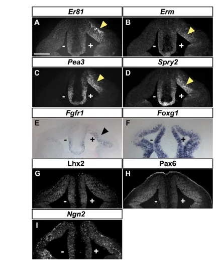
Ectopic expression of Fgf8 induces rostral CR cells Our previous observations suggested that the RM pallium, including the pallial septum, is exposed to the FGF signalling activity of the CoP and the septal VZ (Figs 1-3). We thus examined whether Fgf8, which is the main output of the rostral patterning centre, is involved in the generation of rostral CR cells. We first used a gain-of-function (GOF) approach, whereby a b-gal plasmid was co-electroporated with an Fgf8 expression construct, or electroporated alone in control experiments, in the rostrodorsal pallium of E10.5 embryos, which were then cultivated for 2 days before analysis (see Fig. S4 in the supplementary material). As expected, Fgf8 electroporation resulted in the ectopic induction of several Fgf8 signalling targets, including Er81,Pea3, Erm,Spry2andFgfr1(Fig. 4A-E). Although Fgf8 has a crucial role in patterning the rostral telencephalon (Cholfin and Rubenstein, 2008; Garel et al., 2003; Shimogori et al., 2004; Storm et al., 2006), ectopic Fgf8 did not affect the expression of pallial progenitor markers such as Lhx2, Pax6 or Ngn2(Neurog2) (Fig. 4G-I), nor that of the telencephalic marker Foxg1 (Fig. 4F), as previously reported in an Fgf8 GOF study performed at E11.5 (Shimogori et al., 2004). Hence, the telencephalic identity of the electroporated tissue was maintained upon Fgf8 GOF, but the dorsal pallium adopted a RM pallial identity as shown by the ectopic expression of Erm, Pea3, Spry2and Fgfr1.
Ectopic Fgf8 also markedly increased the number of reelin-expressing cells in the dorsal cortex as compared with the control electroporation or the non-electroporated side (Fig. 5A,B,C⬘,D⬘,E⬘,F⬘). Co-immunolabelling experiments for b-gal, to mark the progeny of electroporated cells, and for rostral CR cell-specific markers showed that most b-gal+, reelin+cells also expressed
[image:4.612.51.297.57.356.2]Tbr1 (Fig. 5D-D), Er81 (Fig. 5F-F) and calretinin (see Fig. S5A,C-C⬙,E-E⬘in the supplementary material), demonstrating that ectopic Fig. 2. Er81 expression in rostral Cajal-Retzius (CR) cells.
(A)Scheme of a coronal section of mouse E11.5 telencephalon at
rostral level. (B,B⬘,C,C⬘,D,D⬘) Co-immunolabelling for Er81 and reelin
(B,B⬘), Er81 and calretinin (C,C⬘), Er81 and p73a(D,D⬘) in the dorsal
and medial cortex (B-D) and the lateral and ventral cortex (olfactory
piriform cortex) (B⬘-D⬘). (B⬙,C⬙, D⬙) Cortical distribution of Er81+cells
that co-express reelin (V, 72.2±5.4%; L, 68.6±7.9%; D, 70.9±7.7%; M, 71.9±8.4%), calretinin (V, 85±2%; L, 81.2±5.3%; D, 71.2±3.8; M,
69.6±6%) and p73a(V, 2.6±0.9%; L, 3.1±0.8%; D, 8±2.4%; M,
33.8±3%). D, dorsal; L, lateral; M, medial; V, ventral. Scale bar:
50mm.
Fig. 3. Er81+CR cells originate particularly from the pallial septum. (A-A⬙) Co-immunolabelling for Er81 and b-galactosidase on
sections of rostral telencephalon of heterozygous Dbx1nls-lacZmouse
embryos at E11.5, showing that some Er81+cells derive from Dbx1+
cells. (B-D)Co-immunolabelling for Er81 and reelin in the dorsal (B,C)
and medial (B⬘,C⬘) cortex of wild type (WT) or Pax6mutant (Pax6 KO) at
E12.5 (see scheme in D). (E)Similar numbers (see Table S1 in the
supplementary material) of rostral CR cells marked by double labelling for Er81 and reelin are found in the dorsal and medial cortex of both
wild-type and Pax6mutant embryos. Scale bars: 100mm in A-A⬙; 25mm
in B-C⬘.
D
E
V
E
LO
P
M
E
N
T
D
E
V
E
LO
P
M
E
N
[image:4.612.312.564.61.260.2]Fgf8 specifically induces CR cells of the rostral type. Moreover, we did not observe any change in Dbx1 or p73alabelling (data not shown; see Fig. S5B,D-D⬙in the supplementary material), indicating that Fgf8 is not sufficient to induce their expression. Co-electroporation of a constitutively active form of FGF receptor 1 (Fgfr1) with b-gal also resulted in the generation of b-gal+cells that
co-expressed reelin and Er81 (see Fig. S6A-A⬘in the supplementary material). Thus, Fgf8 signalling acts cell-autonomously to induce rostral CR cell generation in the rostrodorsal pallium.
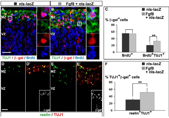
Fgf8 promotes the neurogenesis of rostral CR cells We further analysed the mechanisms by which ectopic Fgf8 expression promotes the generation of rostral CR cells. Ectopic expression of Fgf8 had no significant effect on cell proliferation in electroporated embryos cultivated for 1 or 2 days (see Fig. S7 and Tables S2, S3 in the supplementary material). To determine whether Fgf8 promoted the generation of new neurons, we labelled progenitors in S phase 24 hours after electroporation by a 20-minute exposure of cultured embryos to BrdU, and examined the differentiation of BrdU-labelled progenitors 24 hours later by double labelling for BrdU and TUJ1 [bIII-tubulin (Tubb3) – Mouse Genome Informatics]. Fgf8 expression significantly increased the fraction of BrdU+cells that expressed TUJ1 (Fig. 6A-C; see Table
S4 in the supplementary material), indicating that Fgf8 promotes the generation of new neurons from pallial progenitors.
To determine whether Fgf8 was specifically inducing CR cells or having a more general neurogenic effect, we examined the expression of reelin among Fgf8-induced neurons. Fgf8 expression significantly increased the fraction of TUJ1+, b-gal+electroporated neurons that
co-expressed reelin (Fig. 6D-F; see Table S5 in the supplementary material). Similar results were obtained when a construct expressing a constitutively active form of Fgfr1 was electroporated (see Fig. S6B-B⬘ in the supplementary material). Altogether, these results demonstrate that Fgf8 signalling specifically promotes the generation of CR cells from pallial progenitors in a cell-autonomous manner.
Ectopic expression of Fgf8 in vivo promotes the generation of rostral CR cells
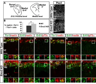
To extend these findings to ectopic expression of endogenous Fgf8, we examined Emx2mutant mice, which present a caudal extension and persistence of the Fgf8,Fgf17andFgf18rostral expression domains, and a concomitant reduction of the caudal Wnt/BMP-secreting telencephalic centre of the CH (Cholfin and Rubenstein, 2008; Fukuchi-Shimogori and Grove, 2003; Kimura et al., 2005; Shimogori et al., 2004). More cells expressed Pea3 within the RM pallium in these mutants than in control embryos (Fig. 7C,C⬘), as previously described (Cholfin and Rubenstein, 2008; Fukuchi-Shimogori and Grove, 2003). We then found a significant increase in the number of Tbr1+, reelin+CR cells in E12.5 Emx2mutant embryos
at rostrodorsal and medial levels (Fig. 7A,B,D,D⬘,G,G⬘; see Table S6 in the supplementary material). Co-labelling for Er81 and reelin showed that the majority of reelin+cells were of the rostral CR type
(Fig. 7E,E⬘,H,H⬘) and that the population of p73a+, Er81–
CH-derived CR cells was in fact reduced in the medial cortex of Emx2 mutant embryos (Fig. 7I,I⬘). We also analysed Gli3mutant mice, in which an upregulation of Fgf8 signalling in the pallium and a loss of p73a+CH-derived CR cells have been reported (Kuschel et al., 2003;
Okada et al., 2008; Rash and Grove, 2007; Theil et al., 1999). We found an increase in Pea3-expressing cells in the RM pallium at E12.5 (see Fig. S1B,B⬘in the supplementary material), as well as clusters of Er81+, Tbr1+, reelin+rostral CR cells in the medial cortex
(see Fig. S1C-F⬘in the supplementary material). These data suggest that the enlarged Fgf8domain in Emx2or Gli3mutant pallium results in an expanded and persistent rostral CR cell progenitor domain, and supports the conclusion that ectopic Fgf8 signalling promotes the generation of rostral CR cells, both in embryo culture and in vivo.
In vitro inhibition of FGF signalling prevents the generation of rostral CR cells
To investigate whether Fgf8 signalling is not only sufficient, but also required for the generation of CR cells, we set up an explant culture system in which Fgf8 signalling could be manipulated pharmacologically. The rostral head of E9.5 (21- to 23-somite) embryos was cultivated on a filter for 2 days in vitro (DIV), during which the explants maintained a dorsoventral organisation, as shown by labelling for Mash1 (Ascl1) and Ngn2 (see Fig. S8 in the supplementary material). TUJ1+neurons were only observed in the
nasal placodes at the beginning of the culture, whereas TUJ1+
neurons were found in the telencephalic part of the explant and particularly at its dorsal periphery after 2 DIV (see Fig. S8 in the supplementary material). Labelling for reelin, Er81 and calretinin showed that most of the TUJ1+neurons generated during the culture
[image:5.612.52.267.56.314.2]were rostral CR cells (see Fig. S8 in the supplementary material). To assess the role of Fgf8 signalling, we used the pharmacological inhibitors SU5402, which inhibits signal transduction by FGF receptors (Mohammadi et al., 1997), and UO126, which prevents phosphorylation of MEK1/2 (Map2k1/2) kinases (Favata et al., Fig. 4. Expression of telencephalic markers following Fgf8
overexpression.(A-I)In situ hybridisation (A-F,I) and immunolabelling (G,H) on coronal sections of E10.5 mouse telencephalon
co-electroporated on the right-hand side (marked by +) with plasmids
expressing nls-lacZand Fgf8 and cultivated for 2 days in vitro (DIV).
(A-E)Fgf8 overexpression results in ectopic expression of the Fgf8
signalling targets Er81, Erm,Pea3,Spry2andFgfr1in the
electroporated area. Note that the ectopic domains of expression are maintained in the MZ (A) or the VZ (B-E), depending on the
endogenous domain of expression. (F-I)Foxg1, Lhx2, Pax6 and Ngn2
show similar expression patterns on the electroporated and
non-electroporated sides. n≥8. Scale bars: 210mm.
D
E
V
E
LO
P
M
E
N
T
D
E
V
E
LO
P
M
E
N
1998). Cultures exposed for 2 DIV to SU5402 (Fig. 8B-B) or UO126 (Fig. 8D-D) showed a strong decrease in TUJ1, calretinin and reelin labelling compared with DMSO-treated control explants in the dorsal part of the explants (Fig. 8A-A,C-C, arrows), whereas the lateral part of the explants was less affected. These results indicate that lowering Fgf8 signalling significantly reduces the rostral CR cell population.
In vivo reduction in Fgf8 signalling decreases rostral CR cell generation
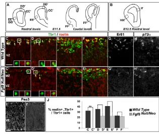
The requirement for Fgf8 signalling in the generation of CR cells was further examined in Fgf8Null/Neomouse embryos, in which
Fgf8 signalling activity in the rostral signalling centre is reduced
(Storm et al., 2006; Storm et al., 2003), as a complete loss of Fgf8 has drastic effects on telencephalic development that preclude examination of CR cells (Meyers et al., 1998). Tbr1+, reelin+CR
cells were completely absent from the RM cortex in E11.5 Fgf8Null/Neomutant embryos (Fig. 9C,C⬘,J; see Table S7 in the
supplementary material), and there was a reduction of this population in the rostrodorsal cortex and piriform cortex in these embryos (Fig. 9D-E⬘,J; see Table S7 in the supplementary material). By contrast, Tbr1+, reelin+CR cells were found in
[image:6.612.53.482.60.264.2]normal numbers in the caudal olfactory cortex (Fig. 9F,F⬘), indicating that Fgf8 signalling is required only rostrally for the generation of CR cells. In addition, Er81 expression was almost completely absent in Fgf8Null/Neomutant embryos (Fig. 9G,G⬘),
Fig. 5. Overexpression of Fgf8 in the dorsal pallium induces the generation of Er81+rostral CR cells.Immunolabelling of sections of mouse
rostral telencephalon electroporated at E10.5 and cultivated for 2 DIV. (A)Control electroporation (nls-lacZ) does not affect reelin expression on the
electroporated side (marked by +) as compared with the non-electroporated side (marked by –). (B)Co-electroporation of Fgf8and nls-lacZleads to
an increase in reelin expression (arrows). The yellow rectangles mark the dorsal cortex area shown at larger magnification in C-F. (C-D)
Co-expression of reelin and Tbr1 in electroporated b-gal+cells demonstrating that ectopic reelin+cells are CR cells. The white rectangles outline the
areas shown at higher magnification to the far right. (E-F) Co-expression of Er81 and reelin in electroporated b-gal+cells shows that ectopic CR
cells have a rostral identity. b-gal, b-galactosidase. For each condition for reelin, n>20, and for Er81/reelin or Tbr1/reelin, n10. Scale bars: 210mm
in A,B; 25mm in C-F.
Fig. 6. Fgf8 overexpression promotes the differentiation of CR cells in the dorsal cortex. Immunolabelling of sections of mouse rostral telencephalon
electroporated at E10.5 and cultivated for 2 DIV. (A-C)Triple
labelling for BrdU, following a 20-minute pulse at 1 DIV and
a 24-hour chase, and for b-gal and TUJ1 to mark newborn
neurons. Ectopic Fgf8 significantly promotes neuronal differentiation in the dorsal cortex (Student’s t-test,
**P0.002; see Table S4 in the supplementary material),
whereas it does not significantly affect the rate of proliferation as measured by the fraction of cells double
labelled for BrdU and b-gal (P0.77). (D-F)Double labelling
for reelin and TUJ1 to mark CR cells shows that ectopic Fgf8 specifically promotes the differentiation of this cell type
(**P0.004; see Table S5 in the supplementary material).
Scale bars: 25mm.
D
E
V
E
LO
P
M
E
N
T
D
E
V
E
LO
P
M
E
N
[image:6.612.50.337.542.743.2]indicating that CR cells generated in the mutant have lost their rostral identity. Moreover, expression of the progenitor marker Pea3 was absent from the mutant RM pallium (Fig. 9I,I⬘), indicating that this territory was not properly specified in Fgf8Null/Neo embryos. We then analysed the p73a expression
pattern at E12.5 to determine whether the decrease in rostral CR cells resulted in an expansion of CH-derived CR cells. However, we did not observe an increase in p73a-expressing cells (Fig. 9H,H⬘), suggesting that CH-derived CR cells had not invaded the rostroventral cortex in Fgf8Null/Neoembryos at the stage examined.
We also asked whether eliminating the Fgf8-secreting rostral signalling centre would affect the generation of rostral CR cells by analyzing embryos with a telencephalon-specific deletion of Fgf8 (Fgf8TelKO) (see Fig. S9 in the supplementary material) (Storm et
al., 2006; Storm et al., 2003). We compared reelin expression in E12.5-13 whole telencephalon from wild-type, Fgf8Null/Neoand
Fgf8TelKO embryos and found a strong reduction in
reelin-expressing cells in the medial and dorsal domains of the telencephalon of both Fgf8mutants as compared with the wild type (see Fig. S9 in the supplementary material). Altogether, these results establish that Fgf8 signalling activity is required for the specification of rostral CR cells originating from the RM pallium.
DISCUSSION
In this study, we have shown that rostral CR cells express the ETS transcription factor Er81, a downstream target of Fgf8 signalling, in addition to reelin, calretinin and p73a. We found that Er81+CR cells
originate largely from the pallial septum, which is under the patterning influence of Fgf8 signalling, suggesting that this pathway could play a role in the specification of this rostral CR cell population. In agreement with this, we showed that ectopic activation of Fgf8 signalling in the dorsal pallium in vitro and in vivo results in ectopic generation of rostral CR cells. We also demonstrated that Fgf8 signalling activity is required in vitro and in vivo for the specification of the RM pallium, including the pallial septum, and for the generation
[image:7.612.51.361.59.332.2]of rostral CR cells. Altogether, our data establish a new role for the rostral signalling centre in the specification of rostral CR cells and further emphasise the involvement of telencephalic signalling centres in the generation of a diverse array of CR cells.
Fig. 7. Increased number of rostral CR cells in the dorsal cortex of Emx2mutant mouse embryos.
(A)Schemes of coronal telencephalic sections
showing the levels analysed at E12.5. (B)The
proportion of CR cells is significantly increased in the
rostrodorsal cortex of Emx2knockout (KO) embryos
(Student’s t-test, **P0.006; see Table S6 in the
supplementary material). (C,C⬘) More cells express
Pea3 in the RM pallium of Emx2knockout than of
wild-type embryos. (D-I⬘) The boxed areas are shown
at high magnification beneath in single channel and
merge. (D,D⬘,G,G⬘) Co-expression of Tbr1 and reelin
identifies CR cells. (E,E⬘,H,H⬘) Mainly Er81 and reelin
are co-expressed in the dorsal and medial cortex
demonstrating that ectopic CR cells in Emx2KO
cortex have a rostral identity. (F,F⬘,I,I⬘) p73a
expression is decreased in the medial cortex, whereas Er81 expression is increased, even at more medial
levels (see scheme in A). Scale bars: 25mm.
Fig. 8. Impairment of FGF signalling with pharmacological inhibitors decreases rostral CR cell generation in telencephalic explants.(A-D) Whole-mount immunohistochemistry for calretinin and TUJ1 and in situ hybridisation for reelin on mouse rostral
telencephalic explants harvested at 22 somites and cultivated for 2 DIV
(see Fig. S8 in the supplementary material). Exposure to SU5402 (B-B
versus A-Acontrol) or UO126 (D-Dversus C-Ccontrol) leads to a
decrease in the generation of CR cells (calretinin+/TUJ1+or reelin+) in
the part of the explant that corresponds to the dorsal telencephalon
(arrow), whereas the neuronal population (TUJ1+cells) is, overall, less
affected. Neurogenesis appears to be less affected by exposure to FGF signalling inhibitors in the part of the explant that corresponds to the
lateral cortex. For each condition for calretinin/TUJ1, n≥15, and for
reelin, n≥5. Scale bar: 400mm.
D
E
V
E
LO
P
M
E
N
T
D
E
V
E
LO
P
M
E
N
[image:7.612.321.558.424.593.2]Er81+CR cells present unique defects in mutant mice
The existence of distinct origins and the fairly limited number of available markers might have complicated the analysis of CR cell subpopulations (Bielle et al., 2005; Garcia-Moreno et al., 2007; Imayoshi et al., 2008; Monuki et al., 2001; Takiguchi-Hayashi et al., 2004; Yoshida et al., 2006; Zhao et al., 2006). Expression of the ETS factor Er81 by rostral CR cells distinguishes them from CR cells located in the subpallium and the caudal pallium, where Er81 is not expressed. We thus used the co-expression of reelin and Er81 to identify rostral CR cells and re-examined the generation of CR cells in Pax6, Emx2and Gli3mutant mice.
Emx2 and Pax6, which cross-repress each other, are considered as positive and negative regulators of CR cell generation, respectively (Bishop et al., 2003; Mallamaci et al., 2000; Shinozaki et al., 2002; Stoykova et al., 2003). We found, however, that a Er81+, reelin+rostral
CR cell population is still present in Pax6mutants, indicating that in contrast to other cell types (Tuoc and Stoykova, 2008), some Er81 -expressing rostral CR cells are not dependent on Pax6 activity.
Our finding that loss of Emx2promotes the generation of rostral CR cells through derepression of Fgf8 signalling (Cholfin and Rubenstein, 2008; Fukuchi-Shimogori and Grove, 2003; Shimogori et al., 2004) is in agreement with the increase in reelin expression observed in Emx2mutant cortex (Mallamaci et al., 2000), although a loss of CR cells in Emx2mutants at later stages has been more widely documented (Bishop et al., 2003; Mallamaci et al., 2000; Yoshida et al., 1997). We can now associate this later phenotype with the loss of the CH-derived subpopulation, which we found already impaired at E12.5.
The generation of CR cells is impaired inGli3mutant embryos, with the loss of caudal signals resulting in a substantial decrease in p73a+CR cells and in a derepression of Dbx1in the dorsolateral
pallium, which adopts a VP-like identity (Friedrichs et al., 2008; Hanashima et al., 2007; Theil, 2005). In agreement with these previous studies, we observed ectopic clusters of Er81+CR cells in
the lateral cortex in theGli3mutant (data not shown). However, we
also noted clusters of Er81+CR cells in the medial cortex, as well as
an increase in Pea3-expressing cells, reflecting the upregulation of Fgf8 signalling in these mutants (Okada et al., 2008; Rash and Grove, 2007; Theil, 2005; Theil et al., 1999). Thus, mutations in patterning genes differentially affect the subpopulations of CR cells because of their distinct effects on the different telencephalic signalling centres.
Multiple roles for Fgf8 signalling in the generation of rostral CR cells
The rostral signalling centre is characterised by the activity of FGF molecules, with Fgf8 playing the main role. We identified two distinct functions of Fgf8 signalling by challenging its activity in vitro and by analysing Fgf8gain- and loss-of-function phenotypes in mice. Firstly, Fgf8 signalling is involved in the specification of a progenitor domain within the RM pallium that gives rise to rostral CR cells, which is in line with its role in rostral telencephalic patterning. Secondly, Fgf8 signalling promotes the generation of rostral CR cells from mitotic progenitors.
[image:8.612.50.365.57.321.2]Our GOF analysis showed that Fgf8 specifically promotes the generation of rostral CR cells. In contrast to a recent study in which ectopic Fgf8 expression resulted in re-patterning of the dorsal pallium into dorsal midline tissue (Okada et al., 2008), we observed that Fgf8-expressing cells maintained the expression of pallial progenitor markers such as Lhx2, Pax6 and Ngn2. These differences between the two studies can be explained by: (1) the competence of the tissue, as Okada et al. performed their experiment in younger embryos than we did (E9.5 as compared with E10.5); (2) the level of Fgf8 overexpression, as they used a stronger promoter to express Fgf8(the pCaggs vector versus pMiw in our study); and (3) the shorter time of exposure to Fgf8 (at 1 DIV versus 2 DIV). The importance of Fgf8 concentration and duration of action in determining its activity has been previously reported in various systems, including the CoP and the mid-hindbrain boundary (Basson et al., 2008; Liu et al., 2003; Sato et al., 2001). Altogether, these data lead us to suggest that a short Fig. 9. Generation of rostral CR cells is reduced in Fgf8Null/Neomutant mouse embryos.
(A,B)Schemes indicating the cortical regions
analysed at E11.5 (A) and E12.5 (B).
(C-J)Immunolabelling of coronal sections of different
cortical regions in wild-type and Fgf8Null/Neomutant
embryos. (C-F⬘,J) Tbr1 and reelin co-expression
identifies CR cells. The proportion of CR cells is significantly decreased in the mutant RM cortex
(C,C⬘,J; see Table S7 in the supplementary material)
and is reduced in dorsal (D,D⬘,J; see Table S7 in the
supplementary material) and piriform cortex (E,E⬘,J;
see Table S7 in the supplementary material), but is
not affected in the caudal cortex (F,F⬘,J; see Table S7
in the supplementary material), indicating that Fgf8 signalling is mainly required in the RM pallium.
(G,G⬘) Er81 is only weakly expressed in the mutant
cortex. (H,H⬘) At E12.5, there is no compensatory
increase in p73a-expressing cells in the mutant
piriform cortex. (I,I⬘) Pea3 expression is lost in the RM
pallium, in agreement with the loss of CR cells in this
area. Scale bars: 25mm.
D
E
V
E
LO
P
M
E
N
T
D
E
V
E
LO
P
M
E
N
exposure to a high Fgf8 concentration promotes a rostral midline identity (Okada et al., 2008), whereas a longer exposure to a lower Fgf8 concentration promotes RM pallial identity.
We have not detected any effect of ectopic Fgf8 on the proliferation of pallial progenitors, which is in agreement with previous studies demonstrating that Fgf8 regulates rostral telencephalic patterning but not cell proliferation (Crossley et al., 2001; Fukuchi-Shimogori and Grove, 2003; Okada et al., 2008; Shimogori et al., 2004). However, we observed that Fgf8 induces neurogenesis, a function that has been previously reported in the eye, in the basal telencephalon (Crossley et al., 2001), and in the developing olfactory system (Bailey et al., 2006; Kawauchi et al., 2005).
CR cells originate from progenitor domains that are not only restricted spatially, but also short lived (e.g. Imayoshi et al., 2008). Our data show that the arrest of production of rostral CR cells after E12.5 might be a consequence of the reduction of Fgf8 signalling in the RM pallium at this stage. Hence, a longer exposure to Fgf8 signalling in Emx2mutants or in electroporated embryos not only induced ectopic rostral CR cell production, but also extended the period of generation of this CR cell population.
In conclusion, our data establish that the rostral telencephalic signalling centre specifies a rostral CR cell population. Our study thus provides new evidence that CR cell progenitor domains are closely associated with telencephalic signalling centres and strengthens the hypothesis that CR cells participate in regional patterning of the cortex (Bielle et al., 2005; Meyer et al., 2004; Meyer et al., 2002). Because they migrate above progenitors at early developmental stages, CR cells are good candidates to provide telencephalic progenitors with instructive cues (Nomura et al., 2008; Soriano et al., 1997) (A.G. and A.P., unpublished). Thus, characterising CR cell subpopulations and their progenitor domains might help to unravel the mechanisms that control the early stages of development of the whole telencephalon.
Acknowledgements
We are particularly grateful to J. Falk for fruitful discussions during the course of this work and to K. Rizzoti and J. Falk for critically reading the manuscript. We thank J. L. L Rubenstein for the Fgf8TelKOembryos and J. Briscoe, C. Goridis, Y. Hashisaki, R. Hevner, E. Monuki, A. Joyner, G. Martin, J. Mason, J. Partanen and S. Price for sharing reagents. We particularly thank the animal husbandry service of the NIMR for their great contribution to this work. C.Z. was supported by a Medical Research Council career development fellowship. J.L. and S.A. were supported by grants from the Swiss National Science Foundation, NCCR Frontiers in Genetics, the Kanton Basel-Stadt, EU Framework Program 7 and the Novartis Research Foundation. A.G. was the recipient of fellowships from the French Ministry of Education and the Association pour la Recherche sur le Cancer, A.P. is a CNRS (Centre National de la Recherche Scientifique) Investigator and their work was supported by a grant from the Agence Nationale de la Recherche (ANR-05-NEUR-007-01 BIS) to A.P. The work in S.G.’s lab was supported by Avenir INSERM, Ville de Paris and EURYI award. The work in F.G.’s lab was supported by institutional funds from the Medical Research Council. Deposited in PMC for release after 6 months.
Competing interests statement
The authors declare no competing financial interests
Supplementary material
Supplementary material for this article is available at
http://dev.biologists.org/lookup/suppl/doi:10.1242/dev.041178/-/DC1
References
Abraham, H. and Meyer, G.(2003). Reelin-expressing neurons in the postnatal and adult human hippocampal formation. Hippocampus13, 715-727. Alcantara, S., Ruiz, M., D’Arcangelo, G., Ezan, F., de Lecea, L., Curran, T.,
Sotelo, C. and Soriano, E.(1998). Regional and cellular patterns of reelin
mRNA expression in the forebrain of the developing and adult mouse. J. Neurosci. 18, 7779-7799.
Bachler, M. and Neubuser, A.(2001). Expression of members of the Fgf family and their receptors during midfacial development. Mech. Dev. 100, 313-316. Bailey, A. P., Bhattacharyya, S., Bronner-Fraser, M. and Streit, A.(2006). Lens
specification is the ground state of all sensory placodes, from which FGF promotes olfactory identity. Dev. Cell11, 505-517.
Basson, M. A., Echevarria, D., Petersen Ahn, C., Sudarov, A., Joyner, A. L., Mason, I. J., Martinez, S. and Martin, G. R.(2008). Specific regions within the embryonic midbrain and cerebellum require different levels of FGF signaling during development. Development135, 889-898.
Bielle, F., Griveau, A., Narboux-Neme, N., Vigneau, S., Sigrist, M., Arber, S., Wassef, M. and Pierani, A.(2005). Multiple origins of Cajal-Retzius cells at the borders of the developing pallium. Nat. Neurosci. 8, 1002-1012.
Bishop, K. M., Garel, S., Nakagawa, Y., Rubenstein, J. L. and O’Leary, D. D. (2003). Emx1 and Emx2 cooperate to regulate cortical size, lamination, neuronal differentiation, development of cortical efferents, and thalamocortical pathfinding. J. Comp. Neurol. 457, 345-360.
Borello, U., Cobos, I., Long, J. E., Murre, C. and Rubenstein, J. L.(2008). FGF15 promotes neurogenesis and opposes FGF8 function during neocortical development. Neural Dev.3, 17.
Buscher, D., Grotewold, L. and Ruther, U.(1998). The XtJ allele generates a Gli3 fusion transcript. Mamm. Genome9, 676-678.
Carney, R. S., Cocas, L. A., Hirata, T., Mansfield, K. and Corbin, J. G.(2009). Differential regulation of telencephalic pallial-subpallial boundary patterning by pax6 and gsh2. Cereb. Cortex19, 745-759.
Chi, C. L., Martinez, S., Wurst, W. and Martin, G. R.(2003). The isthmic organizer signal FGF8 is required for cell survival in the prospective midbrain and cerebellum. Development130, 2633-2644.
Cholfin, J. A. and Rubenstein, J. L.(2007). Patterning of frontal cortex subdivisions by Fgf17. Proc. Natl. Acad. Sci. USA104, 7652-7657.
Cholfin, J. A. and Rubenstein, J. L.(2008). Frontal cortex subdivision patterning is coordinately regulated by Fgf8, Fgf17, and Emx2. J. Comp. Neurol. 509, 144-155.
Crossley, P. H. and Martin, G. R.(1995). The mouse Fgf8 gene encodes a family of polypeptides and is expressed in regions that direct outgrowth and patterning in the developing embryo. Development121, 439-451.
Crossley, P. H., Martinez, S., Ohkubo, Y. and Rubenstein, J. L.(2001). Coordinate expression of Fgf8, Otx2, Bmp4, and Shh in the rostral prosencephalon during development of the telencephalic and optic vesicles.
Neuroscience108, 183-206.
D’Arcangelo, G., Miao, G. G., Chen, S. C., Soares, H. D., Morgan, J. I. and Curran, T.(1995). A protein related to extracellular matrix proteins deleted in the mouse mutant reeler. Nature374, 719-723.
del Rio, J. A., Martinez, A., Fonseca, M., Auladell, C. and Soriano, E.(1995). Glutamate-like immunoreactivity and fate of Cajal-Retzius cells in the murine cortex as identified with calretinin antibody. Cereb. Cortex5, 13-21.
Derer, P. and Derer, M.(1990). Cajal-Retzius cell ontogenesis and death in mouse brain visualized with horseradish peroxidase and electron microscopy.
Neuroscience36, 839-856.
Favata, M. F., Horiuchi, K. Y., Manos, E. J., Daulerio, A. J., Stradley, D. A., Feeser, W. S., Van Dyk, D. E., Pitts, W. J., Earl, R. A., Hobbs, F. et al.(1998). Identification of a novel inhibitor of mitogen-activated protein kinase kinase. J. Biol. Chem. 273, 18623-18632.
Freeman, K. W., Gangula, R. D., Welm, B. E., Ozen, M., Foster, B. A., Rosen, J. M., Ittmann, M., Greenberg, N. M. and Spencer, D. M.(2003). Conditional activation of fibroblast growth factor receptor (FGFR) 1, but not FGFR2, in prostate cancer cells leads to increased osteopontin induction, extracellular signal-regulated kinase activation, and in vivo proliferation. Cancer Res. 63, 6237-6243.
Friedrichs, M., Larralde, O., Skutella, T. and Theil, T.(2008). Lamination of the cerebral cortex is disturbed in Gli3 mutant mice. Dev. Biol. 318, 203-214. Fukuchi-Shimogori, T. and Grove, E. A.(2001). Neocortex patterning by the
secreted signaling molecule FGF8. Science294, 1071-1074.
Fukuchi-Shimogori, T. and Grove, E. A.(2003). Emx2 patterns the neocortex by regulating FGF positional signaling. Nat. Neurosci. 6, 825-831.
Garcia-Moreno, F., Lopez-Mascaraque, L. and De Carlos, J. A.(2007). Origins and migratory routes of murine Cajal-Retzius cells. J. Comp. Neurol. 500, 419-432.
Garel, S., Huffman, K. J. and Rubenstein, J. L.(2003). Molecular regionalization of the neocortex is disrupted in Fgf8 hypomorphic mutants. Development130, 1903-1914.
Gimeno, L. and Martinez, S.(2007). Expression of chick Fgf19 and mouse Fgf15 orthologs is regulated in the developing brain by Fgf8 and Shh. Dev. Dyn. 236, 2285-2297.
Gutin, G., Fernandes, M., Palazzolo, L., Paek, H., Yu, K., Ornitz, D. M., McConnell, S. K. and Hebert, J. M.(2006). FGF signalling generates ventral
telencephalic cells independently of SHH. Development133, 2937-2946.
D
E
V
E
LO
P
M
E
N
T
D
E
V
E
LO
P
M
E
N
Hanashima, C., Fernandes, M., Hebert, J. M. and Fishell, G.(2007). The role of Foxg1 and dorsal midline signaling in the generation of Cajal-Retzius subtypes. J. Neurosci. 27, 11103-11111.
Hevner, R. F., Shi, L., Justice, N., Hsueh, Y., Sheng, M., Smiga, S., Bulfone, A., Goffinet, A. M., Campagnoni, A. T. and Rubenstein, J. L.(2001). Tbr1 regulates differentiation of the preplate and layer 6. Neuron29, 353-366. Hevner, R. F., Daza, R. A., Rubenstein, J. L., Stunnenberg, H., Olavarria, J. F.
and Englund, C.(2003). Beyond laminar fate: toward a molecular classification of cortical projection/pyramidal neurons. Dev. Neurosci. 25, 139-151. Hirsch, M. R., Glover, J. C., Dufour, H. D., Brunet, J. F. and Goridis, C.(2007).
Forced expression of Phox2 homeodomain transcription factors induces a branchio-visceromotor axonal phenotype. Dev. Biol. 303, 687-702.
Imayoshi, I., Shimogori, T., Ohtsuka, T. and Kageyama, R.(2008). Hes genes and neurogenin regulate non-neural versus neural fate specification in the dorsal telencephalic midline. Development135, 2531-2541.
Kawauchi, S., Shou, J., Santos, R., Hebert, J. M., McConnell, S. K., Mason, I. and Calof, A. L.(2005). Fgf8 expression defines a morphogenetic center required for olfactory neurogenesis and nasal cavity development in the mouse.
Development132, 5211-5223.
Kimura, J., Suda, Y., Kurokawa, D., Hossain, Z. M., Nakamura, M., Takahashi, M., Hara, A. and Aizawa, S.(2005). Emx2 and Pax6 function in cooperation with Otx2 and Otx1 to develop caudal forebrain primordium that includes future archipallium. J. Neurosci. 25, 5097-5108.
Kuschel, S., Ruther, U. and Theil, T.(2003). A disrupted balance between Bmp/Wnt and Fgf signaling underlies the ventralization of the Gli3 mutant telencephalon. Dev. Biol. 260, 484-495.
Lee, S. M., Tole, S., Grove, E. and McMahon, A. P.(2000). A local Wnt-3a signal is required for development of the mammalian hippocampus. Development127, 457-467.
Liu, A., Li, J. Y., Bromleigh, C., Lao, Z., Niswander, L. A. and Joyner, A. L. (2003). FGF17b and FGF18 have different midbrain regulatory properties from FGF8b or activated FGF receptors. Development130, 6175-6185.
Mallamaci, A., Mercurio, S., Muzio, L., Cecchi, C., Pardini, C. L., Gruss, P. and Boncinelli, E.(2000). The lack of Emx2 causes impairment of Reelin signaling and defects of neuronal migration in the developing cerebral cortex. J. Neurosci.
20, 1109-1118.
Marin, O. and Rubenstein, J. L.(2003). Cell migration in the forebrain. Annu. Rev. Neurosci.26, 441-483.
Marin-Padilla, M.(1990). Three-dimensional structural organization of layer I of the human cerebral cortex: a Golgi study. J. Comp. Neurol. 299, 89-105. Marin-Padilla, M.(1992). Ontogenesis of the pyramidal cell of the mammalian
neocortex and developmental cytoarchitectonics: a unifying theory. J. Comp. Neurol. 321, 223-240.
Marin-Padilla, M.(1998). Cajal-Retzius cells and the development of the neocortex. Trends Neurosci. 21, 64-71.
Meyer, G., Perez-Garcia, C. G., Abraham, H. and Caput, D.(2002). Expression of p73 and Reelin in the developing human cortex. J. Neurosci. 22, 4973-4986.
Meyer, G., Cabrera Socorro, A., Perez Garcia, C. G., Martinez Millan, L., Walker, N. and Caput, D.(2004). Developmental roles of p73 in Cajal-Retzius cells and cortical patterning. J. Neurosci. 24, 9878-9887.
Meyers, E. N., Lewandoski, M. and Martin, G. R.(1998). An Fgf8 mutant allelic series generated by Cre- and Flp-mediated recombination. Nat. Genet. 18, 136-141.
Mohammadi, M., McMahon, G., Sun, L., Tang, C., Hirth, P., Yeh, B. K., Hubbard, S. R. and Schlessinger, J.(1997). Structures of the tyrosine kinase domain of fibroblast growth factor receptor in complex with inhibitors. Science
276, 955-960.
Molyneaux, B. J., Arlotta, P., Menezes, J. R. and Macklis, J. D.(2007). Neuronal subtype specification in the cerebral cortex. Nat. Rev. Neurosci. 8, 427-437.
Monuki, E. S., Porter, F. D. and Walsh, C. A.(2001). Patterning of the dorsal telencephalon and cerebral cortex by a roof plate-Lhx2 pathway. Neuron32, 591-604.
Nomura, T., Takahashi, M., Hara, Y. and Osumi, N.(2008). Patterns of neurogenesis and amplitude of Reelin expression are essential for making a mammalian-type cortex. PLoS ONE3, e1454.
Ogawa, M., Miyata, T., Nakajima, K., Yagyu, K., Seike, M., Ikenaka, K., Yamamoto, H. and Mikoshiba, K.(1995). The reeler gene-associated antigen on Cajal-Retzius neurons is a crucial molecule for laminar organization of cortical neurons. Neuron14, 899-912.
Ohkubo, Y., Chiang, C. and Rubenstein, J. L.(2002). Coordinate regulation and synergistic actions of BMP4, SHH and FGF8 in the rostral prosencephalon regulate morphogenesis of the telencephalic and optic vesicles. Neuroscience
111, 1-17.
Okada, T., Okumura, Y., Motoyama, J. and Ogawa, M.(2008). FGF8 signaling patterns the telencephalic midline by regulating putative key factors of midline development. Dev. Biol. 320, 92-101.
O’Leary, D. D., Chou, S. J. and Sahara, S.(2007). Area patterning of the mammalian cortex. Neuron56, 252-269.
Paek, H., Gutin, G. and Hebert, J. M.(2009). FGF signaling is strictly required to maintain early telencephalic precursor cell survival. Development136, 2457-2465.
Pellegrini, M., Mansouri, A., Simeone, A., Boncinelli, E. and Gruss, P.(1996). Dentate gyrus formation requires Emx2. Development122, 3893-3898. Rakic, P.(1988). Specification of cerebral cortical areas. Science241, 170-176. Rash, B. G. and Grove, E. A.(2006). Area and layer patterning in the developing
cerebral cortex. Curr. Opin. Neurobiol. 16, 25-34.
Rash, B. G. and Grove, E. A.(2007). Patterning the dorsal telencephalon: a role for sonic hedgehog? J. Neurosci. 27, 11595-11603.
Rice, D. S. and Curran, T.(2001). Role of the reelin signaling pathway in central nervous system development. Annu. Rev. Neurosci. 24, 1005-1039. Sato, T., Araki, I. and Nakamura, H.(2001). Inductive signal and tissue
responsiveness defining the tectum and the cerebellum. Development128, 2461-2469.
Shanmugalingam, S., Houart, C., Picker, A., Reifers, F., Macdonald, R., Barth, A., Griffin, K., Brand, M. and Wilson, S. W.(2000). Ace/Fgf8 is required for forebrain commissure formation and patterning of the telencephalon.
Development127, 2549-2561.
Shimamura, K. and Rubenstein, J. L.(1997). Inductive interactions direct early regionalization of the mouse forebrain. Development124, 2709-2718. Shimogori, T., Banuchi, V., Ng, H. Y., Strauss, J. B. and Grove, E. A.(2004).
Embryonic signaling centers expressing BMP, WNT and FGF proteins interact to pattern the cerebral cortex. Development131, 5639-5647.
Shinozaki, K., Miyagi, T., Yoshida, M., Miyata, T., Ogawa, M., Aizawa, S. and Suda, Y.(2002). Absence of Cajal-Retzius cells and subplate neurons associated with defects of tangential cell migration from ganglionic eminence in Emx1/2 double mutant cerebral cortex. Development129, 3479-3492. Siegenthaler, J. A. and Miller, M. W.(2008). Generation of Cajal-Retzius
neurons in mouse forebrain is regulated by transforming growth factor beta-Fox signaling pathways. Dev. Biol. 313, 35-46.
Soriano, E., Alvarado-Mallart, R. M., Dumesnil, N., Del Rio, J. A. and Sotelo, C.(1997). Cajal-Retzius cells regulate the radial glia phenotype in the adult and developing cerebellum and alter granule cell migration. Neuron18, 563-577. Storm, E. E., Rubenstein, J. L. and Martin, G. R.(2003). Dosage of Fgf8
determines whether cell survival is positively or negatively regulated in the developing forebrain. Proc. Natl. Acad. Sci. USA100, 1757-1762.
Storm, E. E., Garel, S., Borello, U., Hebert, J. M., Martinez, S., McConnell, S. K., Martin, G. R. and Rubenstein, J. L.(2006). Dose-dependent functions of Fgf8 in regulating telencephalic patterning centers. Development133, 1831-1844.
Stoykova, A., Fritsch, R., Walther, C. and Gruss, P.(1996). Forebrain patterning defects in Small eye mutant mice. Development122, 3453-3465.
Stoykova, A., Hatano, O., Gruss, P. and Gotz, M.(2003). Increase in reelin-positive cells in the marginal zone of Pax6 mutant mouse cortex. Cereb. Cortex
13, 560-571.
Super, H., Del Rio, J. A., Martinez, A., Perez-Sust, P. and Soriano, E.(2000). Disruption of neuronal migration and radial glia in the developing cerebral cortex following ablation of Cajal-Retzius cells. Cereb. Cortex10, 602-613. Sur, M. and Rubenstein, J. L.(2005). Patterning and plasticity of the cerebral
cortex. Science310, 805-810.
Takiguchi-Hayashi, K., Sekiguchi, M., Ashigaki, S., Takamatsu, M., Hasegawa, H., Suzuki-Migishima, R., Yokoyama, M., Nakanishi, S. and Tanabe, Y.(2004). Generation of reelin-positive marginal zone cells from the caudomedial wall of telencephalic vesicles. J. Neurosci. 24, 2286-2295. Theil, T.(2005). Gli3 is required for the specification and differentiation of preplate
neurons. Dev. Biol. 286, 559-571.
Theil, T., Alvarez-Bolado, G., Walter, A. and Ruther, U.(1999). Gli3 is required for Emx gene expression during dorsal telencephalon development.
Development126, 3561-3571.
Tuoc, T. C. and Stoykova, A.(2008). Er81 is a downstream target of Pax6 in cortical progenitors. BMC Dev. Biol. 8, 23.
Yoshida, M., Suda, Y., Matsuo, I., Miyamoto, N., Takeda, N., Kuratani, S. and Aizawa, S.(1997). Emx1 and Emx2 functions in development of dorsal telencephalon. Development124, 101-111.
Yoshida, M., Assimacopoulos, S., Jones, K. R. and Grove, E. A.(2006). Massive loss of Cajal-Retzius cells does not disrupt neocortical layer order.
Development133, 537-545.
Yun, K., Potter, S. and Rubenstein, J. L.(2001). Gsh2 and Pax6 play complementary roles in dorsoventral patterning of the mammalian telencephalon. Development128, 193-205.
Zecevic, N. and Rakic, P.(2001). Development of layer I neurons in the primate cerebral cortex. J. Neurosci. 21, 5607-5619.
Zhao, C., Guan, W. and Pleasure, S. J.(2006). A transgenic marker mouse line labels Cajal-Retzius cells from the cortical hem and thalamocortical axons. Brain Res. 1077, 48-53.